CLIN. CHEM. 32/2, 291-295 (1986)
DifferentiatingMuscle Damage from MyocardialInjuryby Means of the
Serum Creatine Kinase (CK) lsoenzymeMB Mass Measurement’TotalCK
ActivityRatio
Magdeielne El Allaf,1 Jean-Paul Chapeiie,1’#{176}
DiaEl
Aliaf,2 Albert Adam,3 Marle-Elisabeth Faymonvllie,4Philippe
Laurent,5and Camille
Heusghem1We immunoenzymometricallymeasured creatine kinase (CK) isoenzyme MB in extracts of myocardium and in ho-mogenatesof five differentskeletal muscles.CK-MB concen-trationsin the former averaged 80.9 jg/g wet tissue; in the skeletalmusclesit varied widely, being (e.g.) 25-fold greater in diaphragm than in psoas. CK-MB in skeletal muscles ranged from 0.9 to 44 ng/U of total CK; the mean for myocardiumwas 202 ng/U. In sera from 10 trauma and 36 bum patients without myocardial involvement, maximum ratios for CK-MB mass/totalCK activityaveraged 7 (SEM 1) ng/Uand 18 (SEM 6) ng/U, respectively.Exceptfor an infant (220 ng/U), the highest ratio we found for serum after muscular damage was 38 ng/U. In contrast, the mean maximumratio determined in 23 cases of acutemyocardial infarction exceeded 200ngJU. Among seven determinations performed
8 to 32 h afteronsetof symptoms,eachinfarct
patient demonstratedat least one ratio 110 ng/U. Ratios observed after infarctwere unrelated to treatment received during the acutephase.We
propose a CK-MB/total CK ratio of 80 ng/U as the cutoffvalue for differentiatingmyocardial necrosisfrom muscularinjury.AdditionalKeyphrases:acutemyocardialinfarction immuno-enzymometnc assay enzymemass vs catalytic activity
The major clinical application of assay of the MB
isoen-zyme of creatine kinase (CK, EC 2.7.3.2) in serum is to assess the possibility and extent of acute myocardial
infarc-tion (AMI) or, more broadly, to differentiate myocardial injury from skeletal muscle damage. The ratio of CK.MB to CK-MM isoenzyme is markedly higher in myocardiuni than
in skeletal muscles, and CK-MB activities in serum are
generally considered as indicative of AM! when they exceed
3to 5% oftotal CK activity (1, 2).
In some studies, however, proportions of CK-MB have exceeded 6% in serum of subjects with multiple trauma or severe burns but without apparent myocardial injury. These unexpectedly high CK-MB contents in skeletal muscles
‘Department of Clinical Chemistry, 2lnatitute of Medicine, and 4Departinent of Anesthesiology, University of Liege, B-4020 Liege,
Belgium.
8Laboratory of Clinical Biology, C.H. Saints-Ode, B-6970
Bacon-foy, Belgium.
5Laboratoire de Physiopathologie de l’Infiammation, Institut Pasteur, Lyon, France.
6Add correspondence to this authorat: Department of Clini-cal Chemistry (J14), University of Liege, 1, rue des Bonnes-Villes, 8-4020 Liege, Belgium.
‘Nonstandard abbreviations: AMI, acute myocardial infarction;
CCU, coronary-care unit; CK, creatine kunase (EC2.7.3.2);MB,CK
isoenzyme; BUS, Burn Unit Skin.
Received July 1, 1985; accepted October 24, 1985.
have called into question the efficiency of assay of this
isoenzyme for diagnosing AM! in patients with muscular
damage (3,4). Moreover, analytical methods measuring the
catalytic activity of CK-MB have produced conflicting re-sults as to the proportions of the CK isoenzymes in the
human tissues (5, 6).Thus we have used a new
immunoen-zymometric assay, designed to determine the mass concen-tration of CK-MB with improved specificity (7), to reexam-ine the CK-MB content of skeletal and heart muscles. We
have also applied this technique to serum specimens taken serially from patients with multiple trauma, burns, or AM!, to appraise the patterns of CK-MB release into the blood and to assess the magnitude of the changes in the serum
concentration of this isoenzyme after muscle and myocardi-urn damage. Finally, we wanted to investigate the value of CK-MB mass measurements for the differential diagnosis of
AM! and skeletal muscle injury.
Materials and Methods
Patients
We studied three distinct categories of patients, from whom blood samples were taken serially during their hospi-tal stay:
.10 trauma patients (nine men and one woman, mean age
40, SD 18 years) admitted with multiple fractures after
motor-vehicle accidents. Criteria of selection of subjects for the study were short delays before hospitalization (<75 mm)
and absence of any clinical evidence of myocardial necrosis
or ischemia. Blood was sampled on admission and every 6 h
during the following two days.
.36 burn patients (20 males and 16 females-including two children, one- and 17-months old-mean age 24, SD 19
years). Myocardial involvement was also ruled out in all
these patients, but the intervals before hospitalization were
less uniform than was the case for the trauma patients
(0.5-7.5 h, mean 3.5, SD 2.9 h). Blood was sampled upon
admission and 24 and 48 h later. The Burn Unit Skin (BUS)
classification was used to rate the severity ofburns (8). The
BUS score was <100% for 21 of these patients, between 100
and 150% for nine, and >150% for six. Seventeen patients survived without complication, 14 survived but contracted
septicemia, and five died within six days.
.23 AM! patients (17 men and six women, mean age 58, SD 9 years) who were admitted to the Coronary Care Unit
(CCU) comprised the third category of patients. AM! was
diagnosed on the basis of a typical clinical history, electro-cardiographic evidence, and the characteristic increase and
decrease of total CK activity in serum. To be included in the study, patients had to reach the CCU within 4 h after the
onsetofchestpain and initial CKvalueshadtobe lessthan 150 UIL. Eleven patients received fibrinolytic therapy (in-tracoronary perfusion of 0.8-1.0 x iO units ofstreptokinase
292 CLINICALCHEMISTRY, Vol. 32, No. 2,1986
within 30 mm) within 15 mm of admission, whereas the other 12 patients were treated in a more conventional
manner (administration of continuous intravenous heparin, 1000 units/h). For all these patients, blood was sampled
upon admission, every 4 h during the first 36 h, and every 12 hthereafter until 72 h.
Tissue Homogenates
From specimens of muscle (diaphragm, psoas,pectoralis major, intercostal, femoralis) and myocardium tissue taken
at autopsy within 6 h after death from three different subjects, homogenates were prepared as previously
de-scribed (9). Inbrief: precisely weighed muscle and myocardi-urn fragments (about 1 g) were homogenized in ice-cold pH
7.0 Tris acetate buffer in an Ultra-Turrax blender. Cell fragments were removed by centrifugation at 4000 x g. The
supernates, kept on ice, were analyzed the same day for total CK activity and CK-MB mass.
Biochemical
Constituents
Total CK. Total CK activities in serum (reference inter-val: 0-100 UIL) were determined at 37#{176}Cwith an optimized
spectrophotometric method (CK UV test, no. 3388; Merck,
Darmstadt, F.R.G.) according to Rosalki (10), by using a discrete analyzer (ABA 100; Abbott Labs., North Chicago, IL 60064). The linearity limit was 1500 U/L. Dilutions were
made in heat-inactivated serum, in the ratio 1:10 or,
excep-tionally, 1:100. In contrast to isotonic saline, heat-inactivat-ed serum has no “activity” effect on CK (11).
CK-MB
CK-MB. We measured CK-MB concentrations with a solid-phase, two-site immunoenzymometric assay
(Tandem-E CKMB; Hybritech Europe S.A., Sart Tihnan, Liege,
Belgium), using the Hybritech ‘Photon” photometer (7). In this technique, samples containing CK-MB are reacted with
a plastic-bead solid phase that is coated with a monoclonal antibody directed toward the M subunit of CK-MB, and with an enzyme-labeled monoclonal antibody directed toward the B subunit of the molecule. After the formation of the solid
phase/CK-MB/labeled-antibody “sandwich”, the bead is
washed, then incubated with enzyme substrate
(p-nitro-phenyl phosphate). The amount of substrate turnover,
deter-mined colorimetrically, is directly proportional to the
con-centration of CK-MB in the test sample. All measurements
were done in duplicate. The mass concentrations of CK-MB
(g/L) were numerically about double the activities (UIL) obtained with the immunoinhibition method at 37#{176}C
(CK-MB UV test, Merck) (12).
Sample Stability
The serum samples collected in the three participating centers (Centre des Br#{252}l#{233}sat Lyon and CCU and Depart-ment of Surgery, Liege) were stored at -22 #{176}Cand trans-ferred within 15 days to the Laboratory of Clinical
Chemis-try of the University of Liege. Total CK activity and CK-MB
mass were measured immediately after thawing. Total CK activity is stable during storage at -22 #{176}C(13). We did not find any statistical differences in the CK-MB mass
concen-trations measured before and after storage at -22 #{176}C(10
replicates) during 30 days for three serum pools with increasing CK-MB content (17,54, and 152 g/L). Moreover, seven successive freezings and thawings of these specimens at approximately 12-h intervals only lowered the CK-MB
mass concentration by 5%.
Results
Patternsof Total CK and CK-MB Release after
injuriesto Muscle and Myocardium
Patients with multiple trauma. In
the
10 trauma patients,total CK activities in serum markedly increased during the
first day of hospitalization and the peak value (mean 15588, SEM 7648 U/L) was recorded after 30 h (Table 1).
The mean curve for CK-MB mass concentrations peaked after 18 h (20.6, SEM 7.7 gfL). CK-MB decreased during the second day after trauma and, in contrast to total CK
activities, had nearly become normal after 48 h (Figure IA). The highest values for the nine CK-MB mass concentrations measured in each patient ranged from 5.5 to 67.1 ,ug/L
(mean 21.4, SEM 7.6 ig/L). Regularly decreasing values were obtained for the CK-MB mass/total CK activity ratio during the period of investigation (Figure IB). Mayimum ratios averaged 7 (SEM 1) ng/U (range 2-13 ngfU).
Patients with burns. In the serum of the 36 burn patients,
mean total CK activities regularly increased during the first two days of hospitalization (Table 1). In contrast, close mean values about 7 1zg/L were obtained for CK-MB mass concen-trations. The highest of the three individual values obtained for CK-MB ranged from 0.1 to 69.0 ug/L (mean 11.7, SEM 3.0 g/L). Because of lower total CK activities, the CK-MB
mass/total CK activity ratios were, however, greater than in the trauma patients at the corresponding measurement times. Maximum ratios averaged 18 (SEM 6) ng/tJ. In our series, only the one-month-old child demonstrated CK-MB mass/total CK ratios >40 ng/IJ: 220 ngfU upon admission
(total CK 76 U/L, CK-MB 16.7 ug/L), 90 ng/tJ (total CK 185 U/L, CK-MB 16.6 gIL) after 24 h, and 63 ng/LJ (total CK
125 UIL, CK-MB 7.9 ug/L) after 48 h. In the 35 remaining
Table 1. Total
CK ActIvItIes and CK-MB Mass Concentrations In PatientswithMultipleTrauma,Burns,and AMI
(Conventional Treatment), at AdmIssIon and 24 and48 h Later
MultIpi. trauma Bums AMI
Adm 24 h 48 h Mm 24 h 48 h Mm 24 h 48 H
Total CK activity, UIL
X±SEM 349±127 13274±6280 14028±6076 1284±372 1840±684 2711±1502 105±18 1283±272 511±99
Range 8-1090 1240-52500 1400-52000 42-8650 19-19000 15-49200 50-260 343-2990 116-1220
CK-MBconcentration, pg/L
X±SEM 1.7±1.2 17.4±6.3 3.3±1.7 7.7±2.4 6.9±2.1 6.8±2.4 2.6±0.9 164.5±30.3 23.9±6.3
Range 0-9.6 3.6-54.4 0.4-14.1 0-69.0 0-61.7 0-64.3 0-8.7 37.3-314.0 0.4-64.2
CK-MBmass/totalCK activity ng/U
X±SEM 4±2 3±1 0.2±0.1 17±7 9±3 7±2 25±98 164±29 32±9
A B BUS score. S <100 (n= 20) CK-MB concentration, jtg/L AdmIssion 6.0± 4.4 24h 3.3±1.9 48h 2.0±0.7
CK-MB mass/totalCK activity, ngIU
Admission 11 ± 3 24h 6 ±2 48h 5 ±2 100-150 (n 9) 4.4 ± 2.9 7.0± 3.1 7.9± 4.5 3 ±1 8 ±4 5 ±2
aResuts for the one-month-oldinfant excluded from this group.
Maximum values Peak Return to normal Maximum value Peak 15.0±1.9 13.0±1.8 Returnto normal 55.5±1.9 FIg.1. Compansonofthemean(±SD) seiumCK-MBconcentrations
(lefi)
orof
themean (±SD)CK-MB/totalCK
ratios(right)ui patients with multiple trauma (-, n= 10) andin patientswithAMI,treatedwithhepatin (-, n= 12)or byIntracoronaryperfusion with streptoidnase (_._ = 11)
patients, the maximum ratios averaged 11 (SEM 2) ngfU
(range 1-38 ng/tJ).
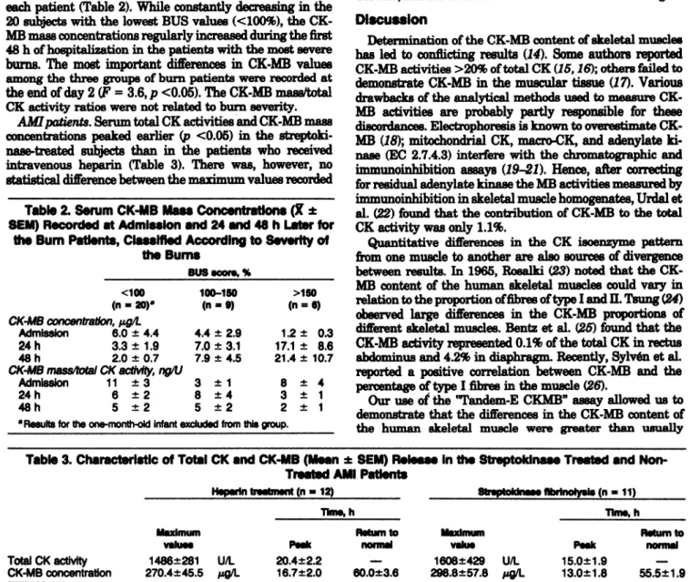
The patient population was subdivided into three differ-ent categories according to the BUS score (8) calculated for each patient (Table 2). While constantly decreasing in the
20 subjects with the lowest BUS values (<100%), the
CK-MB mass concentrations regularly increased during the first 48 h of hospitalization in the patients with the most severe
burns. The most important differences in CK-MB values among the three groups of burn patients were recorded at the end of day 2 (F = 3.6,p <0.05). The CK-MB mass/total CK activity ratios were not related to burn severity.
AMI patients. Serum total CK activities and CK-MB mass concentrations peaked earlier (p <0.05) in the
streptoki-nase-treated subjects than in the patients who received
intravenous heparin (Table 3). There was, however, no statistical difference between the maximum values recorded
Table 2. Serum CK-MB Mass Concentrations (X ± SEM)Recorded at Admission and 24 and 48 h Later for
the Bum Patients, Classified AccordIng to Severity of the Bums
in thetwo groups. The highest oftheseven values obtained for CK-MB mass concentrations between 4 and 28 h after hospital admission in the streptokinase- and heparin-treat-ed group rangheparin-treat-ed from 85 to 693 .tg/L and from 115 to 635 g/L, respectively. The evolution of the CK-MB mass/total CK ratios in the course of AM! was also independent of treatment (Figure 1B). During the corresponding 24-h peri-od, the individual maximum ratios averaged 230 ng/U (range 110-460 ng/U) in the patients who received a fibrino-lytic therapy and 220 ng/IJ (range 120-400 ngfU) in the subjects treated with heparin.
Total CK Activityand CK-MB Mass Concentrationin
Tissue HomogenatesTotal CK activity and CK-MB mass concentration were
determined in homogenates prepared from myocardium and skeletal muscle (psoas, pectoralis major, diaphragm,
femor-alis, and intercostal) specimens obtained at autopsy. In skeletal muscles, the CK-MB mass concentrations ranged from 0.8 (psoas) to 21.4 tg (diaphragm) per gram (wet weight) of tissue (Table 4), and the CK-MB mass/total
CK activity ratio from 0.9 (paces) to 44 ng/IJ (diaphragm).
One gram of myocardium contained, on average, 80.9 ,ug of CK-MB, and the mean CK-MB/total CK ratio was 202 ng/U.
DIscussion
Determination of the CK-MB content of skeletal muscles
has led to conflicting results (14). Some authors reported
CK-MB activities >20% of total CK (15,16); others failed to
demonstrate CK-MB in the muscular tissue (17). Various drawbacks of the analytical methods used to measureCK-MB activities are probably partly responsible for these discordances. Electrophoresis is known to overestimate CK-MB (18); mitochondrial CK, macro-CK, and adenylate
ki-nase (EC 2.7.4.3) interfere with the chromatographic and immunoinhibition assays (19-21). Hence, after correcting
for residual adenylate kinase the MB activities measured by immunoinhibition in skeletal muscle homogenates, Urdal et
al. (22) found that the contribution of CK-MB to the total CK activity was only 1.1%.
Quantitative differences in the CK isoenzyme pattern
from one muscle to another are also sources of divergence
_______
between results. In 1965, Eosalki (23) noted that theCK-MB content of the human skeletal muscles could vary in =6) relation to the proportion of fibres of type land II. Tsung (24)
observed large differences in the CK-MB proportions of 1.2 ± 0.3 different skeletal muscles. Bentz et al. (25) found that the 17.1 ± 8.6 CK-MB activity represented 0.1% of the total CK in rectus 21.4 ± 10.7 abdominus and 4.2% in diaphragm. Recently, Sylven et al.
reported a positive correlation between CK-MB and the 8 ± percentageoftypelfibresinthemuscle(26).
Our use of’ the “Tandem-E CKMB” assay allowed us to demonstrate that thedifferences in the CK-MB content of the human skeletal muscle were greater than usually Table 3. CharacterIstic of Total CK and CK-MB (Mean± SEM) Release In the Streptokinase Treated and
Non-Treated AMI Patients
H.p.rln treatment (n= 12) Strsptoklnaa. fibrinolysis (n= 11)
Time, h Time, h
TotalCKactivity 1486±281 U/L 20.4±2.2 1608±429 U/L
Pectoralls
Myocardlum Picas Diaphragm F.moralls major lntercos
881 938 1051 883
433 851 870 689
370 773 944 784
561 854 955 819
TotalCKactivity,
Uig
406
602 350 925 - 438 1064 X 398 864 CK-MBconcentration, g/g 93.7 0.3 18.0 3.5 12.5 5.3 63.0 0.6 34.0 4.9 7.3 0.9 - 85.9 1.6 12.3 3.1 9.5 2.1 X 80.9 0.8 21.4 3.8 9.8 2.8CK-MBmass/total CK activity, ng/U
231 0.5 20 4 12 5
180 0.6 79 6 8 1
- 196 1.5 33 4 10 3
X 202 0.9 44 5 10 3
Table 4. ActIvity ofTotal CK and Mass Concentration of CK-MB In Human Myocardlum and Skeletal Muscles
294 CLINICALCHEMISTRY, Vol. 32, No. 2,1986
reported: the concentration of CK-MB per gram of wet tissue
was, for example, 24-fold greater in diaphragm than in
psoas. We also showed that the CK-MB content of the
myocardium was only four-fold that in diaphragm. Howev-er, due to the fact that total CK activities per gram of tissue
were higher in skeletal muscles than in myocardium, the
differences in the CK-MB mass/total CK activity ratios were
more important: for four of the five skeletal muscles we
investigated, the CK-MB mass/total CK activity ratio (10 ng/U) was more than 20-fold lower than that for myocardi-urn (202 ng/U).
Published results also differ concerning serum CK-MB
concentrations after muscle injury. In polytrauma patients
without myocardial involvement, OK-MB activities report-edly ranged from 0 to 5.7% (27) or were about 1% of total CK (28). The CK-MB activities in serum of patients with electrical burns, thermal burns, and blunt trauma averaged
8.6,4.6, and 5.7% of total CK, respectively, with an overall range of 0.5 to 22% (4).
In this work, we studied the dynamics of the CK-MB release in the blood after muscular injury. lmmunoenzymo-metric measurements performed at 6-h intervals after ad-mission in the trauma patients indicated an increase and decrease of CK-MB mass concentrations (Figure IA), with maximum values 18 h after admission. In the patients with the most severe burns, CK-MB peaked later (48 h) but the
maximum concentration (mean 21.4 pg/L) was very dose to that calculated for the trauma patients (mean 20.6 ‘L).
Similarly, there was excellent agreement between the ranges of maximum CK-MB concentrations in the trauma patients (5.5 to 67.1 ig/L) and in the burn patients (0.1 to 69.0 ig/L). There was also little difference between the
maximum CK-MB mass/total CK activity ratios recorded
for these two groups: 2 to 13 ng/U (mean 7) in the trauma
patients and 1 to 38 ngfIJ (mean 11) in the burn patients, after exclusion of results for the one-month-old infant. The high ratios (63 to 220 ng/U) found in the latter are likely to be related to an increased CK-MB fraction during develop-ment of the human muscle (29).
The excellent agreement between the range of the
CK-MB mass/total CK ratios in muscle tissue (0.9-44 ng/U) and
the range of the maximum serum ratios measured after muscle injury (1-38 ng/U) in the overall population of
trauma and burn patients must also be emphasized. The
same holds true for the CK-MB mass/total CK activity
ratios determined in myocardium specimens and in the sara
of AMI patients.
Early intracoronary perfusions with streptokinase are
now widely used in AM! patients who reach the CCU
shortly after the attack, in order to recanalize the obstructed
coronary artery and, consequently, to limit the infarcted area (30). Successful fibrinolytic therapy is known to modify
the kinetics of enzyme release, because the increased wash-ing-out from the ischemic area leads to an earlier
appear-ance of the tissue markers in the patients plasma (31,32). Although serum total CK activity and CK-MB mass
concen-trations peaked earlier in the patients treated with strepto-kinase than in those who received heparin (Table 3), the
treatment had no influence on the magnitude of the changes
of CK-MB concentrations and on the evolution of
the OK-MB mass/total CK activity ratios (Figure 1B) in the
course of the disease. The maximum serum CK-MB
concen-trations recorded during the 4th and the 28th hours after admission-that is, given the delays in hospitalization,
about 8 to 32 h after the onset of chest pain-ranged from
115 to 635 ug/L in the patients with conventional treatment and 85-693 pgfL in those who received fibrinolytic therapy.
During this 24-h period, the patients of these two groups
demonstrated at least one CK-MB mass/total CK activity
ratio 110 ng/U.
The use of two monoclonal antibodies, directed towards
the M and B subunits of the CK-MB molecule, makes the Tandem method highly specific. As previously
demonstrat-ed (12), large excess
of
CK-MM and of CK-BB did not interfere with the assay, confirming the results obtained by others with a two-site immunoradiometric assay (33). The fact that no modification of the CK-MB results were ob-served after addition of hemolysates up to a final hemoglo-bin concentration of 1.5 g/L (12) also indicated thatadenyl-ate kinase does ot influence the Tandem assay. Thus, the
immunoenzymometric assay does not require dilution of the
sample when CK-MM is present in large quantities; more-over, it yields reliable results in the presence of adenylate
kinase originating from muscles or erythrocytes. The excel-lent stability of the CK-MB molecule at -22 #{176}Cis an
additional advantage of mass measurements over catalytic
activity determination.
Conclusion
We previously determined the reference range for CK-MB
mass concentrations in presumably healthy subjects to be
0-6 g/L (12). These values were close to those (0-4 g/L)
recommended, however, use of a broader reference interval
(0-9 zg/L) for non-infarct patients. Our study demonstrates
that in fact CK-MB mass concentrations as great as 69 1tgfL may be present in serum of patients with skeletal muscle
injury, and therefore that a cutoff value of 9 ug/L would lead, in trauma patients, to numerous false indications of
AMI. We propose, therefore, that the assessment of myocar-dial necrosis be based on the serum CK-MB mass/total CK
activity ratio rather than on the absolute concentration. None of the trauma or burn patients older than one year
demonstrated a CK-MB mass/total OK activity ratio >40 ng/U at any time during the first 48 h following the
accident. Thus we chose the value of 80 ng/IJ-twofold the
maximum ratio recorded in the patients with muscular injury-as cutoff for differentiating skeletal muscle and myocardiuni necrosis. When several CK-MB mass
measure-ments were performed between 8 and 32 h after the onset of
the symptoms, all AMI patients demonstrated at least one
value 110 ng/U for the CK-MB mass/total OK activity
ratio whether or not they had received fibrinolytic therapy. Consequently, use of the cutoff value of 80 ngfU will completely differentiate AMI patients from those with
trau-ma orburns, thus allowing detection of myocardial necrosis
even in the presence of pre-existing muscular damage. Weare indebted to Mrs. M-C. Aldenhoff and N. Heiligers for their
technical assistance, and to Mrs. B. Cornet for help in the
prepara-tion ofthemanuscript.
References
1. Galen RB, Reiffel JA, Gamhino SR. Diagnosis of acute myocardi-al infarction: relative efficiency of serum enzyme and isoenzyme measurements. J Am Med Assoc 1975;232:145-7.
2. Mercer DW, Varat MA. Detection of cardiac-specific creatune
kinase isoenzyme in sera with normal or slightly increased total creatine kinase activity. Cliii Chem 197521:1088-92.
3. Wilhelm AH, Todd JK. Limited diagnostic value of CK-MB [Letter]. Clin Chem 1977;23:1509-10.
4 Shahangian S, Ash KO, Wahistrom NO Jr, et al. Creatune
kunase and lactate dehydrogenase isoenzymes in serum of patients
suffering burns, blunt trauma, or myocardial infarction. Clin Chem 1984;30:1332-8.
5. Goull#{233}JP, Mechard D, Lame G, et al. Repartition isozymique de la cr#{233}atunekunase dana differents organes humauns. Int#{233}r#{234}ten
pathologie humaine. Ann BiolClin 1979;37:303-7.
6. King IYF, Fu PC, Wishon GM. Persistent creatune kunase MB isoenzyme without cardiac disease. Arch Pathol Lab Med 1978;101:481-2.
7. Chan DW, Taylor E, Frye R, Blitser R-L hnmunoenzymetric assay for creatine kinase MB with subunit-specific monoclonal antibodies compared with an immunological method and electro-phoresis. Cliii Chem 1985;31:465-9.
8. Sachs A, Watson T. The immune consequences of thermal injury: an overview. Chapter 1. In: Nunnemann J, ed. The immune
conse-quences of thermal injury. Baltimore, MD:Williame and Wilkins 1981:1-20.
9. Chapelle JP, Heusghem C. Further heterogeneity demonstrated for serum creatine kinase isoenzyme MM. Cliii Chem
198026:457-62.
10. Rosalki
SB.
An improved procedure for serum creatune phos-phokunase determination. J Lab Clin Med 1967;69:696-705. 11. Farrington C, Chalmers AR. The effect of dilution on creatine kunase activity. Clin Chim Acta 1976;73:217-9.12. Chapelle JP, El Allaf M, Heusghem C. Evaluation of a new
immunoenzymetric assay for CK-MB (Tandem-E CKMB)
[Ab-stract]. Ann Biol Cliii 1985;43:634.
13. Szasz 0, Gerhardt W, Gruber W. Creatune kinase inserum: 5.
Effect of thiols on isoenzyme activity during storage at
various
temperatures. Cliii Chem 197824:1557-63.14. Neumeier D. Tissue specific and subcellular distribution of
creatine kinase isoenzymes. In: Lang H, ed. Creatune kunase isoen-zymes: pathophysiology and clinical application. New York, NY:
Springer Verlag, 1981:85-109.
15. Wilhelm AR, Albers KM, Todd JK. Creatune phosphokinase
isoenzyme distribution in human skeletal and heart muscles. IRCS Med Sci 1976;4:418-20.
16. Thorstensson A, Elwin K, SjOdunB, Karlsson J. Iaoenzymes of
creatine kinase and myokinase in human heart and skeletal muscle. Scand J Cliii Lab Invest 1976;36:821-6.
17. Roberts R, Henry PD, Witteveen SAGJ, Sobel BE.
Quantifica-tion of serum creatune phosphokinase activity. Am J Cardiol 1974;33:650-4.
18. Roberts R, Sobel BE.Isoenzymes of creatune phosphokinase and
diagnosis of myocardial infarction. Ann Intern Med 1973;79:741-3. 19. Fiehn W,SeilerD.Macrocreatune kinase inplasma a cause for
a false positive CK-MB immunoinhibition test. Kiln Wochenschr 1981;59:141-4.
20. Klein B, Jeunelot CL Anion exchange chromatography of
erythrocytic and muscle adenylate kinase and its effect on the
serum creatine kinase isoenzyme assays. Cliii Chem 1978;24:2168-70.
21. Chapelle JP,Heuaghem C. Int#{233}r#{234}tde l’ad#{233}nylatekunase
comme
marqueur
de la n#{233}crosemyocardique. In: Siest G, Galteau MM, eds.Biologieprospective, 4e Coiloque de Pont-#{224}-Mousson.Paris: Mae-eon, 1978:597-601.
22. Urdal P, Urdal K, Str#{248}mmeJR. Cytoplasmic creatine kinase isoenzymes quantitated in tissue specimens obtained at surgery. Cliii Chem198329:310-3.
23. Rosalki SB.Creatine phosphokunase isoenzymes. Nature
(Lon-don)1965207:414.
24. Tsung SH. Creatine kinase isoenzyme patterns in human tissue obtained at surgery. Clin Chem 1976;22:173-5.
25. Bests R,Strom 5, Olin C. OK-MB in serum and in heart and skeletal muscles in patients subjected to mitral valve replacement Eur J Cardiol 1980;12:25-39.
26. Sylv#{233}nC, Janseon E, Olin C. Human myocardial andskeletal muscle enzyme activities: creatune kinase and its isozyme MB as related to citrate synthetase and muscle fibre types. Cliii Physiol
1983;3:461-8.
27. Prellwitz W, Neumeier D. Creatine kinase and CK-MB iaoen-zyme activityin serum of patients after surgical operations, poly-trauma or other damage toskeletal muscle[Letter]. CliiiBiochem 1979;12:225.
28. Lott JA, Stang JM. Serum enzymes and isoenzymee in the
diagnosis and differential diagnosis of myocardial ischemia and necrosis. Cliii Chem 1980;26:1241.-50.
29. Fowall CD, Emery AEH. Changes in creatune kinase and its isoenzymes in human fetal muscle during development. J Neurol Sci 1975;24:483-92.
30. Schwarz F, Faure A, Katus H, et al. Intracoronary thromlioly-sisinacute myocardial infarction: an attempt to quantitate its effect by comparison of enzymatic estimate of myocardial necrosis with left ventricular ejection fraction. Am JCardiol 1983;51:1573-8. 31. Golf SW, Temme H, Kempf KD, et al. Systemic ehort-term fibrinolysis with high dose streptokinase in acute myocardial infarction: time course of biochemical parameters. J Cliii Chem Olin Biochem 1984;22:723-9.
32. Kwong TO, Fitzpatrick PG, Rothbard RL. Activities of some enzymes in serum after therapy with intracoronary streptokinase
in acute myocardial infarction. Cliii Chem 1984;30:731-4.
33. Jackson AP, Siddle K, Thompson RJ. Two-site monoclonal antibody assays for human heart- and brain-type creatine kinase. Cliii Chem 1984;30:1157-62.