MICROBIOLOGY OF AQUATIC SYSTEMS
Cyanobacterial Contribution to Travertine Deposition
in the Hoyoux River System, Belgium
Julia Kleinteich1,2&Stjepko Golubic1,3,4&Igor S. Pessi1&David Velázquez1,5&
Jean-Yves Storme4&François Darchambeau6&Alberto V. Borges6&Philippe Compère7&
Gudrun Radtke8&Seong-Joo Lee9&Emmanuelle J. Javaux4&Annick Wilmotte1
Received: 5 August 2016 / Accepted: 10 January 2017 / Published online: 30 January 2017 # Springer Science+Business Media New York 2017
Abstract Travertine deposition is a landscape-forming pro-cess, usually building a series of calcareous barriers differen-tiating the river flow into a series of cascades and ponds. The process of carbonate precipitation is a complex relationship between biogenic and abiotic causative agents, involving adapted microbial assemblages but also requiring high levels of carbonate saturation, spontaneous degassing of carbon di-oxide and slightly alkaline pH. We have analysed calcareous crusts and water chemistry from four sampling sites along the Hoyoux River and its Triffoy tributary (Belgium) in winter, spring, summer and autumn 2014. Different surface textures of travertine deposits correlated with particular microenviron-ments and were influenced by the local water flow. In all microenvironments, we have identified the cyanobacterium Phormidium incrustatum (Nägeli) Gomont as the organism primarily responsible for carbonate precipitation and
traver-tine fabric by combining morphological analysis with molec-ular sequencing (16S rRNA gene and ITS, the Internal Transcribed Spacer fragments), targeting both field popula-tions and cultures to exclude opportunistic microorganisms responding favourably to culture conditions. Several closely related cyanobacterial strains were cultured; however, only one proved identical with the sequences obtained from the field population by direct PCR. This strain was the dominant primary producer in the calcareous deposits under study and in similar streams in Europe. The dominance of one organism that had a demonstrated association with carbonate precipita-tion presented a valuable opportunity to study its funcprecipita-tion in construction, preservation and fossilisation potential of ambi-ent temperature travertine deposits. These relationships were examined using scanning electron microscopy and Raman microspectroscopy.
Electronic supplementary material The online version of this article (doi:10.1007/s00248-017-0937-7) contains supplementary material, which is available to authorized users.
* Julia Kleinteich
Julia.Kleinteich@gmail.com
1 InBios Center for Protein Engineering, University of Liège,
B-4000 Liège, Belgium
2
Center for Applied Geosciences, University of Tübingen, D-72074 Tübingen, Germany
3
Biological Science Center, Boston University, Boston, MA 02215, USA
4 Palaeobiogeology, Palaeobotany, Palaeopalynology, Department of
Geology, UR Geology B18, University of Liège, B-4000 Liège, Belgium
5
Department of Biology, Universidad Autónoma de Madrid, E-28049 Madrid, Spain
6 Chemical Oceanography Unit, Institut de Physique (B5), University
of Liege, B-4000 Liège, Belgium
7
Department of Biology, Ecology and Evolution (BEE)/Centre of Aid for Research and Education in Microscopy (CAREM), University of Liège, B-4000 Liège, Belgium
8
Hessisches Landesamt für Naturschutz, Umwelt und Geologie, Rheingaustr. 186, D-65203 Wiesbaden, Germany
9 Department of Geology, Kyungpook National University, 1370
Sankyuck-dong, Daegu 702-701, South Korea DOI 10.1007/s00248-017-0937-7
Keywords Calcareous tufa . Culture support . Cyanobacteria . rRNA operon sequencing . Travertine deposition
Introduction
Cyanobacteria are the oldest oxygenic phototrophic microor-ganisms on Earth and the early contributors to most of the primary production and oxygenation of the planet. They have achieved a considerable morphological complexity for pro-karyotic organisms, including multicellularity and cellular dif-ferentiation in structure and function [1]. Cyanobacteria displayed little change in cellular organisation and ecological integration over extended spans of the geologic time [2]. Microbial fossils of cyanobacteria, morphologically similar to modern forms, could be recognised in up to 2.1 Ga (×109years) old deposits [3–7], while traces of photosynthetic microorganisms preserved as laminated mats and stromato-lites influenced the carbonate sediments throughout most of the early history of Earth [8–10]. Today, cyanobacteria con-tinue to play a central role in promoting carbon and nitrogen cycling in marine and freshwater environments, while espe-cially dominating in extreme habitats [11–13]. The study of modern forms helps in the interpretation of fossil records [5]. Involvement of cyanobacteria in deposition of carbonate in freshwater including the build-up of travertine or calcareous tufa occurs worldwide [14–17]. Travertine deposition subdi-vides the river flow by shifting it laterally to form barriers across the riverbed promoting uniform water distribution. The barriers are gradually elevated in the process. Deposition of travertine barriers over time changes the river flow into a series of ponds, accumulating behind the barriers and interconnected by cascades and waterfalls over these bar-riers. It is an ecologically significant landscape-forming pro-cess that creates new environments with a variety of microen-vironments, which, in turn, support diverse specialised macro-and microbiological assemblages. Travertine deposition is subject to cyclic and non-cyclic changes at different scales [18]. The process may improve the water quality along the river as suggested by increased light transmission and spectral shift in light as it penetrates through the water [19]. The loca-tion of travertine deposits and the formaloca-tion of carbonate bar-riers is a sedimentary process guided by the growth response of some benthic organisms, primarily cyanobacteria, algae and mosses, to grow in turbulent water and promote sediment accumulation and consolidation [20]. It is in that sense a bio-genic process, different from sedimentation that follows strict-ly physical laws of depositing particles in calm sections of the river. The resulting travertine barriers are therefore considered to act as freshwater reefs by structure and function [21].
The mechanism of travertine deposition is based on the same principles as the formation of microbialites, sensu
Burne and Moore [22], and stromatolites, sensu Riding [9], where the microorganisms participate by trapping, binding and encrusting the suspended particles carried by the water. The encrustation, which hardens the deposit, is accomplished by in situ carbonate precipitation from carbonate-supersaturated solution, which is also one of the conditions required for deposition of travertine [23]. Soil respiration within the watershed leads to an increase in CO2 concentration in the groundwater, with partial
pressures of CO2 reaching up to values of 50,000 ppm
in the study area (A.V. Borges, unpublished data), more than 12 times above the atmospheric value (400 ppm), causing calcium carbonate dissolution by the groundwater percolating through the base rock. Calcium carbonate (CaCO3) is bound to precipitate again when the CO2
su-persaturated water comes in contact with the air and the CO2 spontaneously degasses toward achieving the
equi-librium with the atmosphere [24]. Gas exchange with the atmosphere is a slow process, but is significantly acceler-ated by the turbulence as the water passes through the rapids and cascades of the travertines. These are also the sites of increased exchange of nutrients and metabolites, and therefore these habitats are dominated by the organ-isms that promote travertine deposition.
Cyanobacteria and microscopic algae are the most com-mon constituents of the biofilms coating the actively accreting travertine surfaces as well as those of mosses and submersed plants. Cyanobacterial trichomes may produce large amounts of exopolymer substances (EPS) generating exopolymer sheaths [25] with structural roles [26]. The microenviron-ments created by the polymeric sheaths can support certain mineral transformations [27]. Mineral precipitates, identified as CaCO3, are common to different cyanobacteria-based
sys-tems [26]. Precipitation of CaCO3can occur within the EPS
sheaths induced by metabolic activities (biologically induced mineralisation) or as result of environmentally driven mineralisation of the organic matrix (biologically influenced mineralisation) [28]. EPS often contain negative charged functional groups, which chelate cations from the running water such as Ca2+[29]. Microsensor studies have document-ed the role of photosynthetic CO2removal by phototrophic
microorganisms [30]. The rates of travertine formation are in the order of millimetre annual increments of the deposit [17]. The travertine deposits are a target for fossilisation studies, including early diagenesis, due to high rates of carbonate pre-cipitation in such systems. The process of fossilisation through calcification has been induced experimentally to study the deterioration of cellular and extracellular (EPS) products under controlled laboratory conditions [31]. Natural fossilisation and early diagenesis have been studied in fresh-water carbonate deposits in carbonate regions for example in the USA and Europe [20, 32, 33] and non-carbonate environ-ments of lacustrine lakes in Antarctica [34].
The investigations of cyanobacterial diversity in biofilms of German karstic rivers by combined morphological and mo-lecular methods [17,35] have shown a number of potential tufa-formers. However, many sequences obtained in those studies were unidentified or found to belong to phylotypes of ambiguous taxonomic identification. Cultivation attempts yielded relatively few strains, of which seven were deposited in a culture collection [36]. Therefore, to date, a direct link between a cultured isolate and natural populations of cyanobacteria involved in the carbonate precipitation in a trav-ertine habitat has not been established.
The goals of the present study are to understand the partic-ular, species-specific contribution of cyanobacteria to the pre-cipitation of travertine or calcareous tufa in the Hoyoux river system in Belgium and further to understand the fossilisation potential of the dominant organism, recorded as mineral and organic textures in carbonate deposits.
Materials and Methods
Site Description
The Hoyoux system, a sub-basin of the Meuse River in Belgium, is 28 km long, has a catchment of ~255 km2, an average slope of 9 m km−1and is partially impacted by human intervention (Fig.1). It is located on the Condroz Inlier. The geology is dominated by Devonian and Lower Carboniferous folded rocks composed of limestone and dolomite bedrocks. Both Hoyoux and Triffoy waterways cross several different strata deposited during Middle and upper Devonian [37]. At the time of sampling, the land cover was 58% cropland, 27% forest and 15% urban. The basin receives an average annual precipitation of 994 mm, and the Hoyoux has an average freshwater discharge of 0.9 m3s−1(seasonal range between 0.2 and 13.0 m3s−1). The Hoyoux drains 31 streams, among which the most representative ones are the Lilot (or Lileau), Triffoy, Vyle, Pailhe, Neuf Moulin and Bonne. In 2008, there were 37 travertine deposition sites along Hoyoux river (over a stretch of about 5 km length) and 19 along the Triffoy (over a stretch of about 1 km length) [37]. Travertine deposition dur-ing our investigation in 2014 was operatdur-ing in both Hoyoux river and its Triffoy tributary, but some impacts due to human intervention have been observed.
Field Analyses and Sampling
Water analyses and travertine sample collection were carried out on travertine barriers at four sites along the Hoyoux River and its Triffoy tributary. Sample collection was carried out throughout an annual cycle on 17 October 2013, 3 February 2014, 20 June 2014, 8 August 2014 and 23 October 2014. Sampling of actively depositing travertine and local water
for chemical analyses took place at four sampling sites (Fig.1), each characterised by a series of carbonate barriers with cascades (except for site 1): Pont de Bonne (site 1) in the upstream part of the Hoyoux River, Moulin de Roiseux (site 2) in the middle part of the Hoyoux, the lower part of the Hoyoux tributary creek Triffoy (site 3) and at Ferme de Barse (site 4) in the lower section of the Hoyoux River. Travertine samples of 1 to 5 cm in diameter were cut to in-clude the active surface as well as the interior to assess differ-ent layers and developmdiffer-ental stages of the deposit. From each sampling site, subsets of collected samples were stored frozen, air-dried or preserved in 3% formaldehyde solution.
Water Chemistry
Water samples were collected in February and October 2014. Conductivity, water temperature, pH and dissolved oxygen were measured in situ at 30 cm depth with a portable field probe (YSI Pro-plus) that was calibrated using standard pro-tocols. Water was collected using a 1.7-L Niskin bottle at 30 cm depth and samples for CH4/N2O were collected in
borosilicate serum bottles, poisoned with HgCl2and sealed
with butyl stoppers and aluminium caps. CH4and N2O
con-centrations were determined by headspace technique and gas chromatography using instrumentation and calibration as de-scribed in Borges et al. [38], with a precision of ±3.9 and ±3.2%, respectively. Samples for the determination of the par-tial pressure of CO2(pCO2) were collected with four 60-mL
plastic syringes, and pCO2was directly determined in the field
by headspace technique and an infra-red gas analyser (Li-Cor Li-840) following the procedure described in Abril et al. [39] with a precision of ±2.0%. Samples for total alkalinity (TA) were filtered on 0.2-μm polyethersulfone (PES) syringe filters and determined by titration [40]. Ammonium (NH4+), nitrate
(NO3−) and soluble reactive phosphorous (SRP) were
deter-mined with standard colorimetric methods [41]. Light Microscopy
Binocular microscopy (up to ×100 magnification; Leica L2) with incident light was used to observe and photo-document the sur-face textures and porosity of carbonate encrustations and for preparation of samples for transmitted light microscopy, scanning electron microscopy and Raman microspectroscopy. Compound light microscopy (Leica DM LB2) was used for identification, photo-documentation and study of the distribution of microor-ganisms within carbonate crusts. Millimetre-size fragments from the surface of the encrustations were treated with dilute (3%) HCl to dissolve CaCO3. Samples for transmitted light microscopy
were prepared from the insoluble residue, observed, measured and photo-documented. The procedure was repeated with frag-ments from the interior of the crust. Diversity and distribution of cyanobacteria was recorded with semi-quantitative estimates in
relation to the total insoluble organic and clay mineral residue (in percent of coverage representing percent of biomass). The pop-ulations were morphometrically characterised by measuring cell dimensions expressed as mean ± SD (n > 30).
Scanning Electron Microscopy
Preparations for SEM were made from subsamples of traver-tine crust preserved in the field with 3% formaldehyde solu-tion in environmental water, according to the following pro-cedures: (a) Subsamples were gradually dehydrated in an eth-anol series, CO2critical point dried (Leica EM CPD300) and
Pt-coated (Balzers SCD 030 sputtering unit) for direct SEM observation of the biofilm and associated mineral precipitates on active travertine surfaces. (b) To remove the organic matter and observe clean mineral surface, subsamples were treated with Na-hypochlorite for 10 days before ethanol dehydration, air-drying and Pt coating. Observations and secondary elec-tron images of both (a) and (b) subsamples were acquired in a SEM Jeol JSM-840A under 22 kV accelerating voltage. (c) Vertical fractures and polished (by use of SiC paper; see be-low)/slightly etched surfaces of sections cut perpendicular to the crust were used for conventional SEM documentation of the mineral texture at depths in the concretions. (d) Subsamples were post-fixed for 2 h in 1% osmium tetroxide (OsO4),‘en bloc’ stained in 1% aqueous uranyl acetate and
gradually dehydrated in an ethanol/propylene oxide series be-fore embedding in epoxy resin (AGAR Low Viscosity Resin Kit, AGAR, R1078, hard mixture) with a step in a vacuum oven to improve resin penetration. These samples were sec-tioned perpendicular to the crust surface and the sections were mirror-polished with SiC paper (ESCIL up to PSA GR. 4000) and with non-aqueous 0.25-μm diamond suspension (ESCIL 1/4PS-1/4MIC), then C-coated in a Balzers MED-010 evapo-rator for back-scattered electron (BSE) imaging in SEM and elemental x-ray microanalysis. Sections included oncoids and encrusted branches to expose profiles and to follow changes of the deposit over time. The observations were realised in an FEI ESEM-FEG XL-30 under high vacuum conditions and 10 kV accelerating voltage. Elemental x-ray dispersive micro-analyses and mappings were performed in the ESEM-FEG with a 129-eV silicon drift detector (Bruker) and the Quantax 800 Esprit 1.9 software (Bruker).
Raman Microspectroscopy
Raman analyses were performed on polished thin sections of travertine and oncoids with a Renishaw In via Raman spec-trometer coupled to a Leica DM 2500 confocal microscope with Ar-ion 50 mW monochromatic 514.5 and 785 nm as laser sources. Laser excitation was adjusted to an on-sample intensity of ca. 2 mW (measured with a Coherent Lasercheck Fig. 1 Map of the research area
with travertine collection sites marked along the Hoyoux and Triffoy rivers, Belgium: 1 Pont de Bonne, 2 Moulin de Roiseux (mid-flow cascades), 3 Triffoy waterfalls, 4 Ferme de Barse. Forests are displayed in dark grey, agricultural land in white
Analyser) and focussed through ×50 and ×100 objectives to obtain a 1–2 μm spot size. Acquisitions were obtained with an 1800 and 1200 mm grating with an air-cooled (−70 °C) 1024 × 256 pixel CCD (charge-coupled device) array detector. This method enabled a 2000 cm−1spectral detection range. Beam centering and Raman spectra calibration were per-formed daily on a Si-Al microprocessor chip with a character-istic Si Raman band at 520.5 cm−1. Spectra were manipulated within Wire 3.4™ software. Point analysis measurements were made in static mode (fixed at 1150 cm−1). Raman map-ping acquisition was also used (a) to detect the distribution of organic matter from the travertine surface through the carbon-ate deposit, to identify minerals and to assess the process of its fossilisation and its effect on calcification and mineral forma-tion and (b) to analyse the nature and preservaforma-tion of UV-protecting pigments [42,43].
Isolation and Culture
Single-trichome cultures were established from different types of travertine material from field sites 2, 3 and 4, sampled during the field trip in February 2014 (Supplementary Table1). Small fragments of travertine rock were distributed homogenously on sterile BG11 [44] agar plates containing 20 μg mL−1 cycloheximide. After visible growth, cyanobacterial trichomes were removed and transferred to a new plate until uni-cyanobacterial cultures were established. Cultures were kept at 15 °C under low light conditions (ca. 2 klux, LS1 in Panasonic Plant Growth Chamber MLR-352). All seven uni-cyanobacterial isolates will be deposited in the public Belgian Co-ordinated Collections of Micro-organisms BCCM/ULC (Strain 2 is available as ULC392 and Strain 10 as UCL393). Genetic information of the 16S rRNA gene plus ITS sequence was obtained from all isolates as described below.
Molecular Characterisation
After collection, samples for DNA analysis were stored frozen at−20 °C. Genomic DNA from environmental material (site 2 and 3, from field trips in June and August, Supplementary Table 1) as well as from cultured isolates (see above) was extracted using the PowerBiofilm DNA Isolation Kit (MOBIO, Carlsbad, USA), according to instructions from the manufacturer. For two samples (MOD14 and 15, from the Triffoy and the Hoyoux, respectively), material collected directly from the travertine surface (upper 1 mm) as well as material from a deeper section were extracted. Two PCR re-actions were performed on all DNA extracts to amplify the 16S rRNA gene as well as the ITS spacer between the 16S rRNA and the 23S rRNA genes. The primer combinations 27F and 809R [45] as well as 359F and 23S30R [46,47] were used in a PCR reaction containing 1× Phusion High-Fidelity
PCR Master Mix (Thermo Scientific, Waltham, USA), sup-plied with 0.5 μM of each primer and 2 μL of DNA per reaction. The cycling conditions for the 27F/809R primer pair were at 98 °C for 2 min, followed by 35 cycles with 98 °C for 30 s, 58 °C for 45 s and 72 °C for 50 s, with a final elongation of 5 min at 72 °C. For the 359F/23S30R primer pair, the PCR conditions were as follows: 98 °C for 2 min, followed by 35 cycles of 98 °C for 30 s, 68 °C for 30 s and 72 °C for 90 s, and a final elongation at 72 °C for 5 min. The amplified fragments were run on an agarose gel and the PCR products purified using the GeneJet PCR Purification Kit (Thermo Scientific, Waltham, USA). The PCR fragments were se-quenced using several primers (27F, 359F, 359R, 809R, 1092R, 1407F, 1497R and 23S30R) on a Sanger Sequencer (3730 DNA Analyzer) at the GIGA sequencing facility (http://www.giga.ulg.ac.be). Individual sequences were trimmed and aligned using Geneious R7.1.7 and compared to the GenBank database using BLAST. To build phylogenetic trees, three most closely related strains and uncultured sequences, respectively, were retrieved by SeqMatch of the Ribosomal Database Project (http://rdp. cme.msu.edu/seqmatch/seqmatch_intro.jsp). The sequences from Arp et al. [17], Cousin and Stackebrandt [35] and Brinkmann et al. [36] were added when they belonged to the clade B of the latter authors, where our sequences are positioned, plus two thin Oscillatoriales. The sequence of Leptolyngbya boryana PCC73110 was selected as outgroup. The sequences were aligned with BioEdit [48]. A first analysis was performed on several long sequences of 1419 bp. However, the tufa sequences from Germany [35], even though closely related to ours, were too short and a second alignment was made with 454 bp to include them. The pairwise sequence differences were calculated with MEGA6 [49]. In both cases, Neighbor-joining trees were built with Treecon [50], with the Kimura correction for multiple muta-tions (selected by MEGA6), indels not taken into account and a bootstrap analysis involving 500 re-samplings. For the ITS sequences, a BLAST analysis was used to find the related sequences, but only the sequences that could be meaningfully aligned were kept in the alignment. Phylogenetic analysis of the ITS sequences was done by the same methods as the 16S rRNA sequence analysis, with the exception that the Jukes and Cantor model [51] was used for nucleotide substitutions.
Results
Hydrology and Water Chemistry
The deposits of ambient temperature travertine or calcareous tufa form a series of cascades in the Hoyoux River and its Triffoy tributary (Fig. 1). Data collected in February corresponded to high water (HW) and, in October 2014, to
low water (LW) periods, with water discharges of 1.1 and 0.5 m3s−1, respectively (for the Hoyoux between Point de Bonne and Ferme de Barse). The water chemistry was analysed for both Hoyoux River and Triffoy tributary (Table1), but no hydrological data were collected for the latter. Both waters are characterised by relatively high pH and total alkalinity with oxygen concentrations close to the atmospheric equilibrium. The pCO2values were significantly
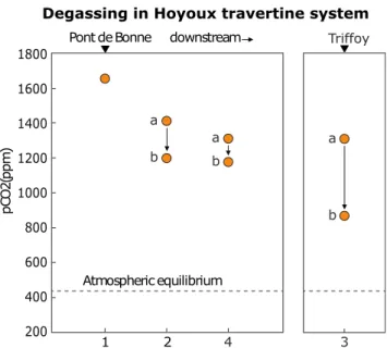
above the atmospheric value (400 ppm) throughout the stud-ied river flow; however, the most significant small-scale de-creases were caused by degassing at the turbulent passages and water falls (Fig.2; Supplementary Fig.1), which are also responsible for the overall longitudinal pCO2decreases and
the associated carbonate precipitation. The waters of both riv-ers were high in NO3−concentrations but comparatively low
in NH4+ and SRP concentrations; they were supersaturated
with respect to CO2, CH4and N2O (Table 1). In February
and October 2014, a decreasing trend in pCO2, CH4 and
N2O wa s o b s e r v e d a l o n g t h e Ho y o u x r i v e r f l o w
(Supplementary Fig.1).
Physical Properties of the Calcareous Crusts
The external shape of the deposit and its internal texture varied in different parts of the system of travertine bar-riers and waterfalls (Fig. 3a). Three types of samples with different calcification patterns were recognised in the field as subjects for this study (Supplementary Fig.2). In the nearly laminar flow over the tops of trav-ertine barriers (Fig. 3b), we observed poorly calcified flat mats, comprised of aquatic mosses and moss proto-nemata, covered by uncalcified or weakly calcified red and green algae (Fig.3c); in the moderate flow between b ar rie rs a n d c as ca de s, we c ol lec te d o nc o ids an d
submersed encrusted wooden branches (Fig 3a and insert). The oncoids were coated by porous carbonate crust of velvet-like consistency and with small granular protrusions (Fig. 3d); in cascades, on sites receiving high water impact (Fig. 3e), we encountered compact crusts with smooth surface of shiny appearance, blue-green to steel-blue in colour, which covered the traver-tine surface or encrusted branches that were trapped there (Fig.3f).
Table 1 Water chemistry in the Hoyoux and Triffoy rivers in February and October 2014
Hoyoux Triffoy
February October February October
Water temperature (°C) 5.9 ± 0.3 10.0 ± 0.1 6.5 ± 0.4 9.7 ± 0.0 Specific conductivity (μS cm−1) 602 ± 3 608 ± 1 644 ± 1 646 ± 3 Total alkalinity (μmol L−1) 4998 ± 8 4944 ± 217 5127 ± 115 4120 ± 559
NO3−(μmol L−1) 429 ± 6 368 ± 1 436 ± 1 372 ± 12 NH4+(μmol L−1) 2.9 ± 1.2 2.4 ± 0.2 2.8 ± 0.8 2.3 ± 0.4 SRP (μmol L−1) 1.2 ± 0.1 1.8 ± 0.1 0.7 ± 0.1 0.9 ± 0.0 pH 8.29 ± 0.20 8.15 ± 0.05 8.48 ± 0.01 8.01 ± 0.25 O2(%) 95.3 ± 5.7 100.0 ± 3.5 101.0 ± 0.4 100.2 ± 0.7 O2(μmol L−1) 363 ± 24 348 ± 11 378 ± 1 353 ± 2 pCO2(ppm) 1348 ± 195 1964 ± 278 1097 ± 312 1264 ± 272 CH4(nmol L−1) 88 ± 45 153 ± 113 40 ± 18 44 ± 23 N2O (nmol L−1) 70 ± 48 66 ± 31 13 ± 6 15 ± 5 pC O 2( ppm )
Degassing in Hoyoux travertine system
downstream Pont de Bonne Atmospheric equilibrium Triffoy 1 2 4 3 a b a b a b 1800 1600 1400 1200 1000 800 600 400 200
Fig. 2 Distribution of CO2partial pressure (pCO2, ppm) before and after
a series of travertine cascades (sites 2–4), illustrating the high carbon dioxide levels responsible for carbonate supersaturation of the water and degassing as condition for carbonate precipitation. Note the pCO2
measured consistently above the atmospheric equilibrium level, with significant drops occurring at the positions of travertine cascades
In the first sample type, it was possible to observe the partial stages of calcification, whereas the carbonate structure in oncoids and tightly compacted crusts in cascades was complete and well organised. The porous velvet-like crust that was coat-ing the surfaces of oncoids (Fig. 4a) and encrusted branches under moderate flow conditions was constructed of calcified tubules that were like tiny needles (Fig.4b), orientated in dif-ferent directions and interconnected, thus leaving pore space between them. These calcareous tubules were formed by calci-fication of extracellular sheaths (EPS) of filamentous cyanobacteria. The tubules became frequently fused into clus-ters and granular protrusions, fractions of a millimetre in size, when fortified by inter-tubular cement (Fig.4c, d). In contrast, the smooth compact crusts (Fig.3f) under high water impact were composed of densely arranged calcareous tubules of the same size as those in the velvet crust but arranged parallel to one another and oriented perpendicular to the crust surface. Such dense crusts often provided a firm coating over porous deposits on the ground (Fig.4e) or encrusted branches (Fig.4f)
and are responsible for dense horizons within travertine deposits with stromatolite-type laminations.
Cyanobacteria Associated with the Calcification Front All calcified microhabitats of the studied travertine (sites 2, 3 and 4) were dominated by a sheathed filamentous non-heterocystous cyanobacterium, identified by phenotype (morphology and ecology) as Phormidium incrustatum (Nägeli) Gomont (Fig. 5), which is characterised by straight trichomes (one trichome per filament) inside thin but firm and distinctive individual sheaths (Fig.5b, arrow). The trichomes were unconstricted at the cross walls, some-times with granulations along them, shortly attenuated at the tips and, when fully differentiated, ending with a flat-conical terminal cell, with thickened outer wall. The cells were 5.92 ± 0.41 (34) μm wide, shorter than wide, 1.41 ± 0.35 (31) μm long, blue-green to olive-green in colour (Fig.5a) when grown in their natural environment Fig. 3 Travertines of the Hoyoux
river system, Belgium. a View of a series of travertine barriers across the river with cascades (see Fig.1, collection site 2) and moderate water flow
(downstream of cascade) Insert: oncoids in the moderate stream (indicated by arrow) between barriers with the encrusted twig (right, 5 cm long). b Arched barrier with smooth overflow. c Poorly calcified mosses and algae from the overflow in (b). d Oncoids and encrusted wooden branches from moderate water flow between the barriers. e Waterfalls with high impact habitats. f Compact encrustation with hard steel-blue surface from high water impact habitats in (e)
on fully illuminated surfaces, but purple to red when grown deeper in the crust or on shaded sites (Fig. 5b). When transferred on agar surface, the Phormidium trichomes reacted with intense gliding movement and showed an in-creased variability in colour and diameter (Fig.5c). Several uni-cyanobacterial strains were isolated from single tri-chomes and maintained in culture. To test for the ability for complementary chromatic acclimation [52], two sub-cultures (Isolate 5 and 6) were grown separately under green and red light (using differently coloured plastic fil-ters; adapted from Tandeau de Marsac [53]). The results were positive, showing a predominance of the blue-green phycocyanobilin pigmentation (Fig.5d) when grown under red light and a change in coloration to purple when grown under green light (Fig.5e), probably due to predominance of phycoerythrobilin pigment.
The porosity of the carbonate crust, especially in the weakly calcified almost laminar overflow over the bar-riers, promoted settlement and/or entrapment of other eu-karyotic and proeu-karyotic microorganisms, present mostly
in small populations (Supplementary Fig. 3). Several eu-karyotic algae have been observed in the crust, notably the large filaments without cross walls of the yellow-green alga Vaucheria sp. often associated with diatoms (Supplementary Fig. 3a, b also in Figs. 7c and 11a) and with the freshwater rhodophyte Batrachospermum sp. in a Chantransia growth stage (Supplementary Fig. 3c). Among eukaryotic organotrophs, a large oomycete has been found trapped and cemented while responding by massive release of oospores, many of them germinating ( S u p p l e m e n t a r y F i g . 3d ) . T h e a c c o m p a n y i n g cyanobacteria include pleurocapsalean taxa characterised by multiple fission and release of small reproductive cells or baeocytes (Supplementary Fig. 3e), including euendolithic Hyella fontana Huber et Jadin, which may have been penetrating the already consolidated carbonate parts of the crust (Supplementary Fig. 3f). Among fila-mentous cyanobacteria, we observed scattered populations of a Lyngbya sp., with filaments significantly narrower than the dominant P. incrustatum (cells 3.2–3.5 μm wide Fig. 4 Cyanobacterial calcareous crusts. a Oncoid covered by two types
of encrustation: nodular (above) and porous velvety (below). b Close-up view of the velvety crust, a porous spatial network of irregularly oriented calcareous tubules produced by Phormidium incrustatum. c Predominantly upward diverging clusters of Phormidium tubules generating a nodular surface. d Section perpendicular to a consolidated
nodular crust covering an oncoid. e A fractured travertine crust showing the transition from a porous velvety to a compact crust (lighter colour) forming a hard top surface. f Concentric changes in encrustation shown on the section of an encrusted twig, collected from a waterfall receiving high water impact. The scale bar in (a) is 5 mm for (a) and (f) and 100μm for (b), (c) and (d)
and 1.0–1.2 μm long, Supplementary Fig.3g), the hetero-cystous cyanobacterium Calothrix sp. accompanied by the pleurocapsalean Xenococcus sp. and densely bent and coiled Leptolyngbya sp. with mildly constricted but con-spicuous cross walls (cells 1.1–1.4 μm wide and 2.5– 3.5 μm long). All these cyanobacteria produced EPS sheaths and envelopes, including some chroococcalean cyanobacteria classified within the genus Aphanocapsa that occurred in small colonies interspersed in the crust. Some of these accompanying taxa may have been trapped together with the leaf detritus; whether they can become permanently established in the crust is not certain. Phylogenetic Relationship among Travertine-Forming Microorganisms
In order to identify the genetic signature of the most prevalent microorganism in the system, the 16S rRNA gene and the ITS fragments from five samples, MOD-14, MOD-14s (surface),
MOD-15, MOD-15s (surface) and MOD 21, were directly amplified by PCR from the environment (site 3 and site 2 in June, site 3 in August) and sequenced. All sequences obtained are listed in Supplementary Table1, together with their origin, GenBank submission numbers and BLAST hits. The se-quence MOD 15 was discarded from further analysis because of bad quality. The amplified 16S rRNA gene sequences MOD 14, MOD 14s and MOD 21 are almost identical and show only 1 bp difference to each other, in addition to four ambiguities with MOD 15s. This finding suggests that a single species or a cluster of very closely related genotypes prevailed during the spring and summer season in at least two locations: Triffoy and Moulin de Roiseux on Hoyoux River.
When subjected to a BLAST search using the GenBank database (Supplementary Table 1), all the 16S rRNA se-quences obtained directly from the environmental samples corresponded with >99.2% (1347 bp) and 100% (656 bp) similarity to the uncultured cyanobacterium clones WB18.8 (GenBank accession no. GQ324964) from a German tufa Fig. 5 Cyanobacterial Phormidium strains detected in the Hoyoux
travertine including Phormidium incrustatum (Nägeli) Gomont, the main architect in the formation of Hoyoux travertines. a Filaments extracted from the illuminated surfaces of travertine oncoids after dissolving the carbonate. b Filaments extracted from the shaded areas of travertine inside the crust. Note the difference in cell pigments. c Trichomes of varying diameters and pigmentation, isolated in raw
culture, actively gliding on the agar surface. d Uni-cyanobacterial culture (Isolate 6) grown under red-filtered light, as part of the complementary chromatic acclimation (CCA) test: the pigmentation enriched in phycocyanobilin. (e) Uni-cyanobacterial culture (Isolate 5) grown under green-filtered light, part of the same CCA testing as in (d): cultures grown under green light filter show pigment shift toward predominance of phycoerythrobilin
system [17, 35]. The habitats in the German study, the Westerhöfer creek in the Harz Mountains, are very similar to those in the Hoyoux system, with high concentrations of cal-cium and magnesium in the water and formation of tufa cas-cades associated with microbial calcification. The detection of the same dominant organism in both systems, now supported by almost identical 16S rRNA sequences, suggests that the same cyanobacterial taxon might dominate several Middle European travertine systems.
Seven distinct Phormidium-type uni-cyanobacterial strains have been obtained in the process of isolating the dominant microorganism of the Hoyoux travertines in culture (Supplementary Table1). The 16S rRNA genes shared a pairwise identity of 98.2% between these seven isolates, showing a larger diversity than the field sample sequences. The 1403-bp fragment of the 16S rRNA gene of uni-cyanobacterial Isolate 8 was found to be 99.8% (BLAST) similar to the uncultured cyanobacterial clone WB18.8 from the German tufa system mentioned above (Supplementary Table1). Moreover, the shorter V3–V4 region of the 16S
rRNA gene of Isolate 8 was 100% identical to the sequences found in the spring and summer environmental samples of the Triffoy (site 3) and the Hoyoux (site 2). This means that Isolate 8 is identical to and representative of the dominant species in the two sampled travertine systems. Since Isolate 8 was generated from a sample taken in February, whereas the direct environmental sequences were obtained in June and August (see above), it can be assumed that the same organism is predominant at these sites throughout the year. Its pheno-typic properties (see also Pentecost [54]) correspond to P. incrustatum (Nägeli) Gomont.
The genetic relationship of the seven isolated uni-cyanobacterial strains to the amplified environmental se-quences as well as to reference strains from public databases is presented in phylogenetic trees based on long and short 16S rRNA gene sequences (Fig.6and Supplementary Fig.4). The shorter alignment was necessary to include short sequences from German travertines. The scales of the trees are very small, and thus all the groups displayed are very similar to each other. Even so, the trees clearly show that the 16S rRNA gene sequence of Isolate 8 is most closely related to those sequences obtained directly from the environmental ma-terial of the Hoyoux travertine system (MODs 14, 14s and 21) as well as to the uncultured WB18.8, CL2.C6, BF.S97 and BF.S143 from the German tufa system. Less closely related, but still in the same cluster, are other environmental sequences (MOD 15 because of one substitution and a few ambiguities) as well as an uncultured bacterium strain Llo_001 (GenBank accession no. FR667299) from epilithic biofilms in a moun-tain lake in Spain [55] and a strain from the epipelon of a Czech lake [56] identified as Phormidium autumnale.
More variation than in the 16S rRNA gene was observed in t h e I T S s e q u e n c e s ( S u p p l e m e n t a r y Ta b l e 1 a n d
Supplementary Fig.5). The ITS region is a highly variable genetic spacer between the 16S and the 23S rRNA genes. The ITS regions of the environmental samples were 94.2% similar to each other, suggesting that slightly different genotypes are present in the travertine system, probably within the same morphotype. The ITS sequences of the isolates were also highly variable, supporting this hypothesis.
Relationship between Cyanobacteria and Carbonate Deposits
The filamentous cyanobacterium, identified as P. incrustatum, was found to predominate in all carbonate-encrusted deposits in the travertines under study. The observed different surface textures of the carbonate crust appear to be due to cyanobacterial growth response to water flow and impact. The porous velvet crust with divergent tubules was found in sections with slow to moderate water flow, where the cyanobacterial filaments were able to grow in any direction. In contrast, in areas of high water impact in cascades, where the crusts are compact and have highest increment rates, the cyanobacterial filaments were directed outward and perpen-dicular to the crust surface.
Individual filaments observed in the loose velvet-like coat-ings under moderate flow conditions form tubular, externally conical calcareous encrustations around their sheaths (Fig.7a). The tips of calcified tubules are“plugged” by tri-chomes (Fig.7a, insert), which move in and out leaving the opening free of sediment while the carbonate precipitation in the surrounding ambient continues to add cement between the tubules (Fig.7b). BSE-SEM images of polished sections of resin-embedded cyanobacteria in their mineral context show carbonate crystal nucleation to be initiated within the EPS sheath (Fig.7c, arrows) from where the crystals expand out-ward (Fig.7d). The calcification starts in the EPS at the fila-ment tip, frequently forming a thin calcareous collar, but al-ways leaving the smooth and uncalcified interior of the sheath (Fig.7e). The external calcification of tubules continues in the surrounding environment and fuses the tubules together (car-bonate cementation), a process that forms nodules (Fig.4c, d) and ultimately solid carbonate tufa, while leaving the interiors of the tubules empty. The intact sheaths lining the interior of tubules are presented by SEM of a fractured travertine rock (Fig.7f). The pores left between filaments are sites of inde-pendent calcium carbonate precipitation also forming rhom-bohedral calcite clusters (Fig. 7g). The relations between P. incrustatum trichomes and carbonate deposit on the hard-crust surfaces from the cascades are shown in the SEM images of critical-point-dried specimens as overview (Fig.7h) and in detail (Fig.7i).
The close relationship between cyanobacteria and carbonate precipitate was further studied by BSE-SEM imaging of resin-embedded travertine samples of
oncoids, a technique which offered high resolution of both biological and mineral structures, also providing the possibility of element identification and mapping (Fig. 8); all pictures represent parts of an oncoid, ori-ented so that the active surface is to the left. At the actively growing surface, P. incrustatum trichomes emerge from their sheaths (Fig.8a, centre of the frame). There is also a trichome fragment (hormogonium) above and an empty tubule below. The thickness of the calci-fied wall of the filament in the frame increases toward the interior of the crust. The image of a P. incrustatum filament with associated calcite grains (Fig. 8b) re-solved intracellular and extracellular properties in lon-gitudinal section. A detailed view of the trichome tip (Fig. 8c) shows the cyanophycine granules along the
cross walls, enhanced due to osmium and uranium con-trast as well as the thickening of the external wall of the apical cell (calyptra ?, white arrow). The x-ray elemen-tal analyses and mapping (Fig. 8d) confirmed calcite mineralogy of the precipitates, the osmium and uranium staining of the trichomes and identified traces of Si in the Phormidium’s EPS sheath (Supplementary Fig. 4). The element analysis also confirmed the presence of silica in trapped diatom frustules and clay particles. The relations between Phormidium tubules and carbon-ate are presented in a section through the centre of an oncoid (Fig. 8e) showing no carbonate precipitation in-side intact tubules. The same can be followed toward the oncoid surface (Fig. 8f, g). However, in oncoids eroded while rolling in the currents, the calcareous Fig. 6 Neighbor-joining tree based on 16S rRNA gene sequences
(1418 bp) of environmental sequences obtained directly from travertine samples, of isolates obtained from travertine material and from the reference strains from public databases, including uncultured sequences. The distance matrix was corrected by the Kimura equation and indels were not taken into account. A total of 500 resampled trees were
calculated for the bootstrap analysis, and the values above 50% are indicated in front of the corresponding nodes. The Hoyoux sequences are in bold and the travertine and tufa sequences from elsewhere are printed in italics. The OTU numbers are taken from Brinkmann et al. [36] and the‘Unid.’ number from Arp et al. [17]
Phormidium tubules are broken and exposed to the en-vironmental water so that the carbonate precipitation takes place inside the tubules as well (Fig.8h).
Progression of the Calcification and Early Diagenesis The calcified wooden branches and twigs incorporated in ertine permit the analysis of relatively recent changes in trav-ertine deposition and reveal the onset of early diagenetic changes. In cross-sections through such deposits, it was pos-sible to observe a pattern of zones of different encrustation density, forming textural layers documenting time-bound changes in the actively of growing surfaces, possibly related
to the environmental conditions (Fig. 9). The same profiles may also reveal early diagenetic alteration of travertine deposits.
Each of the encrusted twigs presented in cross-section showed an external, most recently formed zone of dense carbonate deposit that incorporated void, out-ward radiating Phormidium tubules (Fig. 9a). In the overview of a section (Fig. 9b), this external compact layer (1) includes the surface of the deposit, growing at the time of collection (Fig. 9c). This layer is underlain by a porous travertine layer (2), composed of loosely arranged tubules, similar to the crust observed on oncoids, but less clearly outlined (Fig.9d). The compact Fig. 7 Calcified structures associated with Phormidium incrustatum. a
Conical calcareous tubule produced by Phormidium filament in side view. Insert: Front view of the opening of calcareous tubule with the exiting trichome. Note the fine microcrystalline deposit on the margin of the EPS sheath surrounding the trichome. b Relief formed by several externally cemented calcareous tubules. c Carbonate crystal initiation (nucleation, arrows) within the sheath close to the filament tip. d Filament in cross-section in the middle part of the tubule, showing radial growth of carbonate (C) from the sheath surface (S) outward. e View of the tip of calcified Phormidium sheath in perspective; note the
outward growth of calcite crystals and smoothness of the interior of the tubule. A trapped diatom frustule is at lower left. f Fractured consolidated travertine deposit with tubules nearly in longitudinal section; note the sheath layer lining the lumen of the tubules. g Group of rhombohedral calcites precipitated in travertine pores independent of Phormidium sheath. h Planar overview of the hard travertine surface exposed to water impact in the waterfall showing the tips of Phormidium tubules (compare Fig.3f). i Detail of (h) with several trichomes exiting their tubules. a, b, e, f, g Direct SEM; c, d SEM with BSE (backscattered electronic images); a—insert, h, i SEM of critical-point-dried samples
surface layer (Fig. 9a), when viewed in cross-section (Fig.9e), shows a distinction between calcification that o r i g i n a t e d f r o m c y a n o b a c t e r i a l s h e a t h s a r o u n d Phormidium trichomes (t) and from inter-tubular ce-ment precipitated independently in the pore spaces (p). An arrow points to that boundary.
The zone immediately surrounding the encrusted twig (lay-er 3 in Fig.9b), and therefore the oldest component of the structure, features large calcite crystals (Fig.9f, g), which also comprise the most compact carbonate zone. The encrusted branch is surrounded by a series of dense outward divergent mineral units, marked by periodic upward convex pseudo-stromatolite lines (Fig.9f). The Phormidium tubules, although involved in the original sediment, are here rarely preserved with their lumens partially or completely closed. Their open-ings can be seen only as exits at the upper surfaces of the crystal units toward the internal pore spaces (Fig.9f above). The preserved remains of tubules and the up- and outward diverging orientation (Fig.9g) identify these calcite units as re-crystallised clusters of the once similarly diverging
Phormidium tubules, marking the onset of early diagenetic alteration.
Raman Microspectroscopy of Travertine
Samples of loose travertine coatings of oncoids and encrusted branches, and compact deposits exposed to cascade impact were analysed by Raman microspectros copy (Fig. 10) to examine the alteration of the organic structures incorporated in the sediment. Raman analyses were performed along a pro-file from the active surface to the interior of the carbonate crust that was resin-embedded, sectioned perpendicular to the sur-face and polished (Fig.10a). Local accumulation of the UV-protecting pigment, beta-carotene, has been identified along the contact between calcite grains on the edges of a laminated pseudo-stromatolite texture (Fig.10b). Organic content with still preserved beta-carotene was also recorded in calcified tubules (ca. 2 μm in diameter) and a broken tubule likely resembling a collapsed trichome (<1 μm in diameter, Fig.10c, d). The pigment seemed to be associated with the Fig. 8 Relations between Phormidium incrustatum and carbonate
deposition. a Section perpendicular to the substrate surface showing three Phormidium trichomes emerging from their sheaths and calcareous tubules. The white frame designates the area of the elemental x-ray mapping in (d). b Phormidium filament in its carbonate context. c Detail, note the cyanophycine granules along the cross walls, the narrowed (calyptrate) terminal cell, marked by white arrow and the two-layered EPS sheath with calcite granules included. d Elemental x-ray mapping of the frame outlined in (a). Silicon and aluminium (both marked red) are present in diatom frustules and clay particles trapped between Ca-carbonate grains (in yellow), but it also labels the EPS
sheaths of the cyanobacteria (arrows). e Phormidium tubules in the interior of the dense carbonate crust remain hollow as long as the tube interior is not accessed by carbonate-supersaturated environmental water. f Section through an oncoid showing an interior calcareous block with hollow tubules (right), a solid block with tubules filled with carbonate (centre) and an active incompletely calcified oncoid surface (left). g Detail from the intact surface area of the oncoid in (f) (left), with intact tubules. h Detail of the eroded oncoid with tubule interior filled by carbonate precipitate. All pictures are oriented with the surface of the crust to the left (BSE imaging and x-ray elemental mapping of polished thin sections of samples, stained with OsO4and uranium acetate)
remains of the filament of P. incrustatum; however, due to cellular breakdown and secondary displacement by recrystallisation of the carbonate deposit, a clear localisation could not be ascertained nor could it be assigned to a specific organism. Other calcareous tubules, externally completely cal-cified, showed a weak organic EPS signal indicating that the sheaths remained within the deposit as it was observed by SEM inside the travertine structure (e.g. Fig.7f). The organic matter (OM) or carbonaceous material (CM) content has been detected in orange-brown deeply incorporated tubules by hyperspectral Raman mapping (Fig.10e–h). This signal has been compared with a synthetic CM spectrum and found to correspond to a very immature state of organic matter. The same signal was also observed in orange-brown CM concen-trated along the contact of some recrystallised calcite agglom-erations inside travertine. The finding is consistent with our observations of surface organic residue following the
dissolution of carbonate, which revealed an accumulated or-ganic matter with the predominance of empty sheaths.
In summary, we observed an evolution of the Raman signal from beta-carotene UV protective pigments preserved in the young travertine crust to unspecific CM signal observed deeper inside and therefore older travertine crust. The preser-vation of OM and of calcareous tubules in travertine, on short and long timescales, seems to be poor due to the porous nature of the carbonates prone to oxidation and recrystallisation. However, residual organic matter, usually displaced by recrystallisation, may preserve very locally in a heterogeneous pattern and at a micrometre scale.
Trapping, Binding and Degradation in Travertines In all types of deposits, the calcified network of Phormidium sheaths forms an effective filter in which particles suspended Fig. 9 Textures of travertine crusts. a Vertical section through compact
travertine crust deposited by Phormidium incrustatum under high water impact in a cascade (see area 1 in b); note the parallel, slightly upward-diverging empty tubules that conform to the diameters of Phormidium filaments. b Cross-section of a travertine-encrusted wooden twig (tw— removed from the central hole) and showing changes of travertine deposition in three zones (1, 2 and 3), with the actively growing surface on top. c Side view of carbonate encrusted Phormidium filaments at the active surface of the crust, forming a compact deposit. d Loose, velvety
crust (area 2 in b), as observed in moderate water flow. e Cross-section of the compact, high impact crust (area 1 in b), illustrating carbonate deposit around Phormidium tubules (t) and in pores (p) between filament clusters with an arrow pointing to the boundary. f Pseudo-stromatolite-type of lamination in recrystallised parts of the travertine. g Detail of the recrystallised layer of travertine next to the encrusted twig (tw in b). b Incident light photomicrograph; c, d: SEM of fractured surfaces; a, e, f, g: SEM of polished petrographic thin sections
in the water, including planktonic and benthic microorgan-isms, such as diatoms and other unicellular algae, become trapped, while rapid calcification provides the binding (Fig.11a, b). This property defines travertines as microbialites or stromatolites. Organotrophic bacteria, which decompose autochthonous as well as the extraneous organic matter, are important in this system, where turbulence and aeration sup-port aerobic decomposition, thereby returning some of the CO2into the system [57]. Of particular interest are the bacteria
associated with degradation of the indigenous primary product of the system, especially of the cells and EPS of cyanobacteria, which remain trapped in the carbonate crust. Dense populations of rod-shaped bacteria were found to fill the space previously occupied by trichome cells (Fig.11c) and might be involved in the degradation of cyanobacterial organ-ic matter. The entrapped organorgan-ic compounds are a nutrient source for diverse filamentous and coccoid organotrophic bac-teria that were observed by SEM of critical-point-dried
preparations, including filamentous bacteria (Fig. 11d) and mostly unknown polar attached rod-shaped bacteria (Fig.11e, f).
Discussion
Travertine or calcareous tufa deposits (see Pentecost [54] for definitions) are distributed worldwide [15]. In the studies of complex systems such as the deposition of calcareous tufa or ambient temperature travertines, the questions of biotic versus abiotic factors responsible for the processes involved are com-monly asked and discussed [17,21,23]. In this context, it is useful to assess the degree of biotic versus abiotic influences separately for different phases of the process, starting with (a) the contributions to water chemistry, then to (b) nucleation and formation of minerals and, finally, (c) to the distribution of sediments in the formation of travertine [57]. By using the Fig. 10 Raman spectrum
analysis. a Polished cross-section of a laminated travertine crust with the areas of detailed analysis outlined and numbered. b Finely laminated pseudo-stromatolite texture (zone 1 of a). c Preserved tubule (zone 2 of a). d Raman spectra observed for (b) and (c). e Incorporated‘fossilised’ tubules (zone 3 of a). f Raman hyperspectral mapping of the ‘fossilised’ tubules showing the separation of calcite and carbonaceous matter (CM) in the tubule. g Organic-brown CM accumulations along the contact of some recrystallised cluster of calcite inside the travertine. h Raman spectra of the CM observed in (f) and (g)
example of the Hoyoux river system (Fig.1), which is one of the 12 known travertine depositing rivers in Belgium [14], a link between the chemical and biological processes involved in travertine deposition could be established in this study. The Travertine Deposition in Hoyoux River System The overabundance of carbonate molecules in the waters un-der study has two sources: geological availability of calcium and carbonate from limestone and dolomite bedrocks and the biological availability of CO2from degradation of organic
matter in soils of the entire watershed. Thus, the increase of pCO2above the equilibrium with the atmosphere and the
equivalent carbonate supersaturation of the karstic waters are fuelled by a substantial biogenic input [20] and so is, by ex-tension, the degassing of CO2in the process of re-establishing
the equilibrium with the atmosphere. Pentecost [14], who analysed water chemistry in British karstic regions, came to a similar conclusion. The opposite effect is illustrated in the karstic regions of central Italy, where deforestation and loss of soil was thought to be the main reason why the river Volturno does not precipitate carbonate today, while it has produced extensive travertine platforms in the past [33].
The chemical conditions promoting carbonate precipitation that exist along the studied Hoyoux waterways are promoted by CO2-degassing, stimulated in rapids and waterfalls due to
the increase in surface-to-volume ratio of the air-water inter-phase. The pCO2 values in the Hoyoux and the Triffoy
(<2000 ppm) are at the lower end of the range of values re-ported in other meteogene travertines, ranging between 550 and 31,500 ppm [16,23,58]. The pCO2values in the Hoyoux
and the Triffoy are actually below the average for meteogene travertines of 5200 ppm (distinctly lower than the average of 50,000 ppm for thermogene travertines in hot springs reported by Pentecost [23]). The overall spatial gradient of pCO2in the
Hoyoux was −85 and −183 ppm km−2 in February and October, respectively. These gradients are two orders of mag-nitude lower than those reported in other travertine systems ranging between about−10,000 and −20,000 ppm km−2[23]. The distinction, however, may be related to the fact that the other studies of travertines were conducted directly down-stream of springs, which was not the case in the present study of Hoyoux. Nevertheless, the level of supersaturation and the measured drop of pCO2following each set of cascades along
the river identified the CO2-degassing as the principal driving
force of CaCO3precipitation with a commensurate loss of
carbonate, a part of which may have been incorporated in and contributed to the growth of the carbonate terraces. The relatively high level of nitrate observed in the Hoyoux water-ways may indicate eutrophication, which may lower or even inhibit carbonate precipitation and travertine deposition [32]. The Biological Identity ofPhormidium incrustatum (Nägeli) Gomont
The phenotype of the most dominant calcifying organism is a filamentous non-heterocystous cyanobacterium identified as P. incrustatum (Nägeli) Gomont (Fig. 5), which occupies a wide variety of carbonate depositing microenvironments [17, 20, 33, 54, 57]. The organism was identified by comparing and morphometrically evaluating its phenotypic properties in samples taken from the sites of active carbonate deposition. Fig. 11 Microbial assemblages entrapped in travertine. a Entrapped
diatom (D) assemblage on poorly calcified oncoid surface close to a filament (F) and an empty sheath (S) of Phormidium (upper left corner). b A chrysophyte cyst entrapped inside an oncoid. c Rod-shaped bacteria filling and possibly decomposing a cyanobacterial
trichome. d Assemblage of organotrophic bacteria around entrapped organic matter in a depression on the surface of an oncoid. e Detailed image of polarly attached organotrophic bacteria. f A different assemblage of organotrophic bacteria. a–c BSE images of polished resin-embedded sections; d–f SEM of critical-point-dried samples
Isolation in culture yielded several morphologically similar and genetically closely related uni-cyanobacterial clones (Fig.5c–e). Isolate 8 turned out to be genetically identical with the phenotypically dominant populations at two Hoyoux trav-ertine sites (Fig. 6) as well as with clones from travertine deposits from the Westerhöfer creek in the Harz mountains in Germany [17, 35]. In the latter study, clone WB18.8, which is identical to our dominant sequences, is clustered as ‘Unidentified A’ and is distant from the suspected P. incrustatum identified and sequenced by Arp et al. [17] as ‘Unidentified B’ (clone WB10.1) and from the Tychonema strains that these authors also detected. Our isolates 3 and 2a appear to belong to their OTU5, which is related to the type strain of Tychonema bourrellyi CCAP 1459/11 [59]. The phy-logenetic tree of Brinkmann et al. [36] did not include the clones identical to our dominant strain, and therefore, we can only guess that it belongs or is closely related to their clade B. This would also correspond to the Tychonema/ Phormidium/Microcoleus clade found with a metagenomic study by Schneider et al. [60].
The fact that our culturing, which started with small inoc-ulums placed on standard BG11 [44] 2% agar, from which individual trichomes were isolated, produced several different, albeit related, uni-cyanobacterial strains raised the question of the identity of these strains with those dominating the natural populations. Past experiences have documented the preva-lence of opportunistic organisms favoured by standard culture media at the expense of the field populations that are often adapted to nutrient-deprived extreme life conditions [61] and often do not grow at all in synthetic media [62].
No 16S rRNA gene information of P. incrustatum is pres-ently available in public databases, although its morphotype identification and ecology is well established in modern [20,
54] and ancient travertine settings [33]. We used two indepen-dent approaches to try to iindepen-dentify the dominant strain in the travertine system: (a) by direct amplification of the 16S rRNA gene fragment from the environment and (b) by sequencing the strains we isolated and cultured from that same environ-ment. We selected the cultured strain that was, by its sequence, closest to the environmental one, which also turned out to be the quasi-identical to the strain WB18.8 from Germany in GenBank. Based on morphological identification by LM and SEM observation, correlated with its habitats and ecology and with the observed consistent dominance in travertines where it produces characteristic sedimentary patterns, we can confi-dently conclude that the organism identified as Phormidium incrustatum (Nägeli) Gomont is genetically identical with Isolate 8 obtained in this study. Cyanobacterial taxonomy is in a state of rapid changes by introduction of phylogenetic criteria based mostly on 16S rRNA gene sequencing. The taxonomic revisions are generally based on cultures with new taxa described on the basis of gaps in the sequence record [63,64]. Our results document the need to closely compare the
sequences of cultures and corroborate their identity with those obtained from (direct) sequencing of field populations. The Role ofPhormidium incrustatum
The most important contribution of P. incrustatum and other similarly specialised microorganisms to the formation of trav-ertines lies in their observed habit to grow in rapids where they promote entrapment and binding of suspended particles and cause sediment accumulation. Their activity causes lateral di-version of the water flow as well as an even, lateral distribu-tion of sediment across the riverbed so as to form travertine barriers, thus having a major sedimentological and geological-ly rapid landscape-modifying impact. The barriers differenti-ate the river flow longitudinally into a series of lentic and lotic (lakes and waterfalls) environments each with a variety of microbial habitats and ecological niches [18,20].
Microbial photosynthesis, including that of P. incrustatum, is expected to have contributed to the calcification process [65], but is considered local, and secondary in relation to CO2supersaturation and degassing toward the equilibrium
with the atmosphere, and so would be the seasonal effects of temperature [66]. The relatively low levels of carbonate satu-ration in the Hoyoux system may have been responsible for the dominance of a single cyanobacterium in relation to other compared travertine systems, which show a higher diversity of prokaryotic and eukaryotic microorganisms that are subjected to calcification. Biocalcification appears to be a genetic trait, provided that optimal chemical conditions of carbonate satu-ration are present, as some taxa sharing the same microenvi-ronment calcify while others do not [57,65,67] or may spe-cifically require higher carbonate saturation levels. For exam-ple, a different and species-specific carbonate encrustation was observed in various cyanobacteria and chlorophytes in more diversified travertine systems [20,67,68]. The domi-nance of P. incrustatum in various habitats of the Hoyoux system offered a relatively simple microbial model, where calcification is restricted to one dominant cyanobacterium, while other microorganisms remained uncalcified.
The particular contribution by P. incrustatum involves min-eral nucleation. There is evidence that the EPS produced by this organism competes successfully in attracting new mole-cules of calcium carbonate from the surrounding solution even in the presence of active carbonate mineral surfaces (see Fig.7). The ability of P. incrustatum to initiate nucleation of carbonate on its own organic product (EPS) starts a mineralisation process that complies with a biological rather than mineral template (Figs.7d, e and8e, g).
The calcification appeared to be limited to the external surfaces or the outer layer of cyanobacterial sheaths, while leaving the lumen uncalcified and hollow (Figs. 7e, f and
9a), contributing to the porosity of the deposit. The calcifica-tion expanding from the outer surfaces of EPS sheaths is
consistent with the light-microscopic images (Fig. 5a, b). After the carbonate was removed by dissolution in dilute HCl, the external surfaces of the extracted sheaths were not smooth, but covered by minute irregularly distributed dots, some of which may have marked the positions of calcite nu-cleation sites. In contrast, the interior surfaces of sheaths were smooth as seen in LM as well as in SEM images (Fig.7c–f).
This observation raised the question about the functional dis-tinction between internal and external surfaces of the EPS sheath regarding carbonate nucleation capacity. The resolution of this question came from the study of the dynamics of oncoids rolling in the currents between barriers, which be-came externally abraded. As a consequence, the broken tu-bules become internally calcified as their interior was accessed by the carbonate-supersaturated environmental water (Fig.8h). The above findings show that the EPS sheath of P. incrustatum is externally and internally uniform with re-spect to the attraction of carbonate cations and promotion of crystal nucleation. This is evident also by the fact that the precipitate in the interior of broken tubules started at the in-ternal sheath surface and then proceeds to fill the lumen of the tubule. Thus, the lack of precipitate in the interior of most tubules can be explained by the fact that they remained sealed, initially by the resident trichomes and later by cementation, and thus not accessible to external carbonate-supersaturated water.
The motility of Phormidium trichomes constitutes an adap-tive response property that provides the organism with the ability to avoid environmental hazards, in this case to escape the burial by their own calcified sheaths [68] and by other mineral precipitates derived from supersaturated interstitial waters. This property, if not shared by other microbial constit-uents, provides a critical competitive advantage, which is con-sistent with the observed dominance of this organism in the travertines. In the ecological sense, the travertines may be considered extreme environments, where the rates of encrus-tation and carbonate accumulation constitute the main com-petitive pressure and a limiting parameter. During most of its activity, the trichome is close to the exit of the tubule, gliding up and down within its polysaccharide sleeve and ready to move out and upward with the advancement of carbonate deposition. Its presence acts at the same time as a plug, preventing carbonate precipitation in the interior of its tubule. The combination of gliding motility and the ability to pro-mote carbonate precipitation of P. incrustatum constitutes a mechanism that may explain the observed different textures of the carbonate crust. These appear to be due to cyanobacterial growth response to water flow and impact. The porous velvet crust with diverging tubules was found in sections with slow to moderate water flow, where the maxi-mum carbonate accretion took place at the tips of cyanobacterial filaments (Fig.7a), which were able to grow in any direction, as long as the rate of environmental
precipitation (cementation) remained low (e.g. Fig. 7g). However, the high water impact in cascades, accompanied by elevated rates of environmental calcification, requires a different response. Phormidium trichomes respond by gliding upward to escape the burial, assuming upright position while keeping the pace with the carbonate accretion of the crust. Like other filamentous cyanobacteria, P. incrustatum repro-duces by fragmentation into hormogonia that glide away and start another colony, but the residence time of individual fila-ments and maintenance of an intact tubule may be consider-able as evident from the length of empty tubules inside the consolidated travertines (Figs.8f,9a).
Fossilisation Capacity and Early Diagenesis
The analysis of Hoyoux travertine using Raman spectroscopy (Fig.10) confirmed that carbonate deposition does not support preservation and fossilisation of organic compounds as it pro-motes their complete aerobic decomposition. It did demon-strate, however, the relative recalcitrance in degradation of EPS and UV absorbing pigments that have a better chance to preserve and fossilise.
In addition, the decomposition of organic matter, once in-corporated in travertine, seems to have contributed to early diagenetic alteration of the deposited carbonate. The changes were noticed in a zone of recrystallised mineral fabric along the contact between the carbonate deposit and the encrusted submerged twigs (Figs.3d,4f and9b) with details showing large outward diverging crystal units with pseudo-stromatolite lamination (Fig.9f, g). Similar diagenetic changes have been observed in fossil travertine of Italy, including large crystal units that still preserved tubular patterns of Phormidium on the periphery [20,33] and pseudo-stromatolite layering inside recrystallised units [20]. The Phormidium imprint patterns are comparable with those in modern travertine of the Plitvice Lakes system in Croatia, and they are quite similar to those we now recorded in Hoyoux travertine.
The transitions between the zones with different carbonate textures (Fig.9b) are abrupt, marking interruptions or changes in the process of sediment accretion, including possible differ-ences in the extent or nature of diagenetic alterations in each of the observed zones. The zonal distribution of porosity due to density and orientation of calcareous tubules probably reflects the seasonal changes in water flow providing different condi-tions that promote microbial growth and calcification. The layers immediately surrounding the embedded wooden branches, however, reflect rather the onset of early diagenesis including recrystallisation of the carbonate fabric that modi-fied the texture of the deposit and obliterated the traces left by the original organismal activity. These alterations are likely triggered by the processes of microbial degradation of the incorporated branches and thus proceed in the opposite direc-tion of the accruing travertine deposidirec-tion. The encrusted twigs