PUFAs AND AGING DISORDERS
Linking low docosahexaenoic acid intake to Alzheimer’s disease:
caution recommended
Stephen C. CUNNANE Mélanie PLOURDE Milène VANDAL Erika FREEMANTLE Jennifer TREMBLAY-MERCIER Michel BÉGINResearch Center on Aging, Health and Social Sciences Center, University Institute of Geriatrics of Sherbrooke, Université de Sherbrooke, 1036 Belvédère St. South, Sherbrooke, QC, Canada J1H 4C4
Abstract: Prospective cohort studies and animal models support the concept that low
docosahexa-enoic acid intake is implicated in the etiology or progression of Alzheimer’s disease. However, other studies crucial to this relationship are less encouraging. To date, the few trials using docosahexaenoic acid to treat declining cognition in the elderly have either been very small or, in the largest trial, the beneficial effect was mild and limited to a sub-group of patients. The supplements used in each of these clinical trials contained at least one polyunsaturated fatty acid other than docosahexaenoic acid, so the active ingredient remains unclear. One widely cited study reported markedly lower brain docosahexae-noic acid in Alzheimer’s disease but at least five other much less commonly cited reports have not corroborated this effect. There are numerous inconsistencies or confounders in the data and several challenges to overcome before definitively attributing a specific role to docosahexaenoic acid in the protection of cognitive function in the elderly.
Key words:docosahexaenoic acid, Alzheimer’s disease, omega 3 fatty acids, brain, eicosapentaenoic acid, arachidonic acid
Introduction
As the percentage of elderly people in devel-oped countries increases, awareness is growing rapidly about the pivotal importance of main-taining physical and cognitive autonomy for healthy aging. Alzheimer’s disease (AD) repre-sents the main form of cognitive decline in the adults. As such, preventing or avoiding the main risk factors for AD improves the chances of prolonging cognitive function and quality of life in the elderly. Great efforts are now being made to understand the etiology of AD and to develop strategies to attenuate its significant medical, social and economic impact. Cur-rently, most treatment strategies for AD focus on pharmacological interventions while most strategies for prevention focus on lifestyle risk factors, especially nutrition. According to a recent European report [1], no single strategy has been particularly successful at reducing the incidence or growing socio-economic impact
of AD or other adult forms of significant cogni-tive decline.
There is growing concern that once AD is clini-cally diagnosed it is already too late to initiate effective treatment. Hence, there is a burgeon-ing interest in nutritional approaches to reduce the risk of cognitive decline before it becomes clinically symptomatic. Amongst the various nutritional approaches, one that is gaining a lot of attention at the moment relates to low intake of fish and omega (x) 3 polyunsaturated fatty acids as a risk factor for AD. The x3 fatty acid most studied in the context of AD is docosahexaenoic acid (DHA; 22:6x3), which is found principally in fish and seafood. A possible link between dietary DHA and maintenance of brain function in adults has its origins in the study of infant brain development where DHA is now clearly recognized by most pediatric societies and government regulatory agencies as an essential component of optimal post-natal feeding practice.
At present, we are aware of four reports describing positive but very mild effects of a dietary DHA supplement in AD or other forms of cognitive deterioration in the elderly. In addition, one report showing lower brain DHA levels in AD is frequently cited. These studies compliment a much larger epidemiological lit-erature linking low x3 fatty acid and/or low DHA intake to higher risk of AD, a literature supported by several animal studies (which will not be reviewed here). Despite the apparent
link between low DHA intake and AD, signifi-cant confounders limit interpretation of the studies reporting a clinical benefit of DHA in AD. Several detailed reports of brain fatty acid data in AD actually show no specific change or even a rise in brain DHA, but are not commonly cited. Hence, in the interests of encouraging a more critical perspective on the putative link between DHA and AD, we provide here a brief review of the literature on clinical trials and brain DHA data in AD.
Linking low DHA
and Alzheimer’s disease
A plausible association between DHA and risk reduction for cognitive decline in the elderly is emerging on three main fronts – epidemiologi-cal studies, plasma DHA levels in AD patients, and animal models. A task force of the Interna-tional Association of Nutrition and Aging recently reviewed the literature on nutritional factors associated with aging-related cognitive decline [2]. This report provided details of 15 prospective studies conducted between 1997-2006 that focused on whether the risk of cognitive decline during aging is associated with low intake of total dietary fat, fish, and/or x3 fatty acids from fish. Collectively, these studies followed up an average of 40,000 people over 6 years. Two of the largest of these studies reported no significant associa-tion between x3 or fish intake, but the others
Abbreviations: AA, arachidonic acid
AD, Alzheimer’s disease
DHA, docosahexaenoic acid
EPA, eicosapentaenoic acid
MCI, mild cognitive impairment
MMSE, mini-mental state exam
PE, phosphatidylethanolamine
OCL VOL. 14 N° 3-4 MAI-AOÛT 2007 177
doi:
10.1684/ocl.2007.01
all showed a protective effect of fish and/or DHA on risk of cognitive decline. Other reviews [3] and a more recent epidemiological study [4] also came to the same conclusion. Because of the normally strong positive asso-ciation between plasma DHA and DHA or fish intake, it would be expected that a link between low fish or DHA intake and cognitive decline in the elderly would be reflected by lower plasma DHA in those experiencing cog-nitive decline. Indeed, several studies have found low plasma DHA in AD [5-8], but one study found that AD was accompanied by higher plasma DHA [9].
Clinical efficacy of DHA
in Alzheimer’s disease
Regardless of the plausibility of a relationship that emerges from other types of studies, clearly, the central issue in linking low DHA to risk of AD is whether higher DHA intake has a lasting clinical beneficial effect in preventing or reducing cognitive decline in any form of neu-rodegeneration, most particularly in AD. No other type of study can supplant the impor-tance of a series of independently run, well-designed clinical interventions that collectively support a therapeutic benefit of DHA. In the largest study to date, Freund-Levi et al. [10] recently reported the results of a random-ized, placebo-controlled, double blind trial, in which a mixed supplement containing both DHA (1.7 g/d) and eicosapentaenoic acid (EPA – 20:5x3; 0.6 g/d) was given to patients with mild to moderate AD. The patients continued to receive their usual medication and received the active treatment (DHA + EPA) or placebo for 6 months followed by active treatment of both groups for a further 6 months. In the 174 participants completing the 12 mo trial, no difference in cognitive function between the two groups was observed after the first 6 months. In a small sub-group with relatively high cognitive function at the start of the study, cognitive scores (mini-mental state exam; MMSE) and delayed word recall declined more slowly on the active treatment. More rapid decline of the MMSE score during the first 6 months in those with a starting MMSE of < 24 was noted but was not statisti-cally significant.
Three smaller trials using DHA in AD [11-13] have been reported but two of these studies appear to have only been published in prelimi-nary form [11, 12]. Terano et al. [11] random-ized 20 elderly vascular dementia patients in a nursing home to receive no treatment or a DHA supplement (4.3 g/d for 12 mo). Initially, these patients all had cognitive evaluations consis-tent with mild to moderate dementia. Depend-ing on the cognitive test used, improvement in
the DHA group was seen at 3 and/or 6 mo, but not at 12 mo. Suzuki et al. [12] evaluated the effect of 680-800 mg/d of DHA in 22 elderly patients with cognitive decline of mixed etiol-ogy. In about half the patients, some cognitive parameters improved during the 6 month trial. Kotani et al. [13] evaluated the effect of a 90 d treatment using a mixture providing a total of 240 mg/d of DHA and arachidonic acid (AA; 20:4x6) in patients with AD or mild cognitive impairment (MCI). The proportion of DHA to AA in the mixture was not given. AD patients (n = 8) only received the active treatment. MCI patients received either the active treatment (DHA+AA; n = 12) or a placebo (6 capsules of olive oil providing 240 mg/d; n = 9). Attention and immediate memory scores improved in the MCI group given DHA+AA but not in the other groups.
Boston et al. [14] reported that there was no effect of a 12 wk treatment with 1g/d ethyl-EPA on several parameters of cognition in AD patients who had a starting MMSE score of 19. This report is noted here because it used one of the fish oil x3 fatty acids (EPA) but this highly purified preparation contained no DHA. These clinical studies highlight several chal-lenges associated with investigating a benefi-cial effect of x3 fatty acids in AD. In all four of these trials [10-13], more than one potentially active polyunsaturate was present, so the ques-tion arises as to whether the beneficial effect due to DHA alone, or to the EPA [10-12] or AA [13] also provided with the DHA, or to the combinations of DHA + EPA or DHA + AA. Assuming that half the active treatment in the study by Kotani et al. [13] was DHA, what explains how their results appear to be broadly equivalent to those of Freund-Levi et al. [10] who provided 14 times as much DHA, or those of Terano et al. [11], who provided 36 times as much DHA (120 mg/d versus 1.7 or 4.3 g/d, respectively)? Since different tests of cognitive function were employed and the supplements were given for different periods, were these interventions studies actually comparable? As implied in the study by Freund-Levi et al. [10], are those with higher cognitive function at the start of the trial actually likely to benefit more from a x3 supplement? Are those with lower cognitive function actually at greater risk when given a x3 supplement?
Brain levels of DHA
in Alzheimer’s disease
If DHA supplementation is beneficial in AD or MCI, it seems reasonable to expect that this benefit relates to correcting lower brain DHA. Although we have found no reports of brain DHA levels in humans after DHA supplementa-tion (in AD or otherwise), we are aware of the following reports on brain DHA content in AD:
Bowen et al. [15] appear to be the first to have reported brain fatty acid data in AD. They analyzed fatty acid profiles of phospholipids from frontal grey matter from controls (n = 12), senile dementia (n = 11), AD (n = 4) and Creuzfeldt-Jacob disease (n = 1). Their method pre-dated capillary gas chromatogra-phy so did not permit good separation of spe-cific polyunsaturates. As a result of this impor-tant limitation, AA was grouped with adrenic acid (22:4x6) and DHA was grouped with the two docosapentaenoic acids. These data were reported as ‘polyunsaturates’. They found no age- or disease-related change in brain polyun-saturates or phospholipid concentration. In 1991, Soderberg et al. published the most widely cited paper on fatty acid composition of brain samples obtained at autopsy from 8-10 controls and AD patients [16]. Brain samples were analyzed from four different regions – frontal grey matter, white matter (region not specified), hippocampus and pons. Fatty acids were analyzed from two of the main classes of phospholipids in the brain – phos-phatidylethanolamine (PE) and phosphatidyl-choline, which are rich and poor, respectively, in polyunsaturated fatty acids. The % of DHA in PE was reduced by up to 50% in all four brain regions from patients with AD. The fatty acid changes attributed to AD were not be due to aging alone which, as they reported in the same paper, was not associated with changes in brain fatty acid composition.
In 1993, Skinner et al. [17] reported brain fatty acid composition in 15 AD patients and 10 controls. Grey and white matter of frontal, parietal and parahippocampal regions were studied. Amongst the polyunsaturates, there were no differences due to AD except for 3-4 fold higher levels of adrenic acid in grey matter, as well as higher DHA in parietal white matter.
In 1998, Corrigan et al. [18] reported the fatty acid composition of parahippocampal cortex in 8 AD patients and 6 age-matched controls. AD was confirmed histologically in each case. DHA was not different between AD and con-trols for any of the four phospholipid classes or in the cholesteryl ester class. In the AD samples, adrenic acid was lower in both PE and phos-phatidylserine, and AA was also lower in PE. In the cholesteryl esters of AD brain, a-linolenic acid (18:3x3) was present at only 5% of the level seen in controls.
In 1998, Prasad et al. [19] evaluated brain fatty acid composition in 9 AD patients and 9 age-matched controls. Fatty acids were quantified (nmol/g) in several brain regions and in three phospholipid classes as well as in brain free fatty acids. DHA was 45% lower in PE from the hippocampus and parahippocampal gyrus (both regions analyzed together), and was
24% lower in the phosphatidylcholine fraction of the cerebellum. Lower DHA in hippocampal and parahippocampal PE was actually due to lower PE because, on a percentage basis, DHA was 50% higher in this lipid class. Otherwise, DHA did not differ across lipid fractions or brain regions. This study emphasized the possible importance of lower plasmalogens, a form of PE with vinyl-linked instead of ester-linked fatty acids, in the progression of AD. In AD, a lower concentration plasmalogens was observed but only in the hippocampus [19].
In 1999, Guan et al. [20] reported the phos-pholipid and fatty acid composition of brain samples from AD patients (n = 15; 80 y old) and controls (n = 13; 72 y old). Data were provided for frontal cortex, hippocampus and white matter and were expressed relative to brain DNA. In the AD samples, the content of the two main phospholipids was 16-24% lower in the frontal cortex, an effect due primarily to lower plasmalogen PE. Expressed in relation to brain DNA content, DHA was not different in the various lipid classes of frontal cortex from AD patients compared to the controls. Given that the concentration of plasmalogen PE in the frontal cortex was decreased in AD brain but the DHA concentration in plasmalogen PE was unchanged, the % of DHA in this lipid class actually increased somewhat. In contrast, AA was decreased by 49% and adrenic acid was decreased by 27% in the plasmalogen PE of frontal cortex from AD patients. Oleic acid (18:1x9) was also decreased by 25-75% in the same regions, an effect not reported elsewhere. In 2001, Han et al. [21] reported the fatty acid composition of the various molecular species of PE in several brain regions from AD patients who were separated according to different lev-els of cognitive decline (0.5 to 3 on the clinical dementia rating scale. There were 6 samples in the control group and in each of the four grades of AD. They showed that even at the earliest clinically defined stage of AD, plasmalo-gen PE was decreased by up to 40% in white matter. In gray matter, this decline in PE was less severe but also correlated with clinical severity. DHA concentrations (mg/g protein) were lower in some brain regions as were other fatty acids, including saturates, monounsat-urates and x6 polyunsatmonounsat-urates. The only brain region in which lower DHA was not due to lower PE was in the cerebellum.
Taken collectively, published brain DHA data are available from about 85 AD patients and 54 controls but it is impossible to draw any clear conclusion about a direct link between lower brain DHA and AD. When low DHA was observed, it was not specific for DHA: either AA and/or adrenic acid were also decreased [16, 18, 21], or the lower DHA was a function of a decrease in one of the lipid classes, most
com-monly plasmalogen PE [20]. Increases of up to 50% for DHA [17, 19, 20] and 4 fold for adrenic acid [17] have also been reported for some regions of AD brain. Hence, it is clearly incor-rect to discuss lower brain DHA as if it were a commonly accepted or specific observation in AD. These studies actually seem to agree more about changes in brain phospholipids in AD, particularly the decrease in plasmalogen PE, which may be equally or more important as the possible changes in their fatty acid composi-tion [22]. Therefore, the comment made by Guan et al. [20] nearly a decade ago still seems highly pertinent – ‘these discrepancies are largely due to diagnostic problems, method-ological differences as well as to calculation and presentation’.
Good data from age-matched controls are of critical importance to interpreting changes in brain lipid or fatty acid composition attribut-able to AD. Svennerholm et al. [23] reported age stratified data for brain composition from people living at home who experienced acci-dental deaths. They had 40-50 samples per age group and showed that between the lowest age group (20-39 y) and the highest (80-100 y), brain weights decreased by 20% and brain phospholipid content decreased by about 43%. Nevertheless, no evaluation of cognitive function or fatty acid composition was reported. Soderberg et al. [16] reported age-stratified data showing that aging alone does not change brain fatty acid in either of the two main phospholipid classes of the brain. Therefore, agreement is lacking on even the basic point of whether healthy aging is associ-ated with a change in brain fatty acid compo-sition [16, 23], so there is a real possibility that some of the discrepancies regarding whether or not brain DHA changes in AD may actually be due to difficulty in selecting healthy, age-matched controls or in classifying the AD patients themselves. Subcellular changes in DHA should be explored to see whether they are partially responsible for the wide disparities in reported effects of AD on brain DHA. The reports of lower brain AA and adrenic acid in AD should also not be overlooked because AA may have clinically relevant effects on cogni-tion [13].
Oversimplification
of the relation between DHA
and Alzheimer’s disease?
Assuming that low fish intake does in fact increase the risk of AD, should low brain DHA be necessarily expected in AD or, indeed, that DHA would be clinically effective in AD? DHA synthesis and b-oxidation are both very low in humans [24, 25]. Therefore, unlike in many animal models, in humans, DHA intake does have an important effect on whole body DHA homeostasis [26]. Nevertheless, dietary x3 fatty acid depletion studies show that relative to DHA in blood or other tissues, brain DHA is slower to deplete or to replete after dietary depletion. Hence, in humans, the link between DHA intake and brain DHA is potentially com-plex and it is difficult to make the case that there should necessarily be a direct or simple link between low DHA intake, low plasma DHA and low brain DHA in humans, let alone in AD. Especially in the elderly, factors linked to DHA degradation (peroxidation or loss due to cell death) in the brain may well be as important in determining brain DHA levels as the input vari-ables such as dietary supply of DHA, a trickle of synthesis, or its transport into the brain. Never-theless, the optimization of DHA as a supple-ment for preventing or treating AD depends to some extent on understanding how DHA is utilized by the aging brain (table 1), so as to establish when its use is to be encouraged and when it is contraindicated.
AD has a major impact on the healthcare deliv-ery system and has no widely accepted treat-ment. Exploration of strategies to reduce its risk and possibly improve its treatment is therefore an urgent matter. Notwithstanding the reser-vations we express here about the lack of sup-port from analysis of brain DHA and weak support from clinical trials, DHA still stands to be a key component of such strategies. If the apparent association between low DHA intake and increased risk of cognitive decline is really to bear fruit, several issues need to be kept in focus. Most importantly and as previously pro-posed [27, 28], well designed clinical trials are needed; plausibility and enthusiasm alone can-not supplant this crucial information.
Table 1. Strengthening the putative link between low docosahexaenoic acid (DHA) and Alzheimer’s disease will
require resolution of the following issues.
1. Do the healthy elderly metabolize x3 fatty acids differently than healthy young adults? 2. Does the brain of the healthy elderly contain less DHA than in healthy young adults? 3. In adult humans, is brain DHA content linked to DHA intake?
4. Is the effect of a DHA supplement on performance of cognitive tests dose dependant?
5. Does the dose of DHA affecting performance on cognitive tests depend on background DHA intake? 6. Do other x3 or x6 polyunsaturates contribute to the mild beneficial effect on cognition presently attributed to DHA?
Other issues to be resolved include different definitions of cognitive function, different time courses and durations of treatment, and poten-tial benefits as well as hazards of isolated nutri-ents versus foods, i.e. DHA supplemnutri-ents versus higher intake of fish and seafood. Protective effects against all causes of dementia of as little as 180 mg/d of DHA have been proposed [7], yet this amount is 10% or less of the DHA dose used in two clinical studies [10, 12], and is also well below even the background DHA intake in the latter studies (> 500 mg/d). Very low back-ground DHA intakes, i.e. in the USA, could hypothetically reduce the dose of DHA needed to have a clinical benefit, but this still needs to be established.
Are higher cognitive scores predictive of a bet-ter DHA effect? Oxidative stress is believed to play a role in AD so supplementation with a highly peroxidizable fatty acid like DHA may be contraindicated in some cases, i.e. perhaps when the disease is more advanced. Lower brain AA has been reported in AD and may contribute to the very mild clinical improve-ment reported in MCI, so should AA or other x6 fatty acids [13, 29] be evaluated as a treat-ment for MCI or AD? EPA is normally virtually undetectable in the human brain but it may still have contributed to the mild beneficial effect on AD symptoms attributed to DHA [10-12]. Perhaps further attention should be paid to the clinical efficacy in AD or MCI of pure EPA [14]. Finally, we present preliminary data here sug-gesting that short-term plasma DHA response to a DHA supplement is significantly higher in the healthy elderly than in healthy young adults (figure 1). Much more therefore need to be done to establish whether healthy aging changes x3 fatty acid metabolism; such data will be needed to better interpret DHA effects in disease states affecting the elderly, especially AD. Indeed, DHA or other x3 polyunsaturates are not the only components of fish that could be responsible for protecting the brain against cognitive deterioration – marine foods are rich in other nutrients such as iodine, which may contribute towards maintaining cognitive function in the elderly [30].
Acknowledgements.Financial support for our research contributions in this field was pro-vided by the Canada Research Chairs program (SCC), Canadian Foundation for Innovation, Canadian Institutes for Health Research, Natural Sciences and Engineering Research Council, AFM-Net, Fonds de recherche en Santé du Québec, Health and Social Sciences Center – University Institute of Geriatrics of Sherbrooke, the Research Center on Aging, the Department of Medicine, Université de Sherbrooke (fellowship to MP),
Réseau Québecois de recherche sur le vieillisse-ment, and FORMSAV (studentship to MV). Mary Ann Ryan, Dr. Sébastien Tremblay and Julie Desgagné provided excellent technical support. REFERENCES
1. SOBOCKI P, JÖNSSON B. Wittchen Hans-Ulrich, Olesen J. Cost of disorders of the brain in Europe. Eur J Neurol 2005; 12(suppl 1): 1-27. 2. GILLETTE GUYONNET S, ABELLAN VAN
KAN G, ANDRIEU S, et al. IANA task force on nutrition and cognitive decline with aging.
J Nutr Health 2007; 11(2): 132-52.
3. BOURRE JM. Roles of unsaturated fatty acids (especially omega-3 fatty acids) in the brain at various ages and during ageing. J Nutr Health
Aging 2004; 8(3): 163-74.
4. VAN GELDER BM, TIJHUIS M, KALMIJN S, KROMHOUT D. Fish consumption, n-3 fatty acids, and subsequent 5-y cognitive decline in elderly men: the Zutphen Elderly Study. Am J
Clin Nutr 2007; 85(4): 1142-7.
5. BEYDOUN MA, KAUFMAN JS, SATIA JA, ROSAMOND W, FOLSOM AR. Plasma n-3 fatty acids and the risk of cognitive decline in older adults: the Atherosclerosis Risk in Communities Study. Am J Clin Nutr 2007; 85(4): 1103-11.
6. CONQUER JA, TIERNEY MC, ZECEVIC J, BETTGER WJ, FISHER RH. Fatty acid analysis of blood plasma of patients with Alzheimer’s dis-ease, other types of dementia, and cognitive impairment. Lipids 2000; 35(12): 1305-12.
7. SCHAEFER EJ, BONGARD V, BEISER AS, et al. Plasma phosphatidylcholine docosahexaenoic acid content and risk of dementia and Alzhe-imer disease: the Framingham Heart Study.
Arch Neurol 2006; 63(11): 1545-50.
8. TULLY AM, ROCHE HM, DOYLE R, et al. Low serum cholesteryl ester-docosahexaenoic acid levels in Alzheimer’s disease: a case-control study. Br J Nutr 2003; 89(4): 483-9.
9. LAURIN D, VERREAULT R, LINDSAY J, DEWAIL-LY E, HOLUB BJ. Omega-3 fatty acids and risk of cognitive impairment and dementia. J
Alzhe-imers Dis 2003; 5(4): 315-22.
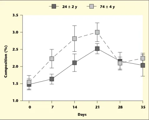
10. FREUND-LEVI Y, ERIKSDOTTER-JONHAGEN M, CEDERHOLM T, et al. Omega-3 fatty acid treat-ment in 174 patients with mild to moderate Alzheimer disease: OmegAD study: a random-ized double-blind trial. Arch Neurol 2006; 63(10): 1402-8. 0 7 14 21 28 35 1.0 1.5 2.0 2.5 3.0 3.5 Composition (%) Days 24 ± 2 y 74 ± 4 y
Figure 1. Changes in the % composition of docosahexaenoic acid (DHA) in plasma total lipids during 21 days
supplementation with 680 mg/d of (DHA) in elderly versus young adults (74 ± 4 versus 24 ± 2 y old, respectively; n = 9-10/group). The supplementation period was followed by a 14 day wash-out. Although starting DHA values were similar and the slopes of the rise and fall of plasma DHA did not differ between the two groups, at d 21, plasma
(DHA) was 42% higher in the elderly (p < 0.05). Compliance was independently verified by adding carbon-13 (13
C)
glucose to the DHA capsules and measuring13C in breath CO
22 h after the capsules were consumed. Participants
were blinded to the presence of the13C-glucose on the capsules and to the reason for breath sampling. This study
suggests that the healthy elderly have a higher short-term plasma response to dietary DHA than do healthy young adults, an effect that is independent of compliance.
11. TERANO T, FUJISHIRO S, BAN T, et al. Docosa-hexaenoic acid supplementation improves the moderately severe dementia from thrombotic cerebrovascular diseases. Lipids 1999; 34(Suppl): S345-S346.
12. SUZUKI H, MORIKAWA Y, TAKAHASHI H. Effect of DHA oil supplementation on intelligence and visual acuity in the elderly. World Rev Nutr
Diet 2001; 88: 68-71.
13. KOTANI S, SAKAGUCHI E, WARASHINA S,
et al. Dietary supplementation of arachidonic
and docosahexaenoic acids improves cognitive dysfunction. Neurosci Res 2006; 56(2): 159-64. 14. BOSTON PF, BENNETT A, HORROBIN DF, BENNETT CN. Ethyl-EPA in Alzheimer’s disease-a pilot study. Prostaglandins Leukot
Essent Fatty Acids 2004; 71(5): 341-6.
15. BOWEN DM, SMITH CB, DAVISON AN. Molecular changes in senile dementia. Brain 1973; 96(4): 849-56.
16. SODERBERG M, EDLUND C, KRISTENSSON K, DALLNER G. Fatty acid composition of brain phospholipids in aging and in Alzheimer’s dis-ease. Lipids 1991; 26(6): 421-5.
17. SKINNER ER, WATT C, BESSON JA, BEST PV. Differences in the fatty acid composition of the grey and white matter of different regions of the brains of patients with Alzheimer’s disease and control subjects. Brain 1993; 116(Pt 3): 717-25.
18. CORRIGAN FM, HORROBIN DF, SKINNER ER, BESSON JA, COOPER MB. Abnormal content of n-6 and n-3 long-chain unsaturated fatty acids in the phosphoglycerides and cholesterol esters of parahippocampal cortex from Alzhe-imer’s disease patients and its relationship to acetyl CoA content. Int J Biochem Cell Biol 1998; 30(2): 197-207.
19. PRASAD MR, LOVELL MA, YATIN M, DHILLON H, MARKESBERY WR. Regional mem-brane phospholipid alterations in Alzheimer’s disease. Neurochem Res 1998; 23(1): 81-8. 20. GUAN Z, WANG Y, CAIRNS NJ, LANTOS PL,
DALLNER G, SINDELAR PJ. Decrease and struc-tural modifications of phosphatidylethanola-mine plasmalogen in the brain with Alzheimer disease. J Neuropathol Exp Neurol 1999; 58(7): 740-7.
21. HAN X, HOLTZMAN DM, MCKEEL JR. DW. Plasmalogen deficiency in early Alzheimer’s dis-ease subjects and in animal models: molecular characterization using electrospray ionization mass spectrometry. J Neurochem 2001; 77(4): 1168-80.
22. FAROOQUI AA, HORROCKS LA, FAROOQUI T. Modulation of inflammation in brain: a matter of fat. J Neurochem 2007; 101(3): 577-99. 23. SVENNERHOLM L, BOSTROM K, JUNGBJER B.
Changes in weight and compositions of major membrane components of human brain dur-ing the span of adult human life of Swedes.
Acta Neuropathol (Berl) 1997; 94(4): 345-52.
24. FREEMANTLE E, VANDAL M, TREMBLAY-MERCIER J, et al. Omega-3 fatty acids, energy substrates, and brain function during aging.
Prostaglandins Leukot Essent Fatty Acids 2006;
75(3): 213-20.
25. PLOURDE M, CUNNANE SC. Extremely limited synthesis of long chain polyunsaturates in adults: Implications for their dietary essentiality and use as supplements. Appl Physiol Nutr
Metab 2007; (in press).
26. ARTERBURN LM, HALL EB, OKEN H. Distribu-tion, interconversion and dose response of n-3 fatty acids in humans. Am J Clin Nutr 83 (suppl): 14675-765
27. ISSA AM, MOJICA WA, MORTON SC, et al. The efficacy of omega-3 fatty acids on cognitive function in aging and dementia: a systematic review. Dement Geriatr Cogn Disord 2006; 21(2): 88-96.
28. MACLEAN CH, ISSA AM, NEWBERRY SJ, et al. Effects of omega-3 fatty acids on cognitive function with aging, dementia, and neurologi-cal diseases. Evid Rep Technol Assess (Summ) 2005(114): 1-3.
29. CORRIGAN FM, VAN RHIJN A, HORROBIN DF. Essential fatty acids in Alzheimer’s disease. Ann
N Y Acad Sci 1991; 640: 250-2.
30. BÉGIN ME, LANGLOIS MF, LORRAIN D, CUNNANE SC. Thyroid function and cognition
during aging. Handbook of Iodine. 2007; (in