O
pen
A
rchive
T
OULOUSE
A
rchive
O
uverte (
OATAO
)
OATAO is an open access repository that collects the work of Toulouse researchers and
makes it freely available over the web where possible.
This is an author-deposited version published in :
http://oatao.univ-toulouse.fr/
Eprints ID : 18169
To link to this article : DOI:10.1002/jor.22889
URL :
http://dx.doi.org/10.1002/jor.22889
To cite this version : Pailhé, Régis and Cavaignac, Étienne and
Murgier, Jérôme and Laffosse, Jean-Michel and Swider, Pascal
Biomechanical study of ACL reconstruction grafts. (2015) Journal of
Orthopaedic Research, vol. 33 (n° 8). pp. 1188-1196. ISSN 0736-0266
Any correspondence concerning this service should be sent to the repository
administrator:
staff-oatao@listes-diff.inp-toulouse.fr
Biomechanical Study of ACL Reconstruction Grafts
R!egis Pailh!e,1Etienne Cavaignac,2J!er^ome Murgier,2Jean-Michel Laffosse,2Pascal Swider21Service de Chirurgie Orthop!edique, H^opital Sud Grenoble, Grenoble Cedex, France,2Service de Chirurgie Orthop!edique et de Traumatologie,
H^opital P.P.R, Toulouse, France
ABSTRACT: There are no published studies describing the strength quadrupled gracilis tendon alone and quadrupled semite-ndinosus tendon alone in the configuration used for anterior cruciate ligament (ACL) reconstruction. The primary objective was to compare the mechanical properties of grafts used for ACL reconstruction during a tensile failure test. The secondary objective was to evaluate the effect of uniform suturing on graft strength. Fifteen pairs of knees were used. The mechanical properties of five types of ACL grafts were evaluated: patellar tendon (PT), sutured patellar tendon (sPT), both hamstring tendons (GST4), quadrupled semitendinosus (ST4), and quadrupled gracilis (G4). Validated methods were used to perform the tensile tests to failure and to record the results. Student’s t-test was used to compare the various samples. The maximum load to failure was 630.8N (! 239.1) for the ST4, 473.5N (! 176.9) for the GST4, 413.3N (! 120.4) for the sPT, and 416.4N (! 187.7) for the G4 construct. Only the ST4 had a significantly higher failure load than the other grafts. The sPT had a higher failure load than the PT. The ST4 construct had the highest maximum load to failure of all the ACL graft types in the testing performed here. Uniform suturing of the grafts improved their ability to withstand tensile loading.
Keywords: biomechanical; graft; ACL; knee
The choice of grafts for anterior cruciate ligament (ACL) reconstruction is not without consequences. One of the advantages of using the hamstring tendons— gracilis (G) and semitendinosus (ST)—is that harvest-ing these tendons leads to lower morbidity than harvesting a bone-patellar tendon-bone graft. Use of the these tendons provides sufficient strength, limits extensor mechanism weakening and lessens anterior knee pain.1–7 However, harvesting the G and ST reduces flexion strength and control over internal rotation.8,9 Recent studies have shown the ST alone can be used as a graft.10–12 However, the ST muscle-tendon unit controls knee rotation in full extension.13 Using the gracilis tendon only should reduce the morbidity induced by harvesting both hamstring ten-dons and preserve the semitendinosus.
Zamarra et al.14 evaluated the potential of using quadrupled gracilis alone or tripled semitendinosus alone to reconstruct the ACL. Authors demonstrated that semitendinosus or gracilis tendon for ACL reconstruction could satisfactorily restore knee kine-matics and the in situ forces to those for the intact ACL. Nonetheless, Abramowitch et al, demonstrated differences between the semitendinosus and gracilis tendons in terms of their quasi-static mechanical and non-linear viscoelastic properties.15 These results were not unexpected given that Kilger et al, in a biomechanical cadaveric study had already showed that gracilis has a higher modulus than the semi tendinosus.16
Among those studies, several others studies have reported on the strength of each individual tendon17–19 but none has determined graft strength in the configu-ration used for ACL reconstruction.There are no
published values for the strength of a quadrupled gracilis (G4) construct or information on the maximum load that a graft in its surgical configuration can withstand before failing.
We wanted to explore the mechanical properties of grafts used for ACL reconstruction. Specifically, we wanted to compare the properties of the tendons currently considered as the gold standard for ACL surgery in athletes with the properties of a quadrupled G4 construct.
The primary objective was to compare the mechani-cal properties of the various grafts used for ACL reconstruction during a tensile failure test. The sec-ondary objective was to evaluate the effect of uniform-ly suturing the patellar tendon graft on its tensile strength. The main outcome measure was the maxi-mum load at failure (N). The secondary outcome measures were stiffness (N.mm"1), and elongation at failure (mm).
MATERIALS AND METHODS
Materials
Thirty cadaver knees from 15 donors were used for the study. The cadavers were stored at 4 ˚C. The donors had a mean age at death of 54 years (range 17–70). The knees had no wounds or macroscopic signs of injury. All knees had joint range of motion measured with a goniometer of at least 0– 130˚. This study was approved by our facility’s institutional review board.
Graft Harvesting
All grafts were harvested at our university’s anatomy laboratory. A midline skin incision was performed. The gracilis and semitendinosus were identified in the lower part of the incision after opening the sartorius aponeurosis. These tendons were detached from their muscle bodies with an open tendon stripper, and then cut from their tibial attach-ment at the periosteum. The patellar tendon was harvested using a standard method20 with tibial and patellar bone blocks.
Graft Preparation
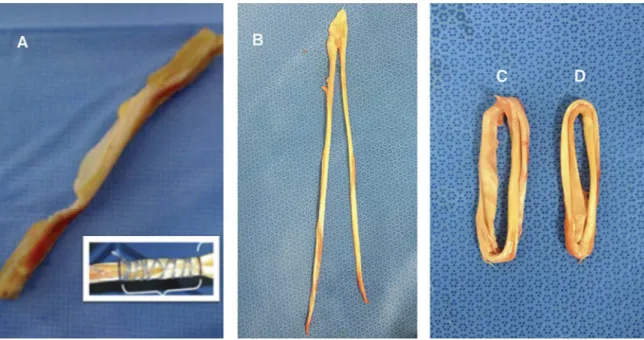
The various grafts were tested under the same conditions used during surgery in terms of graft size and configuration. The surgical techniques were reproduced exactly. The right patellar tendon was left in its native configuration (PT). The left patellar tendon (sPT) was sutured uniformly over its entire length using #2 Vicryl suture (polyglactin 910).
The hamstring tendons from the left knee were used together. After being folded over, they were sutured uniform-ly over their entire length. Vicryl suture was used to bind the four strands together. The gracilis and semitendinosus were harvested separately from the right leg and then prepared separately into quadrupled graft constructs using the TLS1
system (FH Orthopedics, Heimsbrunn, France). These were also sutured uniformly over their entire length. This resulted in four test groups (Fig. 1):
$ Patellar tendons (PT and sPT).
$ Quadrupled hamstring tendon graft (GST4). $ Quadrupled semitendinosus graft (ST4). $ Quadrupled gracilis graft (G4).
The diameter was measured for GST4, ST4, and G4 using a previously described technique21 and the cross-sectional area (CSA) was calculated from the diameter.
Graft Preservation
The prepared grafts were stored at "4 ˚C in a cold freezing solution containing saline and 10% dimethylsulfoxide. They were removed from the freezer the evening before testing and kept at room temperature (21 ˚C) for at least 12h. This processing does not alter the mechanical properties of tendons.20
Methods
Graft Fixation
The graft was gripped at both ends using two serrated jaw clamps22as recommended for tendon allografts23. The clamps were tightened with a torque wrench.
Measurements
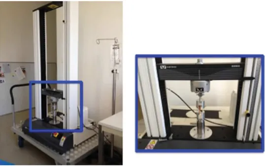
Each set of grips was attached to a materials testing system (Instrom 33001, Instron, Canton, MA) (Fig. 2) to apply tensile loads. Measurements were performed using the system’s software (BlueHill1
, Instrom SA France, Elancourt, France). Each graft was preloaded to 10 N, and then cycled 100 times between 50 and 200 N at 0.5 Hz. A tensile test was then performed using a 10 mm/min crosshead speed until the graft failed. This sequence is a standard, validated test protocol.18 The maximum load at failure (N), elongation at failure (mm) and linear stiffness (N.mm"1) were automatically measured by the software during the failure test. (Supplementary file 1)
Statistical Analysis
The statistical analysis was performed with the Excel 2011 (Microsoft Corp, Redmond, WA) and XLSTAT 2011 (Addinsoft SARL, Paris, France) software packages. The normal distribu-tion of the measured variables was verified using the Shapiro-Wilk test and the homogeneity of variances was verified using Fisher’s f-test and Levene’s test to ensure the conditions had been met for parametric testing. The significance threshold was set at p < 0.05. The descriptive analysis consisted of mean, median and standard deviation values. A comparative analysis was performed using the paired Student’s t-test. Linear regression was performed to assess the correlation between cross sectional area and each biomechanical variable.
RESULTS
Maximum Load at Failure
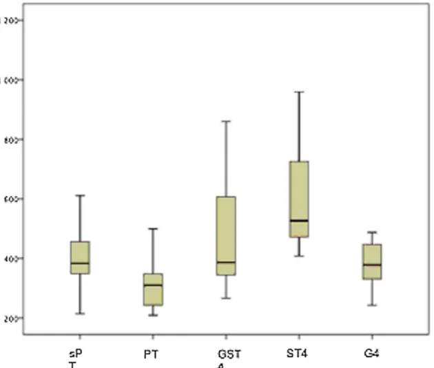
The entire data set is provided in Table 1 and Figure 3. The ST4 construct had the highest mean load at failure, followed by the PT, G4, and GST4. All the PT samples failed at the tendon-bone junction, while the all GST4, ST4, and G4 samples failed midsubstance.
Only the ST4 had a significantly higher failure load than the other grafts (Table 1). The failure loads of the PT, GST4, and G4 were 70%, 62%, and 66% of that of the ST4, respectively. Sutured patellar tendons had a
significantly higher failure load than native PT con-structs. There were no differences between the mean values of the other graft types.
Elongation at Failure
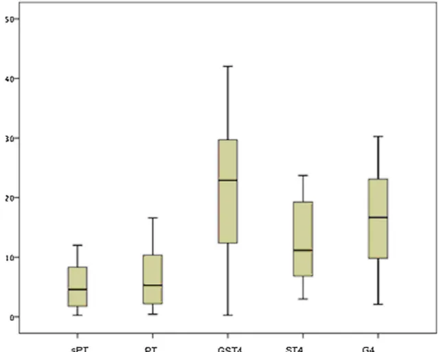
The entire data set is provided in Table 2 and Figure 4. The GST4 construct had the greatest mean elongation at failure, followed by the ST4, G4 and PT.
The mean elongation value of the sutured PT samples was significantly less than the mean value of the GST4, ST4, and G4 groups, but was similar to that of the native PT samples (Table 2). There were no significant differences between the mean values of all the other pairs. The patellar tendon had the smallest elongation at failure of all the tendons evaluated.
Figure 2. Materials testing system (Instron 33001, Instron, Canton, MA) and test configuration used in the current study.
Table 1. Summary of Maximum Load at Failure (N) Measured for All Graft Types and Comparison of Maximum Load at Failure for the Five Types of Fraft
N Minimum Maximum Mean Std. Dev.
sPT 16 214.35 650.35 413.29 120.41 PT 16 208.78 499.24 319.56 92.04 GST4 16 266.14 860.03 473.49 176.90 ST4 16 408.13 1123.44 630.82 239.15 G4 16 242.71 1068.83 416.41 187.68 Paired Differences 95% Confidence Interval
Mean Std. Dev. Std. Error Lower Upper t Sig. (Two-Tail Test) Pair 1 sPT– 93.73 79.09 19.77 51.58 135.88 4.740 .000* Pair 2 sPT– "60.19 232.37 58.09 "184.01 63.62 "1.03 .317 Pair 3 sPT– "217.52 261.06 65.26 "356.63 "78.41 "3.33 .005* Pair 4 sPT– "3.11 245.35 61.33 "133.85 127.62 ".051 .960 Pair 5 GST4– "157.33 341.66 85.41 "339.39 24.73 "1.84 .045* Pair 6 GST4– 57.07 244.96 61.24 "73.45 187.61 .932 .366 Pair 7 ST4–G4 214.41 216.33 54.08 99.13 329.68 3.964 .001*
PT, Patellar tendon; sPT, Sutured patellar tendon; GST4, Doubled gracilis and semitendinosus together; ST4, Quadrupled semitendinosus; G4, Quadrupled gracilis.Paired student’s t-test.Bold value represents values which are statistically significant.*
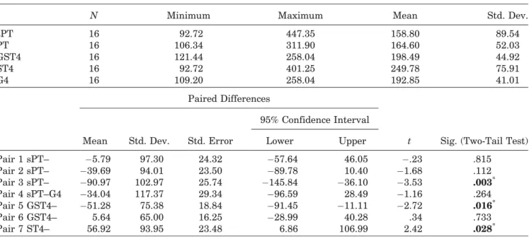
Stiffness
The entire data set is provided in Table 3 and Figure 5. The ST4 construct was the stiffest, followed by the GST4, G4, and PT.
Only the ST4 was significantly stiffer than the other grafts (Table 3). There were no differences between the mean stiffness values of the other graft types.
Cross Sectional Area
The mean CSA was 31.89 þ/" 6.5 mm2for G4, 35.41 þ/ "7.1 mm2 for GST4 and 38.85 þ/" 7.6 mm2 for ST4 (Table 4). The differences between each grafts was statistically significant. Nonetheless there was no correlation between the CSA and maximum load at failure neither elongation at failure or fitness consider-ing all the samples or each graft separately.
DISCUSSION
The aim of this study was to explore the mechanical properties of grafts used for ACL reconstruction. The maximum tensile load that can be withstood by a quadrupled Gracilis construct (G4) was 416 N ! 187
(range: 242–1069 N), which is similar to the reference tendon grafts, namely the patellar tendon and qua-drupled semitendinosus and gracilis (GST4). Only the quadrupled semitendinosus (ST4) had a signifi-cantly higher maximum load at failure than the other tendons. This study also provided validation of uniform suturing significantly increasing graft strength.
Nonetheless these results must be tempered by this study’s limitations. The tensile testing was performed with tissues that had been frozen at "4 ˚C and then thawed. Several studies have explored the effect of freezing and thawing tendons on their mechanical properties.14,24–26 Based on the results of these stud-ies, the mechanical properties of tendons are unaffect-ed when fewer than three, gradual freeze-thaw cycles are performed.
The fact that the age of the donors is higher than that of the patients who typically undergo ligament reconstruction is another study limitation. The effect of age has been evaluated in 82 human patellar tendons from donors between 17 and 54 years of age.27 These tendons were tested at strain rates of either
Figure 3. Box-and-whisker plot of the maximum load at failure for each graft type. PT: Patellar tendon, sPT: Sutured patellar tendon, GST4: Doubled gracilis and semitendinosus together, ST4: Quadrupled semitendinosus, G4: Quadrupled gracilis.
10%/s or 100%/s. Only the specimens tested at 100%/s had a lower modulus of elasticity (25% decrease) in older patients. The other mechanical parameters were unchanged with age.
The fixation method is also another basic consider-ation, as it can affect the results of tensile tests.28 Novel serrated jaw clamps that allow tendons to be tested in a simple and reproducible manner have recently been described by Shi et al.22 Resin-based clamps and cryoclamps are difficult to work with and have not been explicitly validated.29–31 Pap et al.23recently validated a fixation method for auto-grafts that uses the serrated jaw clamps described by Shi et al. by comparing them with other types of clamps. This is the type of clamp used in the current study. It was improved by precisely tightening the grips with a torque wrench.
All the grafts in the current study were preloaded before being pulled to failure. It has been shown that preloading during testing helps to reduce the loss of strength and stiffness that the tendon graft undergoes due to their viscoelastic properties.32 Tendons are temporarily prestrengthened when they undergo pre-conditioning.33 This can be attributed to the progres-sive recruitment of collagen fibers. However this effect was only observed when the preconditioning was less than 1,000 s long. Beyond this period, fatigue micro-fractures can occur and reduce the tendon’s strength. It has been recommended that the preconditioning protocol not exceed 9% elongation, so as to not reduce graft strength.26
In the current study, the quasistatic tensile testing was carried out with a slow crosshead speed so as to not bring the tendon’s viscoelastic properties into play. Tensile strength is reduced when slower elongation speeds are used. When ligaments and tendons are loaded more quickly, there is an increased risk of damaging these structures.34
The maximum load values in the current study were much lower than published values. The leading studies on this topic reported maximum load values of 1719 N ! 1167.80 (range: 456–4546 N), which is nearly three times higher than reported here.17–19It was also surprising to see that this difference did not apply to the stiffness values, which were consistent with our data.
The first reason is related to donor age. The studies reporting the highest failure loads were also those with the youngest donors (20–30 years).17,19,35 The effects of age on tendon strength was described by Noyes et al.36 although the results were not statisti-cally significant. Hashemi et al.37also showed that age reduced tendon strength and presented a linear re-gression model to predict tendon strength using vari-ous parameters, including age.
A second reason revolves around the method used to induce tendon failure. The study with the largest number of ACL grafts and highest published loading values had a significant bias.19 The tensile testing system consisted of pulling on the tendon by dropping a weight from a set height. The maximum load and stiffness were measured using a custom, but
non-Table 2. Summary of Maximum Elongation at Failure (mm) Measured for All Graft Types and Comparison of Maximum Elongation at Failure for the Five Types of Graft
N Minimum Maximum Mean Std. Dev.
sPT 16 .26 12.01 5.13 4.08 PT 16 .42 26.14 7.02 6.81 GST4 16 .26 42.04 21.21 11.55 ST4 16 2.97 23.71 13.06 7.22 G4 16 2.10 45.72 18.03 10.60 Paired Differences 95% Confidence Interval
Mean Std. Dev. Std. Error Lower Upper t Sig. (Two-Tail Test) Pair 1 sPT– "1.89 8.03 2.01 "6.17 2.38 ".944 .360 Pair 2 sPT– "16.08 10.64 2.66 "21.75 "10.41 "6.04 .000* Pair 3 sPT–ST4 "7.93 9.28 2.32 "12.88 "2.99 "3.42 .004* Pair 4 sPT–G4 "12.90 12.60 3.15 "19.62 "6.18 "4.09 .001* Pair 5 GST4– 8.14 14.88 3.72 .21 16.08 2.18 .045* Pair 6 GST4– 3.18 15.87 3.96 "5.27 11.64 .80 .435 Pair 7 ST4– "4.96 12.19 3.04 "11.46 1.53 "1.62 .124
PT, Patellar tendon; sPT, Sutured patellar tendon; GST4, Doubled gracilis and semitendinosus together; ST4, Quadrupled semitendinosus; G4, Quadrupled gracilis.Paired Student’s t-test.Bold value represents values which are statistically significant.* Indi-icates statistically significant difference.
validated accelerometer-based device. These methodo-logical considerations call the validity of their results into question.
The third aspect relates to the elongation speed used in the various studies. Many of these tensile tests used elongation rates greater than 5 mm/s (about 10%/ s). Under these conditions, the tensile tests were not being performed under static conditions, thus bringing the tendon’s viscoelastic properties into play. This could explain the higher maximum load values reported in these studies. We performed an ANOVA test on the published data and found a relationship between the elongation rate and maximum failure load (p ¼ 0.032). However, this potential tendon stiff-ening as the elongation rate increases must still be demonstrated with specific biomechanical studies.
Finally while CSA were significantly different be-tween each graft, we did not observe significant correla-tion between cross seccorrela-tional area and maximum load at failure or stiffness. Recent studies of Abramowitch et al, found differences between the semitendinosus and graci-lis tendons in terms of their quasi-static mechanical and non-linear viscoelastic properties, and the variation of these properties along their length. This suggests that
other morphological and biochemical parameters than CSA have to be investigated.
CONCLUSION
Biomechanical assessment of tendons grafts in the configuration used for anterior cruciate ligament (ACL) reconstruction may have a significant and direct impact on clinical practice. Based on the results of this study, the G4 seems to have bio-mechanical properties that are suited to being used alone during ACL reconstruction. The biomechanical properties of the ST4 are also suitable for use during ACL reconstruction, especially because it had the highest maximum load at failure of all the grafts evaluated. This makes the ST4 our graft of choice for ACL reconstruction. However, the G4 is useful in cases of partial ACL rupture where it can augment the native ligament.30,31,38,39 It could be used in cases of isolated anteromedial bundle tears.32–34,38,40,41 It would be strong enough while also reducing the risk of impingement in the notch because of its smaller volume.12,35 This study’s findings related to implant suturing are consistent with a recent study42 showing that suturing the
Figure 4. Box-and-whisker plot of the maximum elongation at failure for each graft type. PT: Patellar tendon, sPT: Sutured patellar tendon, GST4: Doubled gracilis and semitendinosus together, ST4: Quadrupled semitendinosus, G4: Quadrupled gracilis.
Table 3. Summary of Stiffness (N.mm"1) Measured for All Graft Types and Comparison of Stiffness for the Five Types of Graft
N Minimum Maximum Mean Std. Dev.
sPT 16 92.72 447.35 158.80 89.54 PT 16 106.34 311.90 164.60 52.03 GST4 16 121.44 258.04 198.49 44.92 ST4 16 92.72 401.25 249.78 75.91 G4 16 109.20 258.04 192.85 41.01 Paired Differences 95% Confidence Interval
Mean Std. Dev. Std. Error Lower Upper t Sig. (Two-Tail Test) Pair 1 sPT– "5.79 97.30 24.32 "57.64 46.05 ".23 .815 Pair 2 sPT– "39.69 94.01 23.50 "89.78 10.40 "1.68 .112 Pair 3 sPT– "90.97 102.97 25.74 "145.84 "36.10 "3.53 .003* Pair 4 sPT–G4 "34.04 117.37 29.34 "96.59 28.49 "1.16 .264 Pair 5 GST4– "51.28 75.38 18.84 "91.45 "11.11 "2.72 .016* Pair 6 GST4– 5.64 65.00 16.25 "28.99 40.28 .34 .733 Pair 7 ST4– 56.92 93.95 23.48 6.86 106.99 2.42 .028*
PT, Patellar tendon; sPT, Sutured patellar tendon; GST4, Doubled gracilis and semitendinosus together; ST4, Quadrupled semitendinosus; G4, Quadrupled gracilis.Paired Student’s t-test.Bold value represents values which are statistically significant.
Figure 5. Box-and-whisker plot of the stiffness for each graft type. PT: Patellar tendon, sPT: Sutured patellar tendon, GST4: Doubled gracilis and semitendinosus together, ST4: Quadrupled semitendinosus, G4: Quadrupled gracilis.
graft construct increases its failure load. If these results are confirmed by in vivo studies, surgeons could prepare their grafts in a similar manner. However, it would have to be shown that suturing does not negatively affect the ligamentization pro-cess before this method is adopted clinically.
AUTHORS’ CONTRIBUTIONS
RP did acquisition, and analysis of data. JML did approval of the submitted and final versions. JM did substantial contributions to research design PS drafted the paper and revised it critically. EC did acquisition, and analysis of data. All authors have read and approved the final submitted manuscript.
ACKNOWLEDGMENTS
The authors wish to thank Joanne Archambault, PhD for her editorial support during preparation of this manuscript. Each author certifies that he has no commercial associations (e.g., consultancies, stock ownership, equity interest, patent/ licensing arrangements, etc.) that might pose a conflict of interest in connection with the submitted article.
REFERENCES
1. Bonamo JJ,Krinick RM, Sporn AA. 1984. Rupture of the patellar ligament after use of its central third for anterior cruciate reconstruction. A report of two cases. J Bone Joint Surg Am 66:1294–1297.
2. Maletis GB, Cameron SL, Tengan JJ, et al. 2007. A prospective randomized study of anterior cruciate ligament reconstruction: a comparison of patellar tendon and quadru-ple-strand semitendinosus/gracilis tendons fixed with bioab-sorbable interference screws. Am J Sports Med 35:384–394. 3. Kartus J, Stener S, Lindahl S, et al. 1997. Factors affecting
donor-site morbidity after anterior cruciate ligament
recon-struction using bone-patellar tendon-bone autografts. Knee Surg Sports Traumatol Arthrosc 5:222–228.
4. Christen B, Jakob RP. 1992. Fractures associated with patellar ligament grafts in cruciate ligament surgery. J Bone Joint Surg Br 74:617–619.
5. Risberg MA, Holm I, Steen H, et al. 1999. Sensitivity to changes over time for the IKDC form, the Lysholm score, and the Cincinnati knee score. A prospective study of 120 ACL reconstructed patients with a 2-year follow-up. Knee Surg Sports Traumatol Arthrosc 7:152–159.
6. Sajovic M, Vengust V, Komadina R, et al. 2006. A prospec-tive, randomized comparison of semitendinosus and gracilis tendon versus patellar tendon autografts for anterior cruci-ate ligament reconstruction: five-year follow-up. Am J Sports Med 34:1933–1940.
7. Sachs RA, Daniel DM, Stone ML, et al. 1989. Patellofemoral problems after anterior cruciate ligament reconstruction. Am J Sports Med 17:760–765.
8. Nakamura N, Horibe S, Sasaki S, et al. 2002. Evaluation of active knee flexion and hamstring strength after anterior cruciate ligament reconstruction using hamstring tendons. Arthroscopy 18:598–602.
9. Ohkoshi Y, Inoue C, Yamane S, et al. 1998. Changes in muscle strength properties caused by harvesting of autog-enous semitendinosus tendon for reconstruction of contra-lateral anterior cruciate ligament. Arthroscopy 14: 580–584.
10. Tohyama H, Yasuda K. 1998. Significance of graft tension in anterior cruciate ligament reconstruction. Basic background and clinical outcome. Knee Surg Sports Traumatol Arthrosc 6 Suppl 1 S30–S37.
11. Yasuda K, Tsujino J, Ohkoshi Y, et al. 1995. Graft site morbidity with autogenous semitendinosus and gracilis tendons. Am J Sports Med 23:706–714.
12. Pujol N, Queinnec S, Boisrenoult P, et al. 2013. Anatomy of the anterior cruciate ligament related to hamstring tendon grafts. A cadaveric study. Knee 20:511–514.
13. Collette M, Cassard X. 2011. The Tape Locking Screw technique (TLS): a new ACL reconstruction method using a
Table 4. Diameter and Cross Sectionnal Area
G4 ST4 GST4
D CSA D CSA D CSA
Specimen 1 5.4 31.38 6.1 29.2 5.9 27.33 Specimen 2 6.3 31.17 6.6 34.21 6.5 33.18 Specimen 3 5.8 26.42 6.4 32.16 6.2 30.19 Specimen 4 7.1 39.59 7.9 49.02 7.4 43 Specimen 5 7.5 44.18 8.1 51.53 7.8 47.78 Specimen 6 7.4 43 8.3 54.11 7.9 49.01 Specimen 7 5.4 22.9 6.5 33.18 6.1 29.22 Specimen 8 6.6 34.21 7.5 44.18 7.1 39.59 Specimen 9 6.2 30.19 7.0 38.48 6.6 34.21 Specimen 10 6.9 37.39 7.5 44.18 7.3 41.85 Specimen 11 6.8 36.32 7.4 43 7.2 40.72 Specimen 12 6.2 30.19 6.5 33.18 6.5 33.18 Specimen 13 5.4 22.9 6.9 37.39 5.8 26.42 Specimen 14 6.1 29.22 6.6 34.21 6.5 33.18 Specimen 15 6.1 29.22 6.7 35.26 6.4 32.17 Specimen 16 5.3 22.06 6.0 28.27 5.7 25.52 Mean 6.28 31.9 7.0 38.85 6.68 35.41 Standard Deviation 0.70 6.6 0.68 7.68 0.67 7.15
short hamstring graft. Orthop Traumatol Surg Res 97: 555–559.
14. Zamarra G, Fisher MB, Woo SL-Y, et al. 2010. Biomechani-cal evaluation of using one hamstrings tendon for ACL reconstruction: a human cadaveric study. Knee Surg Sports Traumatol Arthrosc 18:11–19.
15. Abramowitch SD, Zhang X, Curran M, et al. 2010. A comparison of the quasi-static mechanical and non-linear viscoelastic properties of the human semitendinosus and gracilis tendons. Clin Biomech (Bristol, Avon) 25: 325–331.
16. Kilger R, Curran M, Savio L-Y. 2005. A comparison of the biomechanical properties of the semitendinosus, gracilis tendons, and ACL bundles: A consideration for ACL recon-struction. Woo Transactions ORS, 30: Washington, D.C. 17. Noyes FR, Butler DL, Grood ES, et al. 1984. Biomechanical
analysis of human ligament grafts used in knee-ligament repairs and reconstructions. J Bone Joint Surg Am 66:344–352.
18. Yanke AB, Bell R, Lee AS, et al. 2013. Central-third bone-patellar tendon-bone allografts demonstrate superior biomechanical failure characteristics compared with hemi-patellar tendon grafts. Am J Sports Med 41:2521–2526.
19. Handl M, Dr#z!k M, Cerulli G, et al. 2007. Reconstruction of the anterior cruciate ligament: dynamic strain evaluation of the graft. Knee Surg Sports Traumatol Arthrosc 15: 233–241.
20. Vincent J-P, Magnussen RA, Gezmez F, et al. 2012. The antero-lateral ligament of the human knee: an anatomic and histologic study. Knee Surg Sports Traumatol Arthrosc 20:147–152. 21. Cavaignac E, Pailh!e R, Murgier J, et al. 2014. Can the
gracilis be used to replace the anterior cruciate ligament in the knee? A cadaver study.The Knee 21:1014–1017.
22. Shi D, Wang D, Wang C, et al. 2012. A novel, inexpensive and easy to use tendon clamp for in vitro biomechanical testing. Med Eng Phy 34:516–520.
23. Pap K, Hangody G, Szebenyi G, et al. 2014. An easy way to fix tendon allografts into a loading-machine for biomechani-cal testing. ESSKA Congress
24. Woo SL, Orlando CA, Camp JF, et al. 1986. Effects of postmortem storage by freezing on ligament tensile behav-ior. J Biomech 19:399–404.
25. Moon DK, Woo SL-Y, Takakura Y, et al. 2006. The effects of refreezing on the viscoelastic and tensile properties of ligaments. J Biomech 39:1153–1157.
26. Liao H, Belkoff SM. 1999. A failure model for ligaments. J Biomech 32:183–188.
27. Blevins FT, Hecker AT, Bigler GT, et al. 1994. The Effects of Donor Age and Strain Rate on the Biomechanical Properties of Bone-Patellar Tendon-Bone Allografts. Am J Sports Med 22:328–333.
28. Butler DL, Grood ES, Noyes FR, et al. 1984. Effects of structure and strain measurement technique on the material properties of young human tendons and fascia. J Biomech 17:579–596.
29. Cheung JT-M, Zhang M. 2006. A serrated jaw clamp for tendon gripping. Med Eng Phy 28:379–382.
30. Kiss M-O, Hagemeister N, Levasseur A, et al. 2009. A low-cost thermoelectrically cooled tissue clamp for in vitro cyclic loading and load-to-failure testing of muscles and tendons. Medical Engineering and Physics 31: 1182–1186.
31. Riemersa DJ, Schamhardt HC. 1982. The cryo-jaw, a clamp designed for in vitro rheology studies of horse digital flexor tendons. J Biomech 15:619–620.
32. Elias JJ, Kilambi S, Ciccone WJ. 2009. Tension level during preconditioning influences hamstring tendon graft proper-ties. Am J Sports Med 37:334–338.
33. Teramoto A, Luo Z-P. 2008. Temporary tendon strengthen-ing by preconditionstrengthen-ing. JCLB 23:619–622.
34. van Dommelen JAW, Jolandan MM, Ivarsson BJ, et al. 2006. Nonlinear viscoelastic behavior of human knee liga-ments subjected to complex loading histories. Ann Biomed Eng 34:1008–1018.
35. Chandrashekar N, Mansouri H, Slauterbeck J, et al. 2006. Sex-based differences in the tensile properties of the human anterior cruciate ligament. J Biomech 39: 2943–2950.
36. Noyes FR, DeLucas JL, Torvik PJ. 1974. Biomechanics of anterior cruciate ligament failure: an analysis of strain-rate sensitivity and mechanisms of failure in primates. J Bone Joint Surg Am 56:236–253.
37. Hashemi J, Mansouri H, Chandrashekar N, et al. 2011. Age, sex, body anthropometry, and ACL size predict the structur-al properties of the human anterior cruciate ligament. Orthop Res 29:993–1001.
38. Buda R, Ruffilli A, Parma A, et al. 2013. Partial ACL tears: anatomic reconstruction versus nonanatomic augmentation surgery. Orthopedics 36:e1108–e1113.
39. Abat F, Gelber PE, Erquicia JI, et al. 2013. Promising short-term results following selective bundle reconstruction in partial anterior cruciate ligament tears. Knee 20: 332–338.
40. Sonnery-Cottet B, Panisset JC, Colombet P, et al. 2012. Partial ACL reconstruction with preservation of the postero-lateral bundle. Orthop Traumatol Surg Res 98:(8 Suppl) S165–S170.
41. Buda R, Di Caprio F, Giuriati L, et al. 2008. Partial ACL tears augmented with distally inserted hamstring tendons and over-the-top fixation: an MRI evaluation. Knee 15: 111–116.
42. H€oher J, Offerhaus C, Steenlage E, et al. 2013. Impact of tendon suturing on the interference fixation strength of quadrupled hamstring tendon grafts. Arch Orthop Trauma Surg 133:1309–1314.