C
ONTROL OF
I
NFLAMMATION IN
E
XPERIMENTAL
B
OVINE
P
NEUMONIC
P
ASTEURELLOSIS
F.Bureau1, M.L. Van de Weerdt1, E. Hanon2 , P-P Pastoret2, P.Lekeux1 1 Laboratory for Functional Investigation,
2 Laboratory of Virology, Faculty of Veterinary Medicine, University of Liege, Liege, Belgium
ABSTRACT
An experimental model of bovine pneumonic pasteurellosis was used to compare the effects on lung mechanical, gas-exchange, biochemical and cellular parameters before and after a nonsteroidal and a steroidal antiinflammatory drug. Eight Friesian calves were inoculated with Pasteurella haemolytica. After randomisation, 4 calves were treated with a nonsteroidal drug (ketoprofen) and 4 calves with a short-acting dexamethasone solution, all in combination with antibiotics for three days. The calves were inoculated a second time, three days after the last treatment. Flow cytometry was used to differentiate blood mononuclear cells and to measure oxidative burst capacity of antiinflammatory cells in venous blood and in brochoalveolar lavage samples before and after inoculation and treatment. After the first inoculation, all calves had increased respiratory rates, rectal temperatures, increased neutrophil blood count, and a decline of the arterial pressure in oxygen and lung compliance. After treatment, rectal temperature dropped significantly (P<0.05) and a higher arterial pressure in oxygen was found in both groups. However, there was only in the ketoprofen group a significant reduction of the tachypnea. After the second inoculation, a significant drop of the arterial pressure in oxygen and a significant higher respiratory rate was seen only for the dexamethasone group, while a significant neutrophilia was found only in the ketoprofen group. No important treatment differences of oxidative burst activity were evident using the flow cytometry technique due to the large within treatment group vari- ability. In this model oflung inflammation, there was a significant higher impairment of the lung mechanics and the arterial pressure in oxygen after the second Pasteurella challenge for the dexamethasone group compared to the ketoprofen treated calves.
Introduction
In young feeder calves, lung pathology represents 45 to 80% of all diseases and is an important cause of economical loss in the beef industry 1,8,34. The ultimate cause of death is generally
thought to be due to a secondary bacterial infection, of which the major contribu- tors are Pa steurell a haemolytica or Pa steurella multocida 1,30,36. Therefore, antimicrobial therapy is
considered as the basis of the control of most bovine pneumonias. However, in acute cases, the inflammatory reaction in the lungs can be so severe that it interacts negatively with the gas exchange processes and therefore with the survival rate. Indeed, the pulmonary dysfunctions and lesions are generated not only by the microorganisms but also by the released endogenous pro-inflammatory mediators 16,33,35. In these conditions, the modulation of lung inflammation
also plays an important role in an effective control of bovine pneumonia.
For a practitioner, the only available drugs for modulation of inflammation are non specific mediators antagonists, i.e. steroidal (SAID) and non-steroidal antiinflammatory drugs (NSAID). SAID's are known to be powerful antiinflammatory drugs. However their potential side effects on the immune system make their use inappropriate in micro-organism-induced pneumonias, mainly because of a higher risk of recurrence 6,27. On the other hand, NSAID's have been re-
ported to have a good efficacy/toxicity ratio in the treatment of bovine pneumonias 31.
It was intended to investigate the clinical, lung mechanical, blood biochemical parameters and broncho-alveolar cell types in an experimental pasteurella model, after randomising animals to an antibiotic plus ketoprofen treatment or an antibiotic plus dexamethasone treatment. A Pasteurella reinoculation was performed six days after the first one in order to test the potential delayed effects of the treatments. Furthermore, it was the intention to measure the effect of the inoculation and the treatments on the oxidative burst activity of phagocytes and on the relative distribution of peripheral blood mononuclear cells (PBMC), all by means of flow cytometry.
Materials and Methods
ANIMALS
The data were collected from 8 male Friesian calves, weighing 42 to 50 kg, starting the protocol at age 4 to 5 weeks. All calves came from a calf-farm where they were housed for the previous three weeks with about 40 calves of their age group. To enter the trial they were not allowed to have received any antibiotic treatment while staying in their feedlot. Before starting the experiment, the calves were declared free from cardiorespiratory or infectious perturbations with the use of clinical and lung function tests. The calves were cared for in accordance with the Belgian law regarding the protection of experimental animals.3 Two days after arrival of the animals at the University, the experi- ment was started. The protocol was approved by the Ethical Committee, Faculty of Veterinary Medicine, University of Liege.
PASTEURELLA STRAIN
A strain of Pasteurella haemolytica biotype A, serotype 1, was obtained from lung tissue of a 3 week old calf with pneumonia. It was rehydrated from freeze- dried ampoules and incubated at 37°C in a Brain Heart Infusion solution (BHI) for 24 hours. One drop of the suspension was incubated on a blood-agar plate for another 24 hours. Ten colonies were resuspended in a 100 ml BHI solution which was incubated and agitated for five hours at 37°C. Precisely after five hours this BHI suspension was used as the inoculum. A growth curve was obtained by following the BHI solution spectrophotometrically at 600 nm during the five hours. This confirmed previous experience where the Pasteurella population was at the top of its logarithmic growth at 5 hours. A pasteurella population in logarithmic growth is necessary to have maximal cytotoxin production .4 Quantitative plate count at 5 hours growth revealed a concentration of 6.2.108 colony-forming units per ml.
PROTOCOL
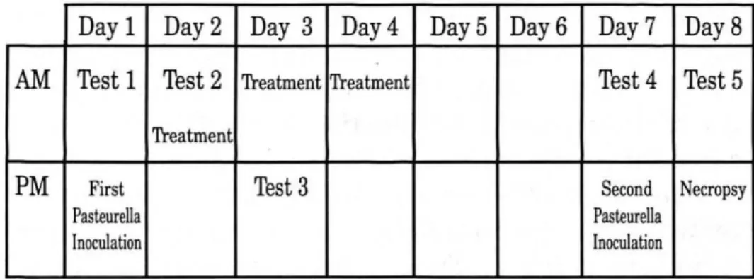
The protocol's schedule for inoculation and testing is given in Fig 1. At Day 1, a control measurement (Test 1) was performed before the first inoculation. The next morning, the inoculation effect was examined in Test 2, which was followed by the first treatment of colistin (52000 IU/kg I.M.) (Colivet S forte, Prodivet, Belgium) as an antibiotic, selected on basis of the result of an antibiogram, and ketoprofen (Group A) (3 mg/kg I.M.) (Ketoprofen 10%, Rhône Merieux, Brussels, Belgium) or a dexamethasone solution (Group B) at a label dose of 1.58.10-2 mg/kg (I.M.) (Voren solution, Boehringer Ingelheim, Brussels, Belgium) after randomising. The morning of day 3 the calves received a second treatment followed by Test 3 to evaluate the treatment effect. One day later a last treatment was given. In total the calves were treated for three days with a single daily injection of the selected drugs. Six days after the first inoculation Test 4 was performed and followed by a second inoculation. The next day, after Test 5, the calves were euthanized using a curare-like agent and an overdose of barbiturates (T61, Hoechst, Brussels, Belgium), and a necropsy was performed.
Figure 1. Experimental design: each of the eight calves, randomly allocated to two groups of 4 calves, was inoculated twice with Pasteurella haemolytica with an interval of 5 days. All animals were treated for three days after the first inoculation with an antibiotic in combination with a non-steroidal (Group A) or a steroidal anti-inflammatory treatment (Group B). Five "tests" were performed, before and after inoculation and treatment, during which clinical, lung mechanical, gas-exchange and biochemical parameters were recorded. A broncho-alveolar lavage was performed at each test to measure the oxidative burst capacity of phagocytes . The experiment was concluded with a necropsy.
2 2
INOCULATION
Both right and left lung were inoculated on day 1 and day 7 (Fig 1) by depositing 4 ml of the inoculum in the main right and left bronchus each and 1 ml in the bronchus of the cranial lobe of the right lung through a polyethylene catheter that was placed via the biopsy port of an endoscope (Pentax PMEII/PUE GI video endoscope).
TESTS
At each of the five tests, indicated on Fig 1, the following parameters were measured :
Clinical parameters: Rectal temperature, respiratory and heart rates (RR, HR), abnormal sounds on pulmonary auscultation, depression or loss of appetite, presence of tachypnea and dyspnea and the presence of cough and/or nasal discharge were recorded.
Arterial blood gas levels: Arterial blood was collected anaerobically through puncturing of the axillary artery with a 21 G heparinized needle after shaving the jugularis region down to the pectoralis region. Arterial pressure in oxygen (PaO2) and the arterial pressure in carbon dioxide
(PaCO2) together with the pH and the base excess were measured with correction to body
temperature 14 (blood gas analyser, model 995 Hb, AVL, Leuven, Belgium).
Respiratory mechanical parameters: Respiratory mechanical parameters were recorded as previously described. 15 Briefly, the respiratory airflow (V) was measured by use of a heated
pneumotachograph Fleish mounted on a mask and coupled to a differential pressure transducer with identical catheters. The V signal was integrated with respect to time to give tidal volume (Vt). Pleural pressure (Ppl) was estimated via a balloon tipped catheter located in the middle third of the oesophagus. Transpulmonary pressure (Ptp) was electrically obtained by subtracting oesophageal pressure from airway opening pressure and was used for the subsequent calculations. All signals, Ptm, Vt and V were fed into a computer (Hemodynamic Respiratory System, ACEC, Belgium), which derived mean pulmonary function values from measurements on 5 regular, successive, and artefact free respiratory cycles; RR and Vt were measured, whereas dynamic lung compliance (CLdyn) and total pulmonary resistance (RL) were calculated by use of the method described by Rodarte and Rehder.26
Blood sample: A venous blood sample was collected on EDTA tubes for a basic haematological profile, yielding counts of red blood cells, hematocrit, haemoglobin, white blood cells, platelets, lymphocytes, monocytes, neutrophils , basophils and the total protein level.
Flow cytometry was used to obtain precise information on the proportion of macrophages, B lymphocytes, T lymphocytes (CD4,CD8, -), and on their morphologic features (size, granularity). To differentiate the PBMC subpopulations, we used the anti-CD2 (CC42), anti-CD4 (CC8), anti-CDS (CC63) and the anti-CD- (CC51) mononuclear antibodies which were kindly provided by W.I. Morisson (Compton, UK). We also used the anti-macrophage (IL-A24), kindly provided by N. MacHugh (Ilrad, Nairobi, Kenya) and the anti-B (1H4), kindly provided by J.-J. Letesson (FNDP, Namur, Belgium). Blood was collected by jugular venipuncture and mixed with heparin. The PBMCs were then isolated on Ficoll-Hypaque (Pharmacia) (density = 1.077 g/ml) density gradient and washed three times with PBS. About 106 cells were incubated for 30 min at
MAb. The cells were then washed with PBS containing 5% FCS and further incubated in PBS containing 5% FCS and FITC-conjugated secondary antiserum at 37° C for 30 min. After an additional wash with PBS containing 5% FCS, the cells were resuspended in PBS and analysed by flow cytometry.
Flow cytometry was also used to determine the oxidative burst activity (OB) of phagocytes in a venous blood sample. The intracellular oxidation of non fluorescent DHR (Dihydrorhodamine 123) to green rhodamine 123 by hydrogen peroxide and peroxidases is the most sensitive method for the analysis of the OB response by flow cytometry.28 DHR was purchased from
Molecular Probes (Eugene, OR, USA). Neutrophil and macrophage cell groups can be differentiated by the large difference in relative OB activity.28 Approximately 106 cells in RPMI
1640 were incubated with 1µM of DHR. After 5 min at 37°C, cells were incubated with 10-7 M of PMA (Phorbol 12-Myristate 13-Acetate) at 37°C during 30 min. Following this, 2.5 µg of Prodium Iodide (Pl) was added to each tube immediately before flow cytometry analysis. Controls included the omission of PMA or the preincubation of the cells with diphenyliodonium (a specific inhibitor of OB).
Bronchoalveolar lavage (BAL): A polyethylene catheter (external diameter 4 mm, internal diameter 1.8 mm) was introduced via a nostril into the trachea and pushed forward till it was wedged in a bronchus. Next, 50 ml of a warm phosphate buffered saline solution (PBS) (25°C) was injected, immediately followed by a negative pressure on the same syringe to recover as much of the injected fluid as possible. The lavage solution was first filtered with a cell strainer, after which 50 ml PBS was added. The solution was then centrifugated at 250 g for 10 min at 5°C. The cells were resuspended in 1 ml of water for 5 sec and immediately transfused in 50 ml of PBS. A second time the remaining white blood cells were centrifuged at 250 g for 10 min after resuspension in 2 ml RPMI. The slides were examined microscopically after staining with Giemsa. Oxidative burst acivity was measured following the same procedures as described above for the venous blood sample.
Analysis by flow cytometry: Flow cytometric analysis was performed using a Becton-Dickinson fluorescence-activated cell sorter (Facstar Plus), equipped with an argon laser (ILT air cooled with 100 mW excitation lines at 488 nm). Dead cells and debris were excluded from the analysis by the use of PI and the conventional scatter gating method. Fluorescein and PI emission signals were collected by using appropriate filters at 530 nm (band pass 30) and 575 nm (band pass 26), respectively. Five thousand events per sample were collected in list mode, stored, and analysed by the Consort 32 system (Becton-Dickinson).
Necropsy
Lung tissue was sampled for bacteriologic analysis, to confirm the presence of Pasteurella haemolytica, and for histopathology. Sections of lung tissue were fixed in 10% phosphate-buffered formalin, embedded in paraffin, stained with hematoxylin and eosin and examined by light microscopy.
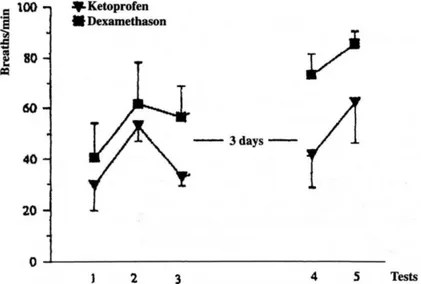
Figure 2. Mean respiratory rate with standard error. A significant improvment for Group AS was found after treatment (p = 0.0051). After the second inoculation, the mean respiratory rate was significantly lower in Group A (62 breaths/min) compared to Group B (85.3 breaths/ min) (p = 0.0485).
Figure 3. Mean arterial oxygen level with standard error. There was only for Group B a significant decrease of the arterial oxygen level found after the second inoculation (p = 0.0078).
Statistical Analysis
Descriptive analysis were performed by graphically presenting for every calf each of the recorded parameters from Test 1 to Test 5. Plots were conducted with the treatment groups mean value at each test for every parameter, accompanied by a 95% confidence interval of the mean. For statistical analysis, unpaired, paired T-tests, and analysis of variance were used at an a-level of 0.05.
Results
One day after the first inoculation, each calf had a marked decline in CLdyn, in addition to an
elevated RR (Fig 2). A marked decline of the PaO2 (Fig 3) and an elevated temperature was also
found in both groups. The blood profile showed a rise in the total white blood cell count and a rise in the absolute count of neutrophils. A significant decrease in mean cell size (p = 0.05) (see * Table 2) was seen after the first inoculation in neutrophils and macrophages in the BAL.
At Test 3, twenty-four hours after the first treatment and two hours after the second treatment were instituted, the body temperature dropped significantly in both groups (Group A: p=0.012, Group B : p=0.013). However it was only in Group A that the tachypnea improved significantly (p=0.005) (Fig 2) next to a significant improvement of the expired minute volume (p=0.003). In both groups there was a trend for PaO2 to rise (Fig 3). At Test 4, a significant difference between
the two treatment groups was detected in the neutrophil blood level (p=0.01), with a mean level of 1.9.109 cells/L in Group A and a mean of 6.4.109 cells/L in Group B (Table 1).
At the second inoculation, RR rose significantly higher in Group B (mean of 85.3 breaths/min) than in Group A (mean of 62 breaths/min) (p=0.049) (Fig 2). A significant drop of the PaO2 was
found only in Group B (p=0.008) (Fig 3). For the neutrophil blood count, only Group A showed a significant increase (p=0.014) (Table 1). This phenomenon was also found for the total lymphocyte blood count, where again only Group A showed a significant rise after the reinoculation (p=0.033) (Table 1).
The lungs were removed at necropsy and a map was made from the macroscopic lesions. Lesions were mostly located in the cranial and middle lobes of the right lung and were either extensive or multifocal. The general pattern was a "marbled" lung, with clear delineation between normal and affected areas. On sectioning, lesions were firm and oedematous with many haemorrhages . In 3 calves multifocal abscesses (diameter 5 mm) were found in the cranial right lobules, one calf in Group A and two calves in Group B. Translucent exudate outlined individual lung lobules. The index score of lung tissue damages was two-times greater in Group B (10.6 ± 6.7 (SD)) than in Group A (5.8 ± 3.5 (SD)), but the difference was not significant at a 95% confidence level. Microscopic investigation was done on lung tissue samples from macroscopically normal and abnormal parts.
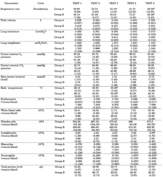
Table 1. Respiratory and hematologic parameters recorded from the experimental calves, means and standard errors ( ).
Group A: Ketoprofen group, Group B: Dexamethasone group.
a: a significant improvement of the respiratory rate (p=0.005) and the expired minute volume (p=0.003) was found
for group A after treatment. b: The temperature dropped significantly after treatment in Group A (p=0.012) and in
Group B (p=0.013). c: A significant difference between the two treatment groups was detected for the neutrophil
blood level (p=0.01) before the second inoculation. d: The respiratory rate was significantly higher in Group B
compared to Group A after the second inoculation. e: A significant drop of the Pa02 was found after the second
inoculation only in Group B (p=0.008). f: Only for Group A, a significant increase in lymphocyte count (p=0.033) and
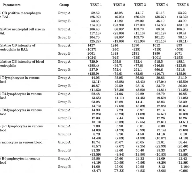
Table 2. Relative oxidative burst measurements of blood - and BAL leukocytes, together with the relative distribution of PBMC subpopulations from test 1to test 5 for the ketoprofen group (Group A) and the dexamethasone group (Group B), means and standard deviations. ( ).
There was a significant decrease (P=0.05) of the neutrophil cell size in both groups after the first inoculation (). Only
in the dexamethasone group, a significant decrease of the % of B-lymphocytes occurred at Test 5 compared to the baseline test (Test 1) ().
For each calf, two different patterns were found. Several lobules showed enlarged septa, con- gestion with a presence of many macrophages but still with intact alveoli pointing to a subacute histiocytic pneumonitis. In other lobules, on the other hand, an acute haemorrhagic bronchoalveolar pneumonia was found with alveoli filled with pus and haemorrhage. The bronchial and bronchiolar epithelia and submucosa were infiltrated with neutrophils.
Cell types recovered by BAL were macrophages, degenerated cells, epithelial cells, neutrophils and lymphocytes. A mean of 17 ml lavage fluid of the 50 ml volume injected was recovered. The quantitative assess- ment of cell types by Giemsa staining was not possible as bovine lymphocytes and macrophages could not be differentiated reliably by light microscopic observation.13·27 However, at Test 1, before the first inoculation, macrophages were large and
readily distinguishable from lymphocytes. At inoculation 1the large majority of cells in the lavage were macrophages (estimated semi- quantitatively from 76% to 95%). At Test 2, after the first inoculation, the neutrophils became the predominant cell type in 6 of 8 calves. At Test 3, 4 and 5 cells became less differentiable and estimation turned out to be unrepeatable by different individuals on the same smear.
The measurements of relative OB activity by flow cytometry did not show any particular pattern, indicating no inoculation effect or treatment effect. A high variability was detected within each treatment group for the relative OB intensity of neutrophils and macrophages in the BAL (Table 2), and for the neutrophils and monocytes in the venous blood samples (Table 1). The mean relative OB intensity for neutrophils and for macrophages was significantly higher (p = 0.05) in BAL than for neutrophils and monocytes in a venous blood sample. Mean relative fluorescence intensity for neutrophils in venous blood was 541.9 ± 57.0 (SE) and in lavage 1924.0 ± 207.9 (SE). In monocytes, the relative OB intensity was 16.3 ± 1.6 (SE) in venous blood and 48.1± 5.9 (SE) for macrophages in lavage.
No significant differences were detected between the two treatment groups and no overall pattern could be established for the percentile-measurements in venous blood, of monocytes, CD2 T lymphocytes , CD4 T lymphocytes, CDS T lymphocytes and - T lymphocytes (Table 2). For these cell groups, there was a high variability measured within each treatment group. The percentage (%) of B lymphocytes however, showed a significant drop in Group B at test 5 (7%), compared to the baseline test (test 1: 14%) (see Table 2). The mean % of B lymphocytes did not change significantly during the experiment for Group A.
The other parameters measured are given in Table 1. No statistical differences were found between the two groups from these parameters mainly because of the high within group variability.
Discussion
Intrabronchial inoculation with Pasteurella haemolytica consistently resulted in severe acute pneumonia as evidenced by the alterations in pulmonary function, haematological values and histopathological findings. Interestingly, while the intention was to infect the complete lung by controlling the bronchial inoculation endoscopically, at necropsy, lesions were found predominantly in the cranial and middle lobes of the right lung. This coincides with pathological findings in clinical cases of lung pasteurellosis. 25 Until present no clear explanation has been
given for this anatomical distribution of pathogenic lung lesions in calves. The results show that a mechanical explanation through easy access does not play a major role because in this experiment each part of the lung was equally infected. Also experiments investigating cellular and biochemical differences in BAL in healthy and pneumonic calves between cranial and caudal lobes could not establish any differences. 13,21,24 Other mechanisms need to be further
investigated to explain this anatomical predisposition.
Initially, in the first three hours after inoculation, there is sequestration of blood neutrophils into the lungs.32 One day after the inoculation a significant neutrophilia was found, indicating a
The reduced CLdyn is considered as an important feature of endotoxaemia 11,19 and bacteremia. 5
The histopathological findings suggest that changes in CLdyn could have been the result of
altered pulmonary hemodynamics, interstitial and alveolar oedema and finally cellular flux into the air spaces. No changes in lung resistance were found confirming necropsy findings as no major abnormalities were seen in large airways.
The hypoxemia after the inoculation was not accompanied by changes in PaCO2. Therefore
alveolar hypoventilation was excluded as being the cause of the hypoxemia. The latter was consequently due to ventilation-perfusion (V/Q) inequality, diffusion impairment, or a combination of these causes.37 Previous investigations involving bovine endotoxaemia
demonstrated that pulmonary oedema may lead to diffusion impairment and to consecutive hypoxemia .23 The drop of PaO2 may be considered, together with the rise in body temperature,
as the important stimulus for the substantial increase in respiratory rate.
All these effects, i.e. high rectal temperature, low CLdyn, hypoxemia and tachypnea are resulting
from the endotoxemic effects of Pasteurella haemolytica growth by its liberation of cytotoxins. 17
This may underline the use of an antiinflammatory treatment next to an antibiotic treatment to effectively counteract the cytotoxin induced dysfunctions.
Three days after the last treatment there was a significantly higher blood neutrophil level in the dexamethasone group while after the second inoculation only in the ketoprofen group a significant neutrophilia and rise in the blood lymphocyte level was found. Pruett et al 22 also
noticed an increase in peripheral blood neutrophils after an injection with dexamethasone explained by a reduced margination of neutrophils. 27 Even if cattle are considered a
corticosteroid resistant species based on corticosteroid induced lymphopenia, 27 apparently,
their was still, three days after the last dexamethasone treatment, an effect on the leukocyte population as seen by the small increase in the peripheral neutrophils and lymphocytes after the second inoculation compared to the significant rise of these cell groups in the ketoprofen treated group. The significance of these differences in leukocyte reaction, resulting in a higher risk of set-backs in dexamethasone treated calves needs further investigation.
Mammalian phagocytes inactivate and destroy in- vading microorganisms and other foreign cells with the formation of oxygen-derived toxic products. This mechanism is called the oxidative burst (OB). The influences on the different white blood cell types after a dexamethasone injection in cattle has been well documented. 10,18,27 The ability for
glucocorticoids to inhibit the recruitment ofleukocytes and monocytes-macrophages into affected areas is considered to be a very important factor in their anti-inflammatory actions.2,20
In this experiment, there exists a high individual variability in the formation of oxygen radicals, by BAL cells and white blood cells, following a certain stimulus. Dyer et al 9 described a
dose-dependent increase of the superoxide anion production in pulmonary alveolar macrophages after challenging with P haemolytica. No influence of the inoculation or the treatment on the OB metabolism could be detected due to this variability when using a small sample size: This could be partly explained by differences in immunity status for P haemolytica between the calves as Williams et al 39 found an OB response of peritoneal macrophages depending on the immunity
status for Ehrlichia risticii in mouse. The higher mean fluorescence intensity for the relative OB of BAL cells than of white blood cells reflects a higher capability of oxygen radicals formation in the lungs for neutrophils and macrophages. This difference existed already before inoculation
which means that these cells are in a pre-activated state in the lungs concerning their OB metabolism.
Lymphopenia after glucocorticoid administration has been reported 29,38 but glucocorticoids did
not cause a selective depletion of B lymphocytes in white blood cells.38 In this experiment only
the % of B lymphocytes of white blood cells dropped significantly in Group B and not in Group A. At the same time no significant changes were detected for the % of T lymphocytes. This is in contrast with the literature, where the effects on T lymphocytes, as a suppressed blastogenesis and a selective depletion in man, following a corticosteroid administration, are well documented .7,12 However, no changes of the % of B lymphocytes in white blood cells or even an enhanced B
lymphocyte function was reported. 7,12 A reduced antibody production after corticosteroid
administration is also explained as an effect of T lymphocyte suppression and not by an influence on the B lymphocyte population. 22
The reduced cell size of neutrophils and macrophages after the first inoculation is probably due to the appearance of younger cells in the blood stream after challenge, which have not yet phagocytosed vacuoles and are consequently smaller.
In conclusion, the significant differences between the two treatment groups occurred predominantly at the second inoculation, instored 6 days after the first inoculation and 3 days after the last treatment. This second challenge showed a significantly higher impairment of the lung mechanics and the arterial pressure in oxygen for the dexamethasone treated group compared to the ketoprofen treated calves.
Acknowledgments
The authors wish to thank Dr L. Devrieze for providing the Pasteurella strain, Dr. J. Pringle and Prof C. Burvenich for their advice and Dr. C. Husson, M. Leblond, I. Sbaï, M. Motkin and A. Trolin for their technical assistance.
References
1. Adlam C., Rutter J.M. Pasteurella en Pasteurellosis, Academic Press Limited, London, 1989, 201.
2. Balow J.E., Rosenthal A.S. Glucocorticoid suppression of macrophage migration inhibitory factor. J. Exp. Med. 1973, 137, 1031-1039.
3. Belgian Ministry of Agriculture Arrêté Royal relatif à la protection des animaux d'expérience. Moniteur Belge, 5 janvier 1994, pp 111-156.
4. Berggren K.A., Baluyut C.S., Simonson R.R., Hemrick W.J., Maheswaran S.K. Cytotoxic ef- fects of Pasteurella haemolytica on bovine neutrophils. Am. J. Vet. Res. 1981,42, 1383-1388.
5. Byrne K., Cooper K.R., Carey P.D., Berlin A., Sielaff T.D., Blocher C.R., Jenkins J.K., Fisher B.J., Hirsch J.I., Tatum J.L., Fowler A.A., Sugerman H.J. Pulmonary compliance : early assessment of evolving lung injury after onset of sepsis. J.Appl. Physiol. 1990, 69, 2290-2295.
6. Christie B.M., Pierson R.E., Braddy P.M., Flalk D.E., Horton D.P., Jensen R., Lee E.A., Remenga E.E., Rutt K.G. Efficacy of corticosteroids as supportive therapy for bronchial pneumonia in yearling feedlot cattle. Bovine Pract. 1977, 12, 115-117.
7. Claman H.N. Corticosteroids and lymphoid cells. N. Engl. J. Med. 1972, 287, 388-397.
8. Drummond R.O., Lambert G., Smalley H.E., Terril C.E. CRC Handbook of pest management in agriculture, CRC Press, Roca Raton, vol 1, 1981, 111.
9. Dyer R.M., Benson C.E., Boy M.G. Production of superoxide anion by bovine pulmonary macrophages challenged with soluble and particulate stimuli. Am. J. Vet. Res. 1985, 46 (2), 336-341.
10. Eckblad W., Stiller D., Woodard L., Kuttler K. Immune responses of calves antigenically stimulated and challenge exposed with anaplasma marginale during tick infestation or treatment with dexamethasone. Am. J. Vet. Res. 1984, 42, 1192-1197.
11. Esbenshade A.M., Newman J.H., Lams P.M. Respiratory failure after endotoxin infusion in sheep : lung mechanics and lung fluid balance. J. Appl. Physiol. 1982, 53, 967-976.
12. Fauci A.S. Immunosuppressive and anti-inflammatory effects of glu- cocorticoids. In: Glucocorticoid Hormone Action, Baxter J.D., G.G. Rousseau (Eds), Springer-Verlag, Berlin, 1979, pp 449-465.
13. Heilmann P., Muller G., Finsterbusch L. Lobäre Depostion radioaktiv markierter Pasteurella-multocida -Aerosole in den Lungen von Ferkeln und Kiilbern. Arch. Exper. Vet. Med. 1988, 42, 490-501.
14. Kelman J. R., Nunn J.F. Nomograms for correction of blood P02, PC02 and base excess for time and temperature. J. Appl. Physiol. 1966, 21, 1484-1490.
15. Lekeux P., Hajer R., Breukink H.J. Pulmonary function testing in calves: Technical data. Am. J. Vet. Res. 1984, 45, 342-345.
16. Lekeux P., Genicot B., Gustin P. Treatment of pulmonary dysfunctions. In: Pulmonary Function in Healthy, Exercising and Diseased Animals, Lekeux P. (Ed.), V.D.T. Publications, Gand, 1993, pp 351-384. 17. Linden A., Desmecht D., Amory H., Daube G., Lecomte S., Lekeux P. Pulmonary ventilation, mechanics, gas exchange and haemodynamics in calves following intratracheal inoculation of Pas teurella haemolytica. J. Vet.Med .A 1995, 42, 531-544.
18. Muscoplat C.C., Shope R.E., Chen A.W., Johnson D.W. Effects of corticosteroids on reponses of bovine peripheral blood lymphocytes cultured with phytohaemagglutinin. Am. J. Vet.Res. 1975, 36, 1243-1244 19. Olson N.C., Brown T.T. Effects of endotoxemia on lung water and hemodynamics in conscious calves. Am. J. Vet. Res. 1985, 46, 711-718.
20. Parillo J.E., Fauci A.S. Mechanisms of glucocorticoid action on immune processes. Annu. Rev. Pharmacol. Toxicol. 1979, 19, 179-201.
21. Pringle J.K., Viel L., Shewen P.E., Willoughby R.A., Martin S.W., Valli V.E.O. Bronchoalveolar lavage of cranial and caudal lung regions in selected normal calves: cellular, microbiological , immunoglobulin, serological and histological variables. Can. J. Vet. Res. 1988, 52, 239-248.
22. Pruett J.H., Fisher W.F., Deloach J.R. Effects of dexamethasone on selected parameters of the bovine immune system. Vet. Res. Commun. 1987, 11, 305-323.
23. Reeves J.T., Daoud F.S., Estridge M. Pulmonary hypertension caused by minute amounts of endotoxin in calves. J. Appl. Physiol. 1972, 33, 739-743.
24. Reinhod P., Muller G., Kreutzer A., Gerischer A., Putsche R. Diagnostische Aussagefahigkeit gesunder und pneumonie kranker Kiilber. J. Vet. Med. A 1992, 39, 404-418.
25. Rehmtulla A.J., Thomson R.G. A review of the lesions in Shipping Fever of cattle. Can. Vet. J. 1981, 22, 1-8.
26. Rodarte J.R. Rehder K. Dynamics of respiration. In: Handbook of Physiology, Section 3, The Respiratory System, vol 3, Mechanics of Breathing, Part 1, Fishman A.P., Fisher A.B. (Eds), American Physiological Society, Bethesda, 1986, pp 131-144.
27. Roth J. A., Kaeberle M.L. Effect of glucocorticoids on the bovine immune system. J. Am. Vet. Med .Assoc. 1982, 180 (8), 894-901.
28. Rothe G., Emmendorffer A., Oser A., Roesler J., Valet G. Flow cytometric measurement of the respiratory burst activity of phagocytes using dihydrorhodamine 123. J. Immunol. Methods 1991, 138, 133-135.
29. Schalm O.W., Lasmanis J., Carroll E.J. The use of a synthetic corticoid on experimental coliform (Aerobacter aerogenes) mastitis in cattle: The response of leukocytes and the effect of hormone-induced neutrophilia. Am. J. Vet. Res. 1965, 26, 851-857.
30. Schimmel D., Fodor L., Stein I., Putsche R. Ergebnisse zur Typisierung und Virulenzprufung von Pasteurella-haemolytica-Feldstammen. Arch. Exper. Vet. Med. 1990, 2, 295-300.
31. Selman I.E., Allan E.M., Dalgleish R.G., Gibbs H.A., Shoo M.K. Proceedings of the International Symposium on Nonsteroidal Antiinflammatory Agents, Orlando, 1986, 23.
32. Slocombe R.F., Derksen F.J., Robinson N.E. Interactions of cold stress and Pasteurella haemolytica in the pathogenesis of pneumonic pasteurellosis in calves: changes in pulmonary function . Am. J. Vet. Res. 1984a, 45, 1757-1770.
33. Slocombe R.F., Malark J., Ingersoll R., Derksen F.J., Robinson N .E. Importance of neutrophils in the pathogenesis of acute pneumonic pasteurellosis in calves. Am. J. Vet. Res. 1985, 46, 2253-2258.
34. USDA Agriculture Statistics Board, NASS, May 1992.
35. Vestweber J.G., Klemm R.D., Leipold H.W., Johnson D.E., Bailia W.E. Clinical and pathologic studies of experimentally induced pasteurella haemolytica pneumonia in calves. Am. J. Vet. Res. 1990, 51, 1792-1798.
36. Visser I.J.R., Zalsman C., Wouda W. Pasteurella haemolytica serotypen bij runderen. Tijdschr. Diergeneeskd. 1992, 117, 35-37.
37. West J.B. Vascular diseases. In: Pulmonary Pathophysiology - The Essentials, 3rd edn, West J.B. (Ed.), Williams & Wilkins Company, Baltimore, 1987, pp 112-133.
38. Wilkie B.N., Caoili F., Jacobs R. Bovine lymphocytes: erythrocyte rosettes in normal, lymphomatous and corticosteroid treated cattle. Can. J. Comp. Med. 1979, 43, 22-28.
39. Williams N.M., Cross R.J. Respiratory burst activity associated with phagocytosis of Ehrlichia risticii by mouse peritoneal macrophages. Res. Vet. Sci. 1994, 57, 194-199.