HAL Id: dumas-03077739
https://dumas.ccsd.cnrs.fr/dumas-03077739
Submitted on 16 Dec 2020
HAL is a multi-disciplinary open access
archive for the deposit and dissemination of sci-entific research documents, whether they are pub-lished or not. The documents may come from teaching and research institutions in France or abroad, or from public or private research centers.
L’archive ouverte pluridisciplinaire HAL, est destinée au dépôt et à la diffusion de documents scientifiques de niveau recherche, publiés ou non, émanant des établissements d’enseignement et de recherche français ou étrangers, des laboratoires publics ou privés.
Brain and heart microvascular disease evaluated with
cerebral MRI and myocardial perfusion entropy in
SPECT, a neurovascular stroke patients’ evaluation
Victor Mathieu
To cite this version:
Victor Mathieu. Brain and heart microvascular disease evaluated with cerebral MRI and myocar-dial perfusion entropy in SPECT, a neurovascular stroke patients’ evaluation. Human health and pathology. 2020. �dumas-03077739�
AVERTISSEMENT
Ce document est le fruit d'un long travail approuvé par le
jury de soutenance.
La propriété intellectuelle du document reste entièrement
celle du ou des auteurs. Les utilisateurs doivent respecter le
droit d’auteur selon la législation en vigueur, et sont soumis
aux règles habituelles du bon usage, comme pour les
publications sur papier : respect des travaux originaux,
citation, interdiction du pillage intellectuel, etc.
Il est mis à disposition de toute personne intéressée par
l’intermédiaire de
l’archive ouverte DUMAS
(Dépôt
Universitaire de Mémoires Après Soutenance).
Si vous désirez contacter son ou ses auteurs, nous vous
invitons à consulter la page de DUMAS présentant le
document. Si l’auteur l’a autorisé, son adresse mail
apparaîtra lorsque vous cliquerez sur le bouton « Détails »
(à droite du nom).
Dans le cas contraire, vous pouvez consulter en ligne les
annuaires de l’ordre des médecins, des pharmaciens et des
sages-femmes.
Contact à la Bibliothèque universitaire de Médecine
Pharmacie de Grenoble :
1
UNIVERSITÉ GRENOBLE ALPES UFR DE MÉDECINE DE GRENOBLE
Année : 2020
BRAIN AND HEART MICROVASCULAR DISEASE EVALUATED WITH CEREBRAL MRI AND MYOCARDIAL PERFUSION ENTROPY IN SCINTIGRAPHY:
A NEUROVASCULAR STROKE PATIENTS’ EVALUATION
THÈSE
PRÉSENTÉE POUR L’OBTENTION DU TITRE DE DOCTEUR EN MÉDECINE DIPLÔME D’ÉTAT
Par : Victor MATHIEU
THÈSE SOUTENUE PUBLIQUEMENT À LA FACULTÉ DE MÉDECINE DE GRENOBLE
Le : 01/12/2020
DEVANT LE JURY COMPOSÉ DE
Président du jury :
M. Pr VANZETTO Gérald Membres :
M. Pr BARONE ROCHETTE Gilles (directeur de thèse) M. Pr DETANTE Olivier
M. Dr MAURIN Marion M. Dr DJAILEB Loïc
L’UFR de Médecine de Grenoble n’entend donner aucune approbation ni improbation aux opinions émises dans les thèses ; ces opinions sont considérées comme propres à leurs auteurs.
6
Remerciements
A mon jury de thèse :
A mon président de thèse, Professeur Gérald VANZETTO,
Merci pour votre accompagnement tout au long de l’internat, et tout ce que vous m’avez appris notamment lors de mon semestre en post USIC, votre sens clinique, la décortication des ECG et votre relationnel avec les patients ont été un exemple pour moi, merci de me faire l’honneur de présider cette thèse.
A mon directeur de thèse, Professeur Gilles BARONE-ROCHETTE,
Merci pour ton implication à l’élaboration de cette thèse, de m’avoir remis dans le bon chemin quand je m’écartais, le tout à distance. Ça a été un plaisir de travailler avec toi, sur cette thèse et dans la pratique Clinique, j’espère que ce travail répondra à tes attentes.
Au Professeur Olivier DETANTE,
Merci de me faire l’honneur d’être dans mon jury de thèse, ça a été très agréable de travailler avec vous, merci pour vos réponses à mes sollicitations, j’espère que ce travail vous intéressera.
Au Docteur Marion MAURIN,
Merci d’avoir accepté de faire partie de mon jury de thèse, merci pour les nombreuses fois où je t’ai sollicité pour avoir un œil d’échographiste aguerri pour les échographies du bip, j’espère que ce travail sur la filière neuro-cardio t’intéressera.
Au Docteur Loïc DJAÏLEB,
Merci pour (une fois de plus) accepter de travailler sur une thèse de cardio, merci de me faire l’honneur d’être dans mon jury de thèse, j’ai apprécié de travailler avec toi, merci pour ton accueil chaleureux et naturel, merci pour ton implication dans ce travail.
7
Aux médecins qui m’ont formé,
A Olivier et Florian, auprès de qui j’ai fait mes premiers pas d’interne, à Charlotte, ça a été un plaisir de faire 6 mois au 8ème C avec toi.
A Stéphanie pour ta bonne humeur pendant les 6 mois à l’USIC, toujours avec une tasse de café au lait sur l’ordinateur ;)
A Estelle, merci pour ton dynamisme entrainant, et ta bonne humeur.
A Pierre-Vladimir, merci pour tout ce que vous m’avez appris en échographie et à toujours guetter les effets indésirables des médicaments, les conversations improbables avec les patients et les leçons sur le fructose vont me manquer.
A Muriel, tu m’as transmis la passion pour l’insuffisance cardiaque chronique, grâce à toi je ne mettrai plus de beta bloquant dans l’amylose !
A Marianne, Elizabeth, Cécile, « Sansan », Véro et toute l’équipe kiné pour ce semestre en rééducation cardiaque, merci tout particulier à Sandrine pour ta bonne humeur et ta folie qui font beaucoup de bien aux patients et à tes collègues. Merci à Noé et Yoan pour le coaching quand je m’incrustais dans le club cœur et santé !
A Hélène, votre expérience et votre calme en toutes circonstances m’ont impressionné, et sont une source d’inspiration.
A toute l’équipe de « mon USIC », Mireille, Manue, Greg, Dimitry (petits anges partis trop tôt, de l’USIC ;)) Elisa, mme Sorel, mme Da Rocha et tous les autres qui ont fait de ce semestre avec mon Guigui un stage de CCU qui restera gravé dans ma mémoire.
Aux équipes soignantes du 8ème B et 8ème C, merci pour votre accueil et la bonne humeur dans le service, on y est si bien qu’on y reste volontiers tard le soir quand on débute l’internat !
A tous les médecins et les équipes d’Annecy pour m’avoir accueilli et formé en fin d’internat, je suis ravi de signer pour deux ans avec vous !
A l’équipe de réanimation d’Annecy pour ce semestre inter COVID dans une ambiance géniale, je terminerai en disant que le patient va bien puisque les valves vont bien, le rythme est sinusal.
A Marie Blanche pour ta gentillesse et ton humanité, toujours à penser au bien-être des patients, c’est une chance d’avoir travaillé avec toi.
A Nico pour ta patience quand tu m’apprenais la pose de voies centrales, au début laborieuses, maintenant je suis un pro !
A toute l’équipe de rythmologie, à Adrien et ton grand sourire colgate, et le nombre incalculable de sollicitations rythmologiques auxquelles tu réponds toujours présent.
8
A Carole, merci pour ce que tu m’as appris en échocardiographie, merci pour tous les surnoms que tu as pu me trouver au cours de l’internat !
A Aude et Caroline, pour votre travail formidable, votre bonne humeur, votre disponibilité et votre pédagogie envers les plus jeunes.
A Maryline, Hubert et toute la télécardio, à toutes les infirmières de consult, ça a été un plaisir de travailler avec vous.
A toute l’équipe des ARCs, et les moments de suspens pour savoir si oui ou non on peut inclure le patient dans EVAOLD, merci à Clémence pour ta participation à la thèse.
Aux secrétaires, Solenne, et Marion merci de m’avoir aidé pour mon recrutement de patients et merci pour votre sympathie !
A notre promo de cardio, Elodie ton nettoyage un peu maniaque de verres et couverts à l’internant va me manquer, Sara, toujours prête pour aller boire un coup le soir pour décompresser, occupes-toi bien de mon USIC, Arnaud et Lucie et les bons moments après le DU à Lyon. A Marjo toujours motivée pour se faire des bons petits restos Grenoblois. A tous les internes de cardio, Guigui, Antoine, Robin, Estelle, Charles Eric, Laura, Lou et les internes devenus « grands », Léa, Anne, Laurianne, Aure Elyse toujours prête à aider un jeune interne qui débute avec le bip, Antoine, Thomas, grâce à chacun de vous qui mettent une bonne ambiance à l’internat de cardio.
A Amandine, Sarah, Apopo, Thomas, Flo, Camille pour notre semestre à Annecy et nos cafés sur notre terrasse privée.
A mes amis, ma famille,
A Ben, Nono, Rikou aux longs doigts, Mathieu (c’est COMMENNNT ?), Océ, Tat, Emy, Thomas, Andrée, bref toute la mif, que nos soirées à Brizon reviennent vite, Baptiste nous attend. Merci pour avoir toujours été là depuis tant d’années. Alex et Andrée on attend l’invitation à Châteauneuf !
A Got et Beutbeut, la fine équipe, et nos virées du vendredi soir (quand on était encore jeunes !), à tous les bons moments à bricoler, les déménagements, le ping, Dax, Bayonne et j’en passe.
A mon Beubeu, mon fidèle de toujours, merci pour les 400 coups qu’on a pu faire, une cohabitation fructueuse à la coloc de Vallières avec nos messages codés, à Nelly, malgré ton grand âge, tu as amené un vent d’air frais, un petit coup de jeune il y a déjà quelques années ! Merci de m’avoir fait une petite place dans la famille, fier d’être témoin de mon Beubeu et le parrain de la petite Loulou, longue vie à nous tous !
Au professeur bricole et son élève, à Lisou, Nabil et Cléo, on veut voir votre maison ! Nelly, Villecroze 2021 ?
9
A Pauline et Carole, bande de jeunes fougueuses ! A nos bons repas et nos dancing parties !
A toute la famille Andrén, pour les bons moments en famille qui rechargent les batteries à chaque séjour en Suède.
A toute la famille Mathieu, avec une pensée pour François, Gérard, et Henri. Au plaisir des futures cousinades, et de vous recevoir Marie Hélène et Chantal, avec des apéricubes en apéritif et des fromages de chèvres stéphanois sélectionnés spécialement par Marie Hélène.
A Luss, tu partages ma vie depuis le début de ces longues études et tu me rends heureux tous les jours, tu t’es si bien occupée de moi pendant la confection de cette thèse que je vais presque regretter de ne plus avoir de mémoire, thèse ou autre à écrire. Que la suite soit aussi bien que ces dix premières années, dans une grande ferme en pierre à la campagne !
A Maman, qui a toujours été là pour me soutenir, toujours attentionnée et à m’aider quand j’en avais besoin, on a de la chance de t’avoir comme mère, tu es une Maman merveilleuse !
A Mimmi, mon f***ing bro, ma vieille branche ! J’ai de la chance d’être ton frère, merci pour tous ces moments cosy au coin du feu, à tous les moments qu’on a vécu en Suède, les escapades fougueuses parisiennes, vivement la prochaine avec Caro et Edouard !
A Titti, notre petite arrivée « à la dernière minute » qui illumine la maison par sa créativité et sa touche artistique, un plaisir de t’aider pour les DM de maths ou physique le dimanche soir à 22h.
A Ben Laden, oui tu peux aller te mouiller les bras, et non tu ne peux pas reprendre une canette.
A Papa, je suis fier d’être ton fils, j’aurai tellement voulu que tu sois là pour ce moment où je deviens médecin, tu me manque et j’espère que de là-haut toi aussi tu es fier de moi.
10
Summary
Remerciements ... 6 Summary ... 10 Abbreviations ... 11 Abstract ... 13 Résumé ... 14 Introduction... 16 Methods ... 20 Study population ... 20 Stroke check up ... 20 Primary endpoint... 21 Secondary endpoint ... 21Assessment of myocardial microvascular statement ... 23
SPECT imaging protocol and analysis ... 23
Assessment of Myocardial Perfusion Entropy (MPE) ... 24
Cerebral small vessels disease assessment ... 26
Statistical analysis ... 29
Results ... 30
Patient’s characteristics ... 31
SPECT analysis ... 32
Neurological assessment and cerebral images analysis ... 33
Primary endpoint... 34
Coronary disease screening ... 34
Discussion ... 35
The systemic microcirculatory disease hypothesis ... 35
Primary endpoint... 38
Literature review ... 38
Coronary artery disease screening in patients with atherothrombotic stroke. ... 41
Limits ... 44
Conclusion ... 46
Bibliography ... 48
11
Abbreviations
ARWMC: Age Related White Matter Changes
CABG: Coronary Artery Bypass Graft
CAD: Coronary Artery Disease
CFR: Coronary Flow Reserve
CHUGA: Centre Hospitalier Universitaire Grenoble Alpes
CMD: Coronary Microvascular Disease
CT: Computerized Tomography
IMR: Index of Myocardial Resistance
INOCA: Ischemia with Non-Obstructive Coronary Artery
LAD: Left Anterior Descending (artery)
LV: Left Ventricle
LVEDV: Left Ventricular End Diastolic Volume
LVEF: Left Ventricle Ejection Fraction
LVESV: Left Ventricular End Systolic Volume
MACE: Major Adverse Cardiovascular Event
MPE: Myocardial Perfusion Entropy
MRI: Magnetic Resonance Imaging
12
RCA: Right Coronary Artery
SPECT: Single Photon Emission Computed Tomography
TOE: Trans Oesophageal Echocardiography
13
Abstract
Introduction- Microvascular disease has been described in many organs, and is associated with poor outcome. The hypothesis of a diffuse organ damage is not answered, in particular the
correlation of cerebral and myocardial microvascular dysfunction. This study is searching for a
correlation between cerebral small vessels disease and coronary microvascular disease.
Methods- We included 38 patients who had a stroke from atherothrombotic origin hospitalized in the CHUGA stroke unit who were addressed to cardiology day hospital for a check-up
including myocardial SPECT. All patients had a neurologic microvascular assessment using
the Age-Related White Matter Changes scale on MRI or CT scan. SPECT images were analysed
with a software attributing an entropy score, corresponding to the heterogeneity of pixels
intensity of the myocardium. Coronary artery disease screening using SPECT was evaluated
after a stroke in selected patients.
Results- We included 38 patients (mean age 64.2 years old +/- 10.9, 81% of men) who had a cerebral and myocardial microcirculation assessment after a stroke. No correlation has been
found between ARWMC score and myocardial perfusion entropy, Spearman correlation -0,10
p = 0,51.
Among all patients, 27.1% had abnormal SPECT and 20 % had a coronary angiogram, 9.1%
had at least one severe coronary stenosis and 7.3% were revascularized, 80% of patients who
underwent coronary angiogram had a treatment adaptation.
Conclusion- No association was found between cerebral microvascular disease and myocardial perfusion heterogeneity, using myocardial perfusion entropy. Coronary artery disease screening
in selected patients who had a stroke is efficient, leading to coronary revascularisation and
14
Résumé
Introduction- L’atteinte microvasculaire est décrite dans de nombreux organes et est associée à un pronostic défavorable. Une atteinte microcirculatoire systémique est suspectée, plusieurs
organes pouvant être atteints simultanément. L’association entre une atteinte microvasculaire cérébrale et myocardique n’a pas encore été prouvée, cette étude recherche une corrélation entre
l’atteinte microcirculatoire de ces deux organes.
Méthodes- Nous avons inclus 38 patients qui ont été hospitalisés dans l’unité neurovasculaire du CHUGA, pour un AVC ischémique d’origine athérothrombotique présumée. Ces patients
ont eu une évaluation de l’atteinte cérébrale microcirculatoire en utilisant le score ARWMC sur un scanner ou une IRM cérébrale. L’étude de l’atteinte myocardique a été réalisée en analysant l’hétérogénéité de perfusion myocardique en scintigraphie myocardique, appelée entropie de perfusion myocardique. Une corrélation entre ces deux scores a été recherchée, et la
performance du dépistage de la coronaropathie a été évaluée chez cette population de patient
ayant présenté un AVC ischémique athérothrombotique.
Résultats- Nous n’avons pas retrouvé de corrélation entre le score ARWMC et l’entropie de perfusion myocardique, corrélation de Spearman -0,10, p=0,51. Parmi nos patients, 27.3%
avaient une scintigraphie pathologique, 20 % des patients ont bénéficié d’une coronarographie,
9.1% avaient au moins une sténose significative et 7.3 % ont été revascularisé. Parmi les
patients ayant eu une évaluation coronaire invasive, 80% ont bénéficié d’une adaptation thérapeutique personnalisée à l’issue de celle-ci.
Conclusion- Avec notre méthode d’évaluation, nous n’avons pas trouvé de corrélation entre les troubles de la microcirculation myocardique et cérébrale. Le dépistage de la coronaropathie
par scintigraphie chez des patients à haut risque cardiovasculaire ayant présenté un AVC
15
aboutissant à une revascularisation par angioplastie ou chirurgicale. Il permet aussi une
adaptation thérapeutique personnalisée chez des patients présentant un athérome coronaire et
16
Introduction
Cardiovascular disease is the first cause of mortality worldwide responsible of 15.2 million of
death (1) whether 31% of total mortality. It is the first cause of mortality in Europe too (2).
Macrovascular disease is now well known, whether it be cerebral, myocardial or peripheral
arterial disease, sharing the same risk factors, each one having a different impact on the location
of the organ damage. Major cardiovascular risk factors include the age depending on age, high
blood pressure, diabetes mellitus, dyslipidaemia, smoking, family history of premature
cardiovascular disease, and to a lesser extent, obesity, physical inactivity and metabolic
syndrome (3–6).
Each risk factor has a different impact for developing either cerebrovascular, myocardial or
peripheral artery damage (7,8). So, pathophysiological mechanisms are common, but also
specific to develop one or another disease. The common mechanism of cardiovascular disease
is the development of atherosclerosis. Macrovascular disease is well studied, but with the
development of imagery, biology including molecular biology, nuclear medicine, some disease
which were idiopathic, have now found a common explanation: the microvascular dysfunction.
Microvascular dysfunction is described in several organs including brain and heart.
The cerebral microvascular disease is highlighted by CT scan or MRI with parenchymatous
damages of white matter, and deep grey nuclei. The lesions seen in sectional imaging is called
leukoariosis, which means capillary rarefaction (9). Theses lesions correspond to a pathological
point of view, to a loss of smooth muscle cells from the tunica media, deposits of fibrohyaline
material, narrowing the lumen, and thickening of the vessel wall (10). These findings are
secondary to chronic ischaemic lesions caused by small vessels disease. This ischaemia is
extended to oligodendrocytes leading to their apoptosis, and the hypothesis that blood brain
17
pathologies like vascular dementia, Alzheimer disease, CADASIL (Cerebral autosomal
dominant arteriopathy with subcortical ischaemic strokes and leukoencephalopathy) which is a
model of microvascular cerebral disease (12). White matter intensities, are proved to be
associated with ageing, diabetes and hypertension (13). The presence of cerebral small vessels
disease is associated with a more than two-times increased risk of dementia at the age of 75.
Leukoariosis is also associated with mood (14), cognitive (15,16), gait (17) and urinary
dysfunction, affecting the quality of life of the patients, and having an impact on their autonomy,
indirectly having a cost impact on the society (18). A correlation has been found between the
progression of leukoariosis and the decline in cognitive performance (19), thus, discovering
white matter intensities should have an incidence on the patient’s care, and it could be a marker
of progression of dementia when it is diagnosed and associated with leukoariosis.
Coronary microvascular disease has been described in many cardiopathies, resulting in a four
grades classification made by Camici et al. (20) CMD in the absence of myocardial diseases
and obstructive coronary artery disease, CMD in myocardial diseases without obstructive CAD,
CMD in obstructive CAD, and finally iatrogenic CMD. CMD is an entity discovered in the last
three decades, but is still a current issue, the first precise definition has been made recently by
the COVADIS (Coronary Vasomotion Disorders International Study Group) in 2017 (21). With
the development of functional pharmacological tests during a coronary angiography, we found
out that this category of patients had a lower response to vasodilator molecules indicating a
microcirculation dysfunction with a loss of flow auto regulation (22,23). Coronary
microvascular disease is an underestimated disease, partly due to a lack of systematic
procedures in patients addressed to the catheterization laboratory for a suspected cardiac
ischemia, without finding any coronary stenosis. In trials studying patients with myocardial
ischemia without significant coronary stenosis, more than 60 % of patients had abnormal
18
impairment proved with Coronary Flow Reserve and/or Index of Microvascular Resistance
(24). In another study, authors found out that 51% of men and 54% of women with suspected
coronary artery disease had a coronary microvascular dysfunction, associated or not with
coronary stenosis (25). CMD is associated with a poor prognosis, with cardiovascular outcomes
including Myocardial Infarction, stroke, hospitalizations and death (26,27).
In 2000 Al Suwaidi & al showed that patients with severe endothelial dysfunction had 14% of
major cardiac events after 28 months follow up against 0% in the mild and normal endothelial
function (27). A meta-analysis published in 2017 by Brainin et al, including 26 prospective
cohort studies, with a large panel of cardiopathies (angina, heart failure, hypertrophic
cardiomyopathy, aortic stenosis, left atrial enlargement) proved that in all these groups, CMD
was associated with significant more MACE than patients with normal microcirculation (28).
Another study found out that patients with CMD had 30 % risk of developing CAD or an acute
coronary event at 10 years. In 2018, a study proved that patients with persistent angina without
obstructive CAD had a limited quality of life, but patients with a proved CMD had an improved
quality of life under specific treatment (29).
Microvascular dysfunction is a pathology identified in almost every organ (30–33), often
sharing the same risk factors, but the question of a systemic disease is still not answered. Every
single condition (ageing, diabetes, hypertension, smoking, obesity…) is not systematically
associated with microvascular dysfunction. Some usual atherosclerosis risk factors like the sex
condition, is inversed when studying microvascular dysfunction, with a higher proportion of
female in CMD (34). Many studies tried to test if one organ damage is associated with other
organs dysfunctions (35). Many correlations have been found, but discordant findings are also
discovered (36), making this subject debated, in particular between cerebral and myocardial
19
In this context, we made a prospective study to search a correlation between cerebral small
vessels disease, and coronary microvascular dysfunction. The purpose of this study is to search
20
Methods
We realized a prospective monocentric observational study at the CHUGA, using a cohort of
patients include from 01/01/2016 to 01/02/2020.
Study population
The study population is constituted of a very high cardiovascular risk in secondary prevention,
who had an ischemic stroke from atherosclerotic aetiology presumed. We included every patient
hospitalized in neurology unit for a stroke, who had the cardiac check up in the cardiology day
hospital of the CHUGA. The stroke is confirmed by a sectional imaging (CT scan or MRI),
excluding haemorrhagic strokes, with an atherosclerotic origin presumed. This presumption is
based on the aetiology work-up after the stroke, finding atheroma on the supra aortic arteries,
and at least one major cardiovascular risk factor, and the absence of an embolic origin (EKG,
absence of embolic arrythmia at the scope or the rhythm holter, no cardio embolic proof on the
TTE and/or TOE, and no coagulation abnormality). The atheroma can be seen on the vascular
doppler, on the CT scan or MRI.
Patients with a RANKIN score > 4 were excluded from the study, their functional capacity and
their dependence were not justifying a coronary disease screening.
Stroke check up
A systematic check-up is recommended after a stroke (37). Complementary exams are done to
search for a carotid or vertebral artery stenosis, and to search for a cardio embolic disease. This
check-up contains a CT scan of supra aortic arteries with contrast product injection,
electrocardiogram, doppler echography of cervical carotid arteries, a rhythm monitoring for 24
to 48 hours, a blood pressure holter, a transthoracic echocardiography. If there is a strong
21
be completed with a bubble test. To precise the cardiovascular risks, a diabetes mellitus
screening should be done with fasting plasma glucose, HbA1c or glucose tolerance test, a
complete lipid blood sample is also indicated. A single photon emission computed tomography
can be proposed for screening obstructive coronary disease, this screening is not recommended
systematically but some statement paper consider it in specific categories of patients with a high
cardiovascular risk. (38)
In our protocol, these complementary exams are done in two times, one part during the
hospitalization in stroke unit, the second part in cardiology day hospital. SPECT are done in the
cardiology day hospital unit from one to three months after the stroke, this delay respects a
restricted time interval between the neurological et cardiological evaluation, et permit a stress
test for the SPECT either with dipyridamole either with dobutamine.
Primary endpoint
The primary endpoint is the correlation score between the neurological score of leukoariosis,
named Age-Related White Matter Changes, and a myocardial perfusion heterogeneity score
evaluated with SPECT images. ARWMC is a quotation score of leukoariosis evaluated on
cerebral MRI or CT scan (39). Myocardial perfusion heterogeneity is evaluated with short axis
acquisitions SPECT images, evaluated by a program giving a Myocardial Perfusion Entropy
score.
Secondary endpoint
The secondary endpoint of our study is to assess the screening of CAD with the use of SPECT,
and assess the revascularisation rate in the cohort of neurological patients. To evaluate the
efficiency of the cardiologic check-up in neurological patients, we included all patients
addressed to cardiology day hospital with a SPECT programmed from 01/01/2016 to
22
embolic stroke who had a high cardiovascular risk were included. We analyse the rate of
pathologic SPECT, the rate and the results of coronary angiogram, the revascularisation rate
23
Assessment of myocardial microvascular statement SPECT imaging protocol and analysis
Stress tests and SPECT MPI were performed according to the recommendations of the
European Association of Nuclear Medicine and European Society of Cardiology (40) and to the
previously described routine protocols in use in our institution (41,42). Briefly, at the end of
stress (exercise test or dipyridamole injection, 0.56mg/kg over 4 minutes), 74 - 92.5 MBq of
Thallium-201 were injected intravenously, and initial gated-SPECT images were recorded 10
minutes later (DST-XLI, Sopha Medical, Paris, France). Three hours following stress, 37.5
MBq of Thallium-201 was injected at rest and gated-SPECT MPI was performed 15 minutes
later (rest reinjection imaging). Images were acquired using a dual-head rotating digital gamma
camera with a 180° circular orbit (32 projections, 30 sec each). A uniform imaging
pre-treatment for the reconstruction of raw MPI data was applied, and images were reconstructed
with a Butterworth filter (cut-off value, 0.3; roll-off value, 20) and reoriented to obtain
transaxial sections of the left ventricle according to the three standard cardiac planes, resulting
24
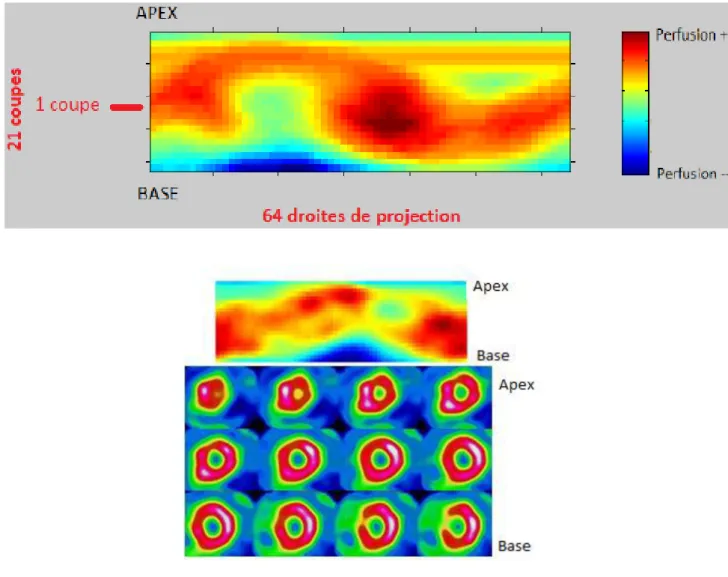
Assessment of Myocardial Perfusion Entropy (MPE)
Dicom files corresponding to stress and rest short-axis images were extracted from the
acquisition system and used for the determination of MPE using an algorithm developed in
Matlab software (Mathworks, Natick, MA, USA), typically leading to 23-31 short axis slices
extracted / patient. Briefly, short axis slices were selected starting from the slice containing the
highest number of pixels with non-zero values. Starting from this slice and going either in the
basal and apical directions, slices considered for analysis were included as long as they
contained at least 1 pixel which intensity was >73% or >65% higher than the maximum pixel
value observed over the whole LV reconstructed volume. Included slices were then interpolated
to obtain a fixed total of 21 slices/patient. On each of these 21 slices, the position of the LV
center was automatically estimated according to the methodology described by Soneson et al
(43). The LV center was used to convert the short-axis cartesian images into polar images using
64 interpolating angles to draw interpolation lines. The pixel of highest intensity along each of
the 64 interpolation lines was selected and used to build a 64 x 21 pixels 2D map of myocardial
perfusion (Figure 1). The 2D map of myocardial perfusion was then normalized from 0 to 1000
using the highest intensity pixel value in order to allow inter-patient comparisons and was
further thresholded to 500 and 850 as previously described (44) MPE was finally computed
from stress and rest 2D perfusion maps from each patient using the formula:
where M was the number of intensity value and pk was the probability associated with pixel
intensity k.
Entropy quantifies the information contained in an image and can be viewed as a measure of
25
required to encode an image without distortion. The entropy is therefore maximal for a uniform
distribution, and equals zero for a constant image.
This method has not proved yet the correlation with IMR or CFR, but it has proved a poor
prognosis in patients (45).
26
Cerebral small vessels disease assessment
Cerebral small vessels disease cannot be diagnosed directly on sectional imaging. The lesions
seen correspond to the consequence of white matter ischemia due to microvascular dysfunction,
also called leukoariosis. These lesions are located in the sub cortical white matter and in deep
grey nuclei. These white matter changes can be observed either on CT scan and on cerebral
MRI with T2 and FLAIR sequences. These sequences are systematically done when the exam
is done to search for a stroke. In CT scan, the lesions are hypodensities, in MRI it corresponds
to hypersignals. A few scores have been established to quantify the extent of leukoariosis, the
mean one is called Age Related White Matter Changes (39,46,47). This score attributes a 0 to
3 score judging the severity of the leukoariosis, in 5 regions in both hemisphere giving un score
out of 30. This score is validated on MRI and on CT scan. In our study, both MRI and CT scan
are used because some patients only had a CT scan to confirm the stroke.
The scale of ARWMC is given below:
White matter lesions:
- 0: no visible lesions
- 1: focal lesions (>5mm)
- 2: beginning confluence of lesions
- 3: Diffuse involvement of the entire region, with or without involvement of U fibers
Basal Ganglia lesions
- 0: No lesions
- 1: 1 focal lesion (>5mm)
- 2: >1 focal lesion
27
The different regions in both hemispheres evaluated are: frontal lobe, parieto occipital lobe,
temporal lobe, infra tentorial region, basal ganglia (containing striatum, globus palidus,
thalamus, internal and external capsula, insula).
Infarcted regions are excluded from the quotation. The final score is out of 30. The ARWMC
score is evaluated for the five first patients in double reading with Pr Detante (Neurology
Physician), then in single reading.
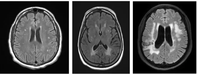
Figure 2: stage 1 (at the left) to stage 3 (at the right) of ARWMC scale, on FLAIR MRI
28
Fazekas score was evaluated in all patients. Fazekas score is a simpler method to assess
leukoariosis on one frame of a CT scan or a MRI. Fazekas score attributes from 0 to 3 depending
on severity, the periventricular leukoariosis. (46,48) Fazekas score is attributed on one single
sectional image containing lateral ventricles, frontal and parieto occipital lobes.
The Fazekas scale is detailed below:
- 0: no lesions
- 1: focal lesions
- 2: beginning confluence of lesions
29
Statistical analysis
Analysis was performed using SPSS 21 software (SPSS Inc., Chicago, IL). Continuous
variables are expressed as mean ± SD or median (25th, 75th percentile) and discrete variables
as percentage. Relationships between the variables were assessed using Pearson's correlation
30
Results
From the January 1st 2016 to February 1st 2020, 371 patients were addressed to cardiology day
hospital for post stroke check-up. Among them, 64 had a check-up with a myocardial SPECT
planned. In these 64 patients, 8 were excluded because of a transient ischemic attack, 7 patients
had cardio embolic stroke, 5 patients didn’t come to their convocation, 3 patients refused the
SPECT, 2 patients had cerebral imaging in another hospital, one had migraine. Finally, we
included 38 patients in our study.
Figure 3: Flow chart
371 patients who had cardiology check up at the cardiology day
hospital from 01/01/2016 to 28/02/20
64 patients were addressed for check up including myocardial
SPECT
38 patients were included
5 patients didn’t come 3 refused to have a SPECT
9 patients had no stroke 7 patients had cardio embolic stroke
2 patients had no cerebral imaging available
31
Patient’s characteristics
In our 38 patients, the mean age was 64.2 +/- 10.9 years old, 31 (81%) were men.
Patient characteristics are summarized in Table 1.
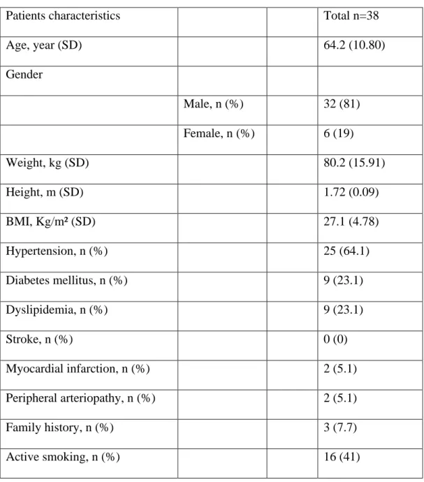
Patients characteristics Total n=38
Age, year (SD) 64.2 (10.80) Gender Male, n (%) 32 (81) Female, n (%) 6 (19) Weight, kg (SD) 80.2 (15.91) Height, m (SD) 1.72 (0.09) BMI, Kg/m² (SD) 27.1 (4.78) Hypertension, n (%) 25 (64.1) Diabetes mellitus, n (%) 9 (23.1) Dyslipidemia, n (%) 9 (23.1) Stroke, n (%) 0 (0) Myocardial infarction, n (%) 2 (5.1) Peripheral arteriopathy, n (%) 2 (5.1) Family history, n (%) 3 (7.7) Active smoking, n (%) 16 (41)
32
SPECT analysis
In SPECT, 13.2% of stress images quality was excellent, 84.2% was good and 2.6 % acceptable,
55.3% of images had no artefacts, 36.8% had mild artefacts and 7.9% had moderate artefacts.
Patients had normal LVEF (61.3%), normal volumes (LVTDV 72.6 mL; LVTSV 28.8mL).
SPECT and Myocardial Perfusion Entropy values are summarized in table 2
SPECT characteristics Total n=38
Pathologic SPECT, n (%) 10 (26.3)
Ischemia %, m (SD) 10.3 (6.8)
Necrosis %, m (SD) 1.7 (2.7)
SRS m (SD) 3.4 (2)
SSS m (SD) 4.4 (3.1)
LVEF m (SD) rest stress
61.3 (9.7) 59.5 (8.9)
LVEDV mL, m (SD) rest stress
72.6 (27.3) 64.9 (17.5)
LVESV mL, m (SD) rest stress
28.8 (16.1) 27.1 (12)
Entropy
Decubitus non gated 4.4256 (0.3315)
33
Neurological assessment and cerebral images analysis
The average NIHSS score of the patients was 5.9 (+/- 5.3), the RANKIN score was 1.7
(+/- 1.5), and the average maximal diameter of the stroke was 23 mm (+/- 1.7). The mean
ARWMC score was 5.2 (+/- 4.9), and the mean FAZEKAS score was 1.0 (+/- 0.96).
Cerebral imaging characteristics are summarized in table 3.
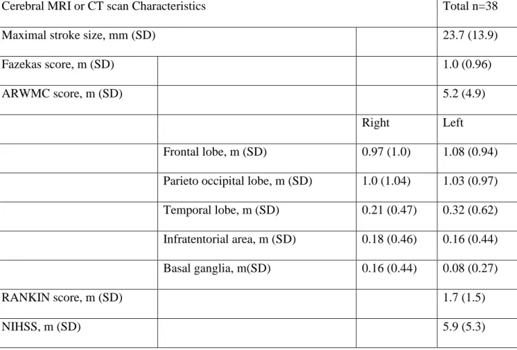
Cerebral MRI or CT scan Characteristics Total n=38
Maximal stroke size, mm (SD) 23.7 (13.9)
Fazekas score, m (SD) 1.0 (0.96)
ARWMC score, m (SD) 5.2 (4.9)
Right Left
Frontal lobe, m (SD) 0.97 (1.0) 1.08 (0.94)
Parieto occipital lobe, m (SD) 1.0 (1.04) 1.03 (0.97)
Temporal lobe, m (SD) 0.21 (0.47) 0.32 (0.62)
Infratentorial area, m (SD) 0.18 (0.46) 0.16 (0.44)
Basal ganglia, m(SD) 0.16 (0.44) 0.08 (0.27)
RANKIN score, m (SD) 1.7 (1.5)
NIHSS, m (SD) 5.9 (5.3)
34
Primary endpoint
There was no correlation between ARWMC and MPE scores, spearman correlation was -0.10,
p = 0.52.
In all risk factors studied, no association neither with ARWMC, neither with Myocardial
perfusion entropy was found.
Coronary disease screening
Among the 64 patients who were addressed to the cardiology day hospital for a check-up
containing a myocardial SPECT, 55 patients (85.9%) benefited from the exam. Among them,
16 patients had an abnormal SPECT (29.1%), 11 went to coronary angiogram (20%). After
proofreading, 5 patients who had abnormal SPECT were considered of false positive because
of artefacts, or had not enough ischemia to undergo an invasive coronary exploration. For
patients who had a coronary angiogram, 2 had multi vessels disease, one was managed with
medical treatment, one with CABG. Two patients had bitroncular stenosis and were both
revascularized with two stents, one patient had a single vessel stenosis and had one stent. Five
patients had diffuse atheroma without significant stenosis. None of the patients benefited from
a microvascular assessment by invasive method (CFR or IMR).
Eight of the ten patients who had a coronary angiogram had a treatment modification. Three
went from a single to a dual anti platelet therapy. The five others had an intensification of their
cardiovascular lowering risk treatment, including an introduction of beta blocker, angiotensin
conversion enzyme inhibitor, thiazide inhibitor, and modification of the anti-diabetic treatment
with introduction of glucagon like peptide 1 analogue. Four of the six patients who were not
revascularized benefited from a treatment modification to lower cardiovascular risk.
35
Discussion
The systemic microcirculatory disease hypothesis
Microcirculation is defined by a vessel diameter < 200 µm, containing three compartments,
arterioles, capillaries and venules (32). These three components are in a constant regulation to
assess its role, to adapt blood flow and vascular permeability for tissue metabolism. It uses
autocrine, paracrine, endocrine system to communicate, and is under the influence of autonomic
nervous system. Microcirculation is the major determinant for vascular resistance, so it is the
principle place for blood pressure regulation (49). Macro and microcirculation are sharing
similar mechanisms and risk factors such as hypertension, diabetes and glucose intolerance,
dyslipidaemia (50) but microvascular disease can reach patients without macrovascular
damage. Functioning of microcirculation involves blood flow regulation by vasodilation or
constriction, vascular permeability, cellular proliferation and angiogenesis.
The concomitant impairment between macro et microcirculation is not systematic but has been
demonstrated in few circumstances, for example, patients presenting hypertension (51),
diabetes (52) or chronic renal failure (53), had a capillary rarefaction on tissue biopsies. Patients
without proven hypertension but in the upper limit of blood pressure have also been shown to
have less capillaries than people with a lower blood pressure (54).
A corner stone of microvascular disease is endothelial dysfunction. Endothelium can be
considered as a single organ responsible for maintaining homeostasis in each organ (55). It is
the main component of capillaries, and its polymorphism permit son adapt itself to each organ
demand. It can be continuous, discontinuous, fenestrated, sinusoidal, associated with a
basement membrane or subendothelial cells to assure its functions. Endothelium role is to
procure enough nutriments to the cell and to evacuate cellular waste. To permit sufficient
36
regulate leucocytes migration by dint of LEUcocytes Adhesion Molecules (LEUCAM), which
contain integrins, selectins, and immunoglobulins (56).
Endothelium has a major place in regulating hemostasia in both ways. The secretion of NO and
prostacyclin will have anti thrombotic and vasodilation action. Tissular plasminogen activator,
secreted by endothelium, produces activated plasmin which degrade fibrin then having an
antithrombotic action. Thrombomudulin which is an endothelial transmembrane protein, with
its co-factor thrombin is able to activate protein C, and then inactivate factors V and VIII
implicate in the coagulation cascade. In the other way, endothelial cells can secrete Willebrand
factor, tissular factor and plasminogen activator inhibitor and will lead to the clot formation and
coagulation activation. In physiological condition, the two systems are in balance to avoid
spontaneous thrombosis, but able to activate coagulation systems if the capillary is damaged
(57).
Microcirculation, by the action of endothelium, is the most important determinant of vascular
resistance and blood flow auto regulation (49). The NO and prostacyclin produced by
endothelial cells will have a vasodilation action on smooth muscle, at contrary, the production
of endothelin or thromboxane A2 will promote vasoconstriction. This blood flow regulation is
mediated by tissular metabolism, shear stress, autonomic nervous system, and by circulating
hormones like angiotensin II and endothelin.
The systemic microvascular disease hypothesis is based on endothelial dysfunction which will
lead to a loss of blood flow auto regulation and an inadequacy between the needs and
contributions of the cell. With the impairment of endothelial function, we understand that this
trouble will impact every organ. Endothelial dysfunction will unbalance all the mechanisms in
37
A model of systemic microvascular disease is obesity (58,59). It is recognised as a risk factor
for developing endothelial dysfunction. The role of white adipose tissue is to stock mobilizable
energy in opposition to brown adipose tissue which regulate thermogenesis. In obesity, the
accumulation of adipocytes in white adipose tissue will cause an increase in cellular size from
50 to 150-200 µm. This will lead to a diminution of oxygen diffusion, and associated with
vascular rarefaction, will cause chronic hypoxia (60,61). The consequence will be a change of
adipocyte secretion, a diminution of adiponectin secretion, which is an insulin sensibiliser
cytokine reducing diabetes risk, lower atherogenisis, and having an anti-inflammatory role (62).
Adipocytes will also secrete more leptin, a satiety hormone, which have lower effects in obese
patients. Leptin stimulates sympathic tonus favouriting vasoconstriction and endothelial
dysfunction. Other cytokines are overproducted in obese patients like resistin, TNF alpha,
interleukine 6 and 18, having a pro inflammatory role (60).
Insulin resistance is also implicated in endothelial dysfunction. Insulin is implicated in many
signalisation routes. In one hand, insulin activates eNOS (endothelial Nitric Oxyde Synthase)
through a PI 3 kinase protein, and will product NO (60). In the other hand, insulin activates
ERK ½ way, resulting in endothelin production, which the most powerful vasoconstrictor agent
known. Endothelin has also an inflammatory role, stimulating cellular proliferation and fibrosis.
For healthy people, insulin effects are in favour of PI 3 kinase and NO synthesis, but for obese
patients, the balance is in favour in the other signalising way, causing inflammation, fibrosis
and vasoconstriction.
As proved before for obese patients, microvascular impairment is the endpoint of many cellular
pathways and cannot be considered as single factor issue. Obesity is one of the risk factors, but
few others have been identified (36,51,63), and as many of them a secondary to modifiable risk
38
Primary endpoint
In our study, we didn’t find any correlation between ARWMC score and Myocardial Perfusion
Entropy in a population of patients who suffered from an ischemic stroke.
Literature review
The studies interesting coronary microvascular disease and cerebral small vessels disease are
few.
Weidmann et al proposed to study cerebral perfusion with a technetium 99 HMPAO brain
SPECT in patients presenting cardiac syndrome X (64). Their definition for Syndrome X was
symptoms of myocardial ischemia on exercise test without any significant coronary stenosis,
and presenting a slow flow at the contrast product injection during coronary angiogram. No
microvascular measurement was done at the coronary angiogram. Many exclusion criteria were
applied, among them any type of cardiopathy (valvular, hypertrophic, dilated or restrictive),
hypertension, diabetes and patients with neurologic symptoms, or with a history of cerebral
ischemia, cerebral disorder, and pathologic findings on carotid artery sonography. They
included 90 patients with syndrome X, 76% had brain perfusion abnormalities, that were
predominant in men (81% vs 69.8%). Their findings are interesting because of the high
prevalence of perfusion abnormalities.
PAI et al made a similar study using a technetium 99 ethyl cysteinate dimer to assess cerebral
perfusion (65). They made a study including 30 patients with syndrome X (typical anginal chest
pain with a positive stress ECG test finding and normal coronary arteries on angiography,
without CFR or MRI performed). They had a dipyramidol stress and resting thallium 201
myocardial perfusion SPECT and were divided in two groups, normal vs abnormal SPECT.
Patients with altered left ventricle ejection fraction, dyslipidaemia, overweight, neurological
39
pathological myocardial perfusion SPECT had hypoperfusion lesions on brain SPECT vs 20%
in patients with normal myocardial SPECT (p value < 0.001).
The last study we found on the brain and myocardial perfusion was made by Sun et al. (66).
Their study is very close to Weidmann’s, they included 40 patients with typical anginal chest pain with positive exercise test and normal coronary arteries on angiography. Patients with
altered LVEF < 50%, regional wall motion abnormalities, hypertension, diabetes, systemic
vascular disease, patients who smoked and patients with any cerebral disorder or symptom were
excluded. Patients were divided into two groups, one with abnormal Thallium 201 myocardial
perfusion SPECT, and the other one with normal SPECT. 92 % of patients with abnormal
myocardial SPECT had multiple hypoperfusion lesions on Technecium 99m HMPAO brain
SPECT vs 12 % in the normal myocardial SPECT group (p<0.001). The design of this study is
very close to Weidmann’s, but they used a more performant gamma camera for the brain SPECT, with a better resolution.
In these three studies, only Weidmann’s study was fulfilling the CMD definition. These studies are suggesting a correlation between cerebral and myocardial perfusion abnormalities, but their
choice to exclude patients with diabetes, hypertension and overweight is objectionable since
they are risk factors identified for microvascular impairment. When excluding the more
important and more frequent risk factors of small vessels disease (hypertension, diabetes,
smoking), the conclusions cannot be extended to the patients we often face, presenting an
advanced age, diabetes, hypertension and smoking. Another thing to underline, is the young
population they included: in Sun’s study, the mean age was 45 YO, in Pai’s study they were
from 37 to 50 YO (no mean communicated) and in Weidmann’s study the mean age was 55
YO. Choosing young patients without the principle risk factors identified for small vessels
disease, may select a disease with microvascular disorder, but not representing the disease we
40
Finally, their brain perfusion method is debatable, the European Association of Nuclear
Medicine Neuro-imaging Committee (ENC) published guidelines in 2009 for the use of brain
SPECT (67). HMPAO and ECD SPECT can be used to assess brain perfusion in patients
presenting mild cognitive impairment or dementia, but they precise that these methods do not
provide absolute quantitative flow values but rather estimate relative regional flow differences
based on the comparison of count density ratios between various regions. Johnson et al found
out in a study with 5 years of follow up, that brain SPECT at baseline evaluation was more
pathological in patients with Alzheimer disease and in patients who will develop Alzheimer
disease, that in control group (68). In the same study, no brain SPECT pattern could differentiate
patients who will develop cognitive impairment without Alzheimer disease, from the control
group without cognitive decline.
In 2018, a Japanese study found out that patients with mild to moderate white matter changes
on MRI had regional perfusion abnormalities in brain SPECT, and hypertension was strongly
associated with WMC (69).
In contrary to our study, Weidman, Pai and Shung included patients with a diagnostic of
syndrome X, then find an association is easier than in our study, because the study population
is not the same. In our study, we included patients who presented a stroke, without knowing if
a cerebral or a myocardial microvascular impairment is present. Then our findings are
depending on the prevalence of both organ damage.
To our knowledge, no studies have been using cerebral MRI to assess cerebral microvascular
disease consequences, and test the correlation with CMD.
Vuorinen et al. followed 69 patients with macrovascular coronary heart disease for a mean time
41
volume compared to control group, whereas no difference was found in the evaluation of white
matter.
Ikram et al. used the Rotterdam study cohort and evaluated dementia and white matter lesions
between patients with unrecognized myocardial infarction and patients without MI. Dementia
and white matter lesions were more important in patients who had a silent myocardial infarction
(71).
Brunelli et al. published a case control study, including 16 patients with syndrome X and 16
controls (72). The syndrome X diagnostic was made on a positive exercise test in patients
suffering from typical angina chest pain, with normal coronary arteries on angiography, and
excluding spasm with ergonovine test. They studied cerebral blood flow reserve using Xenon
133 inhalation and its measurement with 32 epicranial probes, before and after acetalozamide
intravenous administration. They found strictly the same cerebral blood flow reserve in the two
groups (increase of 29.0 % +/- 14% vs 29.5 % +/- 11% in the control group).
Coronary artery disease screening in patients with atherothrombotic stroke.
In our study, we found out that 15 SPECTs had abnormal patterns (27.3%), but after
proofreading by our nuclear physicians, only 11 exams were considered as pathological so the
prevalence of pathological SPECT in our cohort is 20%. Nine percent of the patients had severe
coronary stenosis, the other patients had intermediate stenosis or diffuse atheroma. Finally, 4
patients were revascularized.
Nigoghossian et al. (73) found a similar prevalence (15%) of abnormal dobutamine stress
echography in a similar cohort of patients who had an ischemic stroke attributed to
atherosclerosis. As in his study, all of our patients with pathologic stress test imaging didn’t
have a significant stenosis at the coronary angiogram. Maybe these patients may benefit from
42
In another study, Arenillas et al. (74) included 65 patients with a stroke or a TIA associated with
an intra cranial significant stenosis, and searched for silent myocardial ischemia. They found
out that 52 % of their patients had an abnormal myocardial SPECT, which is higher than our
rate. They published their study without knowing if patients with abnormal SPECT benefited
of a coronary angiogram, then the false positive rate of the SPECT is unknown, but during
exercise test, only 5% were positive because of clinical angina pectoris, and 20% with ST
segment depression. The authors didn’t mention the proportion of abnormal SPECTs due to artefacts.
In 2009, Ovbiagele et al. (75) made a feasibility study of screening CAD in patients after an
ischemic stroke, with a Framingham score > 20 %. The main result is that they proved the
difficulty to organise a systematic screening in this category of patients, because only 44% of
patients who had a good indication for non-invasive ischemic test, benefited of a stress
dobutamine echography. The two reasons for this lack of performance were first the patient’s
noncompliance, and the second one was the physician’s refusal. Forty-two percent of patients
didn’t come to their convocation, a few of them because of insurance issues, but most of them didn’t come because of a lack of motivation and/or a lack of information of the goal of this exam. In our cohort, we suffered from the same problem, since 11 of 60 patients didn’t come to their convocation. In Ovbiagele’s study, 33 % of dobutamine echography didn’t carry out
because of the physician’s refusal, then the problem that can be pointed out is the lack of information of the physicians about CAD in patients after a stroke.
The American Heart Association with the Amercian Stroke Association published a statement
paper in 2003 regarding CAD screening in patients with TIA or stroke (76). Ischemic tests
should not be systematic after a stroke but some population may benefit of it. There are few
reasons for a CAD screening in this population. First, the prevalence of CAD, symptomatic or
43
to 40 % of these patients had silent ischemia (77,78). More recently in a meta-analysis published
in 2016 including more than 50 000 patients who presented a first TIA or a first stroke,
significant coronary stenosis was found in 30 % of the patients, and the risk of myocardial
infarction the first year after a stroke was 3% (79).
Second, after a stroke, cardiac mortality is significant, after analysing studies including 30 000
patients, cardiac mortality is from 2 to 5% at 90 days after stroke (but authors precise that these
deaths concern mainly patients with known CAD) (80,81).
In longer follow up studies, the incidence of myocardial event (myocardial infarction, cardiac
death) is around 2 to 3 % per year during the three years after a stroke or TIA (82,83).
The long-term prognosis of patients is also strongly influenced by CAD. In the Rochester study,
the cumulative incidence of myocardial infarction or sudden death after a stroke was 10.6% at
5 years, and even up to 17.4% at 5 years in the northern Manhattan study (84,85).
Few studies showed a high mortality in the three first months after a stroke due to the actual
stroke or complications linked to it and to the hospitalization, but the mortality after this period
is significantly linked to cardiac death. Between 1 and 5 years after a first stroke, the first cause
of mortality was cardiac deaths (41%), after 5 years non vascular deaths become first but cardiac
deaths are still more important than death because of recurrent stroke (33% versus 23 %) (86).
The conclusion of the ASA AHA statement is that a systematic coronary risk evaluation should
be done after a stroke. It is based on the same guidelines as people in primary prevention, by
calculating a coronary risk score at 10 years, using Framingham algorithm or SCORE score
(87). A reduction of the number of cardiovascular risk factors should be done. Systematic
non-invasive ischemic test is not recommended, but patients with a high risk at 10 years and patients
with a significant carotid stenosis should be considered for either exercise ECG, SPECT,
exercise or dobutamine echography. This statement paper is a little outdated and an update
44
PRECORDIS score, associating Framingham score and cervicocephalic significant
atherosclerosis which was associated with an occult > 50 coronary stenosis on CTA, and the
value of the score was associated with the severity of CAD (88). This PRECORDIS Score may
be an easy tool accessible in every stroke unit to identify the patients who would benefit from
a CAD screening.
More recent studies interested on cardiac computed tomography angiography or coronary artery
calcium score to diagnose asymptomatic CAD, and predict poor prognosis in patients after a
stroke. A Korean prospective cohort with 1893 patients with ischemic stroke found out that
MACE was associated with asymptomatic coronary stenosis diagnosed on cardiac CTA (89).
Cardiac CTA may be a tool to diagnose obstructive CAD, but this exam fails to recognise
INOCA since it is an anatomic evaluation only.
Limits
Our study has few limits. First, the assessment of cerebral small vessels disease is validated and
correlates with poor outcome (10,16,19,90,91) but our myocardial perfusion technic hasn’t
proved a correlation with IMR during coronary angiogram yet with is gold standard. Myocardial
perfusion entropy is associated with poor outcome in patients with diabetes but correlation
between heterogeneity and IMR is poor (Non published data, EVACORY study, NCT0199595).
The second limit is the lack of statistical power because of our too small sample size to find an
association between cerebral and myocardial microvascular dysfunction. Despite an inclusion
period of 4 years, only 64 patients were addressed to cardiology day hospital for stroke check
up with a coronary disease screening with SPECT. Among these patients, some of them didn’t
come to their convocation, and some were excluded from the study, because even if the coronary
disease screening was justified, the origin of the stroke was cardio embolic. Since our cohort
45
none of the two microvascular disease. Then to prove an association between the two organs
damage was even more difficult.
Another limit could have been the single reading of the majority of the cerebral MRI, but the
rating scale of ARWMC is easy to apply, and the strong correlation found between the two
46
Conclusion
In this study, we didn’t find any association between cerebral leukoariosis and perfusion heterogeneity on myocardial SPECT in patients who suffered from a stroke. Further study may
be done using another type of myocardial SPECT analysis, the coronary reserve, to search for
a common disease between the brain and the heart microvasculature. The neurology –
cardiology branch seems to be interesting in selected patients with an acceptable rentability of
48
Bibliography
1. Les 10 principales causes de mortalité. Disponible sur : https://www.who.int/fr/news-room/fact-sheets/detail/the-top-10-causes-of-death
2. Townsend N, Wilson L, Bhatnagar P, Wickramasinghe K, Rayner M, Nichols M. Cardiovascular disease in Europe: epidemiological update 2016. Eur Heart J. 7 nov 2016;37(42):3232-45.
3. EUROASPIRE Study Group. EUROASPIRE: A European Society of Cardiology survey of secondary prevention of coronary heart disease: Principal results. Eur Heart J. 2 oct 1997;18(10):1569-82.
4. Lifestyle and risk factor management and use of drug therapies in coronary patients from 15 countries. Principal results from EUROASPIRE II Euro Heart Survey Programme. Eur Heart J. 1 avr 2001;22(7):554-72.
5. Kotseva K, Wood D, Backer GD, Bacquer DD, Pyörälä K, Keil U, et al. EUROASPIRE III: a survey on the lifestyle, risk factors and use of cardioprotective drug therapies in coronary patients from 22 European countries. Eur J Cardiovasc Prev Rehabil. avr 2009;16(2):121-37.
6. Kotseva K, De Backer G, De Bacquer D, Rydén L, Hoes A, Grobbee D, et al. Lifestyle and impact on cardiovascular risk factor control in coronary patients across 27 countries: Results from the European Society of Cardiology ESC-EORP EUROASPIRE V registry. Eur J Prev Cardiol. mai 2019;26(8):824-35. 7. Amouyel P. Une action sur un nombre limité de facteurs de risque permettrait de réduire au moins de moitié les taux de survenue de maladies cardiovasculaires. Rev Prat. 2005;9.
8. Motreff DP. Facteurs de risque cardio-vasculaire. 2005;7.
9. Longstreth WT, Manolio TA, Arnold A, Burke GL, Bryan N, Jungreis CA, et al. Clinical Correlates of White Matter Findings on Cranial Magnetic Resonance Imaging of 3301 Elderly People: The Cardiovascular Health Study. Stroke. août 1996;27(8):1274-82.
10. Pantoni L. Cerebral small vessel disease: from pathogenesis and clinical characteristics to therapeutic challenges. Lancet Neurol. juill 2010;9(7):689-701.
11. Wardlaw JM, Sandercock PAG, Dennis MS, Starr J. Is Breakdown of the Blood-Brain Barrier Responsible for Lacunar Stroke, Leukoaraiosis, and Dementia? Stroke. mars 2003;34(3):806-12.
12. Chabriat H, Joutel A, Dichgans M, Tournier-Lasserve E, Bousser M-G. Cadasil. Lancet Neurol. juill 2009;8(7):643-53.
49
13. Chamorro A. Periventricular White Matter Lucencies in Patients With Lacunar Stroke: A Marker of Too High or Too Low Blood Pressure? Arch Neurol. 1 oct 1997;54(10):1284.
14. Teodorczuk A, O’Brien JT, Firbank MJ, Pantoni L, Poggesi A, Erkinjuntti T, et al. White matter changes and late-life depressive symptoms: Longitudinal study. Br J Psychiatry. sept 2007;191(3):212-7. 15. Pantoni L, Poggesi A, Inzitari D. The relation between white-matter lesions and cognition: Curr Opin Neurol. août 2007;20(4):390-7.
16. Frisoni GB, Galluzzi S, Pantoni L, Filippi M. The effect of white matter lesions on cognition in the elderly—small but detectable. Nat Clin Pract Neurol. nov 2007;3(11):620-7.
17. Guttmann CRG, Benson R, Warfield SK, Wei X, Anderson MC, Hall CB, et al. White matter abnormalities in mobility-impaired older persons. Neurology. 28 mars 2000;54(6):1277-83.
18. Sado M, Ninomiya A, Shikimoto R, Ikeda B, Baba T, Yoshimura K, et al. The estimated cost of dementia in Japan, the most aged society in the world. PloS One. 2018;13(11):e0206508.
19. Schmidt R, Petrovic K, Ropele S, Enzinger C, Fazekas F. Progression of Leukoaraiosis and Cognition. Stroke. sept 2007;38(9):2619-25.
20. Crea F, Camici PG, Bairey Merz CN. Coronary microvascular dysfunction: an update. Eur Heart J. 1 mai 2014;35(17):1101-11.
21. Kunadian V, Chieffo A, Camici PG, Berry C, Escaned J, Maas AHEM, et al. An EAPCI Expert Consensus Document on Ischaemia with Non-Obstructive Coronary Arteries in Collaboration with European Society of Cardiology Working Group on Coronary Pathophysiology & Microcirculation Endorsed by Coronary Vasomotor Disorders International Study Group. Eur Heart J. 6 juill 2020;ehaa503.
22. Ong P, Camici PG, Beltrame JF, Crea F, Shimokawa H, Sechtem U, et al. International standardization of diagnostic criteria for microvascular angina. Int J Cardiol. janv 2018;250:16-20.
23. Fearon WF, Kobayashi Y. Invasive Assessment of the Coronary Microvasculature: The Index of Microcirculatory Resistance. Circ Cardiovasc Interv. déc 2017;10(12). doi/10.1161/CIRCINTERVENTIONS.117.005361
24. Ong P, Athanasiadis A, Borgulya G, Mahrholdt H, Kaski JC, Sechtem U. High Prevalence of a Pathological Response to Acetylcholine Testing in Patients With Stable Angina Pectoris and Unobstructed Coronary Arteries. J Am Coll Cardiol. févr 2012;59(7):655-62.
25. Murthy VL, Naya M, Taqueti VR, Foster CR, Gaber M, Hainer J, et al. Effects of Sex on Coronary Microvascular Dysfunction and Cardiac Outcomes. Circulation. 17 juin 2014;129(24):2518-27.