The Role of MicroRNA Regulation of Cardiac Ion
Channel in Arrhythmia
par Xiaobin Luo Département de Médecine Faculté de MédecineThèse présentée à la Faculté des études supérieures en vue de l’obtention du grade de
Philosophiae Doctor (Ph.D.) en Sciences Biomédicales
Août 2012
Faculté des études supérieures et postdoctorales
Cette thèse intitulée:
The Role of MicroRNA Regulation of Cardiac Ion Channel in Arrhythmia
Présentée par : Xiaobin Luo
a été évaluée par un jury composé des personnes suivantes :
Dr Eric Thorin Président-rapporteur Dr Stanley Nattel Directeur de recherche Dr Lucie Parent Membre du jury Dr Alvin Shrier Examinateur externe
RÉSUMÉ
La fibrillation auriculaire (FA) est le trouble du rythme le plus fréquemment observé en pratique clinique. Elle constitue un risque important de morbi-mortalité. Le traitement de la FA reste un défi majeur en lien avec les nombreux effets secondaires associés aux approches thérapeutiques actuelles. Dans ce contexte, une meilleure compréhension des mécanismes sous-jacents à la FA est essentielle pour le développement de nouvelles thérapies offrant un meilleur rapport bénéfice/risque pour les patients. La FA est caractérisée par i) un remodelage électrique délétère associé le plus souvent ii) à un remodelage structurel du myocarde favorisant la récurrence et le maintien de l’arythmie. La diminution de la période réfractaire effective au sein du tissu auriculaire est un élément clef du remodelage électrique. Le remodelage structurel, quant à lui, se manifeste principalement par une fibrose tissulaire qui altère la propagation de l’influx électrique dans les oreillettes. Les mécanismes moléculaires impliqués dans la mise en place de ces deux substrats restent mal connus. Récemment, le rôle des microARNs (miARNs) a été pointé du doigt dans de nombreuses pathologies notamment cardiaques. Dans ce contexte les objectifs principaux de ce travail ont été i) d'acquérir une compréhension approfondie du rôle des miARNs dans la régulation de l’expression des canaux ioniques et ii) de mieux comprendre le rôle de ces molécules dans l’installation d’un substrat favorable a la FA.
Nous avons, dans un premier temps, effectué une analyse bio-informatique combinée à des approches expérimentales spécifiques afin d’identifier clairement les miARNs démontrant un fort potentiel de régulation des gènes codant pour l’expression des canaux ioniques cardiaques humains. Nous avons identifié un nombre limité de miARNs cardiaques qui possédaient ces propriétés. Sur la base de ces résultats, nous avons démontré que l’altération de l'expression des canaux ioniques, observée dans diverse maladies cardiaques (par exemple, les cardiomyopathies, l’ischémie myocardique, et la fibrillation auriculaire), peut être soumise à ces miARNs suggérant leur implication dans l’arythmogénèse.
La régulation du courant potassique IK1 est un facteur déterminant du remodelage électrique auriculaire associée à la FA. Les mécanismes moléculaires sous-jacents sont peu connus. Nous avons émis l’hypothèse que l'altération de l’expression des miARNs soit
corrélée à l’augmentation de l’expression d’IK1 dans la FA. Nous avons constaté que l’expression de miR-26 est réduite dans la FA et qu’elle régule IK1 en modulant l’expression de sa sous-unité Kir2.1. Nous avons démontré que miR-26 est sous la répression transcriptionnelle du facteur nucléaire des lymphocytes T activés (NFAT) et que l’activité accrue de NFATc3/c4, aboutit à une expression réduite de miR-26. En conséquence IK1 augmente lors de la FA. Nous avons enfin démontré que l’interférence in vivo de miR-26 influence la susceptibilité à la FA en régulant IK1, confirmant le rôle prépondérant de miR-26 dans le remodelage auriculaire électrique.
La fibrose auriculaire est un constituant majeur du remodelage structurel associé à la FA, impliquant l'activation des fibroblastes et l’influx cellulaire du Ca2 +. Nous avons cherché à déterminer i) si le canal perméable au Ca2+, TRPC3, jouait un rôle dans la fibrose auriculaire en favorisant l'activation des fibroblastes et ii) étudié le rôle potentiel des miARNs dans ce contexte. Nous avons démontré que les canaux TRPC3 favorisent l’influx du Ca2 +, activant la signalisation Ca2 +-dépendante ERK et en conséquence activent la prolifération des fibroblastes. Nous avons également démontré que l’expression du TRPC3 est augmentée dans la FA et que le blocage in vivo de TRPC3 empêche le développement de substrats reliés à la FA. Nous avons par ailleurs validé que miR-26 régule les canaux TRPC3 en diminuant leur expression dans les fibroblastes. Enfin, nous avons montré que l'expression réduite du miR-26 est également due à l’activité augmentée de NFATc3/c4 dans les fibroblastes, expliquant ainsi l’augmentation de TRPC3 lors de la FA, confirmant la contribution de miR-26 dans le processus de remodelage structurel lié à la FA.
En conclusion, nos résultats mettent en évidence l'importance des miARNs dans la régulation des canaux ioniques cardiaques. Notamment, miR-26 joue un rôle important dans le remodelage électrique et structurel associé à la FA et ce, en régulant IK1 et l’expression du canal TRPC3. Notre étude démasque ainsi un mécanisme moléculaire de contrôle de la FA innovateur associant des miARNs. miR-26 en particulier représente apres ces travaux une nouvelle cible thérapeutique prometteuse pour traiter la FA.
ABSTRACT
Atrial fibrillation (AF) is the most frequently-encountered arrhythmia in clinical practice and constitutes a major cause of cardiac morbidity and mortality. The management of AF remains a major challenge as current therapeutic approaches are limited by potential adverse effects and high rate of AF recurrence/persistence. A better understanding of the mechanisms underlying AF is of great importance to improve AF therapy. AF is characterized by impaired electrical and structural remodeling, both of which favors the recurrence and maintenance of the arrhythmia. A key feature in electrical remodeling is the reduced atrial effective refractory period, due to ion channel alteration. Structural remodeling, on the other hand, mainly results from atrial fibrosis. However, the precise molecular mechanisms underlying these remodeling processes are still incompletely understood. The importance of microRNAs (miRNAs) in various pathophysiological conditions of the heart has been well established, but little is known with regard to cardiac arrhythmias. Emerging evidence suggests that dysregulation of miRNAs may underlie heart rhythm disturbances. The aim of the present work was to acquire a comprehensive understanding of miRNA-mediated regulation of ion channels in cardiac arrhythmias. Notably, we will focus on the mechanistic insights of miRNAs related to the control of AF.
Currently available experimental approaches do not permit thorough characterization of miRNA targeting. For this purpose, we performed bioinformatic analyses in conjunction with experimental approaches to identify miRNAs from the database that potentially regulate human cardiac ion channel genes. We found that only a subset of miRNAs target cardiac ion channel genes. Based on these results, we further demonstrated that the dysregulation of ion channel gene expression observed in various cardiac disorders (e.g. cardiomyopathy, myocardial ischemia, and atrial fibrillation) can be explained by the dysregulation of miRNAs. These findings further support the potential implication of miRNAs in arrhythmogenesis under these cardiac conditions.
The upregulation of the cardiac inward rectifying potassium current, IK1,is a key determinant of adverse atrial electrical remodeling associated with AF. The molecular mechanisms underlying this ionic remodeling are poorly understood. We hypothesized that altered miRNA expression is responsible for IK1 upregulation in AF. We found that miR-26
is significantly downregulated in AF and regulates IK1 by controlling the expression of its underlying subunit Kir2.1. Moreover, we demonstrated that miR-26 is under the transcriptional repression of the nuclear factor of activated T cells (NFAT) and enhanced activities of members of the NFAT family, NFATc3/c4, results in miR-26 downregulation, which accounts for IK1 enhancement in AF. Furthermore, we observed that in vivo interference of miR-26 affects AF susceptibility via the regulation of IK1, suggesting an important role of miR-26 in atrial electrical remodeling.
Atrial fibrosis is a major constituent in AF-associated adverse atrial structural remodeling, involving the activation of fibroblasts and cellular Ca2+ entry. Here, we sought to determine whether the Ca2+ permeable channel, TRPC3, plays a role in AF-induced fibrosis by promoting fibroblast activation. Furthermore, we investigated the potential role of miRNAs in this context. We found that TRPC3 channels promote Ca2+-entry, which results in activation of Ca2+-dependent ERK-signaling and consequently fibroblast activation. We also demonstrated that TRPC3 is upregulated in AF and in vivo TRPC3 blockade suppresses the development of AF-promoting substrate. Furthermore, we observed that miR-26 regulates TRPC3 channels via controlling the expression of the underlying channel subunit and is downregulated in AF-fibroblasts. Finally, we showed that the reduced expression of miR-26 is also due to the enhanced NFATc3/c4 activities in AF-fibroblasts and accounts for AF-induced upregulation of TRPC3, suggesting the potential contribution of miR-26 in AF-related adverse structural remodeling process.
In conclusion, our findings emphasize the importance of miRNAs in the regulation of cardiac ion channels. Notably, miR-26 plays a crucial role in AF-associated electrical and structural remodeling via the regulation of IK1 and TRPC3 channel genes. Thus, our study unravels a novel molecular control mechanism of AF at the miRNA level, suggesting miR-26 as a new and promising therapeutic target for AF.
TABLE OF CONTENTS
RÉSUMÉ ... iii
ABSTRACT ... v
TABLE OF CONTENTS ...vii
LIST OF FIGURES AND TABLES ...xii
LIST OF ABBREVIATIONS ...xvii
DEDICATION ...xxii
ACKNOWLEDGEMENTS ... xxiii
STATEMENT OF AUTHORSHIP ... xxiv
CHAPTER 1. INTRODUCTION ... 1
1.1 Overview of Atrial Fibrillation ... 2
1.1.1 Epidemiology of AF ... 2
1.1.2 Classification of AF ... 2
1.1.3 Signs, Symptoms and Predisposing Factors of AF ... 3
1.1.3.1 Signs and Symptoms of AF ... 3
1.1.3.2 Predisposing Factors for AF ... 3
1.1.4 Current Management and Challenges in Treatment of AF ... 4
1.2 Mechanisms of AF ... 6
1.2.1 Physiological Basis of Cardiac Action Potential ... 6
1.2.2 Overview of AF Pathophysiology ... 7 1.2.2.1 Ectopic Mechanism ... 8 1.2.2.1.1 Abnormal Automaticity ... 9 1.2.2.1.2 Delayed Afterdepolarization ... 9 1.2.2.1.3 Early Afterdepolarization ... 9 1.2.2.2 Re-entrant Mechanisms ... 10
1.2.2.3 Relation of basic AF mechanisms to different forms of clinically-encountered AFs... 11
1.3 Atrial Remodeling in AF ... 11
1.3.1 Electrical Remodeling in AF ... 12
1.3.1.1 Upregulation of IK1 ... 12
1.3.1.2 Downregulation of ICaL ... 13
1.3.1.3 Abnormal Ca2+ Handling ... 14
1.3.1.4 Alterations of other K+ currents ... 14
1.3.2 Structural Remodeling in AF ... 15
1.3.2.1 Atrial Fibrosis ... 15
1.3.2.1.1 Profibrotic Factors and Atrial Fibrosis ... 16
1.3.2.1.2 Potential role of TRP Channels in Atrial Fibrosis ... 17
1.3.2.2 Dysregulation of Gap junction protein ... 18
1.4 Inward Rectifier Potassium Channel (IK1) ... 19
1.4.1 Biophysical Properties and Cellular Functions of IK1 ... 19
1.4.2 Molecular Basis of IK1 ... 20
1.4.3 Regulation of IK1 ... 20
1.4.4 Dysregulation of IK1 and its pathophysiological implication in AF ... 21
1.5 Transient Receptor Potential Canonical Channel Type 3 (TRPC3)... 22
1.5.1 Biophysical Properties and Cellular Functions of TRPC3 ... 23
1.5.2 Molecular Basis of TRPC3 ... 23
1.5.3 Regulation of TRPC3 ... 24
1.5.4 Dysregulation of TRPC3 and its potential pathophysiological implication in AF 25 1.6 MicroRNA Biology ... 25
1.6.1 Historical View of MicroRNAs ... 25
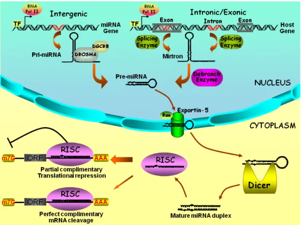
1.6.2 MiRNA Biogenesis ... 26
1.6.3 Action and Biological Functions of MiRNAs ... 27
1.6.3.1 Action of MiRNAs ... 27
1.6.3.2 Biological Functions of MiRNAs ... 28
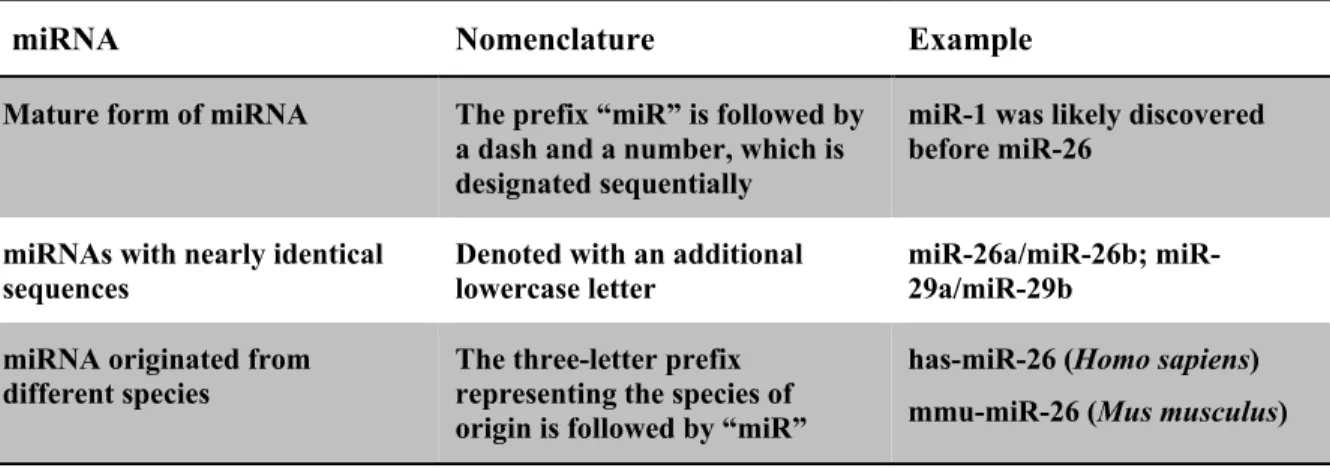
1.6.4 MiRNA Nomenclature ... 29
1.6.5 Determination of MiRNA Targets ... 29
1.6.5.1 Computational Prediction of MiRNA Targets ... 30
1.6.6 MiRNA Expression in the Heart ... 32
1.6.6.1 Cardiac Selectivity of MiRNA Expression ... 32
1.6.6.2 Cell Type Specification of MiRNA Expression in the Heart ... 32
1.6.6.3 MiRNA Detection and Quantification ... 32
1.6.6.3.1 Detection of MiRNAs by Microarray ... 33
1.6.6.3.2 Detection and Quantification of MiRNAs by Deep Sequencing ... 34
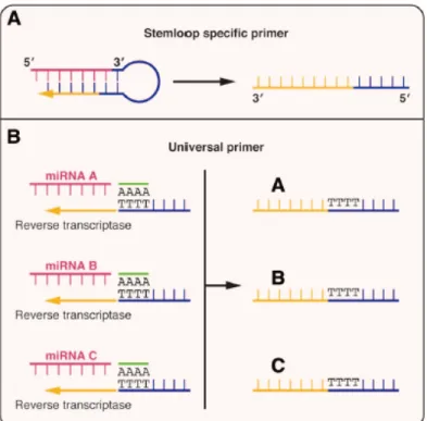
1.6.6.3.3 Quantification of MiRNAs by Real-time RT-PCR ... 34
1.6.6.3.4 Quantification of MiRNAs by Northern blot ... 36
1.6.7 Regulation of MiRNA Expression and MiRNA Interference ... 36
1.6.7.1 Transcriptional Regulation of MiRNA Expression... 36
1.6.7.2 MiRNA Interference ... 37
1.6.7.2.1 In Vitro MiRNA Interference ... 37
1.6.7.2.2 In Vivo MiRNA Interference ... 38
1.6.8 Role of MiRNAs in the Heart ... 40
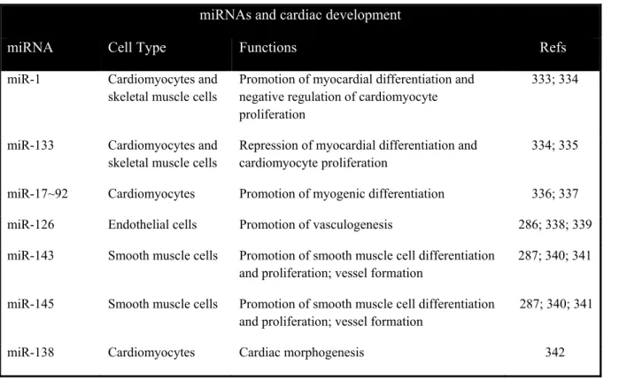
1.6.8.1 Implications of MiRNAs in Cardiac Development ... 40
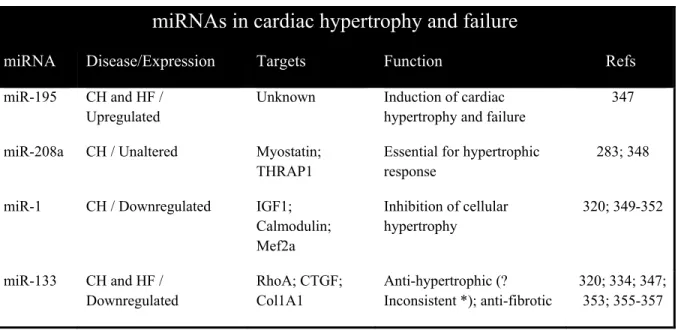
1.6.8.2 Implications of MiRNAs in Cardiac Pathologies... 42
1.6.8.2.1 Roles of MiRNAs in Cardiac Hypertrophy and Heart Failure ... 42
1.6.8.2.2 Roles of MiRNAs in Myocardial Ischemia ... 45
1.6.8.2.3 Roles of MiRNAs in Arrhythmia ... 48
1.7 Rationale for Present Studies ... 50
CHAPTER 2. Overview of the Role of MiRNAs in Regulation of Cardiac Ion Channel Genes and its Potential Arrhythmogenic Implication ... 52
2.1 Regulation of Human Cardiac Ion Channel Genes by MicroRNAs: Theoretical Perspective and Pathophysiological Implications ... 53
Abstract ... 54
Introduction ... 56
Materials and Methods ... 60
Results and Discussion ... 63
References ... 81
Figure Legends ... 93
CHAPTER 3. The Control of Adverse Electrical Remodeling by MiRNAs in AF
... 108
3.1 MicroRNA-26 Governs Profibrillatory Inward-Rectifier Potassium Current Changes in Atrial Fibrillation ... 109 Abstract ... 110 Introduction ... 111 Results ... 113 Discussion ... 121 Methods ... 126 References ... 140 Figure Legends ... 146 Figures ... 152 Supplementary Material ... 160
CHAPTER 4. The Control of Adverse Structural Remodeling by MiRNAs in AF ... 193
4.1 TRPC3-dependent Fibroblast Regulation in Atrial Fibrillation ... 194
Abstract ... 195 Introduction ... 197 Methods ... 198 Results ... 205 Discussion ... 212 References ... 219 Figure Legends ... 224 Figures ... 229 Supplementary Material ... 237
CHAPTER 5. GENERAL DISCUSSION ... 267
5.1 Summary of the Novel Findings in this Thesis ... 268
5.2 Significance of the Major Findings in this Thesis ... 269
5.2.1 Discovery of miR-26 as a novel molecular and signaling mechanism for AF vulnerability and a novel therapeutic target for AF treatment ... 269
5.2.1.1 MiR-26 controls AF by inhibiting IK1-related adverse atrial electrical
remodeling in atrial myocytes ... 269
5.2.1.2 MiR-26 controls AF by inhibiting TRPC3-mediated adverse atrial structural remodeling in atrial fibroblasts ... 271
5.2.1.3 MiR-26 mediates the AF-promoting action of NFAT and is a key factor of two signaling pathways leading to AF ... 272
5.2.1.4 Therapeutic potential of miR-26 for AF ... 275
5.2.2 Discovery of TRPC3 as a novel player in AF ... 275
5.2.3 Systematic identification of miRNA regulation of ion channel genes and its potential implications in arrhythmia associated with heart diseases ... 276
5.3 Future Directions ... 277
5.4 Conclusion ... 279
LIST OF FIGURES AND TABLES
CHAPTER 1
Figure 1. Schematic representation of atrial and ventricular action potentials
with underlying principle ionic currents……….6
Figure 2. Arrhythmogenic mechanisms underlying AF……….8
Figure 3. Cellular mechanisms underlying focal ectopic activity………..8
Figure 4. Mechanisms for reentry………...10
Figure 5. Schematic representation of pathophysiological significance of IK1 upregulation in AF..………...21
Figure 6. Biogenesis and actions of miRNA………...27
Figure 7. Different approaches for miRNA quantification by real-time RT-PCR………...35
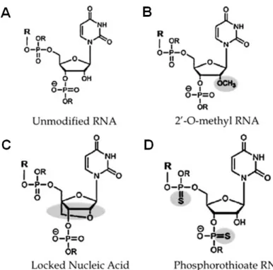
Figure 8. Different chemical modifications of in vivo miRNA knockdown oligos……….39
Table 1. MiRNA nomenclature………...29
Table 2. Summary of miRNAs related to cardiac development……….40
Table 3. Summary of miRNAs related to cardiac hypertrophy and heart failure………42
Table 4. Summary of miRNAs related to myocardial ischemia…………...45
Table 5. Summary of miRNAs related to cardiac arrhythmia………...48
CHAPTER 2
Figure 1. miRNA expression signature in the heart………...95Figure 2. Predicted gene targeting of the top 20 most abundantly expressed miRNAs in myocardium………....96
Figure 3. Predicted gene targeting of the miRNAs deregulated in their expression in cardiac hypertrophy and congestive heart failure (CHF)………...97
Figure 4. Predicted gene targeting of the miRNAs deregulated in their expression in ischemic myocardial injuries………...98 Figure 5. Predicted gene targeting of the miRNAs deregulated in their
expression in experimental atrial fibrillation……….99 Table 1. The genes encoding cardiac cytoplasmic ion channel proteins and
electrogenic ion transporters selected for prediction………...100 Suppl. Figure 1. Verification of relative abundance of miRNAs in human heart by
Realtime RT-PCR……….………...106 Suppl. Figure 2. MiRNA expression changes in rat model of acute myocardial infarction by microarray………...………...107
CHAPTER 3
Figure 1. Downregulation of miR-26 and upregulation of KCNJ2/Kir2.1 in atrial fibrillation (AF)…....………...150 Figure 2. Regulation of Kir2.1-expression by miR-26..……….151 Figure 3. Anti-atrial fibrillation properties of miR-26………152 Figure 4. Role of IK1 in mediating the anti-AF property of miR-26 ..………153 Figure 5. Verification of the ability and specificity of the LNA-miR-Mimic and LNA-miR-Mask…..……….154 Figure 6. Functional evidence for the role of NFAT as a transcriptional
repressor of miR-26 expression…....………...155 Figure 7. Evidence for the interactions between NFAT and the promoter elements of miR-26 host genes………156 Figure 8. Molecular mechanism underlying the AF-promoting effect of miR-26 downregulation.……….………157 Online Figure 1. Alignment of sequences of mature miR-26 family miRNAs of
different species………...……….167 Online Figure 2. Sequences of miR-26a/b and their antisense molecules used in our study…..………..………..168 Online Figure 3. Western blot analysis on the effects of miR-26 on protein levels of several K+ channel pore-forming α-subunits……..……….169
Online Figure 4. Sequences of the antisense to miR-26a..…..…….………..170 Online Figure 5. Schematic illustration of construction of adenovirus vector carrying mouse pre-miR-26a-1..………171 Online Figure 6. One contemporaneous set of wild type controls was performed for all
groups………..172 Online Figure 7. Verification of cellular uptake of Adv-miR-26a-1 in mouse atrial
tissues………173 Online Figure 8. Verification of the effect of the LNA-antimiR-26a and adv-miR-26a on Kir2.1.………...174 Online Figure 9. Sequences of the miR-Mimic and miR-Mask ………....175 Online Figure 10. Identification of transcription start sites (TSSs) and genomic
characteristics of the host genes of the miR-26 family miRNAs using 5’RACE techniques....………...176 Online Figure 11. The constructs used to inhibit the function of nuclear factor of
activated T-cells (NFAT)……...………..180 Online Figure 12. Verification of the efficacy of siRNAs in knocking down the cardiac isoforms of NFAT..……….181 Online Figure 13. Effects of NFAT inhibition on expression of miR-1, determined by qPCR in H9c2 rat ventricular cells…….……….182 Online Figure 14. Temporal changes of miR-26/Kir2.1 during AF development in canine AF model...………...183 Online Table 1. Clinical characteristics of the patients used in our study………....184 Online Table 2. Echocardiographic data for mice subjected to miR-26a
overexpression or knockdown………..…………...186 Online Table 3. Effects of miR-26a manipulation on electrophysiological parameters of mice……….………....187 Online Table 4. Predicted NFAT binding motifs in the promoter regions of the host g e n e s f o r t h e m i R - 2 6 f a m i l y m i R N A s o f v a r i o u s species………..188 Online Table 5. The primers used for PCR-amplification of miR-26 promoter s e q u e n c e s s p a n n i n g N F A T c i s- e l e me n t s for c h r o ma t i n immunoprecipitation assay (ChIP)………..189
Online Table 6. The gene-specific primers (GSP) used for 5’RACE to identify the transcription start sites of the host genes for miR-26 miRNA family members………...190
CHAPTER 4
Figure 1. Roles of TRPC3 in mediating INSC, Ca2+ entry and fibroblast activation………...227 Figure 2. Invovlement of ERK-1/2 phosphorylation in TRPC3-mediated Ca2+
-entry and fibroblast activation.………....228 Figure 3. Atrial remodeling in dogs with electrically-maintained AF……...229 Figure 4. TRPC3-regulation of atrial-fibroblast activation in freshly-isolated fibroblasts………230 Figure 5. TRPC3-regulation of atrial-fibroblast activation in cultured
fibroblasts………231 Figure 6. miR-26 regulation of TRPC3 channels………...232
Figure 7. NFATc3 regulation of miR-26………233
Figure 8. Effects of in vivo TRPC3 blockade on the AF substrate…………234 Suppl. Figure 1. mRNA expression of TRP channels in isolated cardiomyocytes, freshly-isolated fibroblasts or cultured myofibroblasts…………...253 Suppl. Figure 2. Verification of the role of TRPC3 in mediating INSC and Ca2+ entry in fibroblast….……….254 Suppl. Figure 3. Protocol for in vivo TRPC3-blocker (pyrazole-3) treatment study in AF dogs………255 Suppl. Figure 4. Recordings of OAG-induced intracellular Ca2+-response with or without TRPC3 blockade……….256 Suppl. Figure 5. DNA-content histograms and Dean-Jett-Fox model fitting of
fibroblasts with or without TRPC3 blockade………..257 Suppl. Figure 6. TUNEL staining of fibroblasts with or without TRPC3 blockade………...258 Suppl. Figure 7. Immunofluorescent images of rat cardiac fibroblasts cultured with or without Pyr3………...259
Suppl. Figure 8. Suppression of atrial-fibroblast proliferation by TRPC3-knockdown………...260 Suppl. Figure 9. Expression of TRPC1, TRPC3, and TRPM7 subunits in AF-patients, AF-goats, and CHF-dogs with an AF-substrate..………261 Suppl. Figure 10. Recording of surface ECGs and intracardiac electrograms in AF dogs………..262 Suppl. Figure 11. Cardiac functions of AF dogs……….263 Suppl. Figure 12. Verification of miR-26 overexpression and knockdown and sequence alignments of miR-26:TRPC3 and miR-26:AMO26a……….264 Suppl. Table 1. Hemodynamic data of AF dogs………248
LIST OF ABBREVIATIONS
2’-OMe 2’-O-methyl3’-UTR 3’-untranslated region
AAVs Adeno-associated viruses
AAV9 Adeno-associated viruses serotype 9
ACC American College of Cardiology
ACE Angiotensin converting enzyme
AF Atrial fibrillation
AFFIRM Atrial Fibrillation Follow-up Investigation of Rhythm Management
AHA American Heart Association
AngII Angiotensin II
AP Action potential
APD Action potential duration
Arl2 ADP-ribosylation factor-like 2
ATP Atrial tachypacing
ATR Atrial tachycardia remodeling
AT1R Angiotensin II type 1 receptor
Ba2+ Barium ion
Bcl2 B-cell lymphoma 2
Bim BH3-only protein
Ca2+ Calcium ion
[Ca2+]
i Intracellular calcium concentration CaMKII Ca2+/calmodulin-dependent protein kinase II CAMKIIδ Ca2+/calmodulin-dependent protein kinase II delta
C elegans Caenorhabditis elegans
CHF Congestive heart failure
CMs Cardiomyocytes
Col1A1 Collagen 1A1
Cs+ Cesium ion
CTGF Connective tissue growth factor
CV Conduction velocity
Cx40 Connexin 40
Cx43 Connexin 43
DAD Delayed afterdepolarization
DAG Diacylglycerol
Dyrk1a Dual-specificity tyrosine-(Y)-phosphorylation regulated kinase 1a
EAD Early-afterdepolarization
EADs Early-afterdepolarizations
ECM Extracellular matrix
ECs Endothelial cells
ESC European Society of Cardiology
ERK1/2 Extracellular signal-regulated kinase 1/2
ERP Effective refractory period
FasL Fas ligand
Gd3+ Gadolinium ion
GPCR G protein-coupled receptor
HEK293 Human embryonic kidney cell line
HF Heart failure
Hif-1α Hypoxia-inducible factor 1 alpha ICaL Inward L-type calcium current
IGF-1 Insulin-like growth factor-1 IK1 Inward rectifier potassium current
IKACh Acetylcholine-activated inward potassium current IKr Rapidly activated delayed rectifier potassium current IKs Slowly activated delayed rectifier potassium current IKur Ultra-rapidly activating delayed-rectifier K+-current
INa Inward sodium current
IP3 Inositol 1,4,5-triphosphate
IR Ischemia reperfusion
ITGA5 alpha-5 integrin
Ito Transient outward potassium current
La3+ Lanthanum ion
LNA Locked nucleic acid
Mg2+ Magnesium
MEF2A Myocytes enhancer factor 2A
MFs Myofibroblasts
miRNAs microRNAs
MMP-2 Metalloproteinase-2
MuRF1 Muscle specific ring finger protein 1
Na+ Sodium ion
NCX Na+/ Ca2+ exchanger
NFAT Nuclear factor of activated T cells
PAK4 p21-activated kinase-4
PDCD4 Programmed cell death 4
PIP2 Phosphatidylinosital-4, 5-bisphosphate
PKA Protein kinase A
PKC protein kinase C
PLC Phospholipase C
Ppp2r5a Protein phosphatase 2A
PTEN Phosphatase and tensin homolog
Pyr3 Pyrazole
RISC RNA-induced silencing complex
ROCE Receptor-operated Ca2+ entry
ROCK1 Rho-associated coiled-coil containing protein kinase-1
RP Refractory period
RT Reverse transcription
RyR2 Ryanodine receptor 2
SA Sinoatrial
SERCA SR Ca2+-ATPase
siRNAs Short interfering RNAs
SMCs Smooth muscle cells
SOCE Store-operated Ca2+ entry
Spred-1 Sprouty-related EVH1 domain-containing protein 1
Spry1 Sprouty homolog 1
SR Sarcoplasmic reticulum
SRF Serum response factor
TAC Transverse aortic constriction TFIIB Transcription factor II B
TGF-β1 Transforming growth factor β1 TGF-βRII TGF-β receptor type II
THRAP1 Thyroid hormone receptor associated protein 1
TM Transmembrane domains
TRP Transient receptor potential
TRPA Transient receptor potential ankyrin TRPC Transient receptor potential canonical
TRPM Transient receptor potential melastatin TRPML Transient receptor potential mucolipin
TRPP Transient receptor potential polycystin TRPV Transient receptor potential vanilloid
US United States
VSMCs Vascular smooth muscle cells
DEDICATION
This thesis is dedicated to:
My parents, for their unconditional love and support, endless patience and
understanding. Were it not for their sacrifice, this thesis may have never been
completed.
ACKNOWLEDGEMENTS
First of all, I would like to express my deepest appreciation to my supervisor, Dr. Stanley Nattel, for providing me the opportunity to be part of the lab. Were it not for his invaluable support, persistent encouragement, and pertinent guidance, my graduate studies and this thesis would have never been completed. I also want to extend my sincere gratitude to him for inspiring me not only with his rich scientific thoughts but with his philosophy of life.
This work could never have been done without the help and the continued excellence of my wonderful colleagues and friends; Drs. Zhenwei Pan, Hongli Shan, Ling Xiao, Masahedi Harada, Jiening Xiao, Huixian Lin, Ange Maguy, Xiaoyan Qi, and Balazs Ordog. I am also deeply indebted to them for their warmhearted help along the way of my graduate study. I am truly thankful for Kristin Dawson, Mona Aflaky, Artavazd Tadevosyan, and Dr. Ange Maguy for reading the thesis and providing helpful suggestions, as well as the encouragement.
Many thanks go to Jennifer Bacchi, Chantal St-Cyr, Nathalie L’Heureux, and Audrey Bernard, for providing the excellent support and help and making the better working environment for the lab. I would also like to thank the other people in the lab; Patrice Naud, Yu Chen, Takeshi Kato, Jin Li, Sirirat Surinkaew, Patrick Vigneault, Feng Xiong, Hai Huang, and Chia-Tung Wu. The joyfulness they have brought in, was always appreciated.
Finally, I would like to extend my sincere gratitude to Dr. Eric Thorin, Dr. Alvin Shrier, and Dr. Lucie Parent for evaluating this thesis and providing insightful suggestions and constructive comments.
STATEMENT OF AUTHORSHIP
Here is a statement regarding the contribution of coauthors and myself to the three papers included in this thesis.
Chapter 2:
Luo X, Zhang H, Xiao j, Wang Z. Regulation of Human Cardiac Ion Channel Genes by MicroRNAs: Theoretical Perspective and Pathophysiological Implications. Cell. Physiol.
Biochem. 2010;25(6):571-86.
I designed the experiments, performed the miRNA microarray and real-time RT-PCR experiments, partially contributed to bioinformatic works, and analyzed the data. Haijun Zhang took part into the analysis of the bioinformatic results and helped in generating the figures. Dr. Jiening Xiao participated in the miRNA microarray and real-time RT-PCR experiments. Dr. Zhiguo Wang generated the original idea, supervised the work, wrote the manuscript, and finalized the manuscript for publication. More specifically, I generated the Fig. 1A, which is based on the re-analysis of the experimental results of a previous paper (Liang et al, BMC Genomics, 2007), showing the relative expression of all miRNAs in different organs in humans by Realtime RT-PCR. In addition, in collaboration with Dr. Jiening Xiao, we performed Realtime RT-PCR to verify the relative abundance of the cardiac-enriched miRNA reported in Liang’s paper in the healthy human ventricular sample. These data are presented as supplementary figure 1. I also prepared Fig. 1B, which summarizes the total number of ion-channel target genes each cardiac-enriched miRNAs shown in Fig. 1A. Moreover, together with Haijun Zhang, we prepared Fig. 2, which is the cartoon illustration of the cardiac-enriched miRNAs and their corresponding predicted ion-channel target genes. For Fig. 3, I was responsible for the summary and the comparison of all the previously-reported deregulated miRNAs under cardiac hypertrophy and congestive heart failure. For Fig. 4, I compared and summarized the miRNAs changes in ischemic hearts which were reported by three previous publications as well as the miRNA microarray analysis we performed in the hearts of MI rat (the cardiac tissuses of the rat MI model were kindly provided by Dr. Baofeng Yang) and these data are presented in supplementary figure 2. Furthermore, together with Dr. Jiening Xiao, we conducted the
microarray analysis of miRNAs expression profile in the left atrium of a canine AF model (The AF dog atrial samples were also kindly provided by Dr. Baofeng Yang) and I performed Realtime RT-PCR to verify the deregulated miRNAs and prepared Fig. 5. Finally, I summarized the list of ion channel genes selected in the study with their detailed descriptions and prepared Table 1.
Chapter 3:
Luo X*, Pan Z*, Shan H*, Xiao J, Sun X, Wang N, Lin H, Xiao L, Maguy A, Qi X-Y, Li Y, Gao X,Dong D, Zhang Y, Bai Y, Ai J, Sun L, Lu H, Luo X, Wang Z, Lu Y, Yang B, Nattel S. MicroRNA-26 governs profibrillatory inward-rectifier potassium current changes in atrial fibrillation. J. Clin. Invest. (accepted for publication, in press).
Along with Dr. Zhenwei Pan, I designed all the experiments, conducted majority of the luciferase, western blot, real-time RT-PCR, EMSA, and a portion of in vivo experiments, and analyzed the data. During the subsequent revision work of this paper Dr. Hongli Shan has made significant contribution to the in vivo NFAT-overexpression study as well as AF time-course study. Therefore, Dr. Zhenwei Pan, Dr. Hongli Shan and I were considered to have an equal contribution to the present study. Xiao-Yan Luo and Drs. Xuelin Sun, Jiening Xiao, Huixian Lin, Ling Xiao, Deli Dong, Jing Ai, Ange Maguy, Xiaoyan Qi, and Ning Wang performed parts of the luciferase, real-time RT-PCR, CHIP and Western blot analyses. Drs. Hongli Shan, Xuelin Sun, Lihua Sun, and Yunlong Bai conducted patch-clamp recordings. Dr Hang Lu provided the human samples. Drs. Yanjie Lu, Xu Gao and Y.Z. conducted parts of the animal studies. Dr. Zhiguo Wang helped in the conceptualization and design of the studies. Dr. Stanley Nattel and Dr. Baofeng Yang supervised the project and wrote the manuscript. More specifically, I proposed and designed the experiments presented in Fig.1A-F. I designed and performed the Western blot and Realtime RT-PCR experiments as shown in Fig.2A, 2B, 2C and 2F. Together with Dr. Jiening Xiao, we designed and generated the wild-type and mutant luciferase report constructs containing the sequences correspond to the miR-26 binding sites in 3’UTR of KCNJ2 mRNA (Fig. 2E). In addition, I conceived and designed the experiments related to the in vivo assessment of miR-26 overexpression or knockdown on AF vulnerability and worked collaboratively with Dr. Zhenwei Pan on the AF induction experiments in mice
subjected to in-vivo miRNA-26 interference as shown in Fig. 3A. Moreover, I designed the miR-mimics and miR-masks constructs and worked collaboratively with Dr. Pan for the measurement of AF induction rate as well as duration in mice received these constructs (Fig 4A and 4C). Furthermore, I proposed, designed and performed the majority of the experiments related to molecular mechanism underlying the transcriptional control of miR-26 in AF (Fig. 6A, 6C and 6D and Fig. 7A and 7C). I also partially contributed to the works in Fig. 6B and Fig. 7B together with Drs. Jiening Xiao and Huixian Lin. Finally, I carried out bioinformatic analysis or western blot experiments and prepared Suppl. Fig. 1, 2, 3, 4, 9, 11, 12, and 13.
Chapter 4:
Harada M, Luo X, Qi X, Tadevosyan A, Maguy A, Ordog B, Ledoux J, Kato T, Naud P, Voigt N, Shi Y, Kamiya K, Murohara T, Kodama I, Tardif J, Schotten U, Van Wagoner D, Dobrev D, Nattel S. TRPC3-dependent Fibroblast Regulation in Atrial Fibrillation.
Circulation. 2012 Oct 23; 126(17):2051-64.
In this work, I proposed, designed, performed, and analyzed all the experiments related to miRNAs (Figure 6 and Suppl. Figure 12). Dr. Masahide Harada was the primary investigator of this study, performed most of the in vivo and in vitro experiments, and wrote the manuscript. Dr. Xiao-Yan Qi performed the patch clamp experiments. Artavazd Tadevosyan conducted the NFAT immuno-staining study. Dr. Ange Maguy was involved in experiments of western blotting. Dr. Balazs Ordog helped in producing TRPC3 knockdown viruses. Dr. Jonathan Ledoux participated in recording of Ca2+ transient. Dr. Takeshi Kato helped in electrophysiological study of the animal model. Dr. Patrice Naud participated in real-time RT-PCR experiments. Drs. Niels Voigt, Ulrich Schotten, David R. Van Wagoner, and Dobromir Dobrev provided atrial samples from human, goats, and CHF dogs. Drs. Yanfen Shi and Jean-Claude Tardif were involved in echocardiographic measurements. Drs. Kaichiro Kamiya, Toyoaki Murohara, and Itsuo Kodama consulted on the manuscript. Dr. Stanley Nattel generated the original idea, supervised all the aspect of the work, and finalized the manuscript for publication. As the second author of this paper, my major contribution to this paper is to unveil the role of miR-26 in regulation of TRPC3 in AF, for which I proposed the original idea. My findings are important for the understanding of how
TRPC3 is dysregulated in AF and help importantly to the final acceptance of the paper in Circulation. More specifically, I proposed, designed, and performed the experiments (Real-time RT-PCR and bioinformatic analysis) related to the identification of miR-26 as the candidate to regulate TRPC3 in AF, as shown in Figure 6A; I designed and helped Dr. Harada with the cloning of the luciferase constructs of 3’UTR of TRPC3 mRNA bearing the wild-type or mutated miR-26 binding sites. In addition, I designed the miR-26 mimic duplex and miR-26 knockdown oligos and helped in the luciferase experiments, together with Dr. Harada, we measured the luciferase activities of the wild-type and mutated constructs in response to miR-26 overexpression and knockdown in HEK293 cells, the results of which generate Figure 6B. Moreover, I designed, proposed, and, partially contributed to the cell proliferation as well as the Western blot experiments in freshly-isolated fibroblasts subjected to miR-26 over-expression or knockdown and these results are shown in Fig. 6C, 6D and 6E. Furthermore, I proposed and helped in the design of study related to the NFAT-regulation of miR-26 in AF, for which, I partially performed the Realtime RT-PCR and Western blot experiments as shown in Fig 7D and 7E, respectively. Finally, I performed experiments looking at the relative expression of miR-26 in both cardiomyocytes and freshly-isolated fibroblasts and generated the Suppl. Fig. 12.
1.1 Overview of Atrial Fibrillation
Atrial fibrillation (AF) represents the most clinically-encountered sustained cardiac arrhythmia and contributes significantly to cardiac morbidity and mortality. AF increases the risk of developing congestive heart failure (CHF) and stroke, leading to an increased demand on healthcare, and thereby posing a significant socioeconomic burden [1, 2].
1.1.1 Epidemiology of AF
Approximately 0.5% to 1% of the general population is affected by AF [3, 4]. According to a retrospective study in the United States (US), it is estimated that 3.03 million Americans were suffering from AF in 2005 and this number will reach 7.56 million by 2050 [5]. Several epidemiological studies have suggested that the prevalence and incidence of AF increases dramatically in aging population [6-9]. The prevalence of AF increases from 1% in people under 60 to 8% in the population aged 80 or older [4]. It is predicted that the number of AF patients will likely increase by two or three fold within the next two or three decades [10]. The incidence of AF also displays a gender-specific disparity. Based on the Framingham Heart Study, the likelihood of developing AF in men is 1.5 fold greater than that in women with the corrections for age and other predisposing conditions [6]. However, the total number of female AF patients is actually equal to or greater than that of male AF patients because women have longer lifespan [11]. The racial discrepancy of AF has also been reported in several studies, showing that AF is more prevalent in whites than in blacks among the populations either with or without cardiac complications [12-14]. However, the reason for this discrepancy is complex and remains unclear [12, 13, 15].
1.1.2 Classification of AF
The classification of AF was well defined in the 2006 guideline laid out by the joint effort of ACC (American College of Cardiology), AHA (America Heart Association) and ESC (European Society of Cardiology) [16-18]. According to this guideline, AF is classified based on the duration and responses to treatments, which generally falls into three main
categories: paroxysmal, persistent and permanent [16-18]. Paroxysmal AF occurs periodically and is able to self-terminate within a short period (as short as a few seconds or as long as a few hours or days). Persistent AF does not terminate spontaneously but it can be ceased upon proper treatments (either pharmacologic or direct-current electrical cardioversion). Permanent AF, which is irreversible, shows no response to either medication or electrical cardioversion, and hence lasts indefinitely. Permanent AF can arise from paroxysmal and persistent AF [19]. Paroxysmal AF is believed to represent the natural origin of AF. It can gradually develop into persistent or permanent forms through a process termed “atrial remodeling”, in which, the changes in the electrical and structural properties of the atria is caused by AF itself and/or the underlying cardiac conditions [19, 20]. In addition to the above classification, AF can also be classified by the features of the patients: lone AF (AF patients aged under 60 in the absence of underlying cardio-pulmonary disease), valvular AF (AF in these patients is caused by structural changes in the mitral valve or congenital heart diseases), non-valvular AF (AF patients with no sign of rheumatic mitral valve disease, prosthetic heart valve, or mitral valve repair), and secondary AF (AF occurs as a secondary event under pre-existing cardiac disorders) [16-18].
1.1.3 Signs, Symptoms and Predisposing Factors of AF
1.1.3.1 Signs and Symptoms of AF
AF patients can be symptomatic or asymptomatic depending on their awareness of the rapid and irregular heart rate. Clinical presentations of symptomatic AF patients include palpitation (the most common symptom in AF), dizziness, fainting, weakness, fatigue, breath shortness, or angina (chest pain caused by lack of blood supply) [16-18].
1.1.3.2 Predisposing Factors for AF
Substantial evidence has indicated that age is one of the most important risk factors for AF [4-11, 21-23]. Meanwhile, at any given age, gender is also considered as a predisposing factor for AF [8, 9, 21-25]. Other important risk factors predisposing to AF include hypertension [8, 9, 26-29], coronary artery disease [8, 30-32], congestive heart failure [8, 9,
33, 34], valvular heart disease [8, 9, 35, 36], cardiomyopathies [37, 38], myocardial infarction [9, 39-41], diabetes mellitus [8, 9, 42, 43], pulmonary disease [8, 44], postoperative state [45, 46] and hyperthyroidism [47, 48]. AF-promoting phenomenon as seen under the above cardiac conditions is likely due to the accompanied augmentation of atrial pressure and/or atrial dilation [49]. Nonetheless, detailed mechanisms remain largely unknown and merit further investigations.
1.1.4 Current Management and Challenges in Treatment of AF
The goals of AF treatment and management are to correct chaotic and irregular atrial contraction and to prevent thromboembolism and stroke [16-18]. In clinical practice, two main strategies (rhythm control and rate control) are commonly applied to restore normal atrial activities [16-18, 50]. Rhythm control aims to restore and maintain sinus rhythm, whereas rate control focuses on maintenance of normal ventricular response (or the effective heart rate). Furthermore, anticoagulation therapy serves as an important strategy to prevent thromboembolism and stroke during AF treatment and is recommended as a mandatory procedure for both rhythm and rate control treatments [16-18, 50]. Approaches adopted in AF treatment generally fall into two main categories: pharmacological and non-pharmacological. Pharmacological approaches primarily involve using anti-arrhythmic drugs that preferentially control rhythm (Class I and III) or ventricular rate (Class II and IV) or, in some cases, control both rhythm and rate depending on the principle actions of the drug. Non-pharmacological approaches, on the other hand, refer to interventions with attempts to terminate arrhythmia without using pharmacological agents; these include direct current cardioversion (electrical cardioversion), catheter ablation, and other surgical therapies (maze procedure and left atrial appendage obliteration). In general, pharmacological approaches (except amiodarone) are considered as the first-line treatments of AF because of their non-invasive properties, whereas most of the non-pharmacological approaches involve invasive intervention, and thus are considered as the second-line treatment for AF.
In spite of the availability of various approaches for rhythm and rate control strategies, there has been a long debate for clinicians in the choice of intervention. From the
theoretical point of view, rhythm control seems to be a better option. This is based on a notion that reduced ventricle function manifested during AF is able to be reversed once the regular cardiac rhythm is restored, and consequently, the normal heart rate would be retained and thromboembolism would be prevented [51]. However, data from several comparative studies [52-56] and meta-analysis [57] have suggested that rhythm control is suboptimal in terms of mortality due to adverse cardiovascular events [52-56]. In fact, none of these studies showed the expected outcomes from rhythm control [52-56]. Instead, in Atrial Fibrillation Follow-Up Investigation of Rhythm Management (AFFIRM) study, lack of anticoagulants appeared to be the strongest risk factor for stroke, with increased incident of stroke in patients for whom the oral anticoagulation treatment was discontinued while their sinus rhythm was restored [56]. The suboptimal outcome of rhythm control in AF treatment is, probably, in part due to the poor tolerability, limited efficacy, and the potential pro-arrhythmic effect of presently available anti-arrhythmic drugs. Moreover, re-analysis of the cause-specific mortality in AFFIRM study further revealed that several of the anti-arrhythmic drugs used for rhythm control were also correlated with significant increase in mortality due to non-cardiovascular causes [58]. Collectively, it seems that rate control is more preferable than rhythm control in AF treatment. Nevertheless, this conclusion might be oversimplified, given the fact that the currently available population study is limited and that none of these studies shows clear advantages between the two strategies [59]. Taken together, it appears that neither of rate-control, rhythm-control, or anticoagulation strategies is mutually exclusive of each other.
1.2 Mechanisms of AF
1.2.1 Physiological Basis of Cardiac Action Potential
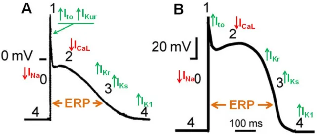
Figure 1. Schematic representation of atrial (A) and ventricular (B) action potentials with underlying principle ionic currents. Inward currents (with downward arrows): INa-sodium current; ICaL-L-type calcium current; Outward currents (with upward arrows): Ito-transient outward current; IKur-ultra rapidly activating delayed rectifier current; IKr and IKs-rapidly and slowly activating delayed rectifier current; IK1-inward rectifier current; Note that IKur is presented only in the atria. Numbers in black indicate the different phases of action potential. ERP: effective refractory period. (Adapted from Ravens U. et al. [60] with modifications).
The normal electrophysiological behavior of the heart is determined by the orderly propagation of electrical impulses resulting in rapid depolarization and slow repolarization, thereby generating action potentials in individual cardiac cells. In order to better understand AF mechanisms, it is essential to review the basic physiology of the cardiac action potential (AP). As the key determinant of the rhythmical contraction of cardiac cells, cardiac AP reflects the alterations of transmembrane potentials which are determined by inward (depolarizing) and outward (repolarizing) currents (Figure 1). Both atrial (Figure 1A) and ventricular (Figure 1B) APs last several hundred milliseconds and consist of five phases: Phase 0, Phase 1, Phase 2, Phase 3, and Phase 4. A rapid depolarization occurs during the phase 0 of AP, which is the result of a large inward current carried by voltage-gated sodium channels (INa) (Figure 1). This rapid depolarization is followed by a phase 1 early repolarization, which is primarily due to the inactivation of INa as well as the activation of the transient outward potassium current (Ito) and the ultra-rapidly activating delayed
rectifier potassium current (IKur) (in atria) (Figure 1A). Repolarization continues in phase 2 of AP, where inward L-type calcium current (ICaL) counteracts with a gradually-increasing outward repolarizing potassium currents that are mainly composed of the rapid delayed rectifier potassium current (IKr) (Figure 1A&B). This results in a long lasting repolarization, constituting a morphological “plateau” shape in cardiac AP (Figure 1A&B). It is noteworthy that the shape of atrial AP (Figure 1A) is normally triangular because of the relatively short plateau phase compared with ventricular action potential (Figure 1B). Phase 3 is the final repolarization step in AP. Both IKr and slow delayed rectifier potassium current (IKs) contribute to terminate the repolarization during this phase. Once repolarization is complete, cardiac cells return to resting membrane potential during phase 4 of AP. The maintenance of the resting membrane potential in phase 4 is determined by the inward rectifier potassium current (IK1). Collectively, increase in inward currents will tend to prolong the action potential duration (APD), whereas increased outward currents abbreviate it. It is important to note that cardiac cells are refractory to the initiation of new APs during phases 0, 1, 2, and part of phase 3 (Figure 1A&B). This is termed the effective refractory period (ERP). During this period, no stimulus regardless of its strength is able to initiate a propagated AP until cells return to phase 4. The ERP acts as a protective mechanism to keep the heart rate in check and coordinate the cardiac muscle contraction, thereby preventing arrhythmia. It is directly correlated with APD. For example, the shorter ERP is often associated with shorter APD.
1.2.2 Overview of AF Pathophysiology
AF is defined as a supraventricular tachyarrhythmia with characteristic chaotic and uncoordinated contraction of atrium, which results in a mechanical dysfunction of the atria. The first modern notions about the mechanism of AF was introduced in the early twentieth century [49]. Over the past century, researches have greatly increased our knowledge in understanding of AF mechanisms; it is now believed that focal ectopic activity (or triggered activity) and reentry (single-circuit reentry or multiple-circuit reentry) are the underlying pathophysiological mechanisms for AF initiation and maintenance [49, 61, 62], as shown in Figure 2.
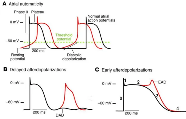
Figure 2. Arrhythmogenic mechanisms underlying AF. A. Ectopic focus. B. Single-circuit reentry. C. Multiple-circuit reentry. (Adapted from Iwasaki Y. et al. [61]).
1.2.2.1 Ectopic Mechanism
Up until Haissaguerre et al. [63] discovered that ectopic beats originating in the pulmonary veins played a pivotal role in AF initiation, the multiple-circuit reentry remained the sole dominant theory for AF mechanism. Abnormal automaticity, delayed afterdepolarization (DAD), and early afterdepolarization (EAD) constitute the principle mechanisms underlying focal ectopic activity as demonstrated in Figure 3 [62].
Figure 3. Cellular mechanisms underlying focal ectopic activity. A. Abnormal automaticity. B. Delayed afterdepolarization. C. Earlier afterdepolarization. Numbers in panel C indicate
the different phases of action potential. (Adapted from Wakili R. et al. [62] with modifications).
1.2.2.1.1 Abnormal Automaticity
Normal atrial cells are fired through the sinoatrial (SA) node pacemaking system, and then return to negative potential (resting potential, ~ -80mV) until the next firing from SA node (Figure 3A, black line). However, when the cell membrane reaches critical potential (or threshold potential, ~ -60mV) before the next normal impulse from SA node, an abnormal spontaneous depolarization may occur, resulting in abnormal automaticity (Figure 3A, red line). It is worth noting that the role of abnormal automaticity as a proarrhythmic mechanism during AF remains unclear [62].
1.2.2.1.2 Delayed Afterdepolarization
Delayed afterdepolarizations (DADs) occur after the complete repolarization of the triggering action potential and account for the most important mechanism underlying focal ectopic activity in AF (Figure 3B) [62]. DADs are caused by excessive diastolic Ca2+ released from sarcoplasmic reticulum (SR, the main cardiac Ca2+ storage organelle). The excessive diastolic Ca2+ are handled by transmembrane Na+/ Ca2+ exchanger (NCX) in an electrogenic manner, extruding one Ca2+ ion while pumping 3 Na+ into the cell. This creates a net inward current, which can depolarize the cell, resulting in an afterdepolarization. Therefore, both increased diastolic Ca2+ and enhanced NCX activity may contribute to DADs. The DAD-related AF is associated with congestive heart failure [64] and genetic defect [65].
1.2.2.1.3 Early Afterdepolarization
Early afterdepolarizations (EADs) occur during phase 2 or 3 of the action potential (Figure 3C). This type of afterdepolarization is primarily caused by recovering of L-type Ca2+
channels from inactivation during the plateau phase due to the excessively-prolonged APD. EAD-related AF has been seen in patients with congenital long-QT syndrome [66].
1.2.2.2 Re-entrant Mechanisms
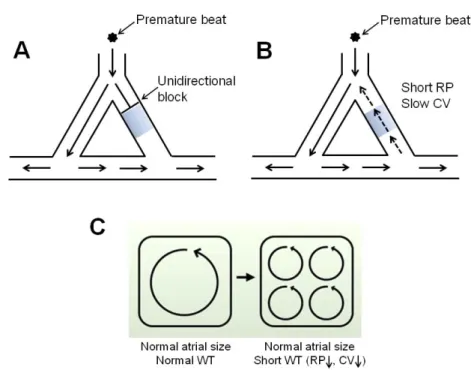
Figure 4. Mechanisms for reentry. A. An unidirectional block occurs when an ectopic impulse dies out in a still-refractory region. B. Conditions for maintenance of reentry. C. Conditions favoring multiple-circuit reentry. RP: refractory period; CV: conduction velocity. Blue-shadow area represents still-refractory region. (Panel C adapted from Nattel S et al. [67] with modifications).
Reentry is conventionally initiated by a premature ectopic beat (Figure 4A). This ectopic beat fails to conduct through a still-refractory region in one direction (“unidirectional block”) (Figure 4A), while conducting in the other direction through a region that is no longer refractory (Figure 4A). However, if enough time has elapsed for the recovery of excitability in the refractory region, the impulse can re-enter this region, resulting in reentry (Figure 4B). Accordingly, shorter refractory period (allowing faster recovery of excitability) and/or slower conduction velocity (gaining more time for the non-refractory region to regain excitability) may favor the maintenance of reentry (Figure 4B). Reentry can sustain either as a single circuit form (Figure 1B) or a multiple-circuit form (Figure 1C). For single circuit reentry, the irregular activity is maintained by rapid regular firing
(Figure 1B), whereas for multiple-circuit reentry, the irregularity is determined by the co-existence of multiple dyssynchronous reentry circuits (Figure 1C). There are two leading theoretical models that conceptualize the mechanism of reentry: leading circuit [68] and spiral wave [69]. While both models predict the presence of vulnerable substrates as the key determinant in reentry sustainability, the leading circle theory fails to explain the clinical observation that blocking of Na+ channel effectively terminates AF [70]. Nevertheless, leading circle theory still remains the widely-accepted notion to explain reentry mechanism. An important concept of leading circle theory is the “wavelength of reentry” [49, 68]. The wavelength (WL) of a reentry circuit refers to the shortest pathlength by which a reentry can be established. It is given as the distance that an electrical impulse travels in one refractory period and is critically determined by refractory period (RP) and conduction velocity (CV), as shown in the equation: WL=RP × CV [70, 71]. Accordingly, a shorter wavelength (as the result of abbreviated RP and/or reduced CV) may allow more available reentry circuits to be accommodated in a given atria, favoring multiple-circuit reentry that sustains AF (Figure 4C).
1.2.2.3 Relation of basic AF mechanisms to different forms of clinically-encountered AFs
The natural course of AF is believed to begin as the paroxysmal form, which is triggered by the ectopic foci originated from pulmonary vein sleeves [63].With time, AF tends to become persistent if ectopic firing is sustained or reversible reentry substrates are developed [62]. If the reentry substrates further become irreversible or fixed, AF becomes permanent [62].
1.3 Atrial Remodeling in AF
The introduction of the concept “atrial remodeling” over the past two decades has greatly advanced our understanding of AF mechanisms. Atrial remodeling refers to the process by which AF, once initiated, alters atrial electrophysiological (in a short term) and/or structural (over a long term) properties in ways that sustain itself [72].
1.3.1 Electrical Remodeling in AF
AF-induced electrical remodeling, which results in trigger activity and functional reentry substrates by altering ion channel/transporter expression and/or function, occurs normally within a short time after AF initiation [73]. Both focal ectopic activity and functional reentry are primarily caused by increased Ca2+ loading due to the very rapid atrial rate (tachycardia) during AF [73]. Therefore, more recently, AF-induced electrical remodeling has been termed as atrial tachycardia remodeling (ATR) [67]. Evidence from animal models and clinical studies has highlighted the importance of ATR in pathogenesis of AF and in the transition from paroxysmal AF to persistent AF [72, 74-81]. A prominent finding in ATR is the abbreviation of refractoriness as the result of APD shortening. Consistently, a shorter atrial APD is found in AF patients compared to patients with sinus rhythm [78-81]. Similar observations have also been seen in animal models of AF [72, 74-76].
The ionic mechanisms underlying APD shortening primarily involve downregulation and/or inactivation of L-type calcium current (ICaL) and enhancement of inward rectifier potassium currents (IK1 and/or IKACh). The functional implication of the deregulation of other ion channels in APD shortening remains obscure. Details regarding the pathophysiological significance of these changes will be described below.
1.3.1.1 Upregulation of IK1
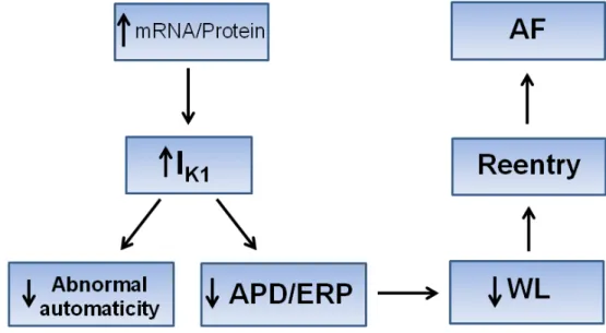
One of the most important electrophysiological changes in AF-related ATR is the upregulation of inward-rectifier potassium channel, particularly, IK1. Increased IK1 has been consistently observed in isolated atrial cardiomyocytes from AF patients [82-89] and experimental AF animal models [90-92]. Interestingly, the functional change of IK1 in AF is in parallel with increased expression of its underlying subunits Kir2.1 at both mRNA and protein levels [83, 85]. The pathophysiological significance of IK1 upregulation in AF is believed to enhance AF sustainability by substantiating reentrant substrates [20, 73]. This is likely attributable to the crucial roles of IK1 in maintenance of resting potential and termination of action potential [67, 73]. Increased IK1 hyperpolarizes the atrial cells, which, on one hand, inhibits the abnormal automaticity, whereas on the other hand, shortens APD and ERP, thereby favoring the reentrant substrates for AF [67, 73, 93, 94]. Recently,
genetic evidence from familial AF as the result of a Kir2.1 gain-of-function mutation further indicates the pivotal role of IK1 upregulation in AF pathogenesis [95]. Nevertheless, the underlying molecular mechanism responsible for enhanced IK1 in AF is largely unknown. Interestingly, a new class of recently-discovered small non-coding RNA, namely microRNAs (miRNAs), has been suggested to be responsible for the upregulation of IK1 in AF [96].
1.3.1.2 Downregulation of ICaL
Another key finding in AF-related ATR is the downregulation of ICaL. ICaL has been consistently found to be downregulated in both clinical and experimental AF paradigms [76, 82, 87, 97-100], accompanied by corresponding decreases of its α-subunit Cav1.2 at mRNA level [101-107]. However, discrepant changes for Cav1.2 protein have been reported [99, 101, 103, 104, 107, 108], implying the complicated underlying mechanisms as well as uncontrollable variables in clinical studies [67]. The observed ICaL reduction in AF is believed to be an adaptive response of atrial cardiomyocytes to calcium overload as a consequence of rapid atrial rate during AF [49]. This adaptive response, in a short term, tends to reduce channels activity, but over a longer term, tends to decrease the expression of the channels at the gene level [49]. As a result, reduced ICaL decreases APD and wavelength, creating reentry substrate for AF perpetuation [49]. Although the molecular mechanism underlying ATR-induced ICaL downregulation has been extensively investigated by many research groups, it is still not completely understood. While some studies indicated that the transcriptional downregulation of Cav1.2 α-subunit is likely due to the activation of Ca2+/calmodulin/calcineurin/NFAT signaling pathway caused by Ca2+ overload [107, 109], others suggested the possible involvements of the downregulation of accessory β1-, β2a-, β2b-, β3- and α2δ2-subunits, impairment of Cav1.2 protein trafficking, and dephosphorylation of the channels due to enhanced PP1 and PP2A activities [85, 99, 102, 110-112]. Furthermore, a recent study demonstrated that miRNAs may potentially contribute to the downregulation of ICaL in AF through a post-transcriptional regulatory mechanism [113]. Taken together, the underlying molecular mechanism for AF-induced ICaL downregulation appears to be a very complex process, involving both transcriptional and post-transcriptional regulations.
1.3.1.3 Abnormal Ca2+ Handling
In addition to the reduction of ICaL, AF-induced electrical remodeling also causes abnormal intracellular Ca2+ handling, which primarily promotes spontaneous diastolic SR Ca2+ release that enhances NCX-mediated Ca2+ extrusion. The NCX-mediated Ca2+ extrusion is an electrogenic process, which produces a net inward (depolarizing) current, causing DADs [62, 73]. Several studies have suggested that the spontaneous diastolic SR Ca2+ release is attributed to the dysfunction of ryanodine receptor 2 (RyR2, cardiac Ca2+ release channel on SR) [114-117], which is likely due to hyperphosphorylation caused either by increased activities of protein kinase A (PKA) and Ca2+/calmodulin-dependent kinase II (CaMKII) [115-118] or by decreased activities of phosphatases [119]. Additionally, abnormal RyR2 release can also be a result of excess SR Ca2+ loading due to abnormal enhancement of SR Ca2+ uptake mediated through SR Ca2+ ATPase (SERCA, Ca2+ uptake pump on SR), which is activated by hyperphophorylated phospholamban [119].
1.3.1.4 Alterations of other K+ currents
IKACh, another important inward rectifier potassium current, is activated upon the stimulation of acetylcholine released from vagal nerve and is responsible for cardiac vagal effects. Activation of the channels hyperpolarizes the cell membrane and shortens APD, contributing to AF pathogenesis. Several studies have shown that the agonist-dependent IKACh is reduced, whereas constitutively-active IKACh (agonist-independent) is significantly enhanced during AF-induced ATR [83, 91, 120, 121]. However, mRNA and protein levels of its underlying subunit Kir3.1 and Kir3.4 are found unaltered in experimental AF animal models [120, 121] but decreased in AF patients [122]. The enhanced constitutively-active IKACh is attributable to increased single channel open probability, which is controlled by the balance between different protein kinase C (PKC) isoforms [123, 124].
Consistent reduction of Ito along with the mRNA and protein of its underlying α-subunit Kv4.3 is found in both AF patients and experimental AF animal models [76, 86, 101, 104]. However, the functional implication of Ito reduction in AF remains to be unclear [67, 73].
Inconsistent changes of IKur have been reported in both experimental AF animal models and AF patients [67], indicating the lack of pathological relevance of this channel in AF. However, given the atrial specific property of IKur, it still remains as a very attractive therapeutic target for the treatment of AF [49].
No changes of delayed rectifier currents IKs and IKr were observed in a canine model of AF [76] and there is lack of information regarding the changes of these currents in humans due to the technical difficulty in acquiring human samples and cell isolation [125].
1.3.2 Structural Remodeling in AF
Atrial structural remodeling (ASR) is another major aspect of atrial remodeling in AF. It is a relatively slower remodeling process compared to electrical remodeling, primarily comprising morphological changes to atrial myocardial structure and atrial architecture [20, 126]. ASR has been observed in AF from both clinical settings and experimental models, and significantly contributes to form the reentry substrates for AF. Consequences of ASR include increased atrial fibrosis, altered connexin expression, myocyte hypertrophy, myocyte apoptosis, and atrial dilation, among which, the first three aspects have been extensively studied.
1.3.2.1 Atrial Fibrosis
Atrial fibrosis, although not exclusively related to AF but appears as a common feature of clinical AF. It is one of the most important arrhythmogenic contributors to AF [73]. Atrial fibrosis has been commonly observed in lone AF [127, 128] and AF-associated pathophysiological conditions, including congestive heart failure (CHF) [129, 130], valvular diseases [131], rheumatic heart disease [132, 133], dilated and hypertrophic cardiomyopathy [134, 135], and senescence [136, 137]. Moreover, a reduced AF stability and delayed atrial structural remodeling process have been observed in some experimental and clinical studies using compounds with known anti-fibrotic effect, such as statins (HMG-CoA reductase inhibitors) [138, 139], angiotensin II type 1 receptor (AT1R) blocker
[140], and fish oil [141]. However, the observed beneficial outcome of these compounds may be a result of the improvements in hemodynamic [73]. Together, these findings suggest an important association between atrial fibrosis and AF but fail to establish a causal significance of atrial fibrosis in AF occurrence and perpetuation. It is also important to note that AF may itself promote atrial fibrosis, which in turn sustains AF [142, 143]. Evidence supporting this notion came from the observations that the quantity of fibrosis positively correlates with the persistence of AF in patients [144], and that atrial tachypacing alone causes atrial fibrosis in a canine ATP model with well-controlled ventricular rate [145]. Cardiac fibrosis is either a reparative or reactive process, which primarily involves excessive accumulation of extracellular matrix (ECM) proteins secreted by myofibroblasts (cells derived from fibroblasts in the presence of various stimuli) [142, 143]. Reparative fibrosis maintains tissue structural integrity by replacing dead cardiomyocytes, whereas reactive fibrosis occurs in response to various cardiac insults and causes interstitial expansion, separating muscle bundles [142, 143]. The resultant fibrotic tissue creates an obstacle to interfere with normal impulse propagation and thus causes conduction abnormalities [129]. In addition, recent studies have suggested that cardiac fibroblasts can electrically influence adjacent cardiomyocytes and thus alter cardiac electrical function [143]. Taken together, the fibrotic remodeling manifested during AF may cause conduction abnormalities and increase fibroblast-cardiomyocyte electrical interaction, favoring AF occurrence and maintenance.
1.3.2.1.1 Profibrotic Factors and Atrial Fibrosis
The precise molecular mechanisms underlying ECM formation during atrial fibrosis remain incompletely understood. Emerging evidence has suggested that several cardiomyocyte and/or fibroblast secreted factors with known profibrotic effects are critically involved [142]. Among these factors, angiotensin II (AngII) and its downstream mediator transforming growth factor β1 (TGF-β1) have been well characterized to contribute to fibroblast differentiation and proliferation, whereas platelet-derived growth factor (PDGF) and connective tissue growth factor (CTGF) have just emerged as potential fibrosis mediators [20, 142].