HAL Id: hal-03113281
https://hal.sorbonne-universite.fr/hal-03113281
Submitted on 18 Jan 2021
HAL is a multi-disciplinary open access
archive for the deposit and dissemination of
sci-entific research documents, whether they are
pub-lished or not. The documents may come from
teaching and research institutions in France or
abroad, or from public or private research centers.
L’archive ouverte pluridisciplinaire HAL, est
destinée au dépôt et à la diffusion de documents
scientifiques de niveau recherche, publiés ou non,
émanant des établissements d’enseignement et de
recherche français ou étrangers, des laboratoires
publics ou privés.
Comprehensive study of 28 individuals with
SIN3A-related disorder underscoring the associated mild
cognitive and distinctive facial phenotype
Meena Balasubramanian, Alexander Dingemans, Shadi Albaba, Ruth
Richardson, Thabo Yates, Helen Cox, Sofia Douzgou, Ruth Armstrong,
Francis Sansbury, Katherine Burke, et al.
To cite this version:
Meena Balasubramanian, Alexander Dingemans, Shadi Albaba, Ruth Richardson, Thabo Yates, et al..
Comprehensive study of 28 individuals with SIN3A-related disorder underscoring the associated mild
cognitive and distinctive facial phenotype. European Journal of Human Genetics, Nature Publishing
Group, inPress, �10.1038/s41431-020-00769-7�. �hal-03113281�
A R T I C L E
Comprehensive study of 28 individuals with
SIN3A-related
disorder underscoring the associated mild cognitive and distinctive
facial phenotype
Meena Balasubramanian1,2●Alexander J. M. Dingemans3●Shadi Albaba4●Ruth Richardson5●Thabo M. Yates 1●
Helen Cox6●Sofia Douzgou7,8●Ruth Armstrong9●Francis H. Sansbury 10●Katherine B. Burke10●
Andrew E. Fry 10●Nicola Ragge6,11●Saba Sharif6●Alison Foster6●Annachiara De Sandre-Giovannoli12,13,14●
Sahar Elouej12●Pradeep Vasudevan15●Sahar Mansour16●Kate Wilson17 ●Helen Stewart17 ●Solveig Heide18●
Caroline Nava18●Boris Keren18●Serwet Demirdas19●Alice S. Brooks19●Marie Vincent 20,21●Bertrand Isidor20,21●
Sebastien Küry 20,21●Meyke Schouten3●Erika Leenders3●Wendy K. Chung22●Arie van Haeringen23●
Thomas Scheffner24●Francois-Guillaume Debray25●Susan M. White 26,27●Maria Irene Valenzuela Palafoll 28●
Rolph Pfundt3●Ruth Newbury-Ecob29 ●Tjitske Kleefstra3 Received: 28 May 2020 / Revised: 15 October 2020 / Accepted: 21 October 2020 © The Author(s) 2021. This article is published with open access
Abstract
Witteveen-Kolk syndrome (OMIM 613406) is a recently defined neurodevelopmental syndrome caused by heterozygous loss-of-function variants in SIN3A. We define the clinical and neurodevelopmental phenotypes related to SIN3A-haploinsufficiency in 28 unreported patients. Patients with SIN3A variants adversely affecting protein function have mild intellectual disability, growth and feeding difficulties. Involvement of a multidisciplinary team including a geneticist, paediatrician and neurologist should be considered in managing these patients. Patients described here were identified through a combination of clinical evaluation and gene matching strategies (GeneMatcher and Decipher). All patients consented to participate in this study. Mean age of this cohort was 8.2 years (17 males, 11 females). Out of 16 patients≥ 8 years old assessed, eight (50%) had mild intellectual disability (ID), four had moderate ID (22%), and one had severe ID (6%). Four (25%) did not have any cognitive impairment. Other neurological symptoms such as seizures (4/28) and hypotonia (12/28) were common. Behaviour problems were reported in a minority. In patients ≥2 years, three were diagnosed with Autism Spectrum Disorder (ASD) and four with Attention Deficit Hyperactivity Disorder (ADHD). We report 27 novel variants and one previously reported variant. 24 were truncating variants; three were missense variants and one large in-frame gain including exons 10–12.
Introduction
Witteveen-Kolk syndrome (OMIM #613406) was first described in 2016 with characteristic distinctive facial fea-tures, microcephaly, short stature, mild intellectual dis-ability (ID) with delayed cognitive and motor development and subtle anomalies on MRI-brain imaging [1]. Although sparsely reported, frameshift as well as missense variants in the Switch-insensitive 3 transcription regulator family member A (SIN3A) (OMIM *607776) have been described in larger neurodevelopmental disorder cohorts, with an overall mild clinical picture [2]. Narumi-Kishimoto et al. presented a further patient with SIN3A frameshift variant and facial features of Witteveen-Kolk with relatively mild ID and normal growth [3].
These authors contributed equally: Meena Balasubramanian, Alexander J. M. Dingemans, Shadi Albaba
These authors jointly supervised this work: Meena Balasubramanian, Tjitske Kleefstra
* Meena Balasubramanian meena.balasubramanian@nhs.net
Extended author information available on the last page of the article
Supplementary information The online version of this article (https://
doi.org/10.1038/s41431-020-00769-7) contains supplementary material, which is available to authorised users.
123456789
0();,:
123456789
The SIN3A gene is located in the chromosome 15 band q24 and is within the shortest region of overlap of various reported 15q24 microdeletions, therefore, is thought to be the critical gene for the atypical 15q24 microdeletion syn-drome [4]. SIN3A encodes a transcriptional regulatory protein, which is associated with scaffolding in the core histone deacetylase complex [5]. In our earlier study, we showed that SIN3A is involved in cortical neurogenesis, supporting the hypothesis that variants in the gene that adversely affect its function lead to a broad range of developmental and neurological problems. We identified an additional 28 patients with SIN3A variants in order to comprehensively define the phenotype with a focus on both developmental and behavioural aspects, as well as investi-gating genotype-phenotype correlations.
Methods
We collected the molecular and clinical features on 28 unpublished individuals with SIN3A variants by a colla-boration facilitated by Deciphering Developmental Dis-orders (DDD study) [6], GeneMatcher [7] and DECIPHER (DECIPHER v9.24: https://decipher.sanger.ac.uk/) [8], in which, several clinical groups independently identified individuals with developmental delay/intellectual disability (DD/ID) and related phenotypes with rare variants in SIN3A during routine diagnostic genetic testing. An application to the DDD study for a Complementary Analysis Project was made, allowing access to anonymised details of patients with SIN3A variants identified through this study (https://www.ddduk.org/). Clinicians of selected patients were then contacted to invite patients and their families to be recruited. Clinical analysis of these patients was per-formed during regular consultations focusing on medical history, physical examination and observational analysis of behavioural features along with reported history by the family. In all patients, exome sequencing and variant fil-tering were performed, according to the routine protocol and diagnostic procedures at each institute.
Identified patients with a class 4 or 5 variants (likely pathogenic or pathogenic variants) in SIN3A, according to the American College of Medical Genetics (ACMG) cri-teria, were approached to participate in this study. Informed consent for publication was obtained from all patients and/ or their guardians. The responsible clinician reviewed medical records of each participant in order to compre-hensively document the phenotype.
The clinical significance of the variants identified was interpreted according to the ACMG [9] guidelines and further review publications [10]. Excluded from the study were: patients with an additional proven genetic diagnosis where the SIN3A variant was not thought to be contributory
or the sole pathogenic finding, those with a chromosomal anomaly explaining or likely to be explaining the pheno-type, and those in whom SIN3A variants were of uncertain clinical significance with no convincing clinical correlation. All the variants reported here have been submitted to DECIPHER database [https://decipher.sanger.ac.uk] and phenotypes are included in DECIPHER database for the DDD patients [Patients 1–13]- DDD identifiers, [https:// decipher.sanger.ac.uk/gene/SIN3A/overview/clinical-info].
Results
Molecular genetics
In this study, we reported 28 patients with variants that adversely affect protein function and which are classified as pathogenic or likely pathogenic variants in SIN3A. The findings of this study expand the variant spectrum pre-viously reported in SIN3A,3. 27/28 patients in this study have novel variants. Patient 15 with c.3310C>T, p. (Arg1104*) variant is a family member of a patient we have previously reported in Witteveen et al. [1]. Predominantly, variants found in our patient cohort are truncating and predicted to result in a protein loss-of-function (25/28). All 24 truncating variants have been classified as variants that adversely affect function (pathogenic) using the ACMG and ACGS (The Association for Clinical Genomics Science) variant classification guidelines using criterions: PVS1, PM2, and PS2 where appropriate [9, 10]. Of the four remaining variants, three were missense (patients 4, 18 and 26; Table 1) and one was a large in-frame gain which included the whole of exons 10, 11 and 12 (patient 24; Table 1). These four variants were all de novo in our patients with a specific and consistent phenotype to SIN3A and were absent from control population data sets in gno-mAD (https://gnomad.broadinstitute.org/). We have also used criterion PM1 at moderate level for two of the three reported missense variants, c.377C>T, p.(Ala126Val) and c.463A>G, p.(Lys155Glu), as they were located within a protein functional domain. These results show that missense causative variants are not clustered in a hot spot within SIN3A (Fig. 1 provides a schematic SIN3A structure and variant locations which, as demonstrated, are distributed throughout the gene). Details of the variants, ACMG cri-terions and classification are listed in Table1.
Assessment of pathogenicity of missense variants in
SIN3A
In order to assess if missense variants in our cohort were predicted to adversely affect the critical functional domains in SIN3A, we used a 3D model based on the solution
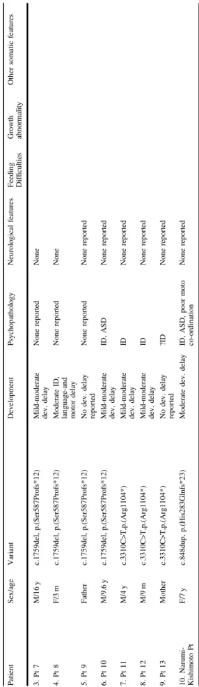
Table 1 Variant Table. No Variant using NM_001145358.1 Inheritance ACMG criterion Classi fi cation Published DECIPHER ID 1 c.1462del; p.(Val488Leufs*7 ) D e novo PS2-T/PVS1/PM2-M Pathogenic Novel 278680 2 c.2764C>T; p.(Arg922*) De novo PS2-T/PVS1/PM2-M Pathogenic Novel 306260 3 c.588del; p.(Asn197Metfs*4) De novo PS2-T/PVS1/PM2-M Pathogenic Novel 271952 4 c.377C>T; p.(Ala126Val) De novo PS2-T/PM1-M/PM 2-M/PP3-S Likely Pathogenic Novel 262798 5 c.1245_1246del; p.(Asn415Lysfs*24 ) D e novo PS2-T/PVS1/PM2-M Pathogenic Novel 293976 6 c.775dup; p.(His259Profs*47) De novo PS2-T/PVS1/PM2-M Pathogenic Novel 263709 7 c.824del; p.(Pro275Hisfs*12) De novo PS2-T/PVS1/PM2-M Pathogenic Novel 307508 8 c.2248_2251del; p.(Leu750Metfs*43 ) D e novo PS2-T/PVS1/PM2-M Pathogenic Novel 266515 9 c.2339del; p.(Ala780Glyfs* 14) De novo PS2-T/PVS1/PM2-M Pathogenic Novel 260388 10 c.889C>T; p.(Gln297*) De novo PS2-T/PVS1/PM2-M Pathogenic Novel 264582 11 c.1715_1722delinsC CCAAGTGTA; p.(Gly572Alafs*11) De novo PS2-T/PVS1/PM2-M Pathogenic Novel 292229 12 c.3323C>G; p.(Ser1108*) De novo PS2-T/PVS1/PM2-M Pathogenic Novel 282105 13 c.3490A>T; p.(Lys1164*) De novo PS2-T/PVS1/PM2-M Pathogenic Novel 282212 14 c.46C>T; p.(Gln16*) Maternal PVS1/PM2-M Pathogenic Novel 421224 15 c.3310C>T; p.(Arg1104*) Unknown PVS1/PM2-M Pathogenic Witteveen et al. [ 1 ] 421225 16 c.2809_2810del; p.(Lys937Glufs*2) De novo PS2-T/PVS1/PM2-M Pathogenic Novel 421226 17 c.1489_1499del; p.(Arg497Cysfs*13 ) D e novo PS2-T/PVS1/PM2-M Pathogenic Novel 421228 18 c.3317T>C; p.(Met1106Thr) De novo PS2-T/PM2-M/B P4-S Likely Pathogenic Novel 421229 19 c.1675C>T; p.(Arg559*) De novo PS2-T/PVS1/PM2-M Pathogenic Novel 421230 20 c.3303C>G; p.(Tyr1101*) De novo PS2-T/PVS1/PM2-M Pathogenic Novel 421231 21 c.1570_1577del; p.(Tyr524Valfs*26) De novo PS2-T/PVS1/PM2-M Pathogenic Novel 421232 22 c.2185_2186del; p.(Leu729Glyfs*8) De novo PS2-T/PVS1/PM2-M Pathogenic Novel 421233 23 c.3441_3445del; p.(Lys1148Argfs*12) De novo PS-2-T/PVS1/PM2 -M Pathogenic Novel 421234 24 c.1318_1737dup; p.(Val580Lysfs*35 ) D e novo PS2-T/PVS1 Pathogenic Novel 421236 25 c.1773G>A; p.(Trp591*) De novo PS2-T/PVS1/PM2-M Pathogenic Novel 421237 26 c.463A>G; p.(Lys155Glu) De novo PS2-T/PM1-M/PM 2-M/PP3-S Likely Pathogenic Novel 421238 27 c.2803C>T; p.(Arg935*) De novo PS-2T/PVS1/PM2-M Pathogenic Novel 421239 28 c.1888dup; p.(Ile630Asnfs*17) De novo PS-2/PVS1/PM2-M Pathogenic Novel 421240 ACMG Criterion applied: PS2-T: De novo (both maternity and paternity con fi rmed) in a patient with the disease and no family history, used at strong level. PVS1: null variant (nonsense, frameshift, canonical ±1 or 2 splice sites, initiation codon, single or multiexon deletion) in a gene where LOF is a know n mechanism of disease. PM1-M: Located in a critical functional domain without benign variation, used at moderate level. PM2-M: Absent from controls in gnomAD database, used at moderate level. PP3-S: Multiple lines of computational evidence support a deleterious effect on the gene or gene product, used at supporting level. PM4-M: Protein length changes as a result of in-frame deletions/insertions in a non-repeat region or stop-loss variants, used at moderate level. BP4-S: Multiple lines of computational evidence suggest no impact on gene or gene product, used at supporting level No corresponds to patient number. DECIPHER ID corresponds to entry of open access variant on https://decipher.sanger.ac.uk [DatabasE of genomiC varIation and Phenotype in Humans using Ensembl Resources].
structure of mouse SIN3A PAH1 bound to the Sin3 inter-action domain (SID) of SAP25 (SIN3A Associated Protein 25) [11]. The pair of amphipathic helices (PAH) domains are predicted as important for the recruitment by and interaction with a diverse number of transcription factors and therefore, critical for protein function [12, 13]. Both altered residues by c.377C>T, p.(Ala126Val) and c.463A>G, p.(Lys155Glu) were predicted to be part of the SAP25 SID-binding surface. We predicted that SIN3A Ala126 with its small hydrophobic side chain formed a pocket for the larger hydrophobic side chain of SAP25 Leu142 and was invariant across species [11].
SIN3A Lys155 is also predicted to form a 2.2A hydrogen bond with the polar Gln143 of SAP25 (orange line in Fig.2) and was also conserved in the PAH1 domain across species.
Along with the lack of normal variation in this region (using missense constraint data in SIN3A from Decipher: https:// decipher.sanger.ac.uk/gene/SIN3A#overview/protein-info), we applied criterion PM1 at moderate level for the classification of both c.377C>T, p.(Ala126Val) and c.463A>G, p.(Lys155-Glu). We also suggest that PM1 can be applied at a moderate level for missense variants within residues p.119–189 of the SIN3A protein providing that the change caused by the variant was not present in other species and also meets the PM2 criteria (see Fig.2).
Finally, the c.3317T>C, p.(Met1106Thr) is within the C-terminal domain of SIN3A, this region is not well char-acterised and the in silico pathogenicity prediction programs suggests a benign effect (REVEL score of 0.105 shows a benign effect). However, this variant was confirmed to be
B B D C C E F F G G A A A L M K K K L J J I I M H H
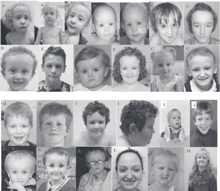
Fig. 1 Characteristic facial appearance of patients with variants in SIN3A. Note the high forehead, small, pointed chin and down-slanting
palpebralfissures. A: Patient 1, B Patient 2, C Patient 4, D Patient 5, E
Patient 9, F Patient 11, G Patient 12, H Patient 13, I Patient 16, J Patient 18, K Patient 25, L Patient 27, M Patient 28.
de novo in patient 18, novel in control populations in gnomAD and with a consistent phenotype to that of the rest of our cohort. No other variants were found in this patient on exome sequencing. Therefore, we classified this variant in addition as likely pathogenic, but without the use of PM1.
Clinical
findings
Table 2 shows a summary of the characteristics of 28 patients (17 males, 11 females) with variants in SIN3A who were included in this retrospective study. The mean age of participants was 8.2 years (range 0.6–67 years). Table 3
provides an overview of salient features described in this cohort in comparison to published literature and Table 4 has detailed phenotypic information on this cohort of patients.
Development
Of 28 patients, 15 (56%) had global developmental delay. Out of 16 patients ≥8 years old, (one patient was not for-mally assessed) seven (44%) had mild intellectual disability (ID), four had moderate ID (25%), and one had severe ID (6%). Four (25%) did not appear to have any cognitive impairment. In 21 patients≥5 years old, some form of motor developmental delay was reported in 13 patients (62%) and in 16 patients (76%) there was some form of language developmental delay. Intelligence Quotient (IQ) was only
formally measured in 6/28 patients and the score ranged from 60 to 100. This suggests that patients within this cohort have low normal intelligence.
Autism spectrum disorder (ASD) is rarely diagnosed before the age of 24 months [14]. In one patient aged 14 months, ASD was thought possible but not formally assessed due to the young age. In three of 23 patients ≥2 years (13%), ASD was diagnosed. Attention Deficit Hyperactivity Disorder (ADHD) is most commonly diag-nosed in children between 6 and 12 years, though this can be diagnosed in a younger age group. ADHD was diag-nosed in four patients≥2 years (17%).
Growth and feeding dif
ficulties
13/28 (46%) patients in our cohort had head circumference at least two standard deviations (SD) below the mean. Weight was less than two SD below the mean in 8/28 patients (29%) and height was less than two SD below the mean in 5/28 patients (18%).15/28 (54%) of patients had feeding difficulties with at least two patients documented to require nasogastric tube feeding mainly in the neonatal period.
Craniofacial features
Fourteen (50%) patients were reported to have craniofacial dysmorphism. In ten patients, the facial gestalt was Fig. 2 Schematic SIN3A protein structure and variant location. ITD: intragenic deletion; Plot of variants done using St. Jude Cloud protein
Table 2 Select phenotypic characteristics of SIN3A cohort compared with previously published literature. Patient Sex/age Variant Development Psychopathology Neurological features Feeding Dif fi culties Growth abnormality Other somatic features 1 DECIPHER 278680 F/9 y c.1462del, p.(Val488Leufs*7) GDD; ataxia; delayed speech N/A Hypotonia Cerebellar atrophy Yes Weight & OFC − 2SD Height − 1SD Scoliosis; strabismus 2 DECIPHER 306260 M/1.2 y c.2764C>T, p.(Arg922*) Language-and motor delay ?ASD Hypotonia?; epilepsy Foci high signal white matter, dilation of lateral ventricles Yes Weight < − 2SD OFC < − 2SD Apnoea; Cow ’s milk protein intolerance, nystagmus secondary to oculocutaneous albinism 3 DECIPHER 271952 F/8.3 y c.588del, p.(Asn197Metfs*4) Mild ID, language-and motor delay ADHD Hypotonia Yes No 4 DECIPHER 262798 M/13.5 y c.377C>T, p.(Ala126Val) GDD language-and motor delay, Developmental coordination disorder Aggressive behaviour, High functioning autism Neonatal hypotonia Yes No Constipation; sacral dimple, Hirsute on back as baby; pes planus joint hypermobility hearing impairment (mixed) 5 DECIPHER 293976 M/10.1 y c.1245_1246del, p.(Asn415Lysfs*24) Moderate ID, language and motor delay None Neonatal hypotonia Yes OFC < − 2SD Pectus excavatum. Unilateral inguinal hernia; Sacral dimple 6 DECIPHER 263709 M/5.4 y c.775dup, p.(His259Profs*47) GDD, language delay None None Unknown Height < − 2SD Weight < − 2SD OFC < -2SD Multiple dental caries; Eczema Failure to thrive; Mild conductive hearing impairment, Obstructive sleep apnoea, Submucous cleft hard palate 7 DECIPHER 307508 M/0.6 y c.824del, p.(Pro275Hisfs*12) GDD, language delay N/A Hypotonia No Height < − 2SD Weight < − 2SD OFC < − 2SD Sensorineural hearing loss 8 DECIPHER 266515 M/7.2 y c.2248_2251del, p.(Leu750Metfs*43) GDD, language-and motor delay None Hypotonia Yes Weight < − 2SD OFC < − 2SD 9 DECIPHER 260388 M/5 y c.2339del, p.(Ala780Glyfs*14) GDD language and motor delay Hyperactive behaviour None Yes Height < − 2SD Weight < − 2SD OFC < − 2SD Poor sleep; Oromotor coordination dysfunction 10 DECIPHER 264582 M/3.2 y c.889C>T, p.(Gln297*) GDD, language-and motor delay None None No
Height unknown Weight unknown OFC
< − 2SD Aplasia cutis congenita over the scalp vertex; Capillary hemangiomas; Reduced subcutaneous adipose tissue 11 DECIPHER 292229 F/7.9 c.1715_1722delinsCCCAAGTGTA, p. (Gly572Alafs*11) GDD, language delay Immature behaviour;? ASD Febrile seizures No No 12 DECIPHER 282105 F/4.6 c.3323C>G, p.(Ser1108*) ADHD, Aggressive behaviour, recurring obsessions Yes Height < − 2SD Weight < − 2SD OFC < − 2SD 13 DECIPHER 282212 M/9.1 c.3490A>T, p.(Lys1164*) Mild ID, language delay ADHD No OFC < − 2SD
Table 2 (continued) Patient Sex/age Variant Development Psychopathology Neurological features Feeding Dif fi culties Growth abnormality Other somatic features 14 DECIPHER 421224 M/25 y c.46C>T, p.(Gln16*) Moderate ID, GDD, language-and motor delay None Epilepsy, thinning of the corpus callosum No No 15 DECIPHER 421225 F/67 y c.3310C>T,p.(Arg1104*) Not formally assessed, possible mild ID None None Unknown OFC < − 2SD 2 episodes DVT cystocele and urethrocele, EVAR prosthesis, hernia cicatricalis, sigmoïd adenocarcinoma 16 DECIPHER 421226 M/11.1 y c.2809_2810del,p.(Lys937Glufs*2) Mild ID, GDD, language- motor delay None None No OFC < -2SD Scoliosis; Burkitt lymphoma (at 8 y ) 17 DECIPHER 421228 F/8.1 y c.1489_1499del, p.(Arg497Cysfs*13) Mild ID,GDD, language- motor delay None Sleeping problems Yes Height < − 2SD Weight < − 2SD OFC < − 2SD Orofacial cleft; conductive hearing loss 18 DECIPHER 421229 M/36.2 y c.3317T>C,p.(Met1106Thr) Moderate ID, language- motor delay None Epilepsy No No 19 DECIPHER 421230 M/22.1 y c.1675C>T,p.(Arg559*) Mild ID ASD, depression, psychosis Sleeping problems Yes No Thickened aortic valve 20 DECIPHER 421231 F/24.2 y c.3303C>G,p.(Tyr1101*) Language delay Multiple Complex DD, anxiety disorder Hypotonia Yes No 21 DECIPHER 421232 F/19 y c.1570_1577del, p.(Tyr524Valfs*26) Normal Schizoaffective disorder Neonatal hypotonia No Unknown Pelvic kidney; Palate defect; Congenital dislocation of the hip 22 DECIPHER 421233 M/10 y c.2185_2186del, p.(Leu729Glyfs*8) Mild motor delay None Hypotonia, epilepsy Unknown No 23 DECIPHER 421234 F/0.6 y c.3441_3445del,p.(Lys1148Argfs*12) Mild motor delay N/A Hypotonia Yes No 24 DECIPHER 421236 M/1.5 y c.1318_1737dup; p.(Val580Lysfs*35) GDD N/A Hypotonia, epilepsy Yes Unknown 25 DECIPHER 421237 M/6 y c.1773G>A, p.(Trp591*) Language and motor delay None Enlarged cerebral spaces Yes Weight < − 2SD 26 DECIPHER 421238 M/11.3 y c.463A>G, p.(Lys155Glu) Severe ID, non-verbal ASD Hypotonia Dysplastic corpus callosum Ventriculomegaly Yes No Multiple VSDs; bilateral iris and chorioretinal coloboma hearing loss; (mixed) immunode fi ciency 27 DECIPHER 421239 F/19 y c.2803C>T, p.(Arg935*) Mild ID, mild language and motor delay ADHD
Hypotonia Headaches Chiari
I malformation No No Joint laxity, recurrent otitis media; asthma; scoliosis 28 DECIPHER 421240 F/6.5 y c.1888dup, p.(Ile630Asnfs*17) Mild GDD,?mild ID None Hypotonia No OFC < − 2SD Joint laxity; Unilateral hypermetropia; multiple dental caries Witteveen et al. [ 1 ] 1. Pt 5 M/9.1 y c.803dup, p.(Leu269Thrfs*37) Moderate -severe ID, language-and motor delay ASD Hypotonia, epilepsy, central apnoea syndrome 2. Pt 6 F/16.4 y c.1010_1013del, p.(Lys337Serfs*16) Mild ID, motor delay ASD, depression, PTSS Hypotonia, epilepsy
confirmed independently by two clinical geneticists with expertise in dysmorphology. Common facial features included a broad, tall forehead; small mouth, thin upper lip with pointed chin and down-slanting palpebralfissures (see Fig. 3). The facial gestalt appears to be similar and poten-tially recognisable but only in the context of reverse phe-notyping with published images, confirming previous findings. Three patients had a palatal defect with one also having a bifid uvula.
Other clinical manifestations
In terms of neuroimaging, given the milder developmental delay phenotype, many patients in the cohort have not had MRI-brain imaging (19/28, 68%). Nine out of a total of nine patients that had brain imaging performed had reports available and in 7/9 (78%) abnormalities were seen. Most common was ventriculomegaly in 2/9, and a hypoplastic/ dysplastic corpus callosum in 2/9. Cerebellar atrophy was reported in two patients and there was one patient had a Chiari 1 malformation.
6/28 (21%) patients were noted to have seizures currently or in their past medical history. Hypotonia was a common manifestation as well, present in 12/28 (43%) patients.
Other reported features included hearing loss (5/28; 18%) of which 1 patient had sensorineural hearing loss, two patients had conductive hearing loss and two patients had mixed hearing loss. Ocular abnormalities reported included strabismus (1), nystagmus secondary to ocular albinism (1), bilateral iris and chorioretinal coloboma (1) and hypermetropia (1).
Of note, 2/28 patients were reported to have a malig-nancy, including sigmoid adenocarcinoma in the 67-year old patient which may be an age-related cancer and 1 younger patient with Burkitt lymphoma (Patient 16 at age
Table 2 (continued) Patient Sex/age Variant Development Psychopathology Neurological features Feeding Dif fi culties Growth abnormality Other somatic features 3. Pt 7 M/16 y c.1759del, p.(Ser587Profs*12) Mild-moderate dev. delay None reported None 4. Pt 8 F/3 m c.1759del, p.(Ser587Profs*12) Moderate ID, language-and motor delay None reported None 5. Pt 9 Father c.1759del, p.(Ser587Profs*12) No dev. delay reported None reported None reported 6. Pt 10 M/9.6 y c.1759del, p.(Ser587Profs*12) Mild-moderate dev. delay ID, ASD None reported 7. Pt 11 M/4 y c.3310C>T,p.(Arg1104*) Mild-moderate dev. delay ID None reported 8. Pt 12 M/9 m c.3310C>T,p.(Arg1104*) Mild-moderate dev. delay ID None reported 9. Pt 13 Mother c.3310C>T,p.(Arg1104*) No dev. delay reported ?ID None reported 10. Narumi-Kishimoto Pt F/7 y c.848dup, p.(His283Glnfs*23) Moderate dev. delay ID, ASD, poor moto co-ordination None reported ID intellectual disability, ASD autism spectrum disorder, ADHD attention de fi cit hyperactivity disorder, GDD global developmental delay.
Table 3 Summary of salient features in SIN3A-related disorder based on current cohort and previously published literature.
Clinical feature Current cohort (n= 28) Witteveen et al. [1] (n= 9) Narumi-Kishimoto [3] (n= 1) Total (n= 38) Intellectual disability 16 7 1 24 Mild 8 5 – 13 Moderate 7 2 1 10 Severe 1 – – 1 Speech delay 11 2 1 13 Hypotonia 12 2 – 14 Feeding difficulties 15 N/R – 15 Short stature 6 2 – 8 Epilepsy 4 1 – 5 Behavioural problems 12 4 1 17
eight). Patient 26 was noted to have T-cell lymphopenia at age 11 associated with immunoglobulin deficiency and significant bronchiectasis.
Discussion
Heterozygous loss-of-function variants in SIN3A were recently described to result in a novel neurodevelopmental syndrome comprising intellectual disability and varying degrees of developmental delay. This syndrome defined as Witteveen-Kolk syndrome was further characterised by subtle brain abnormalities, including corpus callosum dys-genesis and ventriculomegaly, distinctive facial features (a broad, tall forehead; small mouth, thin upper lip with pointed chin and down-slanting palpebralfissures), hyper-laxity and short stature. Furthermore, we showed previously that in vivo functional knockdown of SIN3A leads to reduced cortical neurogenesis, altered neuronal identity and aberrant cortico-cortical projections in the developing mouse brain. Therefore, it is likely that the aberrant cortical development underlies impaired neurodevelopment and leads to cognitive and behavioural problems in patients, varying from paediatric to adult-onset. Here, we summarise
the neurodevelopmental and facial phenotype in an addi-tional 28 patients with SIN3A-related disorder.
Table 2 provides an overview of the patients reported here and all previously reported patients with SIN3A structural variants alone. Supplementary table provides a comprehensive review of all the clinical information avail-able on this cohort. Below is extracted information from these tables.
Genotype-phenotype correlation in
SIN3A
Most of the patients in this cohort (25/28; 89%) have truncating or frameshift variants in SIN3A except three patients with missense variants (Patients 4, 18 and 26) which have been classed as a class 4 (likely pathogenic) variant based on a combination of evidence as previously described (see variant table for further information). It is likely that as previously described haploinsufficiency is the predominant likely mechanism in SIN3A-related disorder but it may be that the phenotype may differ depending on the nature of the SIN3A variant. From the evidence gathered so far, there is no apparent correlation between severity of phenotype and genotype.
Neurodevelopment
As noted in our earlier study [1], the overall intellectual dis-ability seems to be mild, with 11 of the 16 patients (69%) over the age of eight years having either no ID or mild ID. The low prevalence of severe intellectual disability different from the neurodevelopmental phenotype of moderate or severe intel-lectual disability associated with 15q24 microdeletion syn-drome, suggests that additional genes contribute to the cognitive phenotype in the 15q24 deletion syndrome [15].
The median intelligence is relatively high with IQ of 74. Interestingly, all patients in whom intelligence was tested reported a higher verbal than performal intelligence score. This is an importantfinding that clinicians should be aware of, since disharmonic intelligence profiles easily lead to over-estimating of the self-management capabilities of patients. A further study is planned to undertake detailed psychometric assessments and IQ measurements in this cohort.
Behavioural phenotype
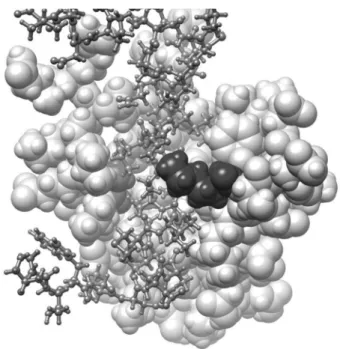
Overall, a third of the cohort had a psychiatric or beha-vioural condition reported, including ADHD, aggressive behaviour, OCD, depression, psychosis, anxiety and schi-zoaffective disorder. In three patients, ASD was concurrent with a psychiatric diagnosis. This is a significant finding, since such neuropathology has a significant impact on the quality of life of patients especially in those adults with milder neurodevelopmental phenotypes. Knowing that Fig. 3 3D model to demonstrate predicted consequences of SIN3A
missense variant in our cohort. This 3D-model is based on the solution structure of mouse Sin3A PAH1 bound to the Sin3 interaction domain (SID) of SAP25 (Sin3A Associated Protein 25) displayed using UCSF Chimera v1.14 (Pattersen et al. 2004). The PAH1 domain of Sin3A (residues 119-189; sphere model, light grey) with residues Ala126 (red) and Lys155 (blue) highlighted. The SAP25 protein SID domain (residues 126-186; ball and stick model, dark grey) binds in the fold formed by the four helices of Sin3A PAH1. Sin3A Lys155 is predicted to form a 2.2A hydrogen bond with the polar side chain of Gln143 of SAP25 (orange line).
patients with variants in the SIN3A gene are at risk for such concerns, early intervention is important to ensure optimal treatment and outcomes.
Of those patients with psychiatric disease, only two had brain imaging done. Interestingly, both those MRIs showed ventriculomegaly, while one also showed delayed myeli-nation. The younger patients in the cohort did not have any neuroimaging done given the milder clinical presentation. However, it is likely that once the diagnosis of a SIN3A-related disorder is made, imaging of the brain should be offered in the context of neurological symptoms rather than routine work-up.
Craniofacial dysmorphism
Our previous study presented evidence for a characteristic facial appearance associated with SIN3A. Some of the patients in this cohort have clear similarities. As with many of the mild and variable neurodevelopmental phenotypes, it remains to be seen whether the facial gestalt is easily identifiable in clinical practice. However, there appears to be a common, emerging facial phenotype with a tall, broad forehead, down-slanting palpebral fissures, triangular face with a pointed chin and a thin upper lip, based on the patient’s photographs (both included and unpublished but shared with the authors due to parental consent for pub-lication of photos being declined).
Interestingly, one patient wasfirst suspected of progeria, because of the typical shape of his neurocranium (Patient 25: J). Following genetic testing, he was diagnosed with a variant in SIN3A, demonstrating that this may be part of the spectrum of the syndrome. Patient 12: G also appears to have a progeric face. Sparse hair and reduced subcutaneous tissue was reported in 3/28 (10%) of patients in this study and note the progeric appearance in at least two of the patients in this cohort. However, no other ectodermal fea-tures were identified. This leads to the possibility of dif-ferential diagnoses including progeroid group of conditions; however, SIN3A-related disorder does not appear to present predominantly with a progeroid phenotype from the large cohort described here.
Other clinical manifestations
14/28 (50%) patients in our study population had either epilepsy, hypotonia, or both. Two out of the three patients with epilepsy who had a brain MRI (of a total of five patients with epilepsy) had abnormalities found. None of the patients with epilepsy from this cohort reported any psychiatric disease.
Interestingly, other commonly reported symptoms in patients with intellectual disability were not reported in our patient population. Constipation for instance, was only
reported in one patient, while hearing loss and refraction abnormalities were also not as prevalent as in other intel-lectual disability cohorts or the 15q24 microdeletion syn-drome [15,16].
Further information also needs to be collected to ascer-tain whether malignancy is a significant association or merely an observation with a large cohort of patients with Witteveen–Kolk Syndrome. In addition, as described above, additional features appear to be emerging from the larger cohort of patients published here and further follow-up is required to see if this is a consistent part of the phenotype.
Conclusion
Patients with disease causing variants in SIN3A usually have mild global developmental delay/ID, with some even having tested IQs in the normal range with variable pene-trance. There are similar facial features in around half of patients for which a targeted molecular evaluation would be feasible. However, it is likely that diagnostic evaluation and identification of SIN3A variants in suspected individuals will be performed using large ID panels or WES/WGS. There is evidence to suggest these patients are at risk for psychiatric- and neurological conditions and therefore, a multidisciplinary team approach should be considered in caring for these patients. Data collected so far seems to suggest additional features such as hypotonia, seizures along with the previously well described neurodevelop-mental association with this disorder.
There is no apparent genotype–phenotype correlation and/ or missense variant hot spot within SIN3A and the missense variants appear to be distributed throughout the gene based on observation of this cohort. As expected, majority of patients in published literature and this cohort appear to have truncating variants reinforcing hap-loinsufficiency as likely mechanism of pathogenicity, although SIN3A missense variants affecting critical func-tional domain in SIN3A also appear to be associated with disease. Further studies of this nature are required to ascertain clinical correlation in this disorder.
Acknowledgements We are grateful to the patients and their families for their cooperation. This study makes use of data generated by the DECIPHER Consortium. A full list of centres who contributed to the
generation of the data is available fromhttps://decipher.sanger.ac.uk/
and via email from decipher@sanger.ac.uk. Funding for the project was provided by the Wellcome Trust and by grants from the Nether-lands Organization for Health Research and Development (ZonMw
grant 91718310 and the Dutch Scientific Organization (NWO, grant
NWA 1160.18.320). WKC is supported by grants from SFARI and the JPB Foundation
DDD statement The DDD study presents independent research com-missioned by the Health Innovation Challenge Fund [grant number
HICF-1009-003]. This study makes use of DECIPHER (http:// decipher.sanger.ac.uk), which is funded by the Wellcome Trust. See
Nature PMID: 25533962 or www.ddduk.org/access.html for full
acknowledgement. We would also like to thank all the families for consenting to this publication.
Author contributions MB and TK designed and supervised the study; MB was responsible for collection of data from UK cohort of patients whilst TK was responsible for data collection from Dutch and inter-national cohort; SA, AD and RR performed all data collection and molecular interpretation of SIN3A variants; all authors contributed to clinical or molecular data collection and approval of submitted manuscript.
Compliance with ethical standards
Conflict of interest The authors declare that they have no conflict of
interest.
Ethics statement This study was performed in adherence to the principles set out in the Declaration of Helsinki. The DDD study has UK Research Ethics Committee approval (10/H0305/83, granted by the Cambridge South REC, and GEN/284/12 granted by the Republic of Ireland REC). Informed consent for publication of clinical and molecular data and inclusion of photos where applicable was obtained from all patients and/ or their guardians included in this study.
Publisher’s note Springer Nature remains neutral with regard to
jurisdictional claims in published maps and institutional affiliations.
Open Access This article is licensed under a Creative Commons Attribution 4.0 International License, which permits use, sharing, adaptation, distribution and reproduction in any medium or format, as long as you give appropriate credit to the original author(s) and the source, provide a link to the Creative Commons license, and indicate if changes were made. The images or other third party material in this
article are included in the article’s Creative Commons license, unless
indicated otherwise in a credit line to the material. If material is not
included in the article’s Creative Commons license and your intended
use is not permitted by statutory regulation or exceeds the permitted use, you will need to obtain permission directly from the copyright
holder. To view a copy of this license, visithttp://creativecommons.
org/licenses/by/4.0/.
References
1. Witteveen JS, Willemsen MH, Dombroski TC, van Bakel NH,
Nillesen WM, van Hulten JA, et al. Haploinsufficiency of
MeCP2-interacting transcriptional co-repressor SIN3A causes mild intel-lectual disability by affecting the development of cortical
integ-rity. Nat Genet. 2016;48:877–87.
2. Stessman HA, Xiong B, Coe BP, Wang T, Hoekzema K,
Fenckova M, et al. Targeted sequencing identifies 91
neurodevelopmental-disorder risk genes with autism and
developmental-disability biases. Nat Genet. 2017;49:515–26.
3. Narumi-Kishimoto Y, Araki N, Migita O, Kawai T, Okamura K,
Nakabayashi K, et al. Novel SIN3A mutation identified in a
Japanese patient with Witteveen-Kolk syndrome. Eur J Med Genet. 2019;62:103547.
4. Mefford HC, Rosenfeld JA, Shur N, Slavotinek AM, Cox VA, Hennekam RC, et al. Further clinical and molecular delineation of the 15q24 microdeletion syndrome. J Med Genet. 2012;49:
110–8.
5. Grzenda A, Lomberk G, Zhang JS, Urrutia R. Sin3: master scaffold and transcriptional corepressor. Biochim Biophys Acta.
2009;1789:443–50.
6. Wright CF, Fitzgerald TW, Jones WD, Clayton S, McRae JF, van Kogelenberg M, et al. Genetic diagnosis of developmental dis-orders in the DDD study: a scalable analysis of genome-wide
research data. Lancet. 2015;385:1305–14.
7. Sobreira N, Schiettecatte F, Valle D, Hamosh A. GeneMatcher: a matching tool for connecting investigators with an interest in the
same gene. Hum Mutat. 2015;36:928–30.
8. Firth HV, Richards SM, Bevan AP, Clayton S, Corpas M, Rajan D, et al. DECIPHER: database of chromosomal imbalance and phenotype in humans using ensembl resources. Am J Hum Genet.
2009;84:524–33.
9. Richards S, Aziz N, Bale S, Bick D, Das S, Gastier-Foster J, et al. Standards and guidelines for the interpretation of sequence var-iants: a joint consensus recommendation of the American College of Medical Genetics and Genomics and the Association for
Molecular Pathology. Genet Med. 2015;17:405–24.
10. Ellard S, Baple EL, Callaway A, Berry I, Forrester N, Turnbull C,
et al. ACGS best practice guidelines for variant classification
2020. Recommendations ratified by ACGS Quality Subcommittee
on 04/02/2020.
11. Sahu SC, Swanson KA, Kang RS, Huang K, Brubaker K, Ratcliff K, et al. Conserved themes in target recognition by the PAH1 and PAH2 domains of the Sin3 transcriptional corepressor. J Mol Biol.
2008;375:1444–56.
12. He Y, Radhakrishnan I. Solution NMR studies of apo-mSin3A and mSin3B reveal that the PAH1 and PAH2 domains are
struc-turally independent. Protein Sci. 2008;17:171–5.
13. Pettersen EF, Goddard TD, Huang CC, Couch GS, Greenblatt
DM, Meng EC, et al. UCSF Chimera—a visualization system for
exploratory research and analysis. J Comput Chem.
2004;25:1605–12.
14. Rydzewska E, Hughes-McCormack LA, Gillberg C, Henderson
A, MacIntyre C, Rintoul J, et al. Age at identification, prevalence
and general health of children with autism: observational study of a whole country population. BMJ Open. 2019;9:e025904. 15. Mefford H, Shur N, Rosenfeld J, Adam MP, Ardinger HH, Pagon
RA et al. 15q24 Microdeletion syndrome. In MP Adam, HH Ardinger, RA Pagon, SE Wallace, LJH Bean, K Stephens & A Amemiya (Eds.), GeneReviews((R)). Seattle (WA). 1993 16. Chaidez V, Hansen RL, Hertz-Picciotto I. Gastrointestinal
pro-blems in children with autism, developmental delays or typical
development. J Autism Dev Disord. 2014;44:1117–27.
Affiliations
Meena Balasubramanian1,2●Alexander J. M. Dingemans3●Shadi Albaba4●Ruth Richardson5●Thabo M. Yates 1●
Helen Cox6●Sofia Douzgou7,8●Ruth Armstrong9●Francis H. Sansbury 10●Katherine B. Burke10●
Andrew E. Fry 10●Nicola Ragge6,11●Saba Sharif6●Alison Foster6●Annachiara De Sandre-Giovannoli12,13,14●
Caroline Nava18●Boris Keren18●Serwet Demirdas19●Alice S. Brooks19●Marie Vincent 20,21●Bertrand Isidor20,21●
Sebastien Küry 20,21●Meyke Schouten3●Erika Leenders3●Wendy K. Chung22●Arie van Haeringen23●
Thomas Scheffner24●Francois-Guillaume Debray25●Susan M. White 26,27●Maria Irene Valenzuela Palafoll 28●
Rolph Pfundt3●Ruth Newbury-Ecob29 ●Tjitske Kleefstra3
1 Sheffield Clinical Genetics Service, Sheffield Children’s NHS
Foundation Trust, Sheffield, UK
2 Academic Unit of Child Health, Department of Oncology &
Metabolism, University of Sheffield, Sheffield, UK
3 Department of Human Genetics, Donders Institute for Brain,
Cognition and Behavior, Radboud University Medical Center, Nijmegen, the Netherlands
4 Sheffield Diagnostic Genetics Service, Sheffield Children’s NHS
Foundation Trust, Sheffield, UK
5 Northern Genetics Service, Newcastle upon Tyne Hospitals NHS
Trust, Newcastle, UK
6 West Midlands Regional Clinical Genetics Service and
Birmingham Health Partners, Birmingham Women’s and
Children’s Hospitals NHS Foundation Trust, Birmingham, UK
7 Manchester Centre for Genomic Medicine, Saint Mary’s Hospital,
Manchester University NHS Foundation Trust, Manchester, UK
8 Division of Evolution and Genomic Sciences, School of Biological
Sciences, Faculty of Biology, Medicines and Health, University of Manchester, Manchester, UK
9 East Anglian Medical Genetics Service, Addenbrooke’s Hospital,
Cambridge, UK
10 All Wales Medical Genomics Service, NHS Wales Cardiff and
Vale University Health Board, Institute of Medical Genetics, University Hospital of Wales, Cardiff, UK
11 Department of Biological and Medical Sciences, Oxford Brookes
University, Oxford, UK
12 Aix Marseille Univ, INSERM, MMG, U1251 Marseille, France
13 Department of Medical Genetics, La Timone Children’s Hospital,
Marseille, France
14 Biological Resource Center (CRB-TAC), Assistance Publique
Hôpitaux de Marseille, La Timone Children’s Hospital,
Marseille, France
15 Leicester Clinical Genetics Service, University Hospitals of
Leicester NHS Trust, Leicester, UK
16 Clinical Genetics Service, St George’s University Hospitals NHS
Foundation Trust, London, UK
17 Oxford Centre for Genomic Medicine, Nuffield Orthopaedic
Centre, Oxford University Hospitals NHS Foundation Trust, Oxford, UK
18 Clinical Genetics Service, GH Pitié-Salpêtrière, Pitié Salpêtrière
Hospital, APHP Sorbonne University, Paris, France
19 Department of Clinical Genetics, Erasmus Medical Centre,
Erasmus University, Rotterdam, the Netherlands
20 Service de Génétique Médicale, CHU de Nantes, 44000
Nantes, France
21 Inserm, CNRS, Univ Nantes, l’institut du thorax, 44000
Nantes, France
22 Departments of Pediatrics and Medicine, Columbia University,
New York, USA
23 Department of Clinical Genetics, Leiden University Medical
Center, Leiden, the Netherlands
24 Klinik für Kinder- und Jugendmedizin, Perinatal- und
Stoffwechselzentrum, Reutlingen, Germany
25 Metabolic Unit—Department of Medical Genetics, CHU &
University Liège Domaine L Sart-Tilman Bât B35, B-4000 Liège, Belgium
26 Victorian Clinical Genetics Services, Murdoch Children’s
Research Institute, Melbourne, VIC, Australia
27 Department of Paediatrics, University of Melbourne,
Melbourne, VIC, Australia
28 Department of Clinical and Molecular Genetics, University
Hospital Vall d´Hebron and Medicine Genetics Group, Valle Hebron Research Institute, Barcelona, Spain
29 University Hospitals Bristol NHS Foundation Trust, Clinical