HAL Id: tel-00804227
https://tel.archives-ouvertes.fr/tel-00804227
Submitted on 25 Mar 2013
HAL is a multi-disciplinary open access
archive for the deposit and dissemination of sci-entific research documents, whether they are pub-lished or not. The documents may come from teaching and research institutions in France or abroad, or from public or private research centers.
L’archive ouverte pluridisciplinaire HAL, est destinée au dépôt et à la diffusion de documents scientifiques de niveau recherche, publiés ou non, émanant des établissements d’enseignement et de recherche français ou étrangers, des laboratoires publics ou privés.
Study of cell host factors involved in Hepatitis C virus
tropism
Daniel da Costa
To cite this version:
Daniel da Costa. Study of cell host factors involved in Hepatitis C virus tropism. Virology. Université de Strasbourg, 2012. English. �NNT : 2012STRAJ071�. �tel-00804227�
UNIVERSITÉ DE STRASBOURG
ÉCOLE DOCTORALE 414, ED des Sciences de la Vie et de la Santé
LABORATOIRE : INSERM U_748
THÈSE
présentée par :
Daniel DA COSTA
soutenue le : 18 septembre 2012pour obtenir le grade de :
Docteur de l’université de Strasbourg
Discipline/ Spécialité: Aspects moléculaire et cellulaire dela Biologie
Option : Biologie moléculaire et cellulaire
Study of cell host factors involved in
Hepatitis C virus tropism
THÈSE dirigée par :
Mr BAUMERT Thomas Professeur, Université de Strasbourg
RAPPORTEURS :
Mr PATEL Arvind Professeur, University of Glasgow
Mme COCQUEREL Laurence Docteur, Université de Lilles
« Même un chemin de mille lieues commence par un
premier pas »
Remerciements
Je remercie les membres du jury qui m’ont fait l’honneur d’évaluer mon travail de
thèse. Le Pr. Arvind Patel et le Dr. Laurence Cocquerel en qualité de rapporteurs externes, ainsi que le Pr. Gilles Prevost qui a accepté d’être mon rapporteur interne. Un grand merci au Pr. Baumert, directeur de l’unité INSERM U748 et mon directeur de thèse de m’avoir donné l’opportunité de travailler sur un sujet qui m’a passionné tout au long de ces trois années.
Mes remerciements les plus chaleureux vont au Dr. Mirjam Zeisel qui a supervisé mes travaux de recherche durant toutes ces années. Nous avons traversé des moments difficiles mais toujours avec le sourire. J’ai beaucoup appris sous ta supervision et je t’en remercie Mirjam. Je te souhaite encore plus de succès que ce que tu as déjà connu dans ta carrière.
Ce manuscrit de thèse marque la fin d’intenses années de recherche mais également le début de nouvelles opportunités. J’ai eu la chance de rencontrer durant
ma thèse et au sein de l’Université de Strasbourg, des personnes qui m’ont
marquées et ont changé la manière dont je concevais mon avenir.
Ces travaux auraient été beaucoup moins agréables à mener sans les redoutables « jokes » de Joachim, notre mentor à tous en la matière !!! Je remercie Laetitia et Marine avec qui j’ai pu partager les délires et les coups de blues. Je remercie tous mes collègues de l’unité : Isabel, Dan, Rajeev, Fei, Nauman, Christine, Roxane, Catherine F., Catherine S., Catherine C., Sarah, Laura, Charlotte, Lamine, Mathieu, Emilie, Marie, Eric S., Eric R., Laurent, Céline, Tao, Jochen, Heidi, Evelyne, Samira, Mélanie, Sylvie, Géraldine, Alizé, Thomas, Marina, Maryse, Bin, Alexandre, Françoise, Christiane, Richard, Nicolas.
Un remerciement particulier à Anne, Jérémy, Patricia, Dominique et Sigis que l’on voit peu souvent mais qui sont indispensables dans l’équipe. Merci pour leur bonne humeur et leur sourire aux croisements de couloirs.
Au cours de ma thèse, j’ai eu l’occasion de connaître l’association des doctorants et docteurs d’Alsace où j’y ai rencontré des personnes formidables qui ont contribué à rendre ces trois années beaucoup plus conviviales. A ce titre, je souhaite tous les remercier. J’ai beaucoup appris à vos côtés et j’ai beaucoup appréciés les longues réunions de bureau qui se sont toujours déroulé dans la franche camaraderie.
Un évènement majeur au court de ma thèse fut l’organisation du Forum BIOTechno 2011 à Strasbourg. Ce fut le premier projet que j’ai eu l’occasion de gérer de « A à Z » et ca restera à toujours une excellente expérience. Un grand merci à Inès, Abdelkader, Laetitia, Marine, Claire qui ont participé à l’organisation du Forum BIOTechno. Un remerciement tout particulier à Marie Kneib et Emilie Audran qui ont
toujours répondu présentes dans les temps difficiles et qui se sont révélées d’une aide inestimable au cours de l’organisation.
Je souhaiterais remercier Norbert, Richard et Benjamin que je connais depuis de nombreuses années. Nos choix professionnels nous ont amené à nous éloigner géographiquement, mais l’amitié qui nous lie nous rassemble toujours pour un week-end ou une semaine, qui au fil du temps, deviennent des souvenirs inoubliables. Merci de votre soutien et de votre amitié.
Je tiens à remercier tout particulièrement la personne qui durant ces trois années a été à mes côtés pour partager mes joies et mes peines. Elle a su me réconforter dans les moments difficiles et a su me montrer ses plus beaux sourires lors de mes réussites. Orphée, merci pour de ta présence.
Je souhaiterais dédier mes derniers remerciements à ma famille qui compte énormément pour moi. Je souhaiterais remercier mes parents qui m’ont soutenu tout au long de ces années. Malgré leur origine, ils ont su nous éduquer et nous mener sur le droit chemin, malgré les dérapages. J’imagine que ca n’a pas été facile, mais vous pouvez être fier des enfants que vous avez élevés. Je tiens à remercier mes frères et sœurs Marie, Mike, Maria et Susu. Je ne suis pas très expressif mais sachez qu’une journée en votre compagnie vaut beaucoup plus que trois années de thèse. Si j’ai l’occasion d’écrire ces lignes aujourd’hui c’est grâce à votre soutien et votre générosité. Vous m’avez ouvert les yeux quand je doutais, vous avez toujours accepté mes choix et vous avez su faire preuve de compréhension. Merci ! Un merci à ceux qui sont venu agrandir la famille Da Costa, Fred, Stéphane, Elodie et bien sûr la relève qui attend Hugo, Liza, Antoine, Auxane et depuis peu Nelson. J’attends avec impatience les vacances tous ensembles. Un grand merci à vous tous et sachez qu’ensemble on forme une famille géniale !!!
Merci à tous ceux qui ont contribués de près ou de loin à ce travail de thèse et que j’ai oublié de citer.
Table of Content
REMERCIEMENTS ... 4
ABBREVIATIONS ... 10
INTRODUCTION ... 14
A. Hepatitis C virus ... 16
i. Structure of hepatitis C virus ... 16
ii. From genome to viral proteins ... 17
iii. The genetic variability of HCV ... 25
iv. Hepatitis C : the disease ... 25
B. The viral life cycle ... 27
i. HCV entry ... 27
ii. HCV replication ... 37
iii. Assembly and release of virions ... 46
C. Model systems to study HCV ... 50
i. In vitro models ... 50
ii. In vivo models... 54
D. HCV tropism ... 59
i. Hepatotropism of HCV ... 59
ii. Extra-hepatic tropism ... 63
iii. Species-specificity of HCV ... 66
AIM OF THE STUDY ... 72
RESULTS ... 76
Reconstitution of the entire HCV life cycle in non-hepatic cells ... 77
Investigation of factors responsible for the HCV species specificity ... 112
DISCUSSION AND PERSPECTIVES ... 128
MATERIAL AND METHODS ... 148
Table of Illustrations
Figures
Figure 1: Schematic representation of HCV ... 17
Figure 2: Organization of HCV genomic RNA ... 19
Figure 3: Worldwide repartition of the different HCV genotypes and prevalence among adults ... 26
Figure 4: HCV entry into hepatocytes ... 35
Figure 5: Biogenesis of microRNAs and function ... 40
Figure 6: Schematic representations of models for the HCV virion assembly process .... 49
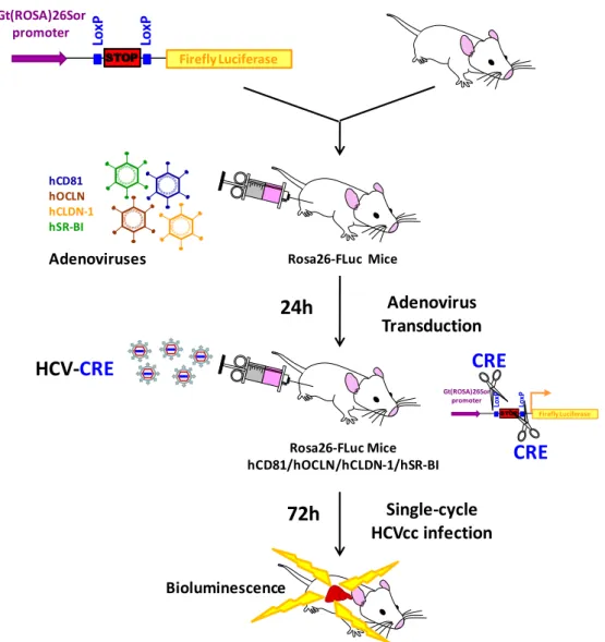
Figure 7: Immunocompetent mouse model to study single-cycle HCV infection ... 58
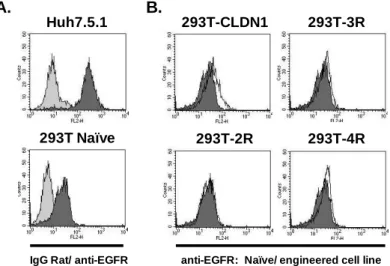
Figure 8: EGFR is not overexpressed during the selection process. A. EGFR expression in naïve cells ... 107
Figure 9: 293T cells engineered to express different sets of HCV entry factors and miR-122 are permissive for HCVcc entry and replication ... 109
Figure 10: HCV replication in 293T-4R/miR122 is associated with sustained viral protein expression ... 110
Figure 11: The amount of intracellular miR-122 does not influence HCV replication in 293T-4R cells ... 111
Figure 12: Endogenous expression of mouse homologue of HCV entry factors on primary and mouse hepatoma cells ... 115
Figure 13: Expression of human HCV entry factors in engineered mouse hepatoma cell lines AML12, BNL-1 and Hepa 1.6 ... 117
Figure 14: Expression of the four human HCV entry factors renders mouse hepatoma cells permissive to HCVpp entry. ... 118
Figure 15: HCVpp entry-permissive mouse hepatoma cells are resistant to HCVcc
infection. ... 119
Figure 16: HCV IRES allows viral RNA translation in mouse hepatoma cells ... 120
Figure 17: MiR-122 expression is not sufficient for robust HCV RNA replication in HCV entry permissive mouse hepatoma cells ... 122
Figure 18: Mouse hepatoma cells constitutively expressing miR-122 are transfected with a
miR-122 mimic or a miR-Control ... 124
Figure 19: HCVcc infection of mouse hepatoma cells upon miR-122 transfection ... 125
Figure 20: Transduction of mouse apoE in engineered mouse hepatoma cells ... 126
Figure 21: Chimeric HCV viruses used in the present study. ... 152
Tables Table 1: Primers used to amplify puromycin and pri-miR-122 genes. ... 150
Table 2: List of primary antibodies used in the present study. ... 152
Table 3: Summary of host cell factors expressed in the cell lines engineered in the present study. ... 154
aa amino acid
apoB, apoE Apolipoprotein B or E
ARF Alternative reading frame
ARFP Alternative reading frame protein
CD81 Cluster of differenciation 81 CLDN Claudin CMV Cytomegalovirus CsA Cyclopsorin A CyP Cyclophilin Da Dalton
DC-SIGN Dendritic cell-specific intercellular adhesion molecule three grabbing non integrin
DGAT-1 Diacyl glycerol acyltransferase 1
ECL Extra cellular loop
ECMV Encephalomyocarditis virus
EGF Epidermal growth factor
EGFR Epidermal growth factor receptor
EphA2 Ephrin receptor A2
HCC Hepatocellular carcinoma
HCV Hepatitis C virus
HCVcc Cell culture derived HCV
HCVpp HCV pseudo-typed retroviral particles
HDL High density lipoprotein
HIV Human immunodeficient virus
HS Heparan sulfate
HVR Hypervariable region
hVAP-33 Human homologue of the 33-kDa vesicle-associated membrane protein-associated protein
IFNAR Interferon alfa receptor
IRES Internal ribosomal entry site
IRF-3 Interferon regulatory factor 3
JFH1 Japanese fulminent hepatitis
KO Knock-out
LD Lipid droplet
LDL Low density lipoprotein
LDLR Low density lipoprotein receptor
LEL Large extracellular loop
LNA Locked nucleic acid
LSEC Liver sinusoidal endothelial cells
L-SIGN Liver and lymph node specific SIGN
LVP Lipo-viro-particles
MAVS Mitochondrial antiviral signaling protein
MEF Mouse embryonic fibroblast
miRNA microRNA
MTP Microsomal triglyceride transfer protein
NANBH Non-A Non-B hepatitis
NPC1L1 Niemann Pick C1- Like 1
NS Non structural proteins
NTR Non translated region
OCLN Occludin
ORF Open reading frame
PEG-IFNα Pegylated interferon alfa
PKI Protein kinase inhibitor
PKR Protein Kinase R
RC Replication complex
RdRp RNA dependent RNA polymerase
RIG-I Retinoic acid inducing interferon gene I
RISC RNA-interference-silencing complex
RNAi RNA interference
RTKs Receptor tyrosine kinase
sE2 Truncated soluble form of the glycoprotein E2
SL Stem loop
SP signal peptidase
SPP signal peptide peptidase
SR-BI Scavenger receptor class B type I
TC-PTP T-cell protein tyrosine phosphatase
TGF-α Transforming growth factor alfa
TLR3 Toll-like receptor 3
TRIF Toll-IL-1 receptor domain-containing adaptor inducing IFN-beta
uPA-SCID urokinase-type-plasminogen activator (uPA)-Severe combined immunodeficiency
Hepatitis C virus (HCV) infection affects more than 160 million individuals worldwide. HCV infection is most often asymptomatic, leading to an evolution of the disease toward chronic hepatitis in 70% of the cases. Chronic hepatitis evolves silently to cirrhosis in 15 to 20% of the infected patients, among which 5% will develop hepatocellular carcinoma.
The treatment for hepatitis C infection is based on pegylated interferon alfa and ribavirin. This treatment, little effective and expensive, is not prescribed to all infected patients, due to its strong side effects. The absence of a vaccine and new medications for the treatment of hepatitis C renders primordial the development of alternative therapies.
New molecule development to treat HCV has been hampered by the lack of in
vitro and in vivo models. Since its discovery in 1989, the first model system allowing
the study of the full HCV life cycle in vitro has been available from 2005 only. Furthermore, among the rare cellular models supporting the entire HCV life cycle, all have hepatic origin, revealing the rigorous hepatic tropism of HCV.
Currently, only chimpanzees and mice, repopulated with human hepatocytes, allow the in vivo study of HCV infection, mouse hepatocytes being naturally resistant to HCV infection. So far, no mouse model easily handleable is available to the scientific community, which slows down the preclinical studies and the launch on the market of effective and more tolerated molecules. Its hepatotropism and its species specificity make HCV a difficult to study virus.
The PhD work presented in this manuscript first focused on factors restricting HCV infection to hepatocytes. In a second part, we have studied HCV infection in mouse hepatoma cells. After a bibliographic summary of the current knowledge on HCV, the original results emanating from our work will be presented and finally, the last part of this manuscript will be dedicated to the discussion of the results as well as conclusions and perspectives of the subject.
A. Hepatitis C virus
HCV has been isolated in 1989 from a complementary DNA bank constructed using the plasma of patients suffering from a non-A and non-B hepatitis (NANBH) (Choo et al., 1989).
i.
Structure of hepatitis C virus
HCV belongs to the Flaviridae family. This family contains single strand RNA viruses of positive orientation which are classified in three different genera: Flaviviruses (Yellow fever virus, West Nile virus and Dengue virus), Pestivirus (classical swine fever virus, Bovine Viral Diarrhoea virus) and Hepacivirus in which the unique member is HCV. However, very recently a virus isolated from dog’s respiratory samples has been shown to be a dog homolog of HCV (CHV, canine hepacivirus) and it has been suggested, according to their similarities that this virus could be a new member of the Hepacivirus genus (Kapoor et al., 2011).
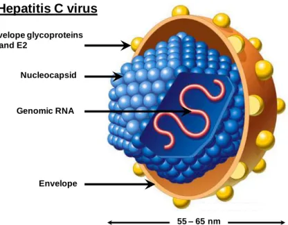
HCV is an enveloped virus with a diameter of 55 to 65 nm (Kaito et al., 1994) containing a nucleocapsid protecting a single positive strand RNA genome of 9600 bases. The HCV genome is constituted of a non-translated region at its 5’ end (5’ NTR) containing the internal ribosomal entry site (IRES), an open-reading frame (ORF) encoding the structural and the non-structural proteins and a non-translated region at the 3’ end (3’ NTR). The HCV envelope derives from the cellular lipid bilayer and contains the viral glycoproteins E1 and E2. The nucleocapsid is composed of the capsid protein (core) which protects the genomic RNA (Figure 1).
HCV circulates within the blood of infected patients under different forms. The viral particle can circulate freely, associated to lipoproteins of different sizes or associated to immunoglobulins. These different forms confer the virus a heterogeneous distribution on a sucrose gradient. The particles found in the low density fractions (1,03 to 1,08 g/ml) and associated to lipoproteins of low density (LDL) or very low density fraction (VLDL), are more infectious than those found in fractions of higher density (1,17 to 1,25 g/ml), which are most often associated to immunoglobulins (Agnello et al., 1999; Andre et al., 2002).
Figure 1: Schematic representation of HCV. HCV is an enveloped virus with a diameter of 55 to 65 nm, containing an icosahedric capsid protecting the positive strand genomic RNA of 9.6 kb. The glycoproteins E1 and E2 are embedded into the lipid bilayer of the viral particle. (© 2001, James. A. Perkins)
The low density particles are called lipo-viro-particles (LVPs) due to their association to lipoproteins. These LVPs are rich in triglycerides and contain, besides the viral capsid containing the HCV genomic RNA, the glycoproteins E1 and E2 and the apolipoproteins B and E (apoB, apoC and apoE) (Diaz et al., 2006).
ii. From genome to viral proteins
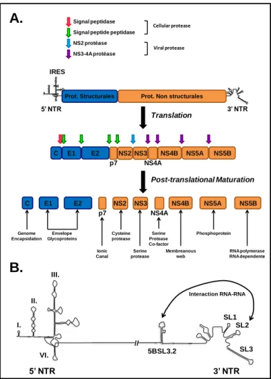
The HCV genome is translated into a precursor polyprotein of 3000 amino acids in the endoplasmic reticulum (ER). This polyprotein is cleaved by cellular and viral proteases in a co and post-translational manner, into 10 viral proteins; (i) the
structural proteins, viral capsid or C protein or core and the envelope glycoproteins
E1 and E2 as well as (ii) the non-structural proteins, p7, NS2, NS3, NS4A, NS4B, NS5A and NS5B (Figure 2A) (Ploss and Dubuisson, 2012).
1. The non-translated regions
The 5’ NTR region, of 341 nucleotides, well structured and conserved, contains elements from the IRES that ensure the translation of the polyprotein and domains important for replication. The IRES corresponds to the domain II. to IV. of the 5’ NTR region as well as the initial part of the ORF sequence (Figure 2B). This structured
Envelope glycoproteins E1 and E2 Nucleocapsid Genomic RNA Envelope Hepatitis C virus 55 – 65 nm
organization of the RNA within the IRES is indispensable to assure the
cap-independent translation of the viral RNA. Studies have revealed that the 5’ NTR
sequence would contain important elements for the translation but also for RNA replication (Astier-Gin et al., 2005; Friebe et al., 2001). Thus, it has been shown that
(i) the domain I. of the 5’ NTR sequence is fundamental for the viral RNA replication
(Friebe et al., 2001), (ii) IRES regions are important for the RNA replication efficiency (Friebe et al., 2001) and finally (iii) the stem loop of the domain II. of the IRES is essential for replication, in addition to its role in the translation process (Appel and Bartenschlager, 2006). It has also been shown that the 5’ NTR of the RNA represents an interaction site for the microRNA-122 (miR-122) which binds to two sequences present between the domain I. and II. of the 5’ NTR region (Jopling et al., 2005). This interaction is so important that its inhibition leads to a drastic decrease in viral load in infected chimpanzees (Lanford et al.). This interaction will be the subject of a following paragraph in the present manuscript.
The 3’ NTR region has a variable size according to the genotypes. It can be subdivided into three distinct domains: (i) a poorly conserved region, (ii) an internal sequence uracil/pyrimidin (poly(U/UC)) of an average length of 80 nt and (iii) the X region of 100 nt, which constitutes the most conserved domain and the most structured of the 3’ NTR region. The X region includes three loops (SL1, SL2 and SL3) and plays a primordial role in the negative strand RNA synthesis during replication (Figure 2B). A new particular structure of the viral RNA has been found within the NS5B coding sequence, this structure called 5BSL3.2 interacts with the stem-loop SL2 during replication of the viral RNA and this interaction is essential for this step of the viral life cycle (Figure 2B) (Friebe et al., 2001; You et al., 2004).
2. The viral proteins
The core protein (capsid protein or C protein), the first protein to be translated and cleaved from the polyprotein, constitutes the unique component of the viral capsid. Its maturation follows a process involving two cellular proteins. During translation, the nascent polyprotein is directed toward the ER. The core protein is then cleaved a first time by the signal peptidase (SP) at the protein junction core-glycoprotein E1, releasing core from the polyprotein to give an immature protein of 23 kDa (Yasui et al., 1998).
Figure 2: A. Organization of HCV genomic RNA. The HCV genome is a positive single strand RNA.
IRES, present in the non coding 5’ end (5’NTR), is upstream of the open reading frame which codes for a polyprotein of 3000 amino acids. The polyprotein is cleaved in a co and post-translational manner by cellular and viral proteases (colored arrows) releasing structural proteins (blue) and non-structural proteins (orange).
B. Schematic representation of non-translated regions of HCV RNA. The domains of the non-translated
regions (NTR) are indicated in each region.
The sequence between amino acids 179 and 191, anchored in the ER membrane, is further recognized and cleaved by the signal peptide peptidase (SPP)
to give the mature form of the 21 kDa core protein (McLauchlan et al., 2002). The
maturation of the core protein by SPP is essential since the insertion of mutations within the recognized sequence or the inhibition of SPP activity by specific drugs, reduce considerably the production of infectious viral particles (Targett-Adams et al., 2008). The mature core protein remains anchored at the ER membrane by its hydrophobe domain present at the C-terminal part, which gives stability to the core protein. The protein moves then to the lipid droplets (LD) at the ER level (McLauchlan et al., 2002). Furthermore, it has been recently shown that diacyl glycerol
A.
B.
I. II. III. VI. SL1 SL2 SL3 // 5BSL3.2 Interaction RNA-RNA 5’ NTR 3’ NTR C E1 E2 p7 NS2 NS4A NS3 NS4B NS5A NS5BProt. Structurales Prot. Non structurales
IRES 5’ NTR 3’ NTR C E1 E2 p7 NS2 NS4A NS3 NS4B NS5A NS5B Genome Encapsidation Envelope Glycoproteins Ionic Canal Cysteine protease Serine protease Serine Protease Co-factor Membreanous web Phosphoprotein RNA polymerase RNA dependente Translation Post-translational Maturation Signal peptidase Signal peptide peptidase NS2 protéase NS3-4A protéase
Cellular protease
acyltransferase 1 (DGAT-1), a factor important for the assembly and release of infectious viral particles and involved in LDs biogenesis, recruits core protein to LDs (Herker et al., 2010). Apart of its structural role, the core protein plays an important role in the assembly of the viral particles. Its N-terminal domain, rich in basic amino acids, interacts with the 5’ NTR region of the viral RNA allowing the formation of the nucleocapsid. It has also been shown that the protein core recruits the non structural proteins involved in the viral RNA replication around the LDs, suggesting a central role of core protein in the morphogenesis of the virions (Miyanari et al., 2007).
The protein core coding sequence encodes, on an alternative reading frame (ARF) the frame +1, another protein of 17 kDa called protein core+1, protein F
(Frameshift) or ARFP (alternative reading frame protein). This protein is localized
in the ER and its half-life does not overpass 10 min because it is rapidly degraded by the proteasome (Xu et al., 2003). A truncated form of the ARFP protein of 8 kDa has been isolated and its expression is inversely proportional to the core protein, suggesting that its expression decreases when the replication increases (Wolf et al., 2008). The presence of antibodies and T cells specific to the ARFP protein insinuated that it is produced in vivo (Dalagiorgou et al., 2011; Gao et al., 2011). The precise function of the ARFP protein is not yet known, but it has been suggested that some effects attributed to the core protein could in fact be due to the expression of the ARFP protein (Branch et al., 2005). Furthermore, Fiorucci et al. have shown that ARFP could modulate the expression of cytokines (Fiorucci et al., 2007).
The envelope proteins E1 and E2 are the constituents of the viral particle envelope. They interact with the host factors present at the cell surface and induce the fusion between the viral particle envelope and the host endosomal membrane. During the polyprotein maturation, E1 and E2 are released by the SP (Dubuisson et al., 2002). E1 and E2 contain each a large N-terminal extracellular domain of respectively 160 and 334 amino acids and a C-terminal transmembrane domain of 30 hydrophobic amino acids (Cocquerel et al., 2000). To be functional, these proteins associate to each other in a heterodimer through their transmembrane domain (Op De Beeck et al., 2001). E1 has a molecular weight of 31 kDa. Its precise role in HCV entry into host cells remains not well understood but it seems that the E1 protein plays a role in the fusion process between the viral envelope and the host endosomal membrane (Lavillette et al., 2007). E2 has a molecular weight of 70 kDa. The role of
this protein is more characterized because it is the primary target of the immune responses, suggesting an important role in the HCV entry process into host cells. E2 includes three hypervariable regions (HVR). HVR1, present at the N-terminal part, is constituted of 27 amino acids. HVR2 is composed of 9 amino acids and more recently a third region has been identified the HVR3 region, encompassed between the HVR1 and the HVR2 regions (Troesch et al., 2006). HVR1 is a hotspot of extreme genetic variability, responsible for the differences in viral particle infectivity in patients and also within a same patient. This high variability allows the virus to escape from host immune responses (von Hahn et al., 2007). The deletion of HVR1 in the E2 glycoprotein renders the virus much less infectious in vivo (Forns et al., 2000), suggesting that this region has a functional role, essentially in the virus attachment to the CD81 entry factor (Bankwitz et al., 2010). Furthermore, it has been noticed that the HVR1 region also interacts with the SR-BI for an efficient entry. Indeed deletion of this region from the E2 protein reduces the susceptibility of HCVpp and HCVcc entry to anti-SR-BI neutralization (Bankwitz et al., 2010; Bartosch et al., 2003b; Scarselli et al., 2002). The structure of HVR1 is relatively well conserved, which suits to its roles in HCV entry into the host cell and as a target of the immune system (Penin et al., 2001). The function of the HVR2 region is less characterized. It seems that it is engaged in the interaction of the glycoprotein E2 with host cell entry factors such as CD81 (Roccasecca et al., 2003). The third hypervariable region, HVR3, is 35 amino acids long and seems to be also implicated in the viral binding to host cell factors (Troesch et al., 2006).
The glycoproteins E1 and E2 form complexes stabilized by disulfide bonds (Vieyres et al., 2010). These proteins are highly glycosylated. Glycan residues are added to E1 and E2 during the polyprotein elongation at the ER level. These glycosylations play a primordial role in the viral particle infectivity (Helle et al., 2010; Lavie et al., 2007). Furthermore, the presence of glycosylations at the envelope protein surface plays a role in the dissimulation of functional domains to neutralizing antibodies (Falkowska et al., 2007; Helle et al., 2011).
The p7 protein is the smallest viral protein of HCV. It is 63 amino acids long and arises from an imperfect cleavage of the E2 protein. Its precise role in the HCV life cycle is not well characterized. It has been shown that p7 has the property to oligomerize thus forming an ion channel (Carrere-Kremer et al., 2002). This protein
could be involved at an early step of the morphogenesis of viral particles upstream of the virions assembly (Jones et al., 2007). Besides, Steinmann et al. have shown that p7 plays a role in the assembly and release of viral particles (Steinmann and Pietschmann, 2010). P7 also appears to be important for HCV infection of chimpanzees (Sakai et al., 2003). Despite its presence in the ER membrane and its role in virion assembly and in viral particle infectivity, it is not clear whether this protein is present at the viral particle surface (Dubuisson, 2007). Furthermore, recently it has been shown that p7 interacts with NS2 for HCV particle assembly, and this interaction regulates core localization (Boson et al., 2011; Jirasko et al., 2010; Ma et al., 2011; Popescu et al., 2011; Stapleford and Lindenbach, 2011; Tedbury et al., 2011). Interestingly, its involvement in virion assembly was independent of its ion channel activity suggesting that p7, apart of its ion channel activity, has another function in HCV particle assembly (Boson et al., 2011; Tedbury et al., 2011).
The NS2 protein is a 21-23 kDa protein. NS2 is a cystein protease associated to the ER membrane (Lorenz et al., 2006). The N-terminal domain of NS2 consists of one or several transmembrane domains; the exact number is still controversial (Jones et al., 2007). The C-terminal domain of NS2, in association with the N-terminal domain of NS3, forms the NS2-3 protease, an enzyme catalyzing the unique cleavage between these two proteins (Lorenz et al., 2006). NS2 is not essential for viral replication but is involved in the assembly of infectious viral particles (Charrin et al., 2009; Jones et al., 2007). There have been several studies very recently supporting the importance of NS2 in viral assembly. It has been shown that NS2 interacts with the ion channel p7, and this interaction regulates the intracellular localization of core (Boson et al., 2011; Jirasko et al., 2010; Ma et al., 2011; Popescu et al., 2011; Stapleford and Lindenbach, 2011; Tedbury et al., 2011). Interestingly, all the mentioned studies could demonstrate that NS2 is able to interact with non-structural and non-structural proteins, thus it has been proposed that NS2 serves as a scaffold for the assembly of viral particles (Ma et al., 2011). The studies have shown that NS2 interacts with the structural proteins E1 and E2 bringing them to the assembly sites close to lipid droplets. They also could detect interactions between NS2 and NS3 and NS5A and found that NS2 was close to replication complex. These results provide a new function for NS2 as a conductor of HCV particle assembly.
The NS3 protein assures several functions during the HCV life cycle. NS3 is a 70 kDa protein associated at its N-terminal domain to the NS4A protein, acting as a co-factor, to form a protein complex having a serine protease activity (Morikawa et al., 2011). The C-terminal end of the protein has a helicase activity indispensable to the viral RNA replication, by contributing to the double strand RNA and the RNA secondary structure unwinding (Raney et al., 2010). It has been recently shown that the helicase activity of NS3 could have a role in the early phase of the viral particle assembly. It seems that this protein recruits NS5A to the LDs, presumed site for viral particles assembly (Ma et al., 2008). The protease activity of the protein complex NS3-4A allows the cleavage of the non-structural proteins present downstream of the NS4A protein. The protease activity is better characterized than the helicase activity, probably due to its importance in the escape from the host cell innate immune response. Indeed, it has been shown that NS3-4A has the capability to cleave several cellular proteins, in particular adaptive molecules of the innate immune response. Foy et al. have shown in 2003 that this protein has the capacity to disrupt the phosphorylation of the transcription factor interferon regulatory factor 3 (IRF-3), the major factor for the induction of the interferon (IFN) response (Foy et al., 2003). The factors recognized and cleaved upstream of IRF-3, have been identified later on and are the mitochondrial antiviral signaling protein (MAVS or IPS-1, VISA or CARDIF) (Li et al., 2005b) and the Toll-IL-1 receptor domain-containing adaptor inducing IFN-beta (TRIF or TICAM-1) (Li et al., 2005a), which are the adaptive molecules of retinoic acid inducing interferon gene I (RIG-I) and Toll-like receptor 3 (TLR3) respectively. More recently, a third protein has been identified as being a
target of NS3-4A, this latter is called T-cell protein tyrosine phosphatase (TC-PTP).
The cleavage of this protein could have the effect of stimulating the activation of the epidermal growth factor receptor (EGFR) and thus increase HCV replication. However the mechanism by which the inhibition of TC-PTP could have a positive
effect on HCV replication is not yet known (Brenndorfer et al., 2009). With its
versatility and its numerous cellular targets, the protein complex NS3-4A is a great therapeutic target. Many molecules have been developed to inhibit the protease activity of NS3-4A, such as the FDA-approved protease inhibitors telaprevir and boceprevir, but the genetic variability of the virus induces resistance to these newly developed inhibitors (Morikawa et al., 2011).
The NS4B protein is a non-structural protein that, few years ago, was not yet characterized. NS4B is a 27 kDa hydrophobic protein anchored in the ER membrane (Hugle et al., 2001). This protein was initially described as being able to induce by its own, drastic rearrangements in the conformation of ER membranes, known as the membranous web (Egger et al., 2002). This membranous web is known to be the scaffold necessary for the replication complex (Gosert et al., 2003). The NS4B protein contains at least 4 transmembrane domains and has the capacity to oligomerize, facilitating the replication complex formation (Yu et al., 2006). Despite the advances on the understanding of the viral life cycle, the function of the NS4B protein remains to be better characterized.
The NS5A protein exists in the host cell as two phosphorylated forms. A basal phosphorylated form of 56 kDa and a hyperphosphorylated form of 58 kDA. This hyperphosphorylation of NS5A is due to cellular kinases such as casein kinase 1
(CK1) and CK2. The N-terminal extremity of NS5A adopts an amphiphil α helix
structure which allows it to anchor into the ER membrane. It could also allow it to form protein/protein interactions essential to the formation of a functional replication complex. NS5A is constituted of three domains I, II and III. The domain I and II are involved in viral replication (Tellinghuisen et al., 2005). Moreover, it has been recently reported that the domain III intervenes into the assembly of the viral particles (Appel et al., 2008) and more recently, it has been demonstrated that NS5A interacts with apoE, a primordial factor for viral particles assembly (Benga et al., 2010; Jiang and Luo, 2009). NS5A has also a role in the regulation of the balance between viral RNA replication activity and virion assembly (Alvisi et al., 2011). It has been suggested that its phosphorylation rate could be the factor which regulates viral RNA replication (Evans et al., 2004; Neddermann et al., 2004).
The NS5B protein is a RNA dependant RNA polymerase (RdRp). Its
C-terminal region (21 amino acids) forms an α helix transmembrane domain allowing
the protein to anchor into the cytosolic part of the ER. This domain is not primordial for the polymerase activity in vitro but is essential for HCV RNA replication in cell culture (Moradpour et al., 2004). The NS5B structure is similar to most of the polymerases, which means a structure in right hand, with subdomains
thumb-palm-finger (Bressanelli et al., 2002). A particularity of the HCV RdRp is that the intense interaction between the subdomains thumb and finger results in a closed conformation of the active site (Moradpour et al., 2007). Furthermore, NS5B oligomerization has been shown to be important in the RNA synthesis. The importance of this protein in the viral RNA replication makes it a major target of new therapeutic molecules for the treatment of HCV infection (Patil et al., 2011).
iii. The genetic variability of HCV
HCV is known to present a high genetic variability. The absence of a NS5B proof reading frame activity explains in part this high variability. However, the strong in vivo
viral replication (1010 to 1012 newly produced virions per day) may also contribute to
this variability (Neumann et al., 1998). Despite the high error rates which can reach
10-3 to 10-4 per nucleotide, the 5’ NTR region is one of the most conserved region of
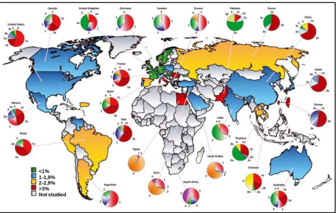
the viral RNA with a homology superior to 90% between the different genotypes (Bukh et al., 1992). According to this high variability, HCV variants are classified into four classes which are genotypes, sub-genotypes, isolates and quasi-species (Farci and Purcell, 2000). To date, HCV is classified in 7 major genotypes (1 to 7) and subdivided into sub-genotypes, classified by letters (1a, 1b, 2a…). Within a same patient, the virus can have a variability of 1 to 5% and be distributed as a quasi-species. The genotype 1a is frequent in North America and in Europe. The genotype 1b has a worldwide repartition and is the most frequently encountered genotype. The genotypes 2a and 2b are present in North Italy and in Japan. The genotype 3 is more frequently encountered in India and in South-East Asia. The genotypes 5 and 6 are relatively rare but can be found in South Africa and South-East Asia, respectively (Figure 3). The genotype identification is fundamental because certain genotypes (1 and 4) are less sensitive to the pegylated interferon-α and ribavirin-based treatment than others (2 and 3); and the length of the treatment is also dependent on the genotype (Maekawa and Enomoto, 2009).
iv. Hepatitis C : the disease
HCV is the principal cause of chronic hepatitis, hepatic cirrhosis and hepatocellular carcinoma. With more than 160 million people infected worldwide (2,3% of the world population), HCV is a major health burden (Negro and Alberti, 2011). The parenteral
route is the principal way of HCV transmission. Before the systematic screening tests of the blood bags, transfusion and transplantation were the principal causes of contaminations.
Currently, the principal cause of contamination in developed countries is the use of drugs through intravenous injections and nasal absorption. In developing countries, the contamination overcomes during surgical acts with contaminated material (Alter, 2007). Contaminations have also been reported through the use of piercings, tattoos or acupuncture (Kim et al., 2011; Tohme and Holmberg, 2012).
Hepatitis C is a progressive disease which evolves from an acute hepatitis to a chronic hepatitis if the infection is not diagnosed. In the long term, chronic hepatitis evolves towards cirrhosis and in 5% of the cases, the cirrhosis leads to hepatocellular carcinoma (HCC).
Figure 3: Worldwide repartition of the different HCV genotypes and prevalence among
adults. (adapted from (Negro and Alberti, 2011))
The acute phase of HCV infection is most often asymptomatic, but 20 to 30% of
the patients present symptoms such as tiredness, nausea, jaundice and anorexia. The incubation time is relatively short (4 to 12 weeks), this phase is followed by an
Not studied <1% 1-1,9% 2-2,9% >3%
increase of the transaminases (ALAT), first signs of a hepatic injury. In 25% of cases, an immune response, fast and efficient, is launched and leads to the natural clearance of the virus (Grebely et al., 2012). Fulminent hepatitis C exists, but these cases remain really rare (1% of the patients). In 70% of the cases, the virus persists within the organism which leads to a chronic hepatitis. This latter is defined by the presence of viral RNA in the blood six months following the primary infection. The chronic infection remains most of the time asymptomatic but can be associated to extra-hepatic syndromes such as cryoglobulinemia, nephropathie or thyroïdian pathologies (Zignego et al., 2007). Approximately a quarter of the chronically infected patients will evolve to cirrhosis. In 1 to 4% of the cases, the disease will evolve to
hepatocellular carcinoma (HCC) (Lauer and Walker, 2001) for which the unique
treatment remains currently the hepatic transplantation, followed by a systematic re-infection of the liver graft (Germani et al., 2012).
B. The viral life cycle
i.
HCV entry
HCV entry is the first step of virus-host cell interactions which leads to a robust infection. HCV entry is a finely orchestrated process involving many viral and cellular factors. So far, HCV entry is one of the best characterized steps of the viral life cycle most likely due to its attractiveness as a target for antiviral therapy (Zeisel et al., 2011b).
1. HCV entry factors
The viral factors involved in HCV entry are the glycoproteins E1 and E2. These proteins have been described previously (cf. A. ii. 2. The viral proteins).
HCV attachment to hepatocytes and viral entry are complex processes including several steps. Using several tools and HCV models, different cell surface factors have been identified as interacting directly or indirectly with HCV. These factors are CD81 (Pileri et al., 1998), the low density lipoprotein receptor (LDLR)
(Agnello et al., 1999), heparan sulfates (HS) (Barth et al., 2003), the scavenger receptor class B type I (SR-BI) (Scarselli et al., 2002), DC-SIGN (dendritic cell-specific intercellular adhesion molecule three grabbing non integrin)/ L-SIGN (DC-SIGNr, liver and lymph node specific) (Lozach et al., 2004) (Pohlmann et al., 2003), claudin-1 (CLDN1) (Evans et al., 2007) and occludin (OCLN) (Liu et al., 2009; Ploss et al., 2009; Yang et al., 2008c). In vivo, HCV enters into the liver through the blood stream via the sinusoids. The interception of viral particles by the liver sinusoid endothelial cells (LSEC) could facilitate the infection of neighboring hepatocytes which are not in direct contact with the circulating blood. This process could involve DC-SIGN, which is expressed in Kupffer cells present next to hepatocytes, and SIGN which is highly expressed in LSECs. It has been shown that DC-SIGN and L-SIGN are able to bind the E2 glycoprotein with high affinity (Gardner et al., 2003; Pohlmann et al., 2003).
At the hepatocyte surface, HS are the first attachment sites for the virus (Barth et al., 2003; Barth et al., 2006). This non-specific binding could facilitate virus access to the host entry factors.
It is known that HCV circulates in the blood stream associated with different lipoproteins such as VLDL and LDL. Thus, the LDLR has been suggested as a binding molecule and/or a host factor for HCV entry (Agnello et al., 1999; Wunschmann et al., 2000). Since retroviral pseudo-particles bearing the HCV glycoproteins (HCVpps, detailed later) are not associated with lipoproteins, studies assessing the role of LDLR using HCVpps did not uncover any major role of LDLR in HCV entry (Bartosch et al., 2003a). Furthermore, no direct interaction between the HCV E2 glycoprotein and the LDLR could be determined (Wunschmann et al., 2000). However, it has been shown that LDLR allows the internalization of HCV, derived from patient’s sera, in HepG2 cells deficient in CD81 expression by interacting with viral particles associated to LDL (Agnello et al., 1999). More recently, Albecka et al. have shown that LDLR has a role in virus binding to cells, but more remarkably, its physiological function plays a role in HCV replication (Albecka et al., 2012). This group also suggests that internalization of HCV through LDLR binding leads to a non-specific internalization and to a non-productive infection, but the physiological function of LDLR is important is HCV replication. Further studies are required to assess the precise role of the low density lipoprotein receptor in both HCV entry and
HCV RNA replication, but it is clear that this receptor does not play a role in the post-binding steps of HCV entry.
CD81 was the first HCV entry factor identified. CD81 is a ubiquitously
expressed protein of 25 kDa including a small and a large extracellular loop (LEL). CD81 has been the first described molecule as interacting with a soluble form of the E2 glycoprotein and shown to be essential for HCV entry (Pileri et al., 1998) (McKeating et al., 2004). It has been shown that CD81 LEL is the domain involved in the interaction with viral glycoprotein E2 since a soluble form of this domain could inhibit both HCVpp entry and the cell-culture derived HCV (HCVcc) infection (Zhang et al., 2004). Several amino acids have been identified in E2 and CD81 as crucial for E2 binding to the CD81 receptor (Bertaux and Dragic, 2006; Owsianka et al., 2001; Patel et al., 2000; Pileri et al., 1998). The in vitro models developed these last years, the HCVpp (Bartosch et al., 2003a) and HCVcc (Lindenbach et al., 2005; Wakita et al., 2005; Zhong et al., 2005) brought remarkable information on the E2-CD81 interactions and allowed to highlight important amino acids present in the E2 glycoprotein at postions 415, 420, 529, 530 et 535 (Dhillon et al., 2010; Owsianka et al., 2006). Furthermore, functional analysis of HCV entry factors required to over passing the species specific barrier of HCV, revealed that human CD81 and human OCLN, is the minimal set of human factors necessary to render mouse cells permissive to HCV entry (Ploss et al., 2009).
SR-BI also named CLA-1, CD36 or LMPII analogous-1 is highly expressed in
steroidogenic tissues (ovaries and adrenal glands) as well as in the liver (Krieger, 2001). This 82 kDa glycoprotein is composed of a LEL which binds to several lipoproteins (HDL, LDL and oxidized LDL (oxLDL)), and is involved in the bi-directional transport of cholesterol to the plasma membrane as well as the selective uptake of cholesteryl ester (CE) from HDL and catabolism of VLDL. SR-BI was first identified as another putative HCV receptor based on its interaction with the soluble form of the E2 glycoprotein and this interaction was localized to the HVR1 domain of E2 (Scarselli et al., 2002). Recent studies indicate that the amino acids responsible for this interaction are amino acids 70 to 87 of the SR-BI protein which interact with the amino acid E210 of the E2 glycoprotein (Catanese et al., 2010). It seems that SR-BI could have a dual role in the HCV entry process, during the attachment phase but
also in a post-attachment step of the HCV entry process (Catanese et al., 2010; Zeisel et al., 2007). Recently, it has been shown that SR-BI has multiple function in HCV entry. It seems that SR-BI plays an attachment function, an access function and an HCV entry enhancement function and only the enhancement function requires E2 binding (Dao Thi et al., 2012). Natural ligands of SR-BI have been shown to modulate HCV infection, HDL are able to increase both HCVpp and HCVcc infectivity while the oxidative form of LDL (oxLDL) inhibit their infection (Bartosch et al., 2005; Voisset et al., 2005; von Hahn et al., 2006). Recent evidence suggests that the physiological properties of SR-BI (lipid transfer and HDL binding) are required for the proper role of SR-BI as an HCV entry factor (Dreux et al., 2009). Altogether, these data indicate that a complex interaction between HCV glycoproteins, lipoproteins and SR-BI is involved in the HCV entry process.
None of the factors previously cited could explain the limited HCV tropism suggesting that other factors were involved in HCV entry. Thus, in the effort to identify the remaining HCV entry factors, Evans et al. have performed a lentiviral based delivery of a complementary DNA bank derived from the highly HCV-permissive human cell line Huh7.5 into the HCVpp non-HCV-permissive 293T cell line. This system allowed identifying CLDN1 as another HCV entry factor (Evans et al., 2007). Upon CLDN1 expression, the HCVpp non-permissive cell line 293T, become permissive to HCV entry. CLDN1 is a 23 kDa protein which contains 4 transmembrane domains. It belongs to the claudin family which includes 24 members in humans. It has been reported that CLDN6 and 9 are also able to support HCV entry (Meertens et al., 2008; Zheng et al., 2007). Claudins are essential components of the tight junctions and they regulate paracellular permissivity and maintain epithelium and endothelium polarity. CLDN1 is expressed in all the epithelial tissues but predominantly in the liver (Furuse et al., 1998). It has to be taken into account that CLDN1 is expressed at the tight junctions of the hepatocytes but is also present at the basolateral surface of these cells (Reynolds et al., 2008). It has been recently suggested that the CLDN1 proteins which are not involved in the tight junctions could play a critical role in HCV entry (Cukierman et al., 2009; Evans et al., 2007) and these proteins could intervene in a post-binding step of the HCV entry process (Krieger et al., 2010). To date, there is no evidence showing an interaction between CLDN1 and HCV. Studies have shown that the first extracellular loop of CLDN1 and
more particularly the residues within the conserved motifs of the claudins W(30)-GLW(51)-C(54)-C(64) are important for HCV entry (Cukierman et al., 2009; Evans et al., 2007). It has been shown that CLDN1 interacts with CD81 in several cell lines and the formation of this complex is essential for HCV infection (Harris et al., 2010; Harris et al., 2008). Mutations within the residues 32 and 48 in the ECL1 domain of CLDN1 disrupt CLDN1 and CD81 association and its function as an HCV entry factor (Benedicto et al., 2009). While expression of CLDN1 in 293T cells renders these cells permissive to HCVpp, its expression in HeLa or HepH cells which both express CD81 and SR-BI do not allow rendering them permissive to HCV entry (Evans et al., 2007), suggesting that other factors are involved in HCV entry process and that remained to be identified.
OCLN was recently identified as another host cell factor critical for HCV entry
(Benedicto et al., 2009; Liu et al., 2009; Ploss et al., 2009). OCLN is a transmembrane protein of 65 kDa containing 4 transmembrane domains. Like CLDN1, OCLN is present in the tight junctions of polarized cells. So far, it remains a matter of discussion whether HCV directly interacts with OCLN at the cell surface or intracellularly (Benedicto et al., 2009). It has been suggested that OCLN, with CD81, was responsible of the species specificity of HCV. Indeed the expression of human OCLN in combination with human CD81 renders mouse cells permissive to HCV entry (Ploss et al., 2009). Of note, a study has demonstrated that glucocorticoïds lead to an increase in OCLN expression in hepatocytes and an increase in HCV entry (Ciesek et al., 2010) while HCV infection induces a down-regulation of OCLN expression thus preventing a super-infection (Liu et al., 2009).
Further studies are required to discriminate the precise role of OCLN in the HCV entry process and its possible interaction(s) with the other known HCV entry factors.
Among all HCV entry factors involved in HCV entry, it has been shown that the minimal set of cell factors allowing HCV entry into non permissive cells are CD81, OCLN, CLDN1 and SR-BI (Ploss et al., 2009).
More recently, through a functional siRNA screen, our laboratory identified 58 kinases as important novel HCV entry cofactors, among which two receptor tyrosine
kinases (RTKs) ephrin A2 (EphA2) and epidermal growth factor receptor (EGFR) (Lupberger et al., 2011). Both EphA2 and EGFR are transmembrane proteins with a large extracellular domain involved in ligand binding and intracellular regulatory domains which contain phosphorylation sites for the regulation of cellular processes. EphA2 regulates cell proliferation, motility and cell proliferation (Lackmann and Boyd, 2008). Its natural ligand is ephrin A1. EGFR, present at the cell surface, can bind to two ligands which are transforming growth factor alfa (TGF-α) and epidermal growth factor (EGF), and regulates cell proliferation, survival, differentiation during development and tumorigenesis (Schneider and Wolf, 2009). The implication of RTKs in HCV entry was studied using the well characterized protein kinase inhibitors (PKIs) dasatinib (EphA2 inhbitor) and erlotinib (EGFR inhitor). The inhibition of EGFR and EphA2 activity in human hepatocytes decreased the susceptibility of these cells to HCVpp entry, suggesting that these RTKs were involved in HCV entry (Lupberger et al., 2011). Further silencing of these RTKs, using specific siRNAs, confirmed the functional role of these RTKs in the HCV entry process, but interestingly did not affect binding of the soluble form of the E2 glycoprotein to human hepatocytes. These results suggest that these RTKs have an important role for HCV entry but this role does not require direct RTK-HCV interaction. Thus it has been proposed that RTKs play a role in post-binding steps of the HCV entry process (Lupberger et al., 2011). It has been further demonstrated that RTKs regulate the interaction between CD81 and CLDN1 to form complexes which are crucial for HCV entry and erlotinib and dasatinib inhibit HCV entry by disrupting CD81-CLDN1 complex formation (Lupberger et al., 2011). Using a kinetic assay to determine the steps where RTKs act on HCV entry, Lupberger et al. demonstrated that these RTKs are involved at late steps of HCV entry and involved in pH-dependent fusion of the viral membrane (Lupberger et al., 2011). Finally, the relevance of these RTKs was confirmed in vivo by delivering PKIs to HCV infected uPA-SCID mice repopulated with human hepatocytes and this demonstrated the clinical potential of erlotinib as a novel antiviral strategy (Lupberger et al., 2011).
While the HCV entry process is becoming more and more characterized, a recent study has identified another HCV entry factor required for HCV infection. Because HCV is naturally associated to cellular lipoproteins, Sainz et al. have assessed the role of the cholesterol uptake receptor Niemann-Pick C1-Like 1
(NPC1L1) as another putative HCV entry factor. NPC1L1 is a cholesterol uptake
receptor containing thirteen transmembrane domains and is about 1332 amino acid long (Yu, 2008). NPC1L1 is present on the apical face of enterocytes and on the canalicular membrane of hepatocytes (Gao et al., 2011; Sainz et al., 2011; Yu, 2008). It is known that NPC1L1 is involved in the cholesterol absorption in enterocytes and is involved in the transfer of cholesterol from canalicular bile to hepatocytes (Sainz et al., 2011; Temel et al., 2007; Yu, 2008). Sainz et al. have shown that the inhibition of NPC1L1 activity, using an antibody and RNAi silencing, leads to a drastic decrease of HCV infection (Sainz et al., 2011). They have identified the first LEL (LEL1) to be the domain important for HCV infection (Sainz et al., 2011). Further drug-mediated inhibition of NPC1L1 using ezetimib, a direct inhibitor of NPC1L1 internalization, demonstrated that NPC1L1 acts at post-binding step(s) of HCV entry but before viral membrane fusion (Sainz et al., 2011). Finally, they have shown that NPC1L1 action on HCV entry is cholesterol dependent, since NPC1L1 had no effect on HCVpp, known not to be associated to lipoproteins, but had more drastic effect on HCVcc bearing a mutation that enhances the association of the viral particles to cholesterol compared to wild-type HCVcc (Sainz et al., 2011). These data suggest that the cholesterol uptake properties of NPC1L1 could reveal important binding domains of the E2 glycoprotein when the lipoviroparticles bind to the host cell surface (Sainz et al., 2011), but this remains to be demonstrated. Thus, the identification of NPC1L1 as a putative HCV receptor brings more information on the complex mechanism of HCV entry. However, it is not yet known how this receptor is involved in HCV entry process, since NPC1L1 is present at the bile canalicular side of hepatocytes while it is believed that HCV entry occurs at the basolateral side of hepatocytes (Farquhar and McKeating, 2008; Lupberger et al., 2012; Zeisel et al., 2011b).
2. HCV entry: a multi-factor process
In a physiological context, HCV, associated with lipoproteins and coming from the bloodstream, interacts with hepatocytes at the basolateral surface of hepatocytes. HS are the first interacting molecules to which HCV binds in a non-specific manner (Barth et al., 2003) (Barth et al., 2006). This step is the first one of a complex process
involving several host factors: SR-BI (Scarselli et al., 2002) (Bartosch et al., 2003b) (Zeisel et al., 2007), CD81 (Pileri et al., 1998), CLDN1 (Evans et al., 2007; Krieger et al., 2010) and OCLN (Ploss et al., 2009) (Liu et al., 2009). It is of importance to note that all the cited entry factors are required and important for a persistent HCV infection. These data suggest that HCV entry follows a finely regulated process through the formation of a HCV-entry factors complex(es) at the host cell surface (Farquhar and McKeating, 2008; Krieger et al., 2010; Zeisel et al., 2007). The formation of such complexes has been first shown using the fluorescence resonance energy transfer (FRET) where the role of the formation of the CD81-CLDN1 complex has been demonstrated to be crucial in HCV entry (Harris et al., 2010; Harris et al., 2008). The fact that only CLDN-1, -6 and -9 are able to interact with CD81 and support HCV entry, suggests that this complex is important for HCV entry (Harris et al., 2010; Krieger et al., 2010). At present, the formation of complexes between other HCV entry factors is not yet known. It has been shown that most of the CLDN1 molecules present at the plasma membrane interact with OCLN, but the relevance of such interaction in HCV entry has not yet been demonstrated (Harris et al., 2010). Moreover, it has been shown that cellular contacts influence CLDN1 and SR-BI expression and favors the formation of HCV entry factor complexes, facilitating HCV internalization (Schwarz et al., 2009).
Up to now, the different events involved in HCV-entry factor interactions, internalization, fusion and replication remain unknown. Using the HCVpp and HCVcc model system, studies have shown that HCV entry into human hepatoma cells and primary human hepatocytes is dependent on clathrin-dependent endocytosis (Blanchard et al., 2006; Codran et al., 2006; Coller et al., 2009).
Furthermore, it has been demonstrated that efficient HCV entry requires an actin-clathrin association (Coller et al., 2009). According to the complexity of HCV entry, it is likely that the internalization of HCV particles is associated with HCV entry factor internalization and it has recently been shown that CD81 and CLDN1 internalize together with HCV (Coller et al., 2009; Farquhar et al., 2012). It is established that the polarization restricts HCV entry and that HCV entry factors involved in the entry process are mostly expressed at the basolateral face of hepatocytes and not those present at the tight junction (Reynolds et al., 2008).
Figure 4: HCV entry into hepatocytes. HCV binds to hepatocytes via highly sulfated heparan sulfates
(HS) and the LDL receptor (LDLR) at the basolateral surface of hepatocytes. Following this interaction, a sequence of events involving several host factors such as SR-BI, CD81, CLDN1 and OCLN takes place. The virion is then internalized in a clathrin-dependent manner. The fusion between the vial membrane and the endosomal membrane leads to the release of the viral RNA into the cytoplasm where the translation and the replication will take place. The HCV viral particles are then assembled and released out of the host cell at the level of ER-lipid droplet interactions. Cell-to-cell transmission is an alternative route of infection of hepatocytes by HCV, allowing escape from neutralizing antibodies present in the extracellular environment. This multistep entry process offers several interesting targets for antiviral therapy as indicated by red boxes. In the figure BC means bile canaliculi. Figure and legend from (Zeisel et al., 2011b).
In agreement with these data, imaging studies suggest that HCV internalization does not occur at tight junctions (Coller et al., 2009). In clathrin-dependent endocytosis, viruses are transported, together with their receptors, in early and late endosomes (Marsh and Helenius, 2006). It has been shown that HCVpp were directed to early and not late endosomes (Meertens et al., 2006). This result is in agreement with recent imaging studies where colocalization between Rab5, an early endosome marker, and HCV particles has been shown (Coller et al., 2009).
Enveloped virus entry is mediated by fusion of the membranes catalyzed by peptide fusion present into viral enveloped glycoproteins (Smith and Helenius, 2004).
Up to now, the mechanisms mediating HCV fusion has not been elucidated, but it has been suggested that the fusion mechanism occurring for other Flaviviridae viruses may apply to HCV (Moradpour et al., 2004). Observations on HCVpp (Bartosch et al., 2003a; Lavillette et al., 2006) and HCVcc (Blanchard et al., 2006; Tscherne et al., 2006), show that membrane fusion is pH-dependant, thus allowing HCV RNA delivery into cytoplasm, supporting the hypothesis of similar fusion mechanisms between HCV and other Flaviviridae viruses. Although HCV entry requires an acidification step, it is worth noting that extracellular HCV is resistant to acid pH treatment (Meertens et al., 2006; Tscherne et al., 2006). Compared to other viruses, HCV fusion is delayed, thus it has been suggested that HCV requires an additional, post-internalization step to deliver its genomic RNA such as: additional low-pH-dependent protein interaction, enzymatic modifications or further HCV particle trafficking to other compartments for efficient fusion (Meertens et al., 2006). Membrane fusion is the last step of the HCV entry process and different fusion assays have been developed to better understand the fusion requirements. Based on an artificial liposome/HCVpp fusion assay, it has been shown that HCVpp fusion is dependent on low-pH, on temperature and is facilitated by cholesterol (Lavillette et al., 2007). Furthermore, the role of the envelope glycoproteins in the fusion process has been highlighted by discovering patient-derived anti-HCV antibodies able to inhibit post-binding steps and membrane fusion (Haberstroh et al., 2008; Kobayashi et al., 2006). However, these fusion assays did not evaluate the role of HCV entry factors in the fusion process. In order to assess the role of viral and cell factors in the fusion process, Kobayashi et al. have developed a cell-cell fusion assay, where viral glycoproteins are expressed on a cell type and the host cell factors on a second one (Kobayashi et al., 2006). This assay confirmed that HCV fusion was dependent on low pH, but more interestingly, it highlighted for the first time the importance of CLDN1 and CD81 in the fusion process. Furthermore, it has been demonstrated that the RTK inhibitors erlotinib and dasatinib, inhibiting EGFR and EphA2 respectively, decreased significantly HCV fusion in a cell-cell fusion assay (Lupberger et al., 2011). So far, the question remains whether HCV entry factors act directly on the fusion process or if they participate in early events for an efficient fusion (Figure 4).
In addition to the primo-infection of hepatocytes via the previously described process, also called “cell-free entry”, it has been demonstrated that another route of
infection exists, called “cell to cell transmission” (Timpe et al., 2008). Indeed, it has
been shown that HCV can be directly transmitted from an infected cell to an adjacent cell through a mechanism also requiring HCV entry factors. SR-BI, CD81, CLDN1 and OCLN seem to be implicated in this transmission mode (Brimacombe et al., 2011; Schwarz et al., 2009; Timpe et al., 2008). It has to be noted that a CD81-independent route of cell-to-cell transmission has been reported (Brimacombe et al., 2011; Timpe et al., 2008; Witteveldt et al., 2009). The cell-to-cell transmission allows the virus to escape from most of the neutralizing antibodies (Brimacombe et al., 2011), which allows the virus to persist in the liver. This transmission mode should be taken into account for the development of future therapeutic molecules and more particularly the development of antibodies targeting HCV host entry factors.
ii. HCV replication
HCV RNA replication follows a process involving several viral and host factors. As for all positive strand RNA viruses, the replication begins with the synthesis of a negative strand complementary to the positive strand which will be the template for the replication of the genome into multiple copies.
1. The replication complex
Infections by a positive strand RNA virus leads most of the time to a rearrangement of the intracellular membranes, a pre-requisite for the formation of a replication complex (RC) which will associate viral proteins and cellular components. The formation of the negative strand and of a complementary positive strand RNA is catalyzed by the NS5B protein, the HCV RdRp (cf the previous chapter on NS5B). The recombinant NS5B protein shows an RdRp activity in vitro but the activity lacks specificity and fidelity to the template. It is thus conceivable that cellular and/or viral factors are required for an optimal replication of the viral RNA and for the formation of the RC.