0066-4804/11/$12.00 doi:10.1128/AAC.01486-09
Copyright © 2011, American Society for Microbiology. All Rights Reserved.
Biochemical and Structural Characterization of the Subclass B1
Metallo-
-Lactamase VIM-4
䌤
Patricia Lassaux,
1Daouda A. K. Traore
´,
2Elodie Loisel,
3Adrien Favier,
4Jean-Denis Docquier,
5Jean Se
´bastien Sohier,
1Cle
´mentine Laurent,
1Carine Bebrone,
1Jean-Marie Fre
`re,
1Jean-Luc Ferrer,
2* and Moreno Galleni
1*
Laboratoire de Macromole´cules Biologiques, Centre d’Inge´nierie des Prote´ines, Universite´ de Lie`ge, Alle´e du 6 Aouˆt B6, Sart-Tilman, 4000 Lie`ge, Belgium1; Laboratoire de Cristallographie et Cristallogene`se des Prote´ines (LCCP), Groupe Synchrotron,2
Laboratoire d’Inge´nierie des Macromole´cules (LIM),3and Laboratoire de Spectrome´trie de Masse des
Prote´ines (LSMP),4Institut de Biologie Structurale Jean-Pierre Ebel, rue Jules Horowitz 41,
Grenoble 38027 Cedex 1, France; and Dipartimento di Biologia Molecolare, Laboratorio di Fisiologia e Biotecnologia dei Microrganismi, Universita` di
Siena, Via Banchi di Sotto 55, I-53100 Siena, Italy5
Received 20 October 2009/Returned for modification 1 January 2010/Accepted 3 October 2010
The metallo--lactamase VIM-4, mainly found in Pseudomonas aeruginosa or Acinetobacter baumannii, was produced in Escherichia coli and characterized by biochemical and X-ray techniques. A detailed kinetic study performed in the presence of Zn2ⴙat concentrations ranging from 0.4 to 100M showed that VIM-4 exhibits a kinetic profile similar to the profiles of VIM-2 and VIM-1. However, VIM-4 is more active than VIM-1 against benzylpenicillin, cephalothin, nitrocefin, and imipenem and is less active than VIM-2 against ampicillin and meropenem. The crystal structure of the dizinc form of VIM-4 was solved at 1.9 Å. The sole difference between VIM-4 and VIM-1 is found at residue 228, which is Ser in VIM-1 and Arg in VIM-4. This substitution has a major impact on the VIM-4 catalytic efficiency compared to that of VIM-1. In contrast, the differences between VIM-2 and VIM-4 seem to be due to a different position of the flapping loop and two substitutions in loop 2. Study of the thermal stability and the activity of the holo- and apo-VIM-4 enzymes revealed that Zn2ⴙions have a pronounced stabilizing effect on the enzyme and are necessary for preserving the structure.
First discovered in Bacillus cereus, a bacterial species of little, if any, clinical relevance (40), metallo--lactamases (MBLs) have rapidly emerged in opportunistic microorganisms such as Bac-teroides fragilis and Pseudomonas aeruginosa. These enzymes show a broad substrate spectrum, including carbapenems (2), and are not susceptible to conventional-lactamase inactiva-tors (37).
Despite the low level of sequence identity within the MBLs, they share a similar fold (8). MBLs have two metal binding sites, and the cofactors in both sites are Zn2⫹ions. Due to its sequence heterogeneity, this group has been divided into three different subclasses (B1, B2, and B3). Acquired enzymes of subclass B1, such as SPMs, SIMs, GIMs, IMPs, and VIMs, have been reported, among which the VIM and IMP types are more distributed and more frequently identified. Indeed, in the last 10 years, their production by bacteria favored major out-breaks of nosocomial strains. To date, 24 blaVIM gene se-quences have been deposited in GenBank, but only VIM-2 has
been biochemically and structurally characterized (13, 17). The VIM family can be divided into five sublineages, VIM-1 (16), VIM-2, VIM-7 (43), VIM-12 (23), and VIM-13 (21), on the basis of their amino acid sequences.
The VIM-4 enzyme was first described in a Pseudomonas aeruginosa isolate from the University Hospital of Thessaly (Larissa, Greece), and this finding was followed by an outbreak in this institution (35, 36). As blaVIMgenes are carried on gene cassettes in class 1 integrons, they can disseminate rapidly (25). Bacteria producing VIM-4 have been reported in several coun-tries (Greece, Italy [28], Sweden [19], Hungary [26], Poland [33], Belgium [6], Tunisia [24], the United States, and Australia [34]) and belong to various species (Pseudomonas aeruginosa [19], Pseudomonas putida [6], Aeromonas spp., Enterobacter cloacae [28], Klebsiella pneumoniae [28], and Acinetobacter bau-mannii [18]). Libisch et al. (27) reported an outbreak of Pseudomonas strains producing the acquired VIM-4 MBL of seven hospitals in Hungary between October 2003 and Novem-ber 2005.
Although a significant amount of biochemical and structural data is available for VIM-2 (13, 17) and other important sub-class B1 enzymes (e.g., IMP-1 and CcrA), little is known about the functional and structural properties of VIM-4. VIM-4 differs from VIM-1 (16) by only one amino acid substitution (S228R), which has been hypothesized to be potentially relevant for sub-strate binding (17). Here, we describe a detailed kinetic, biochem-ical, and structural characterization of VIM-4 and compare its properties to those of the VIM-1 and VIM-2 enzymes.
* Corresponding author. Mailing address for Moreno Galleni: Labo-ratoire de Macromolecules Biologiques, Centre d’Inge´nierie des Pro-te´ines, Universite´ de Lie`ge, Alle´e du 6 Aouˆt B6, Sart-Tilman, 4000 Lie`ge, Belgium. Phone: (32) 4 3663549. Fax: (32) 4 3663364. E-mail: [email protected]. Mailing address for Jean-Luc Ferrer: Laboratoire de Cristallographie et Cristallogene`se des Prote´ines (LCCP), Groupe Synchrotron, Institut de Biologie Structurale Jean-Pierre Ebel (CNRS/ CEA/UJF), rue Jules Horowitz 41, Grenoble 38027 Cedex 1, France. Phone: 33 (0) 4 38 78 59 10. Fax: 33 (0) 4 38 78 51 22. E-mail: [email protected].
䌤Published ahead of print on 13 December 2010.
1248
on December 23, 2014 by UNIV DE LIEGE
http://aac.asm.org/
MATERIALS AND METHODS
Chemicals.Buffers, Chelex 100, and bovine serum albumin (BSA) were
ob-tained from Sigma-Aldrich (Steinheim, Germany). Kanamycin, ZnCl2, and
tet-racycline were purchased from Merck (Darmstadt, Germany). Imipenem was from Merck Sharpe & Dohme Research Laboratories (Rahway, NJ), nitrocefin was from Oxoid Ltd. (Basingstoke, United Kingdom), and ampicillin and aztreo-nam were from S.A. Bristol-Myers Squibb (Belgium). Chloramphenicol, carben-icillin, benzylpencarben-icillin, and EDTA were purchased from Sigma (St. Louis, MO). Cephalothin was from Eli Lilly Laboratories (Indianapolis, IN), cefuroxime was from Glaxo Group Research (Greenford, United Kingdom), meropenem was a gift from ICI Pharmaceuticals (Macclesfield, United Kingdom), biapenem was a gift from Wyeth Lederle (Tokyo, Japan), and fura-2 was from Invitrogen (Carls-bad, CA).
Bacterial strains, plasmids, and culture media.The blaVIM-4gene was cloned
in a pEt9a plasmid. E. coli BL21CodonPlus(DE3) (Novagen Inc., Madison, WI) was used as the host for metallo--lactamase gene expression. Medium compo-nents were from Difco Laboratories (Detroit, MI).
Protein production.Protein production was performed as described by Studier (42). The expression plasmid pET9a/VIM-4 was transformed into E. coli BL21CodonPlus(DE3) cells. A 100-ml P 0.5G medium overnight culture of these cells was used to inoculate 12 times 200 ml of autoinducing medium ZYP5052
supplemented with 100g/ml kanamycin, 50 g/ml chloramphenicol, and 25
g/ml tetracycline. The cultures were grown overnight at 37°C with shaking. The cells were collected by centrifugation, resuspended in 150 ml of 15 mM HEPES buffer, pH 7.2 (buffer A), and lysed using a Emulsiflex C3 cell disrupter (Avestin,
Germany). The cell debris was removed by centrifugation (10,000⫻ g for 30 min
at 4°C), and the crude protein solution was dialyzed overnight at 4°C against 15 liters of buffer A. The amount of enzyme was estimated by measuring the activity of the crude extract against that of benzylpenicillin (final concentration, 1 mM). Cation-adjusted Mueller-Hinton (MH) medium (agar and broth) was used to determine MICs, as recommended by the CLSI (9).
Purification of VIM-4.The dialyzed solution was loaded onto a 150-ml ion-exchange Q Sepharose FF column (Pharmacia Biotech) previously equilibrated with buffer A. The elution of VIM-4 was performed using a linear NaCl gradient
(0 to 1 M) over 10 column volumes of buffer A. Fractions containing-lactamase
activity were pooled, and solid ammonium sulfate was added to yield a final concentration of 1 M. The sample was loaded onto a butyl-Sepharose column (20 ml) (Pharmacia Biotech) previously equilibrated with buffer A containing 1 M ammonium sulfate. VIM-4 was recovered in the flowthrough. The third purifi-cation step consisted of size-exclusion chromatography (Sephacryl-100 column). The final protein concentration was determined by using the molar extinction
coefficient at 280 nm (ε280⫽ 28,420 M⫺1䡠 cm⫺1), calculated with the help of the
ProtParam program (ExPASy Proteomics Server, http://expasy.org/). Electrospray ionization mass spectrometry (ESI-MS) was performed on a Micromass (Waters) Q-TOF Ultima global Quattro Ultima apparatus on the GIGA platform.
Determination of kinetic parameters.Hydrolysis of the substrates was moni-tored by following the absorbance variations as described previously (29, 30), using an Uvikon 860 spectrophotometer connected to a microcomputer via a
RS232 serial interface. To determine the Zn2⫹dependence of the VIM-4
activ-ity, 1 mM benzylpenicillin was used as the substrate. Apparent dissociation
constants between Zn2⫹ions and VIM-4 were obtained by fitting our data with
the help of the following equation:
A⫽K2⫻ K3⫻ E0⫻ AM⫹ K3⫻ 关Zn兴 ⫻ E0⫻ AD
K2⫻ K3⫹ K3⫻ 关Zn兴 ⫹ 关Zn兴2
where A, AM, AD, K2, K3, E0, and [Zn] are the measured activity, the monozinc
activity, the dizinc activity, the dissociation constant of the second Zn2⫹, the
dissociation constant of the third Zn2⫹, the enzyme concentration, and the Zn2⫹
concentration, respectively.
The impact of the presence of increasing concentrations of Zn2⫹(0, 50, and
100M) was further analyzed using various -lactam compounds. Kinetic
pa-rameters were determined at the best Zn2⫹concentration. Enzyme dilutions
were prepared in a reaction buffer composed of buffer A supplemented with 20
g/ml of BSA and 50 M ZnCl2. The steady-state kinetic parameters (Kmand
kcat) were determined with the integrated Henri-Michaelis equation or the
Hanes-Woolf plots. Kmvalues below 20M were determined as Kivalues in
competition experiments with nitrocefin as the reporter substrate (11). For the
reactions characterized by high Kmvalues, kcat/Kmwas calculated as described in
reference 29. The reported kcat/Kmvalues are the means of at least three
exper-iments.
Preparation and characterization of apo-VIM-4.Apo-VIM-4 was prepared by
diluting 50l of 40 M VIM-4 in 450 l of 20 mM HEPES, pH 7.5, 20 mM
EDTA, and 0.2 M NaCl (buffer 1). The mixture was incubated for 10 min at room
temperature and was concentrated by centrifugation to a final volume of 50l
using a Microcon YM-10 centrifuge. Two more cycles of dilution/concentration with buffer 1 were performed. The chelating agent was then removed by three cycles of dilution/concentration with metal-free buffer containing 20 mM HEPES, pH 7.5, and 1 M NaCl previously stirred in the presence of 50 g/liter Chelex 100 (Bio-Rad Life Science Research). Two additional cycles of dilution/ concentration were performed to decrease the salt concentration using similarly treated 20 mM HEPES, pH 8.0, and 0.2 M NaCl. The residual activity of the enzyme was measured as described above.
The circular dichroism (CD) spectra of the holo- and apoenzymes (0.2 mg/ml) were recorded on a Jasco J-810 spectropolarimeter. The spectra were scanned at 25°C with a scanning speed of 20 nm/min at wavelengths ranging from 200 to 250 nm.
TSA.Thermal shift assay (TSA) experiments were carried out using an IQ5
96-well-format real-time PCR instrument (Bio-Rad) as described by Attali et al. (1).
1H HET-SOFAST NMR.One-dimensional HET-SOFAST nuclear magnetic
resonance (NMR) experiments were performed on a DirectDrive 600 spectrom-eter (Varian, Inc.) equipped with a triple-resonance cold probe as described by Schanda et al. (41).
Crystallization and structure determination.Crystals were obtained at 20°C from a mixture of 2l 5-mg/ml protein solution (10 mM HEPES, pH 7.2, 50 M
ZnCl2) and 1l 1.55 M ammonium citrate, pH 7.0. The crystals were flash frozen
in a liquid nitrogen stream. One crystal was then exposed to X rays on the ID14-1 beam line of the European Synchrotron Radiation Facility. The diffraction data were indexed, integrated, and scaled by using XDS software (22). Five percent of the reflections were set aside for cross-validation. As VIM-4 differs from VIM-2 by only 12 residues, the structure of VIM-4 was solved using a molecular re-placement approach with the Phaser program (32) and the structure of VIM-2 (Protein Data Bank accession number 1KO3) as the starting model. Several cycles of manual rebuilding using the Coot program (15) and refinement using the REFMAC (CCP4) program (10) were carried out. Water molecules were added manually by examining the environment around the electron densities that
were present in both Fo⫺ Fcand 2Fo⫺ Fcmaps, where Foand Fcare the
observed and calculated structufactor amplitudes for each reflection h, re-spectively.
Protein structure accession number.Coordinates and structure factors have been deposited in the Protein Data Bank with accession number 2whg.
RESULTS
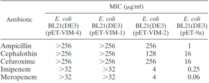
Determination of MICs. Compared to E. coli BL21(DE3) (pET-VIM-2), E. coli BL21(DE3)(pET-VIM-4) exhibited de-creased susceptibility to ampicillin, narrow-spectrum cephalo-sporins, and carbapenems. Susceptibility to those compounds was identical between E. coli BL21(DE3)(pET-VIM-4) and E. coli BL21(DE3)(pET-VIM-1) (Table 1).
Production and purification of VIM-4.The E. coli BL21Codon Plus(DE3) strain, chosen to produce VIM-4, allowed
produc-TABLE 1. In vitro antimicrobial susceptibility profile of E. coli BL21(DE3) carrying recombinant vectors in which the
blaVIM-4gene was cloneda
Antibiotic MIC (g/ml) E. coli BL21(DE3) (pET-VIM-4) E. coli BL21(DE3) (pET-VIM-1) E. coli BL21(DE3) (pET-VIM-2) E. coli BL21(DE3) (pET-9a) Ampicillin ⬎256 ⬎256 256 1 Cephalothin ⬎256 ⬎256 128 16 Cefuroxime ⬎256 ⬎256 256 16 Imipenem ⬎32 ⬎32 4 0.25 Meropenem ⬎32 ⬎32 4 0.06
aData for strains carrying the empty vector or carrying the cloned bla
VIM-1and
blaVIM-2alleles are shown for comparison.
on December 23, 2014 by UNIV DE LIEGE
http://aac.asm.org/
tion of 120 mg of VIM-4 per liter. Three purification steps were needed to purify VIM-4 to homogeneity. Approximately 25 mg of purified enzyme was obtained per liter of culture (20% overall purification yield). Upon SDS-PAGE, VIM-4 migrated with an apparent molecular mass of about 25,000 Da, and the preparation was estimated to be 95% pure. The mo-lecular mass of VIM-4 determined by mass spectrometry was 25,392 Da, in good agreement with the theoretical value (25,391.2 Da).
Zn2ⴙ
dependence of VIM-4 and functional properties.With benzylpenicillin, the enzyme activity increased with the Zn2⫹ concentration up to 50M and was slightly inhibited at higher concentrations (Fig. 1). For example, working at a [Zn2⫹] of 300M, the enzymatic activity dropped by a factor of 40%. In fitting curve 1, we have determined the second and the third dissociation constants of Zn2⫹for VIM-4. Indeed, the disso-ciation constant of the first Zn2⫹ (K1) was predicted to be much lower than 1M. We determined by inductively coupled plasma-mass spectrometry that the protein in the absence of added Zn2⫹(50 nM) was in a monozinc form; in the presence of 50M Zn2⫹, the dizinc form was obtained. Thus, we can confirm the very low dissociation constant of the first zinc binding site (Zn1) to the enzyme, and then K1can be negligi-ble. We then fitted the data using a two-parameter equation and then determined the values of K2and K3to be 8.5⫾ 2.3 M and 350 ⫾ 180 M, respectively.
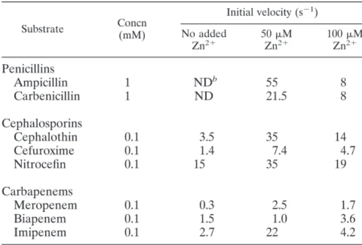
The impact of the Zn2⫹concentration (0, 50, and 100M) on activity was also determined with other-lactam substrates (Table 2). For all tested compounds, with the exception of biapenem, the enzyme exhibited the best activity in the pres-ence of 50M Zn2⫹. It is noteworthy that these conditions are the same as those used for the characterization of VIM-1 and VIM-2 (13, 25). The steady-state kinetic parameters (Kmand kcat) of VIM-4 were thus determined in the presence of 50M Zn2⫹(in the buffer) against a representative set of-lactam compounds (Table 3).
VIM-4 exhibited a broad-spectrum activity profile with im-portant differences depending on the tested substrate. As has been observed with all MBLs described so far, only aztreonam escaped its action. With penicillins, kcat/Kmvalues range from 71 to 3,100 mM⫺1䡠 s⫺1. The individual kinetic parameters of VIM-4 against ampicillin could not be determined, since the initial velocities remained proportional to the substrate con-centrations up to 3 mM. VIM-4 was less efficient than VIM-2
against ampicillin. The catalytic efficiencies of both enzymes were similar with benzylpenicillin, but VIM-4 was 100-fold more efficient than VIM-1 against benzylpenicillin, thanks to both a lower Kmvalue and a higher kcatvalue. In the case of
ampicillin, despite higher Kmand kcatvalues, the kcat/Kmvalue was not significantly different from that of VIM-1. With ceph-alosporins, the Kmvalues were low, whereas the turnover rates exhibited higher values, so that VIM-4 was overall more effi-cient than VIM-1 and VIM-2. Two exceptions could be ob-served: first, the kcat/Kmvalues for cefuroxime were similar for
VIM-4 and VIM-1, and second, VIM-4 was almost as efficient as VIM-2 against nitrocefin. With carbapenems, the kcat/Km
ratios ranged from 230 to 23,000 mM⫺1䡠 s⫺1, resulting from a combination of low Kmvalues and very low turnover rates, a
characteristic behavior of the VIM-type enzymes. The pH de-pendence of the VIM-4 activity on benzylpenicillin was mea-sured at various Zn2⫹concentrations (data not shown). At pH values lower than 5, the VIM-4 enzyme is poorly active in all tested buffers, whatever the Zn2⫹ concentration. At pH 6, VIM-4 exhibited maximal activity at 100 M Zn2⫹, and at higher Zn2⫹ concentrations, the activity decreased. Between pH 7 and 8, VIM-4 presented its maximal activity at 50M Zn2⫹, and above 50M Zn2⫹, the activity gradually decreased. At a pH of⬎8, VIM-4 presented the same efficiency in the presence of 50 to 400M Zn2⫹, but at 1 mM Zn2⫹, its activity was reduced by 50%.
VIM-4 apoenzyme. The specific activity of the apoenzyme against benzylpenicillin was 1.2% of that of the native protein activity at 50M Zn2⫹. When VIM-4 and denatured apo-VIM-4 were added with free fura-2, a chromophoric chelator, there was no shift in fura-2 absorbance. Both spectra could be perfectly superimposed, showing the apoenzyme to be Zn2⫹ free. Under our experimental conditions, the apoenzyme could not be completely reactivated. A maximum of 56% activity was recovered upon addition of 10 Zn2⫹ equivalents to 40 M apoenzyme. With Ca2⫹, Cd2⫹, and Co2⫹ions, a maximum of 3% of the initial activity was recovered.
The native protein, alone or in the presence of excess Zn2⫹, and the apoprotein were gradually heated, and the unfolding process was monitored with the fluorescent probe
Sypro-or-TABLE 2. Zn2⫹impact on catalytic efficiency of VIM-4a
Substrate Concn (mM) Initial velocity (s⫺1) No added Zn2⫹ 50ZnM2⫹ 100ZnM2⫹ Penicillins Ampicillin 1 NDb 55 8 Carbenicillin 1 ND 21.5 8 Cephalosporins Cephalothin 0.1 3.5 35 14 Cefuroxime 0.1 1.4 7.4 4.7 Nitrocefin 0.1 15 35 19 Carbapenems Meropenem 0.1 0.3 2.5 1.7 Biapenem 0.1 1.5 1.0 3.6 Imipenem 0.1 2.7 22 4.2 a
Experiments were done in triplicate. Standard deviations were always⬍10%.
b
ND, not determined.
FIG. 1. Influence of the Zn2⫹concentration on the activity of pu-rified VIM-4 at 1 mM benzylpenicillin.
on December 23, 2014 by UNIV DE LIEGE
http://aac.asm.org/
ange (Fig. 2). The native VIM-4 exhibited one transition tem-perature (melting temtem-perature [Tm]) at 58.5 ⫾ 0.1°C. The
addition of a 10:1 ratio of Zn2⫹ions to VIM-4 triggered an important modification of the protein stability with two tran-sitions, one at 44.4⫾ 0.3°C and the other one at 56.6 ⫾ 0.3°C. The behaviors of the apo- and holoenzymes in the presence of 25 molar equivalents of EDTA showed that both unfolding processes were identical, with a single transition at 31.8 ⫾ 0.2°C, which reflected a 27°C decrease in Tms. The addition of a 1:1 to 10:1 ratio of Zn2⫹, Cd2⫹, Co2⫹, and Ca2⫹ to the apoenzyme form had no effect upon the stability of the protein (data not shown).
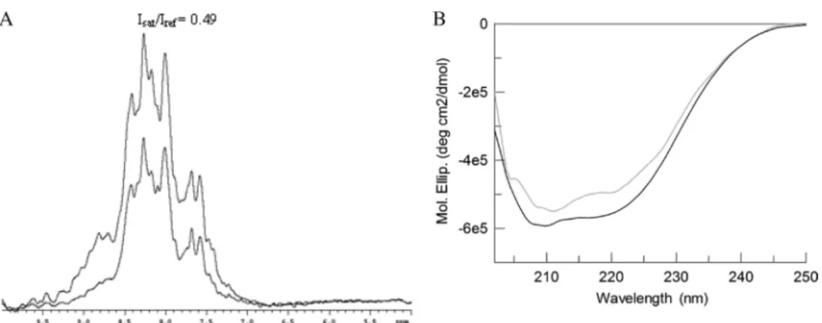
In order to measure the degree of structure in native, metal-free, and reactivated VIM-4, one-dimensional (1-D) HET-SOFAST experiments were performed. VIM-4 presented a NOEvalue of 0.49 (Fig. 3A), which suggested a high degree of
flexibility in solution. As underlined by the thermal shift assay, the apoenzyme form of VIM-4 was highly destabilized; indeed, the measured⌵⌷⌭value was 0.57. The apoenzyme form in the presence of 5 equivalents of ZnCl2 exhibited a NOE value
(0.51) consistent with a more structured protein. The destabi-lization of apo-VIM-4 was also confirmed by the far-UV CD spectra of the native and apoenzyme forms, which exhibited significant differences (Fig. 3B).
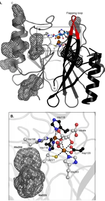
Three-dimensional structure of VIM-4. The structure of VIM-4 in the presence of 50M Zn2⫹was refined to a reso-lution of 1.9 Å (Table 4). The crystals adopted a C-2 space group with two molecules in the asymmetric unit. Residues 26 to 31 and 262 to 266 were not solved, as no density was observed for them; thus, the observed polypeptide chain of VIM-4 con-sisted of residues 32 to 261. The final model included a total of 218 water molecules, two Zn2⫹ions, and one citrate anion per molecule. Figure 4 shows that VIM-4 has the typical␣/␣ fold of MBLs, consisting of a core of sheets surrounded by ␣ helices. In this monomeric enzyme, two Zn2⫹ions are found in the active site located at the bottom of the-sheet core. Zn1
and Zn2 had tetrahedral and hexagonal coordinations (6
li-gands), respectively. The latter is different from the trigonal bipyramidal coordination (5 ligands) usually observed in MBLs (Fig. 4B). This can be explained by the presence of the citrate anion in the active site. The water molecule that usually serves as a bridging ligand between the two metals was replaced by a citrate carboxylate group. Moreover, the three carboxylate groups of citrate interacted with the side chains of the Asn231, Phe61, Arg228, and Tyr67 residues. The MBL conserved motif H (N/Q)116-X-H118-X-D120 and the residues that bind Zn2⫹ ions were present in VIM-4, with the first Zn2⫹binding site containing the usual three His ligands and the second site
TABLE 3. Kinetic parameters of the purified VIM-4 metallo--lactamase compared with those of VIM-1 and VIM-2a
Substrate
kcat(s⫺1) Km(M) kcat/Km(mM⫺1䡠 s⫺1)
VIM-4 VIM-1 VIM-2 VIM-19 VIM-4 VIM-1 VIM-2 VIM-19 VIM-4 VIM-1 VIM-2 VIM-19
Penicillins Ampicillin ⬎210 35 125 NDb ⬎3,000 920 90 ND 71 38 1,400 ND Carbenicillin 100 170 190 ND 31 75 210 ND 3,300 2,300 900 ND Benzylpenicillin 650 30 280 1,300 180 840 70 300 3,100 36 4,000 5,000 Cephalosporins Cephalothin 500 280 130 ND 14 55 11 ND 36,000 5,100 12,000 ND Cefuroxime 120 330 8 ND 11 42 20 ND 6,000 7,700 400 ND Nitrocefin 690 95 770 ND 22 17 18 ND 31,000 5,600 43,000 ND Carbapenems Meropenem 7 13 5 25 8 50 2 15 875 260 2,500 2,000 Biapenem 15 8.5 8.5 ND 66 7.5 15 ND 230 1,100 550 ND Imipenem 70 0.2 34 250 3 1.5 9 40 23,000 130 3,800 6,000 Monobactam, aztreonam ⬍0.01 ⬍0.01 ⬍0.01 ND ⬎1,000 ⬎1,000 ⬎1,000 ND ⬍0.01 ⬍0.01 ⬍0.01 ND
aData for VIM-1, VIM-2, and VIM-19 are from Franceschini et al. (16), Docquier et al. (13), and Rodriguez-Martinez et al. (39), respectively. Individual kinetic
parameters are the means of three measurements. Standard deviations were always⬍10%.
bND, not determined.
FIG. 2. Thermal stability of VIM-4. Thermal unfolding of 40M VIM-4 (heavy black line) and apo-VIM-4 (thin gray line) was followed in the presence of Sypro-orange fluorescent probe. ZnCl2(400M;
thin black line) or 1 mM EDTA (heavy gray line) was added to VIM-4. The signal of the buffer is represented by black dots. These sets of curves are representative of three independent experiments. au, arbi-trary units.
on December 23, 2014 by UNIV DE LIEGE
http://aac.asm.org/
containing Asp120, Cys221, and His263. The other ligands in the second site were the hydroxyl and carboxylate groups of the citrate anion.
The native VIM-4 structure was compared to the VIM-2 structure. Consistent with the high degree of similarity in the amino acid sequences, the overall folds of VIM-4 and VIM-2
were quite similar (root mean square deviation [RMSD], 0.545 Å). Interestingly, the Zn2⫹ions in VIM-2 and VIM-4 were in identical positions, with their ligands being almost perfectly superimposed. The RMSD value obtained from a fit including only the atoms in a radius of 10 Å around Asp120 was 0.487 Å, underlining the better structural conservation of the active site than of the overall fold. Most of the 12 residues which differ in VIM-4 and VIM-2 were in the C-terminal part of the protein and were surface exposed (Fig. 4A). They were distant from the active site, with the exception of the two residues at posi-tions 223 and 224 (in loop L3). Despite identical sequences, the so-called flapping loop (residues 58 to 68) of VIM-4 was not positioned as it is in VIM-2, resulting in a narrower active site in VIM-4.
Only the Ser228Arg substitution distinguished VIM-4 from VIM-1. This substitution is located at the entry of the active-site cavity of the protein (Fig. 5). In the case of VIM-4, the entry is thus narrowed and positively charged compared to VIM-1.
DISCUSSION
The study of the Zn2⫹dependence of VIM-4 revealed some important features. The enzyme activity is clearly optimal at 50 M Zn2⫹. The maximal activity is obtained between pH 7 and
8 in HEPES buffer. This feature is similar to the pH curve obtained for the hydrolysis of benzylpenicillin and cephalospo-rins by BcII (7). As pH and Zn2⫹dependence is observed for all MBLs, it seems to be important to define standard exper-imental conditions for comparison of their catalytic behaviors. Inhibition at a high Zn2⫹concentration (300M) was ob-served. This is the first report highlighting inhibition of a B1 enzyme at a high Zn2⫹concentration. This inhibition could be explained by nonspecific interactions between the surface of the enzyme and the Zn2⫹cations, as has already been observed for VIM-2 or other MBLs (4, 17). This Zn2⫹fixation could either strongly stabilize the enzyme and impair the active-site mobility or strongly destabilize the enzyme and disorganize the active site.
The kinetic parameters at 50M Zn2⫹confirmed the broad activity spectrum of VIM-4, which includes all-lactam
com-FIG. 3. Comparison of the structural properties of holo- and apo-VIM-4. (A) Spectra of the reference and saturated intensities (Isatand Iref,
respectively) of the native protein from HET-SOFAST experiment of the native protein. (B) Circular dichroism spectra of the apoenzyme for (gray) and native form (black) VIM-4 are shown. Mol. Ellip., molar ellipticity.
TABLE 4. Data collection and refinement statistics of the native VIM-4
Parameter
Value(s)
Apo-VIM-4 VIM-4 wild type
Data collection
X-ray source ESRF/ID14-1
Space group C 1 2 1 Cell dimensions a, b, c (Å) 141.39, 46.22, 105.99 ␣, , ␥ 90.0, 105.237, 90.0 Wavelength 0.934 Resolution (Å) 47.30–1.9 (2.0–1.9)a No. of reflections 52,283 Redundancy 3.6 (3.3) Rmerge b 0.098 (0.44) I/ 8.92 (2.59) Completeness (%) 98.6 (95.7) Refinement Resolution (Å) 47.30–1.9 No. of reflections 49,667 Rworkc/Rfreed 0.19/0.25
No. atoms (AUe) 3,974
Protein 3,509
Ligand 26f
Cofactor 4
Water 435
B factor for protein 26.52 RMSDs
Bond lengths (Å) 0.015 Bond angles (°) 1.424 a
Values in parentheses refer to the highest-resolution shell.
b
Rmerge⫽ ¥ 兩I ⫺ 具I典兩/¥具I典, where I is the intensity of individual reflections.
c
Rwork⫽ ¥h兩Fo⫺ Fc兩/¥h兩Fo兩.
d
Rfreewas calculated with 5% of the diffraction data selected randomly and
excluded from refinement.
e
AU, arbitrary units.
f
One molecule of citrate anion per molecule of VIM-4.
on December 23, 2014 by UNIV DE LIEGE
http://aac.asm.org/
pounds with the exception of aztreonam. The comparison of the kinetic parameters of VIM-1, VIM-2, and VIM-4 obtained under the same conditions revealed that VIM-4 had similar activity to that of VIM-2 for almost all tested substrates and better activity than that of VIM-1. It has previously been shown that for enzymes with the S228R substitution, e.g., VIM-2, the Kmvalue for ampicillin was lower than the values for the other
B1 enzymes (17). Despite this mutation, however, VIM-4 pre-sents a high Kmvalue for ampicillin. VIM-4 exhibits a lower Km value for benzylpenicillin. Because benzylpenicillin and ampi-cillin differ only by an amino group on the C-6 side chain, the position of the flapping loop in a narrow conformation for VIM-4 could explain this behavior. VIM-19, another VIM-1 variant, possesses two substitutions (S228R and N215K) (38). Its catalytic efficiency toward benzylpenicillin and meropenem was similar to that of VIM-4 (Table 3) (39). Interestingly, VIM-4 was more efficient against imipenem. This underlines the finding that the N215K substitution does not have a direct impact on the broadening of the activity spectrum of MBL enzymes.
Only a few data on the role of bound Zn2⫹ ions on the stability of MBLs have been collected. With the subclass B2 CphA enzyme, differences in the fluorescence emission and circular dichroism spectra were obtained between the apo-, mono-, and dizinc forms (20). The thermal denaturation of the CphA apoenzyme exhibits a Tmof 45°C, which is lower than
the Tms of the mono- and dizinc forms (Tms, 55°C and 60°C,
respectively) (20). In another member of the metallo- -lactamase superfamily, glyoxalase II, Zn2⫹ appears to be essential for the maintenance of the native structure, as its binding occurs during the refolding process (14). The study of the apoenzyme confirmed the Zn2⫹ dependence of VIM-4 stability. The addition of 1 or 2 equivalents of Zn2⫹ions to the apoenzyme form of VIM-4 did not reveal any effect on the stability or activity. This irreversibility has already been de-scribed for some MBLs, such as for the apo-MBL from B. fragilis, which cannot be reactivated by addition of Zn2⫹(12). Likewise, the study of IMP-1 (DK4) by Juan et al. (21) and Yamaguchi et al. (44) has revealed the impossibility to obtain a dizinc enzyme by addition of Zn2⫹to the apoenzyme. The structural differences between the apo- and dizinc forms of VIM-4 revealed by the1H HET-SOFAST NMR and CD
ex-periments confirm the important structural role of Zn2⫹in the VIM-4 enzyme.
FIG. 4. Structure of VIM-4. (A) Three-dimensional structure of VIM-4 enzyme. Both Zn2⫹ ions are represented as orange spheres, Zn2⫹ligands (gray or black sticks) are shown, and the locations of the VIM-4/VIM-2 mutations are shown as meshes. (B) Active site of the dizinc structure. Citrate and ligands are represented as sticks. The figures were generated with the PyMOL program.
FIG. 5. Comparison of the electrostatic charges in the VIM-4 and VIM-1 active sites. Zn2⫹ions are represented as black spheres, and the citrate anion is represented as green sticks. The active-site topology of the enzyme is a tunnel. The mutation is positioned at one side of the active-site cavity, where arginine 228 is located and interacts with the citrate carboxylate.
on December 23, 2014 by UNIV DE LIEGE
http://aac.asm.org/
Finally, the structure of VIM-4 confirmed an overall fold characteristic of MBLs with an␣/␣ sandwich structure. In-terestingly, but not uncommonly, we observed a citrate anion in the active site of the-lactamase. Citrate has been observed in the active site of at least one member of each class of -lactamase. In all cases, the citrate was provided by the crys-tallization buffer and was bound to the active site. Despite these multiple interactions, citrate did not have significant in-hibitory activity against VIM-4 (5).
The structural comparison of VIM-1, VIM-2, and VIM-4 has revealed some interesting features. First, VIM-4 and VIM-2 possess an arginine at position 228 in loop L3 posi-tioned in the active-site channel. At this position, VIM-1 has a serine with a small and noncharged lateral chain. This sole feature is responsible for the important differences between the individual kinetic parameters of these enzymes, with the Kmvalues of VIM-4 usually being lower than those of VIM-1. Biapenem, a bulky substrate with a charged lateral chain on C-2, cannot be strongly distorted, and this can impair its proper positioning in the active site of VIM-4 due to the bulk and the positive charge of the Arg228. This can explain the different Kmvalues of VIM-4 and VIM-1 for biapenem.
For VIM-1 and VIM-4, Lys224, a catalytically important residue, is replaced by His224. In IMP-1 (31) and CphA (3), the side chain of Lys224 binds to the carboxylate moiety on C-4 or C-3 of the substrate. The side chain of the histidine residue is much shorter than that of Lys, preventing its interaction with this carboxylate. As already proposed for VIM-2 (17), Arg228 of VIM-4, with its long side chain, may replace Lys224 and interact with the carboxylate of-lactams. Mutations between VIM-4 and VIM-2 are in the C-terminal part of the protein and are mainly exposed to the solvent on the protein surface. Only the V223I and H224Y substitutions occur close to the active site, and both belong to loop 2. In the inhibition of VIM-2 by the rac-2-omega-phenylpropyl-3-mercaptopropionic acid (phenylC3SH) complex, loop 2 undergoes important changes (44). Those two mutations (His and Val), which have shorter side chains than Lys and Ile, may impair the interaction between loop 2 and the substrate; thus, they may change the affinity for some substrates. Another difference between VIM-2 and VIM-4 is the flapping loop, which seems to be more closed in the latter, which would decrease the accessibil-ity to the active site for bulky antibiotics, as for biapenem, and have an impact on catalysis.
In conclusion, this study revealed a Zn2⫹and pH depen-dence of the VIM-4 enzyme, in agreement with observations made on other subclass B1 MBLs. Our data highlight the need to define the experimental conditions used to study MBLs. Finally, we have proved the structural role of Zn2⫹on the enzyme VIM-4, and our data suppose that VIM-4 cannot exist as an apoenzyme in vivo, as the deletion of Zn2⫹is fatal to the protein.
ACKNOWLEDGMENTS
We thank N. Otthiers for the N-terminal experiments and Thierry Vernet and Claire Durmort for the TSA experiments.
P.L. was supported by the Fonds pour la Formation a` la Recher-che dans l’Industrie et dans l’Agriculture, an EMBO short-term fellowship, and a traveling grant from the University of Lie`ge for her stay at the IBS.
REFERENCES
1. Attali, C., et al. 2008. Streptococcus pneumoniae choline-binding protein E interaction with plasminogen/plasmin stimulates migration across the extra-cellular matrix. Infect. Immun. 76:466–476.
2. Bebrone, C. 2007. Metallo-beta-lactamases (classification, activity, genetic organization, structure, zinc coordination) and their superfamily. Biochem. Pharmacol. 74:1686–1701.
3. Bebrone, C., et al. 2008. Mutational analysis of the zinc- and substrate-binding sites in the CphA metallo-beta-lactamase from Aeromonas hy-drophila. Biochem. J. 414:151–159.
4. Bebrone, C., et al. 2009. The structure of the dizinc subclass B2 metallo-beta-lactamase CphA reveals that the second inhibitory zinc ion binds in the histidine site. Antimicrob. Agents Chemother. 53:4464–4471.
5. Beck, J., et al. 2009. Discovery of novel lipophilic inhibitors of OXA-10 enzyme (class D beta-lactamase) by screening amino analogs and homologs of citrate and isocitrate. Bioorg. Med. Chem. Lett. 19:3593–3597. 6. Bogaerts, P., et al. 2008. Nosocomial infections caused by multidrug-resistant
Pseudomonas putida isolates producing VIM-2 and VIM-4 metallo-beta-lactamases. J. Antimicrob. Chemother. 61:749–751.
7. Bounaga, S., A. P. Laws, M. Galleni, and M. I. Page. 1998. The mechanism of catalysis and the inhibition of the Bacillus cereus zinc-dependent beta-lactamase. Biochem. J. 331(Pt 3):703–711.
8. Carfi, A., et al. 1995. The 3-D structure of a zinc metallo-beta-lactamase from Bacillus cereus reveals a new type of protein fold. EMBO J. 14:4914– 4921.
9. Clinical and Laboratory Standards Institute. 2006. Methods for dilution antimicrobial susceptibility tests for bacteria that grow aerobically, 7th ed. Approved standard. CLSI document M7-A7. Clinical and Laboratory Stan-dards Institute, Wayne, PA.
10. Collaborative Computational Project No. 4. 1994. The CCP4 suite: programs for protein crystallography. Acta Crystallogr. D Biol. Crystallogr. D50:760– 763.
11. Cornish-Bowden, A. 2001. Fundamentals of enzyme kinetics. Portland Press Ltd., London, United Kingdom.
12. Crowder, M. W., Z. Wang, S. L. Franklin, E. P. Zovinka, and S. J. Benkovic. 1996. Characterization of the metal-binding sites of the beta-lactamase from Bacteroides fragilis. Biochemistry 35:12126–12132.
13. Docquier, J. D., et al. 2003. On functional and structural heterogeneity of VIM-type metallo-beta-lactamases. J. Antimicrob. Chemother. 51:257–266. 14. Dragani, B., et al. 1999. Unfolding and refolding of human glyoxalase II and
its single-tryptophan mutants. J. Mol. Biol. 291:481–490.
15. Emsley, P., and K. Cowtan. 2004. Coot: model-building tools for molecular graphics. Acta Crystallogr. D Biol. Crystallogr. 60:2126–2132.
16. Franceschini, N., et al. 2000. Purification and biochemical characterization of the VIM-1 metallo-beta-lactamase. Antimicrob. Agents Chemother. 44: 3003–3007.
17. Garcia-Saez, I., J. D. Docquier, G. M. Rossolini, and O. Dideberg. 2008. The three-dimensional structure of VIM-2, a Zn-beta-lactamase from Pseudo-monas aeruginosa in its reduced and oxidised form. J. Mol. Biol. 375:604– 611.
18. Giannouli, M., et al. 2009. Molecular epidemiology of carbapenem-resistant Acinetobacter baumannii strains in intensive care units of multiple Mediter-ranean hospitals. J. Antimicrob. Chemother. 63:828–830.
19. Giske, C. G., M. Rylander, and G. Kronvall. 2003. VIM-4 in a carbapenem-resistant strain of Pseudomonas aeruginosa isolated in Sweden. Antimicrob. Agents Chemother. 47:3034–3035.
20. Hernandez Valladares, M., et al. 1997. Zn(II) dependence of the Aeromonas hydrophila AE036 metallo-beta-lactamase activity and stability. Biochemis-try 36:11534–11541.
21. Juan, C., et al. 2008. Characterization of the new metallo-beta-lactamase VIM-13 and its integron-borne gene from a Pseudomonas aeruginosa clin-ical isolate in Spain. Antimicrob. Agents Chemother. 52:3589–3596. 22. Kabsch, W. 1993. Automatic processing of rotation diffraction data from
crystals of initially unknown symmetry and cell constants. J. Appl. Crystal-logr. 26:795–800.
23. Kontou, M., et al. 2007. Molecular cloning and biochemical characterization of VIM-12, a novel hybrid VIM-1/VIM-2 metallo-beta-lactamase from a Klebsiella pneumoniae clinical isolate, reveal atypical substrate specificity. Biochemistry 46:13170–13178.
24. Ktari, S., et al. 2006. Emergence of multidrug-resistant Klebsiella pneu-moniae isolates producing VIM-4 metallo-beta-lactamase, CTX-M-15 ex-tended-spectrum beta-lactamase, and CMY-4 AmpC beta-lactamase in a Tunisian university hospital. Antimicrob. Agents Chemother. 50:4198–4201. 25. Lauretti, L., et al. 1999. Cloning and characterization of blaVIM, a new integron-borne metallo-beta-lactamase gene from a Pseudomonas aerugi-nosa clinical isolate. Antimicrob. Agents Chemother. 43:1584–1590. 26. Libisch, B., et al. 2004. Isolation of an integron-borne blaVIM-4 type
me-tallo-beta-lactamase gene from a carbapenem-resistant Pseudomonas aeruginosa clinical isolate in Hungary. Antimicrob. Agents Chemother. 48: 3576–3578.
27. Libisch, B., et al. 2006. Molecular epidemiology of VIM-4
on December 23, 2014 by UNIV DE LIEGE
http://aac.asm.org/
lactamase-producing Pseudomonas sp. isolates in Hungary. Antimicrob. Agents Chemother. 50:4220–4223.
28. Luzzaro, F., et al. 2004. Emergence in Klebsiella pneumoniae and Entero-bacter cloacae clinical isolates of the VIM-4 metallo-beta-lactamase encoded by a conjugative plasmid. Antimicrob. Agents Chemother. 48:648–650. 29. Matagne, A., J. Lamotte-Brasseur, and J. M. Frere. 1993. Interactions
be-tween active-site serine beta-lactamases and so-called beta-lactamase-stable antibiotics. Kinetic and molecular modelling studies. Eur. J. Biochem. 217: 61–67.
30. Matagne, A., et al. 1990. The diversity of the catalytic properties of class A beta-lactamases. Biochem. J. 265:131–146.
31. Materon, I. C., Z. Beharry, W. Huang, C. Perez, and T. Palzkill. 2004. Analysis of the context dependent sequence requirements of active site residues in the metallo-beta-lactamase IMP-1. J. Mol. Biol. 344:653–663. 32. McCoy, A. J. 2007. Solving structures of protein complexes by molecular
replacement with Phaser. Acta Crystallogr. D Biol. Crystallogr. 63:32–41. 33. Patzer, J., et al. 2004. Pseudomonas aeruginosa strains harbouring an
un-usual blaVIM-4 gene cassette isolated from hospitalized children in Poland (1998-2001). J. Antimicrob. Chemother. 53:451–456.
34. Peleg, A. Y., J. M. Bell, A. Hofmeyr, and P. Wiese. 2006. Inter-country transfer of Gram-negative organisms carrying the VIM-4 and OXA-58 car-bapenem-hydrolysing enzymes. J. Antimicrob. Chemother. 57:794–795. 35. Pournaras, S., et al. 2003. Hospital outbreak of multiple clones of
Pseudo-monas aeruginosa carrying the unrelated metallo-beta-lactamase gene vari-ants blaVIM-2 and blaVIM-4. J. Antimicrob. Chemother. 51:1409–1414. 36. Pournaras, S., A. Tsakris, M. Maniati, L. S. Tzouvelekis, and A. N. Maniatis.
2002. Novel variant (bla(VIM-4)) of the metallo-beta-lactamase gene
bla(VIM-1) in a clinical strain of Pseudomonas aeruginosa. Antimicrob. Agents Chemother. 46:4026–4028.
37. Prosperi-Meys, C., et al. 1999. Interaction between class B beta-lactamases and suicide substrates of active-site serine beta-lactamases. FEBS Lett. 443: 109–111.
38. Robin, F., N. Aggoune-Khinache, J. Delmas, M. Naim, and R. Bonnet. 2010. Novel VIM metallo-beta-lactamase variant from clinical isolates of Entero-bacteriaceae from Algeria. Antimicrob. Agents Chemother. 54:466–470. 39. Rodriguez-Martinez, J. M., P. Nordmann, N. Fortineau, and L. Poirel. 2010.
VIM-19, a metallo-beta-lactamase with increased carbapenemase activity from Escherichia coli and Klebsiella pneumoniae. Antimicrob. Agents Che-mother. 54:471–476.
40. Sabath, L. D., and E. P. Abraham. 1966. Zinc as a cofactor for cephalospo-rinase from Bacillus cereus 569. Biochem. J. 98:11C–3C.
41. Schanda, P., V. Forge, and B. Brutscher. 2006. HET-SOFAST NMR for fast detection of structural compactness and heterogeneity along polypeptide chains. Magn. Reson. Chem. 44(Spec. No.):S177–S184.
42. Studier, F. W. 2005. Protein production by auto-induction in high density shaking cultures. Protein Expr. Purif. 41:207–234.
43. Toleman, M. A., K. Rolston, R. N. Jones, and T. R. Walsh. 2004. blaVIM-7, an evolutionarily distinct metallo-beta-lactamase gene in a Pseudomonas aeruginosa isolate from the United States. Antimicrob. Agents Chemother.
48:329–332.
44. Yamaguchi, Y., et al. 2007. Crystallographic investigation of the inhibition mode of a VIM-2 metallo-beta-lactamase from Pseudomonas aeruginosa by a mercaptocarboxylate inhibitor. J. Med. Chem. 50:6647–6653.