D
E
V
E
LO
P
M
E
N
T
INTRODUCTIONCollagens are triple-helical molecules that form the major structural elements of almost all tissues outside the nervous system. Collagen types I, II and III are evolutionarily related, and comprise the quantitatively major fibril-forming (fibrillar) collagens (Olsen, 1991). They form organotypic matrices by tissue-specific expression and by assembly of the collagen triple-helix into a variety of supramolecular aggregates (Birk et al., 1991). Collagen I is the major extracellular matrix (ECM) protein of bone, tendons, ligaments, and the dermis of the skin. Collagen II is the major collagen of cartilage, but it is also present in the vitreous of the eye and in the nucleus pulposus of the intervertebral disc (Zhu et al., 1999). Collagen III is a component of many stretchable tissues, such as the vascular wall, dermis, lung, heart and gastrointestinal tract (De Paepe and Malfait, 2004). It can co-polymerize with collagen I to form heterotypic fibrils and it is co-expressed in varying proportions with collagen I at many locations, with the exception of bone (Niederreither et al., 1995). Collagen I contains two ␣1(I) chains and one ␣2(I) chain, whereas collagens II and III are homotrimers of ␣1(II) and ␣1(III) chains, respectively (Olsen, 1991). Structural
mutations in collagen genes have serious consequences for tissues where single collagen types are major constituents. Collagen I mutations cause osteogenesis imperfecta (Rauch and Glorieux, 2004). Chondrodysplasias, sometimes with ocular anomalies, such as Stickler syndrome, are caused by collagen II mutations (Chan et al., 1995; Cole, 1997; Snead and Yates, 1999; Tiller et al., 1995), and collagen III mutations cause the vascular subtype of the Ehlers-Danlos syndrome (type IV), which is characterized by aortic aneurysms and skin fragility (De Paepe and Malfait, 2004).
Fibrillar collagens are made as a precursor (procollagen) and consist of a long triple-helical (collagenous) domain flanked at each end by globular non-collagenous propeptides (Olsen, 1991). The three major fibrillar procollagens share similar biosynthetic mechanisms. Following intracellular assembly of the triple helix, the procollagen molecules undergo proteolytic excision of each propeptide either prior to or following secretion (Canty et al., 2004; Olsen, 1991). The C-propeptide is excised by BMP1, an astacin-like protease (Kessler et al., 1996). ADAMTS2 (for A Disintegrin-like And Metalloprotease domain with Thrombospondin type 1 motifs 2) can excise the N-propeptide of all three major fibrillar collagens (Tuderman and Prockop, 1982; Wang et al., 2003). The processed collagen monomers assemble into fibrils and tissue-specific supramolecular aggregates. Because of a common biosynthetic pathway, it was expected that defects in ADAMTS2 would have broader effects than structural mutations involving individual procollagen chains. Instead, ADAMTS2 mutations lead to a limited clinical syndrome – dermatosparaxis (fragility of skin) in animals and the Ehlers-Danlos syndrome-type VIIC (EDS-VIIC or the dermatosparactic type) in humans (Colige et al., 2004; Colige et al.,
Regulation of procollagen amino-propeptide processing
during mouse embryogenesis by specialization of
homologous ADAMTS proteases: insights on collagen
biosynthesis and dermatosparaxis
Carine Le Goff1, Robert P. T. Somerville1, Frederic Kesteloot2, Kimerly Powell1, David E. Birk3, Alain C. Colige2
and Suneel S. Apte1,*
Mutations in ADAMTS2, a procollagen amino-propeptidase, cause severe skin fragility, designated as dermatosparaxis in animals, and a subtype of the Ehlers-Danlos syndrome (dermatosparactic type or VIIC) in humans. Not all collagen-rich tissues are affected to the same degree, which suggests compensation by the ADAMTS2 homologs ADAMTS3 and ADAMTS14. In situ hybridization of
Adamts2, Adamts3 and Adamts14, and of the genes encoding the major fibrillar collagens, Col1a1, Col2a1 and Col3a1, during
mouse embryogenesis, demonstrated distinct tissue-specific, overlapping expression patterns of the protease and substrate genes.
Adamts3, but not Adamts2 or Adamts14, was co-expressed with Col2a1 in cartilage throughout development, and with Col1a1 in
bone and musculotendinous tissues. ADAMTS3 induced procollagen I processing in dermatosparactic fibroblasts, suggesting a role in procollagen I processing during musculoskeletal development. Adamts2, but not Adamts3 or Adamts14, was co-expressed with
Col3a1 in many tissues including the lungs and aorta, and Adamts2–/–mice showed widespread defects in procollagen III processing. Adamts2–/–mice had abnormal lungs, characterized by a decreased parenchymal density. However, the aorta and collagen fibrils in the aortic wall appeared normal. Although Adamts14 lacked developmental tissue-specific expression, it was co-expressed with
Adamts2 in mature dermis, which possibly explains the presence of some processed skin procollagen in dermatosparaxis. The data
show how evolutionarily related proteases with similar substrate preferences may have distinct biological roles owing to tissue-specific gene expression, and provide insights into collagen biosynthesis and the pathobiology of dermatosparaxis.
KEY WORDS: ADAMTS, Metalloprotease, Procollagen, Dermatosparaxis, Ehlers-Danlos syndrome
Development 133, 1587-1596 (2006) doi:10.1242/dev.02308
1Department of Biomedical Engineering and Orthopaedic Research Center, Lerner
Research Institute, Cleveland Clinic Foundation, 9500 Euclid Avenue, Cleveland, OH 44195, USA. 2Laboratory of Connective Tissues Biology, Center of Biomedical
Integrative Genoproteomics, University of Liège, 4000 Sart Tilman, Belgium.
3Department of Cell Biology, Thomas Jefferson University, Philadelphia, PA 19107,
USA.
*Author for correspondence (e-mail: aptes@ccf.org)
D
E
V
E
LO
P
M
E
N
T
1999; Lapiere and Nusgens, 1993; Nusgens et al., 1992; Petty et al., 1993; Smith et al., 1992; Wertelecki et al., 1992). Collagen fibrils in the dermis of dermatosparactic calves, humans and mice are thin, branched and ‘hieroglyphic’ on cross-section, instead of being round and of relatively uniform diameter (Li et al., 2001; Nusgens et al., 1992). Although this disorder is largely caused by anomalies in skin (dermal) collagen, there is only a modest effect on other collagen I-rich tissues, such as bone, tendon and muscle (Lapiere and Nusgens, 1993). Processing of procollagen II in the cartilage of dermatosparactic calves and mice is only minimally compromised, or not at all (Fernandes et al., 2001; Li et al., 2001).
It was hypothesized that enzymes closely related to ADAMTS2 may process procollagen I in tissues other than skin (Lapiere and Nusgens, 1993); this is supported by the observation that in bovine dermatosparaxis, the residual processed procollagen is cleaved at the expected site (Colige et al., 2002). We previously found that ADAMTS3, could process the amino-propeptide of procollagen II (Fernandes et al., 2001). Subsequently, it was shown that another homologous enzyme, ADAMTS14, could process procollagen I (Colige et al., 2002), and that ADAMTS2 could process procollagen III (Wang et al., 2003). Nevertheless, a comprehensive understanding of how these enzymes participate in collagen biosynthesis in vivo, particularly during embryogenesis, as the first collagenous matrices are laid down, has not been achieved.
ADAMTS2, ADAMTS3 and ADAMTS14 constitute an evolutionarily related cluster of ADAMTS proteases, the procollagen amino-propeptidases (PNPs). They have an identical domain structure and a unique modular composition not present in any other enzyme. The active site sequence in their catalytic domain (HETGHVLGMEHD) differs from that of the other ADAMTS proteases. This, and previous experimental evidence, suggested that their catalytic activities are very similar (Colige et al., 2002; Fernandes et al., 2001; Wang et al., 2003). Here, we demonstrate, through the temporal and spatial analysis of mRNA encoding ADAMTS2, ADAMTS3 and ADAMTS14, as well as of that encoding procollagen I, II and III, that there is highly coordinated developmental expression of two of these enzymes and their procollagen substrates during mouse embryogenesis. We demonstrate that ADAMTS3 can process procollagen I. Together with its prominent expression in some procollagen I rich tissues and in cartilage, ADAMTS3, and not ADAMTS2, may be the major procollagen I and II aminopropeptidase from a developmental perspective. ADAMTS2 is essential for procollagen III processing in mice, with possibly important implications for human dermatosparaxis.
MATERIALS AND METHODS Riboprobes
cDNA clones suitable for in vitro transcription of mouse pro-␣1(I) collagen (Col1a1) and mouse pro-␣1(III) collagen (Col3a1) were provided by E. Vuorio (University of Turku, Finland) (Niederreither et al., 1995). The mouse pro-␣1(II) collagen (Col2a1) cDNA was provided by Dr Naomi Fukai (Harvard Medical School). The Adamts2 probe was generated by subcloning a 600 base pair (bp) NotI-PstI fragment from IMAGE clone AA832579 into pBluescript II (Stratagene, La Jolla, CA). The following primers were used to amplify Adamts3 and Adamts14 cDNA fragments by RT-PCR of mouse 17.5 days post-coitum (dpc) embryo cDNA.
Adamts3 (amplifying a 528 bp fragment corresponding to the unique
C-terminal domain of mouse Adamts3), 5⬘-CTGAAGAGCTGGTCTCAGTG-3⬘ (forward) and 5⬘-TGGACTTGTCACCCAAACATG-5⬘-CTGAAGAGCTGGTCTCAGTG-3⬘ (reverse); and
Adamts14 (producing a PCR product of 310 bp that corresponds to the
unique C-terminal domain), 5⬘-AGGTGGGTGACAGAAGAGTGG-3⬘ (forward) and 5⬘-AGGCTGCAGATCTGCACCATG-3⬘ (reverse).
The PCR products were cloned into pGEMT-Easy (Promega, Madison, WI). Radioactive sense and antisense cRNA probes were generated by in vitro transcription using T3, T7 or SP6 RNA polymerases in the presence of [35S]-UTP (Perkin-Elmer).
In situ hybridization
In situ hybridization was performed on tissue sections of mouse embryos of gestational age 7.5 to 17.5 dpc. Embryos, including the surrounding uterus at 7.5 dpc and 9.5 dpc, were fixed overnight in 4% paraformaldehyde, embedded in paraffin wax, and sectioned at 6 m. The sections were hybridized to [35S]-UTP labeled riboprobes, as previously described
(Somerville et al., 2003). Slides were dipped in NTB-2 emulsion (Kodak, Rochester, NY) and exposed at 4°C for 2 to 10 days. Nuclei were stained with Hoechst 33258 (Sigma, St Louis, MO). Dark-field (autoradiographic signal) and fluorescence microscopy (nuclear staining) images were captured on a Leica DMR upright microscope (Leica Microsystems, Wetzlar GmbH), with a Retiga Exi Fast 1394 cooled CCD camera with RGB liquid crystal color filter slider (Q Imaging, Burnaby, BC, Canada). Images were viewed with ImagePro Plus software (Media Cybernetics, Silver Spring, MD). Silver grains visualized on dark field microscopy were given a red pseudocolor and superimposed on nuclear fluorescence (blue).
ADAMTS3 antibodies
A polyclonal antibody was generated in rabbits by immunization with a C-terminal peptide from human ADAMTS3 (CKKDGKIIDNRRPTRSSTLER-COOH), coupled to keyhole-limpet hemocyanin (Alpha Diagnostics International, Houston, TX). The antiserum was affinity-purified against the peptide coupled to NHS-sulfolink (Pierce).
Cloning of cells expressing ADAMTS3 and co-culture with dermatosparactic fibroblasts
HEK 293F cells (Invitrogen, Carslbad, CA) were maintained in growth medium (DMEM supplemented with antibiotics and 10% fetal bovine serum). Clones stably expressing the ADAMTS3 expression plasmid pSHTS3 were isolated as previously described (Fernandes et al., 2001). Culture media were analyzed by western blotting with ADAMTS3 polyclonal antibody at a 1:1000 dilution, and clones expressing the highest levels of ADAMTS3 were used in subsequent studies. Peptide N-glycosidase F (PNGaseF, New England Biolabs, Beverly, MA) treatment of conditioned medium was carried out as previously described (Somerville et al., 2003). Dermatosparactic calf fibroblasts were cultured alone or co-cultured with equivalent numbers of HEK293 cells transfected with the empty plasmid vector (a negative control), or with the plasmid vector expressing either ADAMTS3 or ADAMTS2 (as a positive control) (Colige et al., 2002). Co-culture experiments were performed in DMEM supplemented with 2% FCS and ascorbic acid (50 g/ml) in the absence of G418. After 24 hours, the culture medium and the cell layer were collected separately and denatured in Laemmli sample buffer with or without 0.1 M DTT. The characterization of collagen polypeptides was performed by western blotting using type I or type III collagen-specific antiserum and ECL. These antisera were produced by repeated immunization of rabbit and guinea pig with bovine collagen I or collagen III, respectively. These antisera, used at a 1:2000 dilution, were highly specific and did not cross-react with other collagen types (data not shown).
Histology, transmission electron microscopy (TEM) and mCT of
Adamts2-deficient (Adamts2–/–) mice
Adamts2–/–mice in the 129Sv strain were kindly provided by D. Prockop
(Tulane University) and were used under approved institutional protocols. Genotype analysis was by PCR as previously described. Two-month-old mice were killed by CO2inhalation, and tissues and organs were isolated for
histological analysis and for transmission electron microscopy. Lungs were fixed in 10% formalin under inflation as previously described (Oblander et al., 2005), followed by routine histology, including Hematoxylin and Eosin stain, Hart’s stain for elastin and the Mallory trichrome stain. The skulls and hind limbs of the Adamts2–/–mice and wild-type littermates were analyzed
by micro-computerized tomography (mCT), followed by three-dimensional reconstruction of the images. Samples for TEM were prepared as previously described (Birk and Trelstad, 1986). Thick sections (1 m) were cut and
D
E
V
E
LO
P
M
E
N
T
stained with methylene blue-azure B. Thin sections were cut using a Leica UCT ultramicrotome and a diamond knife. The lung was stained with 2.5% (w/v) ethanolic uranyl acetate followed by 0.04% (w/v) bismuth subnitrate. The aorta was stained with aqueous 2% (w/v) uranyl acetate followed by lead citrate. Sections were examined and photographed at 80 kV using a Tecnai 12 transmission electron microscope equipped with a Gatan US1000 2K Ultrascan digital camera.
Procollagen processing in Adamts2–/–mice
Individual organs were isolated from Adamts2–/– mice and wild-type
littermates. These were either ground in liquid nitrogen and suspended in washing buffer [0.25 M sucrose, 20 mM EDTA, 2.5 mM NEM, 0.5 mM PMSF, 50 mM Tris (pH 7.5)] or directly solubilized in Laemmli sample buffer (aorta). After centrifugation (20000 g, 10 minutes), supernatants were discarded and pellets washed again as described above. Pellets were then suspended in 0.15 M extraction buffer [0.15 M NaCl, 50 mM Tris (pH 7.5)], rotated for 2 hours at 4°C and centrifuged (20000 g, 10 minutes). Extracts were collected and pellets sequentially extracted in 1 M extraction buffer [1 M NaCl, 50 mM Tris (pH 7.5)] and 0.1 M acetic acid. Adequate amounts of the various samples were then dialyzed at 4°C in 0.1 M acetic acid, lyophilized, denatured in Laemmli sample buffer with or without 0.1 DTT, and analyzed by SDS-PAGE. Sponge granulomas were induced by the subcutaneous insertion of circular PVA sponges (10 mm diameter, clinical PVA sponges grade 3, M-PACT Worldwide Management, Eudora, KS) re-hydrated in PBS. After 4 weeks, mice were sacrificed and sponges were collected. Histology demonstrated the presence of abundant granulation tissue containing fibroblasts and blood vessels (data not shown). The sponges were ground, washed as described above, and then proteins were extracted in Laemmli sample buffer, separated by SDS-PAGE and western blotted with anti-collagen III as described above.
RESULTS
The distribution of the fibrillar collagen mRNA was consistent with previous reports (Niederreither et al., 1995). Thus, Col1a1 was primarily expressed in bone, skin, perichondrium, periosteum, tendons and the adventitia (outermost layer) of blood vessels. Col2a1 was expressed almost exclusively in cartilage. Col3a1 expression was prominent in the vascular wall, lung mesenchyme, peritoneal lining and the wall of visceral organs. Col3a1 was co-expressed with Col1a1 in skin, but not in bone. None of the sense probes for any of the collagen or ADAMTS genes gave a signal above background levels.
Adamts2 and Adamts3 are expressed in the maternal tissues at day 7.5 of gestation
Northern analysis of commercial embryo RNA blots had shown the highest expression of both Adamts2 and Adamts3 at 7 dpc (Fernandes et al., 2001). However, in situ hybridization clarified that there was no expression in the embryo itself, whereas there was strong expression in maternal tissues, specifically in the decidual reaction and the myometrium of the uterus. Prominent Col1a1 expression was seen in these tissues. Col1a1 and Adamts3 expression overlapped significantly in the decidual reaction (Fig. 1). The Adamts2 expression pattern was different, but also overlapped with Col1a1 (Fig. 1). The prominent co-expression of Col1a1 with these biosynthetic enzymes reflects the intense remodeling of the uterus that occurs during pregnancy. These data suggest that the previously described northern analysis findings resulted from the inclusion of a significant amount of maternal tissue.
Adamts3, but not Adamts2 or Adamts14, is co-expressed with Col2a1 in cartilage
Most of the skeleton is formed from cartilage models that undergo endochondral ossification. Cartilage is first formed around 12 dpc in mesenchymal condensations that begin to express procollagen II and
aggrecan. We have previously shown that ADAMTS3 processed procollagen II in vitro, and that ADAMTS3 mRNA was present at higher levels than ADAMTS2 mRNA in cultured chondrocytes by RT-PCR (Fernandes et al., 2001). However, in that study, the spatial and temporal association of Adamts3 and Col2a1 was not determined. Adamts3 mRNA was associated with Col2a1 mRNA expression from the initiation of chondrogenesis (Fig. 2A) and, subsequently, throughout skeletal development in all cartilage models, whether in the appendicular, axial or craniofacial skeleton (Fig. 2A-G). Chondrocytes in developing growth plates differentiate into hypertrophic chondrocytes and, as they do so, they suppress collagen II production and produce an abundance of a short-chain collagen, type X (LuValle et al., 1989), whose N-propeptides are not processed. Interestingly, Adamts3 was downregulated as chondrocytes became hypertrophic (Fig. 2C,E-G). After 12 dpc, pre-cartilaginous mesenchymal tissue is located in the perichondrium around cartilage (Zhu et al., 1999); The Adamts3 expression domain extended beyond the limits of the Col2a1 expression domain into the perichondrium (Fig. 2B-G). Neither Adamts2 nor Adamts14 were detectable by in situ hybridization in cartilage at any developmental stage examined.
Adamts3, but not Adamts2 or Adamts14, is co-expressed with Col1a1 during bone and musculotendinous development
During endochondral ossification, as well as during intra-membrane ossification, osteoblasts deposit collagen I in the osteoid, which is subsequently mineralized to form bone. During both types of osteogenic processes, Adamts3 was co-expressed with Col1a1 (Fig. 3A,B). In addition, Adamts3 was co-expressed with Col1a1 in perichondrium, including that around the hypertrophic zone of cartilage (Fig. 2C, Fig. 3A,C). Musculotendinous structures develop around the skeleton and synthesize abundant collagen I late in development; Adamts3 was expressed in these structures at many locations in the skeleton at 17.5 dpc (Fig. 2D,E, Fig. 3B-D). Neither Adamts2 nor Adamts14 were detectable by in situ hybridization in bone or musculotendinous tissue during embryonic development. Adamts2–/–mice have a normal skeleton and teeth mCT analysis of two-month-old mice in whom skeletal maturity was attained, demonstrated no consistent anomalies in either the appendicular, axial or craniofacial skeleton. The overall morphology of the skull, as well as the surface features such as vascular foramina and the dimensions of its component bones, appeared no different from that of wild-type littermates (Fig. 4A). Severe abnormalities of the secondary dentition have been documented in EDS-VIIC patients (De Coster et al., 2003; Malfait et al., 2004). Incisors from Fig. 1. Adamts2 and Adamts3 expression overlap with Col1a1 in the 7.5 dpc uterus. Note that Adamts3 expression is similar to that of Col1a1, whereas Adamts2 expression is different but has partial
overlap. Asterisk indicates the embryo, which did not show expression. Scale bar: 100 m.
D
E
V
E
LO
P
M
E
N
T
Adamts2–/– mice appeared normal, although molar teeth from
Adamts2–/–mice showed a subtle loss of surface contour when
compared with the wild-type littermates (Fig. 4A). Although Adamts2 expression was not detectable during tooth development, Adamts3, but not Adamts14, was highly expressed in the dental papilla and at lower levels in ameloblasts (Fig. 4B).
ADAMTS3 rescues a procollagen I processing defect in dermatosparactic skin fibroblasts
HEK293F cells stably transfected with an ADAMTS3 expression plasmid, but not vector-transfected cells, produced an ~130-150 kDa protein that was reactive with the affinity-purified ADAMTS3 antibodies. The reactive band migrated faster after digestion with PNGase F as expected, since ADAMTS3 is predicted to have a N-linked carbohydrate (Fig. 4C). Type I collagen produced by dermatosparactic calf skin fibroblasts (DS) in culture consists of pro
␣1(I) and pro ␣2(I) in the cell layer, and pro ␣1(I), pro ␣2(I) and pN␣1(I) in the medium, demonstrating the absence of significant type I aminoprocollagen peptidase activity (Fig. 4D). In co-culture with control HEK293F transfected cells (Ct), pro ␣1(I) and pro ␣2(I) are also the most abundant products, but pC␣1(I), pN␣1(I), ␣1(I) and pN␣2(I) are also detected, especially in the conditioned medium. These data illustrate the presence of low amino- and carboxy-procollagen peptidase activity. This is probably related to the synthesis of low levels of BMP1, ADAMTS3 and/or ADAMTS14 by DS, enzymes that may be more active in the high-confluence conditions observed in co-cultures than in monocultures. HEK293F cells expressing ADAMTS2 were used as a positive control. When co-cultured with DS, fully processed ␣1(I) and ␣2(I), and a significant amount of pC␣1(I), were found associated with the cell layer. In the conditioned medium, only collagen molecules fully processed at the N terminus [␣1(I), ␣2(I), pC␣1(I) and pC␣2(I)]
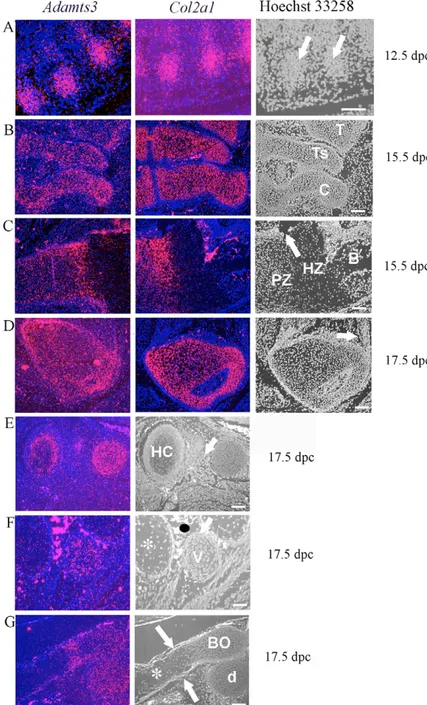
Fig. 2. Co-expression of Adamts3 and Col2a1 in musculoskeletal tissues. (A-G) In situ hybridization is used to show expression of Adamts3 in developing cartilage from 12.5 dpc to 17.5 dpc. In A-D, images include hybridization of Col2a1 to adjacent serial sections. Hoechst 33258 stained images are included on the far right for morphological correlation; the age of the embryo from which the section was taken is shown on the right. (A) Expression in initial sites of chondrogenesis in the developing ribs (arrows). (B) Expression in primordial cartilage of the ankle joint. T, tibia; Ts, Talus; C, calcaneus. (C) Expression in the proximal humerus growth plate. Note that expression is restricted to the proliferating zone (PZ). Hypertrophic zone chondrocytes (HZ) do not express
Adamts3. Col2a1 is not expressed in the bone of the
primary ossification center (B). Adamts3 is expressed in the perichondrium (arrow) and bone. (D) Proximal femur. The arrow indicates a tendon insertion site where Adamts3, but not Col2a1 is expressed. (E-G) 17.5 dpc skeletal elements, demonstrating that Adamts3 is not expressed by hypertrophic chondrocytes. (E) Absence of Adamts3 expression in the hypertrophic chondrocytes (HC) of the manubrium sterni. Note Adamts3 expression in developing intercostal muscle (arrow). (F) Ribs and vertebral lateral process. The rib elements contain hypertrophic chondrocytes (asterisk) that do not express Adamts3. By contrast, the lateral process of a thoracic vertebra (v) has small chondrocytes and these express Adamts3. The black oval indicates a spurious signal from erythrocytes. (G) Sagittal section through the basi-occiput (BO) and the dens (d) of the second cervical vertebra. Note the strong expression of Adamts3 in regions containing small chondrocytes and absence from the hypertrophic zone (asterisk). Expression is seen in the perichondrium (arrows). Scale bar: 100 m.
D
E
V
E
LO
P
M
E
N
T
Fig. 3. Co-expression of Adamts3 and Col1a1 in the musculoskeletal system. All images are from 15.5 and 17.5 dpc embryos; Hoechst 33258-stained images are shown in the right-hand panel. (A) Expression in the appendicular skeleton (proximal humerus). Note that Adamts3 expression overlaps with Col1a1 in bone (B) and in the perichondrium around the hypertrophic zone and the periosteum (around bone). Chondrocytes in the proliferating zone (PZ) and hypertrophic zone (HZ) are indicated; arrows indicate mRNA in muscle. (B) Expression in the craniofacial skeleton. Note co-expression of Col1a1 and Adamts3 in bone (B) in the mandible and maxilla, as well as in developing masseter muscle (mt). (C) Expression in the proximal femur. The trochanteric fossa (TF), which contains the obturator tendons, is indicated by the arrow. The left panel in this figure is the same as that in Fig. 2D. (D) Expression in the developing Achilles tendon at 15.5 dpc. The image on the left shows Adamts3 expression in the ankle cartilage (C) and tendon (arrow); that on the right shows the Achilles tendon at a higher magnification (boxed area in middle panel). Scale bar: 100 m.
Fig. 4. Adamts3, not Adamts2, has a role in procollagen I processing in the skeleton. (A) Normal craniofacial
development, bone structure and minimal tooth involvement in
Adamts2–/–mice. mCT analysis and
three-dimensional reconstruction of skulls of Adamts2–/–mice and
wild-type (+/+) littermates do not show differences in overall patterning, size, dimensions or organization of any craniofacial elements. The lower panels show tooth
morphology from littermates. Note the poorly defined topography of molars from Adamts2–/–mice,
although the overall morphology is similar. (B) Adamts3, but not
Adamts2 or Adamts14, was
strongly expressed during tooth development in the dental papilla (DP), and was expressed less strongly in the ameloblasts (arrow). (C) Expression of ADAMTS3 in stably transfected 293 cells.
Conditioned medium from cells was analyzed by western blotting with a polyclonal antibody to ADAMTS3. Note that upon treatment with PNGase F, ADAMTS3 migrates faster. Untransfected cells showed no protein. Molecular mass markers are indicated on the right. (D) Rescue of the procollagen I processing defect by ADAMTS2 and ADAMTS3 in dermatosparactic calf skin fibroblasts (DS). DS were plated alone or mixed with an identical number of HEK-293 cells stably transfected with an empty expression vector (Ct), or with the same vector encoding ADAMTS2 (TS2) or ADAMTS3 (TS3). After two days in culture, western blotting of a non-reducing SDS-PAGE gel was used to detect type I collagen. Note that the antibody used detects both ␣1 and ␣2 chains, but reacts more strongly with the ␣1 chain. The identity of collagen polypeptides associated with the cell layer or recovered from the conditioned culture medium is indicated between the two panels. (E) Rescue of the procollagen III processing defect by ADAMTS2 and ADAMTS3 in DS. Samples used in D were also analyzed by western blotting using an anti-collagen III antiserum. The lower amount of collagen synthesized by DS cultured alone, as compared to co-cultures, was repeatedly observed and may be related to the lower level of confluence in the monoculture. Alternatively, HEK-293 cells may secrete factors activating the synthesis of procollagen. Scale bar: 100 m.
D
E
V
E
LO
P
M
E
N
T
were detected, illustrating a high aminoprocollagen peptidase activity. In DS/ADAMTS3-HEK293F cell co-culture, analysis of the collagen pattern demonstrates a high aminoprocollagen peptidase activity, although it is lower than that observed in presence of ADAMTS2.
The same co-culture samples were investigated for type III procollagen processing (Fig. 4E). A significant amount of fully processed ␣1(III) was observed in the presence of ADAMTS2. Procollagen III processing was also detected when DS were co-cultured with cells expressing ADAMTS3.
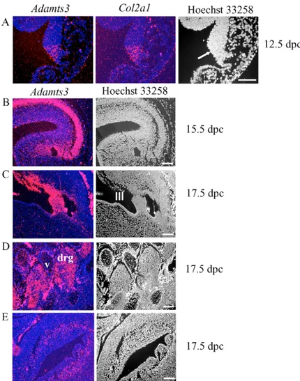
Adamts3 is expressed at extraskeletal locations Col2a1 expression has been previously reported at locations that are not associated with cartilage formation (Cheah et al., 1991). We found Adamts3 to be precisely co-expressed with Col2a1 at one such location in the hindbrain at 12.5 dpc (Fig. 5A). In addition, Adamts3 was highly expressed at other locations where Col2a1, Col1a1 or Col3a1 gene expression was absent. At 15.5 dpc, Adamts3 was highly expressed in the developing cerebral cortex (Fig. 5B), and at 17.5 dpc it was strongly expressed in the subcommisural organ, a specialized ependymal zone between the third and fourth ventricles (Fig. 5C), and in dorsal root ganglia (Fig. 5D). We have not investigated Adamts3 expression in the vitreous humor, another extraskeletal site of Col2a1 expression. Adamts3 expression was seen in the wall of the urinary bladder (Fig. 5E).
Adamts2, but not Adamts3 or Adamts14, is co-expressed with Col3a1
Adamts2 was co-expressed with Col3a1 in the wall of large arteries, such as the aorta, carotid and pulmonary arteries, throughout mouse embryogenesis (Fig. 6A,B). By contrast, Col1a1 was expressed only in the adventitial (outermost) layer of arteries (data not shown). Expression of both Col3a1 and Adamts2 was highest in the lung mesenchyme (Fig. 6B,C). Adamts2 was also co-expressed with Col3a1 at other sites, such as the palate (Fig. 6D), the intestinal wall, peritoneum and mesentery (Fig. 6E), the wall of the urinary bladder (Fig. 6F) and the skin (see Fig. 8A). Adamts14 showed no co-expression with Col3a1, although expression of Adamts3 in the wall of the urinary bladder partially overlapped with the expression of both Adamts2 and Col3a1 (Fig. 5E, Fig. 6F). With this exception, Adamts3 was not otherwise co-expressed with Col3a1.
Procollagen III processing is decreased in Adamts2–/–mice
As only Adamts2 was substantially co-expressed with Col3a1, and because ADAMTS2 processes procollagen III, we investigated whether procollagen III processing was affected in Adamts2–/–
mice. Collagens were extracted from various mouse organs and western blotting was used to evaluate procollagen III processing. The different types of extraction gave essentially similar results. In all cases, a substantial proportion of procollagen III was
Fig. 5. Expression of Adamts3 outside the
musculoskeletal system. Hoechst 33258-stained images are shown in the right-hand panel. (A) Co-expression of
Adamts3 and Col2a1 in the floor of the fourth ventricle in
the hindbrain (arrow). (B) Expression of Adamts3 in the cerebral cortex. (C) Adamts3 expression in the ependyma of the subcommissural organ. III, third ventricle. (D) Adamts3 in dorsal root ganglia (drg). Note the sparse expression in the vertebral cartilage (v) that comprises mainly hypertrophic chondrocytes. (E) Expression of
Adamts3 in the inner musculature of the urinary bladder.
Note, in B-E, Adamts3 was not co-expressed with Col2a1. Scale bar: 100 m.
D
E
V
E
LO
P
M
E
N
T
unprocessed in the Adamts2–/–mice, whereas it was extensively or completely processed in wild-type mice (Fig. 6G). To investigate whether procollagen III processing by Adamts2 occurred in inflammation-induced fibrosis, proteins were analyzed from granulomas induced in Adamts2–/– and wild-type mice by subcutaneous implantation of a PVA sponge. The analysis demonstrated that most of the newly synthesized procollagen III induced in the granulomas of wild-type mice was processed, whereas little or no procollagen III processing occurred in the granulomas induced in Adamts2–/–mice (Fig. 6H).
Adamts2–/–mice have distal airspace distension The lung was the most prominent site of developmental expression of Adamts2, where its distribution corresponded with that of Col3a1 (Fig. 6B,C). Lungs from two-month-old Adamts2–/–mice and wild-type littermates were evaluated histologically after fixation under inflation to optimize parenchymal distension. Adamts2–/–lungs had a consistent decrease in parenchymal density at both 2 months (Fig.
7A) and 2 weeks (data not shown). This emphysema-like appearance was unaccompanied by inflammatory cells or obvious fibrosis. Histochemical detection of elastin and collagen (data not shown) did not reveal any abnormalities and the ultrastructure showed the presence of normal basement membranes (Fig. 7B), elastic fibers and surfactant-producing pneumocytes (data not shown). By contrast, the histology (Fig. 7C) and electron microscopy of the aorta from Adamts2–/–mice did not show consistent changes from that of
the wild-type littermates. In both the lung and aorta (Fig. 7D) of Adamts2–/–mice, the shape and diameter of collagen fibrils appeared
to be unaffected.
We investigated whether the processing of both procollagen I and procollagen III were impaired in the aorta and lungs from Adamts2–/–mice. Although it is not possible to comment from this
data on the relative proportion of the two collagens in these tissues, it is clear that both newborn and adult Adamts2–/–mice demonstrated
significantly less processing of both collagens than did their wild-type littermates (Fig. 7E,F).
Fig. 6. Co-expression of Adamts2 and Col3a1, and deficiency of procollagen III processing in Adamts2–/–mice. (A,B) Adamts2 is
co-expressed with Col3a1 in the arterial wall, as shown at two developmental stages: 12.5 dpc carotid artery (A) and 17.5 dpc pulmonary artery (B). L, lung. (C) Expression of Col3a1 and Adamts2 in lung. Note the expression in lung mesnchyme but not in the epithelium of bronchial tubes (asterisk). (D) Co-expression of Adamts2 and Col3a1 in the mesenchyme of the palate (P). Adamts2 is also expressed in the perichondrium of the
basisphenoid (BS), at which location Col3a1 is absent. (E) Co-expression of Adamts2 and Col3a1 in the peritoneum lining the liver (L, arrow), and mesentery of the gut (G) and pancreas (Pa). (F) Co-expression of Adamts2 and Col3a1 within the subepithelial layer of the developing urinary bladder. Scale bar: 100 m. In D-F, Adamts2 expression is weaker than in lung and arteries, and Col3a1 expression is also significantly weaker and more dispersed. (G) Defective procollagen III processing in tissues from Adamts2–/–mice. Western blot analysis of procollagen isolated from mouse
tissues with 0.15 M NaCl is shown. The tissue origin of the sample and the respective genotype is shown above each lane; procollagen III and collagen III molecular species are indicated on the left. Scanning densitometry of the different collagen III signals was used to calculate the proportion of fully processed molecules in the various tissues (lower panel). (H) Defective procollagen III processing in newly synthesized collagen in
Adamts2–/–mice. Protein was extracted from sponge granulomas and western blotting used to define the molecular species of procollagen III that
D
E
V
E
LO
P
M
E
N
T
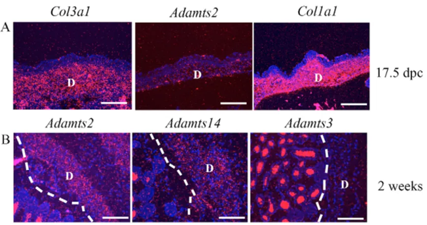
Adamts2 and Adamts14 are co-expressed in mature mouse dermis
In embryonic skin, low-level Adamts2 expression was noted, but no expression of Adamts3 or Adamts14 was detected (Fig. 8A). However, in two-week old mice, both Adamts2 and Adamts14 were detected in the dermis, but not Adamts3 (Fig. 8B).
DISCUSSION
To rigorously establish the physiological significance of a protease-substrate interaction in vivo requires that the protease and protease-substrate should be co-expressed, or that they should co-localize in tissues, and that in the absence of the protease, cleavage of the substrate should be reduced or absent. In addition, failure to cleave the substrate should have a significant impact on a biological system. We applied these criteria to exploring the roles of the procollagen N-proteinases during mouse embryogenesis. The results suggest that ADAMTS2 is the major procollagen III amino-propeptidase in mice, but that it appears to share the procollagen I processing function with ADAMTS3. Previous literature had suggested that a significant proportion of procollagen III in tissues was unprocessed and that the procollagen III-processing enzyme was not the same enzyme that processed procollagen I (Halila and Peltonen, 1986; Halila et al., 1986; Peltonen et al., 1985; Tuderman and Prockop, 1982). Wang et al. recently demonstrated that ADAMTS2 could process procollagen III in vitro, but the in vivo relevance was unclear
(Wang et al., 2003). The overlapping expression of Adamts2 and Col3a1, and the substantial reduction in procollagen III processing in Adamts2–/–mice provides solid proof that ADAMTS2 is the major procollagen III-processing enzyme.
The role of ADAMTS2 in processing procollagen III may further contribute to the loss of mechanical integrity of dermatosparactic skin, as collagen I and III are both present in the dermis. In situ hybridization revealed prominent Col3a1 expression during lung development, in accordance with a previous study suggesting that, in mice, collagen III is a crucial structural component of the lung (Shiomi et al., 2003). The pseudo-emphysematous appearance of the lungs and the near-total lack of lung procollagen III processing in Adamts2–/–mice strongly suggests a vital requirement for collagen III in the lung. Lung abnormalities may result from a fragility of the pulmonary walls, similar to skin fragility in dermatosparaxis. Neither Adamts3 nor Adamts14 were expressed in the lung and, therefore, there was no compensating processing enzyme. However, in the aorta, which is severely affected by structural collagen III mutations, the near-total lack of procollagen III processing had no apparent effect on histology and collagen ultrastructure at 2 months of age. It is possible that changes may be seen in older mice, or by the induction of stress on the aortic wall in appropriate in vivo models.
The data suggest that ADAMTS3 probably makes a significant contribution to procollagen I processing during musculoskeletal development in mice. Expression of Adamts3 in many Col1a1-Fig. 7. Histology and ultrastructure of lung and aorta in Adamts2–/–mice and wild-type (+/+) littermates. (A) Comparative lung histology
from Adamts2–/–and their wild-type (+/+) littermates. The distal air spaces are much larger in the mutant mice, but there is no inflammatory
exudate. (B) Ultrastructure of an alveolar wall, demonstrating comparable basement membranes (arrows) in Adamts2–/–and wild-type mice. E,
erythrocyte. (C) Methylene blue-azure B stain for elastin in Adamts2–/–and wild-type aortas demonstrates comparable aortic wall thickness and
elastin lamellae (arrow). (D) Electron micrograph of collagen fibrils in cross section (in area marked by the asterisk) demonstrates comparable size and shape, and uniformity of fibril diameter. Scale bar: 100 m in A,C; 500 nm in B,D. (E) Western blot analysis of procollagen III (above) and I (below) processing in mouse lungs. Samples from wild-type and mutant newborn and adult mice are illustrated. (F) Western blot analysis of procollagen III (above) and I (below) processing in the mouse aorta. The various molecular species of collagen are indicated.
D
E
V
E
LO
P
M
E
N
T
expressing tissues, such as bone and tendon, may explain why the most severe deficiencies in procollagen I processing in mouse dermatosparaxis are in the dermis. Together with our previous analysis of bovine dermatosparactic cartilage that demonstrated complete procollagen II processing, the data suggest that ADAMTS3 is probably the major procollagen II-processing enzyme, and predict that Adamts3 mutations will lead to a chondrodysplasia or osteochondrodysplasia. Although Col2a1 and Col1a1 expression are generally mutually exclusive in tissues, Adamts3 expression overlapped with that of both collagen genes. Although we detected Adamts14 mRNA by RT-PCR of total embryo RNA (data not shown), in situ hybridization with three non-overlapping riboprobes showed no discernible developmental expression.
Is it possible that the PNPs have substrates other than the fibrillar procollagens? Although Adamts2 expression was associated with procollagen gene expression, we noted strong expression of Adamts3 in the nervous system at locations that lacked procollagen I, II or III mRNA. For instance, Adamts3 was highly expressed in the sub-commisural organ (SCO), a specialized ependymal organ located at the entrance to the aqueduct of Sylvius. Its major secretory product is SCO-spondin, a glycoprotein that undergoes proteolytic processing prior to polymerization into the Reissner fiber of the spinal cord (Gobron et al., 1996). It is possible that ADAMTS3 might also be a SCO-spondin-processing enzyme.
Can the major fibrillar procollagen amino-propeptides be processed by other proteases? The highly conserved PNP modular organization (except for the variable C-terminal module) is different from all other proteases and is probably highly specialized for binding to triple-helical collagens. Heat-denatured collagen is not efficiently processed by ADAMTS2 (Tuderman et al., 1978), suggesting a need for spatially stringent interactions between the ancillary domain and the triple helix. Because residual procollagen processing in dermatosparactic skin occurs at the same cleavage site as in normal skin (Colige et al., 2002), it supports processing by an enzyme(s) closely related to ADAMTS2. It is clear from the Adamts2–/–mice that absence of ADAMTS2 leads to a significant
loss of processing of procollagen III in many tissues. At these locations, neither Adamts3 nor Adamts14 are expressed, and, thus, do not compensate for its absence. Thus other enzymes do not appear to compensate significantly. A splice variant of collagen II, termed IIA is expressed in pre-cartilaginous mesenchyme and in perichondrium (Ryan and Sandell, 1990; Ryan et al., 1990). Ultrastructural and biochemical studies suggest its N-propeptide is not excised during development, suggesting perhaps that it is resistant to the action of these proteases (Zhu et al., 1999). By
contrast, matrix metalloproteinases (MMPs) have been shown to cleave the propeptide from collagen IIA at numerous sites other than the PNP bond (Fukui et al., 2002).
ADAMTS2 mutations in humans lead to characteristic craniofacial changes and decreased growth; features that are absent in Adamts2–/–
mice. As individuals with EDS-VIIC age, the skin fragility is somewhat ameliorated and there is increasing joint laxity, which is not a prominent feature in early childhood. Adamts2–/–mice also have fragile skin, but they are not significantly growth retarded and have no craniofacial dysmorphism (Li et al., 2001). These differences between EDS-VIIC and mouse dermatosparaxis are compatible with the different deployment of functionally equivalent enzymes in humans and mice through the transcriptional regulation of their genes. The expression of ADAMTS3 and ADAMTS14 may change with age in humans, perhaps explaining how the properties of dermatosparactic skin change as affected individuals get older.
In addition to skin fragility, humans with EDS-VIIC manifest with a variety of other anomalies. Easy bruising, a universal sign in patients with EDS-VIIC, can be partly attributed to the ability of ADAMTS2 to process procollagen III, and to our observation, albeit in mice, that it is the only PNP expressed at significantly high levels in the walls of blood vessels. The procollagen III-processing defect in the absence of ADAMTS2 appears to be dramatic in most mouse tissues. We were particularly concerned that procollagen III processing in the mouse aorta was defective. Although histology and electron microscopy did not reveal specific anomalies in the aortic wall, and no aortic or other vascular aneurysms have been hitherto noted in EDS-VIIC patients, our findings suggest a need for the monitoring of EDS-VIIC patients as they age. Bladder rupture and intestinal perforations have been reported in individuals with EDS-VIIC. Our studies demonstrate co-expression of procollagen III and ADAMTS2 in the developing mouse at these locations. A recent clinical study emphasized the abnormal permanent dentition of individuals with EDS-VIIC, including hypodontia (De Coster et al., 2003). Adamts2–/–mice have mild dental changes and compensation
by Adamts3 may account for this.
Most of the 19 ADAMTS proteases can be assigned to phylogenetically distinct clades (Apte, 2004; Huxley-Jones et al., 2005; Nicholson et al., 2005). Recent analysis of ADAMTS gene evolution suggests that genes within mammalian ADAMTS clades arose by duplication of their primitive ancestors (Huxley-Jones et al., 2005; Nicholson et al., 2005). We speculated that, following duplication, closely related genes, such as ADAMTS2, ADAMTS3 and ADAMTS14, were highly conserved to provide subfunctionalization (retention of molecular function but with diversification of expression patterns) rather than
Fig. 8. Procollagen N-propeptidase expression in skin. D, dermis; the white dashed line indicates the line of demarcation of epidermis and dermis. (A) Co-expression of Col3a1 and Col1a1 in the dermis of the skin of a 17.5 dpc embryo. Adamts2, but not Adamts14 or Adamts3, mRNA was detected in the dermis. (B) Adamts2 and
Adamts14 mRNA, but not Adamts3 mRNA, is detectable
D
E
V
E
LO
P
M
E
N
T
neofunctionalization (acquisition of new functions) (Huxley-Jones et al., 2005). The ability of all three PNPs to process procollagen I, but specialization in this regard as a result of differential gene regulation, supports this hypothesis. Other ADAMTS clades may exhibit the same phenomenon.
We thank D. Prockop for Adamts2–/–mice; E. Vuorio, B. de Crombrugghe and
N. Fukai for cDNA probes; Amanda Allamong for histology; Katie Jungers for assistance with in situ hybridization; Kaitlin Petrella for assistance with electron microscopy; and Craig Bennetts for mCT scanning. The studies were supported by grants from the National Institutes of Health (AR49590 to S.S.A. and AR44745 to D.E.B.). A.C.C. is supported by the Belgian Fonds National de la Recherche Scientifique (grant 3.4562.03), and F.K. by a Télévie grant (7.4.561.03.F). Histology and mCT was supported by a Core Center for Musculoskeletal Disorders award from NIH (AR050953).
References
Apte, S. S. (2004). A disintegrin-like and metalloprotease (reprolysin type) with thrombospondin type 1 motifs: the ADAMTS family. Int. J. Biochem. Cell Biol. 36, 981-985.
Birk, D. E. and Trelstad, R. L. (1986). Extracellular compartments in tendon morphogenesis: collagen fibril, bundle, and macroaggregate formation. J. Cell Biol. 103, 231-240.
Birk, D. E., Silver, F. H. and Trelstad, R. L. (1991). Matrix Assembly. In Cell Biology of Extracellular Matrix (ed. E. D. Hay), pp. 221-254. New York: Plenum Press.
Canty, E. G., Lu, Y., Meadows, R. S., Shaw, M. K., Holmes, D. F. and Kadler, K. E. (2004). Coalignment of plasma membrane channels and protrusions (fibripositors) specifies the parallelism of tendon. J. Cell Biol. 165, 553-563. Chan, D., Rogers, J. F., Bateman, J. F. and Cole, W. G. (1995). Recurrent
substitutions of arginine 789 by cysteine in pro-alpha 1 (II) collagen chains produce spondyloepiphyseal dysplasia congenita. J. Rheumatol. Suppl. 43, 37-38.
Cheah, K. S., Lau, E. T., Au, P. K. and Tam, P. P. (1991). Expression of the mouse alpha 1(II) collagen gene is not restricted to cartilage during development. Development 111, 945-953.
Cole, W. G. (1997). Abnormal skeletal growth in Kniest dysplasia caused by type II collagen mutations. Clin. Orthop. Relat. Res. 341, 162-169.
Colige, A., Sieron, A. L., Li, S. W., Schwarze, U., Petty, E., Wertelecki, W., Wilcox, W., Krakow, D., Cohn, D. H., Reardon, W. et al. (1999). Human Ehlers-Danlos syndrome type VII C and bovine dermatosparaxis are caused by mutations in the procollagen I N-proteinase gene. Am. J. Hum. Genet. 65, 308-317. Colige, A., Vandenberghe, I., Thiry, M., Lambert, C. A., Van Beeumen, J., Li,
S. W., Prockop, D. J., Lapiere, C. M. and Nusgens, B. V. (2002). Cloning and characterization of ADAMTS-14, a novel ADAMTS displaying high homology with ADAMTS-2 and ADAMTS-3. J. Biol. Chem. 277, 5756-5766.
Colige, A., Nuytinck, L., Hausser, I., Van Essen, A. J., Thiry, M., Herens, C., Ades, L. C., Malfait, F., Paepe, A. D., Franck, P. et al. (2004). Novel types of mutation responsible for the dermatosparactic type of Ehlers-Danlos Syndrome (Type VIIC) and common polymorphisms in the ADAMTS2 gene. J. Invest. Dermatol. 123, 656-663.
De Coster, P. J., Malfait, F., Martens, L. C. and De Paepe, A. (2003). Unusual oral findings in dermatosparaxis (Ehlers-Danlos syndrome type VIIC). J. Oral Pathol. Med. 32, 568-570.
De Paepe, A. and Malfait, F. (2004). Bleeding and bruising in patients with Ehlers-Danlos syndrome and other collagen vascular disorders. Br. J. Haematol. 127, 491-500.
Fernandes, R. J., Hirohata, S., Engle, J. M., Colige, A., Cohn, D. H., Eyre, D. R. and Apte, S. S. (2001). Procollagen II amino propeptide processing by ADAMTS-3. Insights on dermatosparaxis. J. Biol. Chem. 276, 31502-31509. Fukui, N., McAlinden, A., Zhu, Y., Crouch, E., Broekelmann, T. J., Mecham, R.
P. and Sandell, L. J. (2002). Processing of type II procollagen amino propeptide by matrix metalloproteinases. J. Biol. Chem. 277, 2193-2201.
Gobron, S., Monnerie, H., Meiniel, R., Creveaux, I., Lehmann, W., Lamalle, D., Dastugue, B. and Meiniel, A. (1996). SCO-spondin: a new member of the thrombospondin family secreted by the subcommissural organ is a candidate in the modulation of neuronal aggregation. J. Cell Sci. 109, 1053-1061. Halila, R. and Peltonen, L. (1986). Purification of human procollagen type III
N-proteinase from placenta and preparation of antiserum. Biochem. J. 239, 47-52. Halila, R., Steinmann, B. and Peltonen, L. (1986). Processing of types I and III
procollagen in Ehlers-Danlos syndrome type VII. Am. J. Hum. Genet. 39, 222-231. Huxley-Jones, J., Apte, S. S., Robertson, D. L. and Boot-Handford, R. P.
(2005). The characterisation of six ADAMTS proteases in the basal chordate Ciona intestinalis provides new insights into the vertebrate ADAMTS family. Int. J. Biochem. Cell Biol. 37, 1838-1845.
Kessler, E., Takahara, K., Biniaminov, L., Brusel, M. and Greenspan, D. S. (1996). Bone morphogenetic protein-1: the type I procollagen C-proteinase. Science 271, 360-362.
Lapiere, C. M. and Nusgens, B. V. (1993). Ehlers-Danlos type VII-C, or human dermatosparaxis. The offspring of a union between basic and clinical research. Arch. Dermatol. 129, 1316-1319.
Li, S. W., Arita, M., Fertala, A., Bao, Y., Kopen, G. C., Langsjo, T. K., Hyttinen, M. M., Helminen, H. J. and Prockop, D. J. (2001). Transgenic mice with inactive alleles for procollagen N-proteinase (ADAMTS-2) develop fragile skin and male sterility. Biochem. J. 355, 271-278.
LuValle, P., Hayashi, M. and Olsen, B. R. (1989). Transcriptional regulation of type X collagen during chondrocyte maturation. Dev. Biol. 133, 613-616. Malfait, F., De Coster, P., Hausser, I., van Essen, A. J., Franck, P., Colige, A.,
Nusgens, B., Martens, L. and De Paepe, A. (2004). The natural history, including orofacial features of three patients with Ehlers-Danlos syndrome, dermatosparaxis type (EDS type VIIC). Am. J. Med. Genet. A 131, 18-28. Nicholson, A. C., Malik, S. B., Logsdon, J. M., Jr and Van Meir, E. G. (2005).
Functional evolution of ADAMTS genes: evidence from analyses of phylogeny and gene organization. BMC Evol. Biol. 5, 11.
Niederreither, K., D’Souza, R., Metsaranta, M., Eberspaecher, H., Toman, P. D., Vuorio, E. and De Crombrugghe, B. (1995). Coordinate patterns of expression of type I and III collagens during mouse development. Matrix Biol. 14, 705-713.
Nusgens, B. V., Verellen-Dumoulin, C., Hermanns-Le, T., De Paepe, A., Nuytinck, L., Pierard, G. E. and Lapiere, C. M. (1992). Evidence for a relationship between Ehlers-Danlos type VII C in humans and bovine dermatosparaxis. Nat. Genet. 1, 214-217.
Oblander, S. A., Zhou, Z., Galvez, B. G., Starcher, B., Shannon, J. M., Durbeej, M., Arroyo, A. G., Tryggvason, K. and Apte, S. S. (2005). Distinctive functions of membrane type 1 matrix-metalloprotease (MT1-MMP or MMP-14) in lung and submandibular gland development are independent of its role in pro-MMP-2 activation. Dev. Biol. 277, 255-269.
Olsen, B. R. (1991). Collagen Biosynthesis. In Cell Biology of Extracellular Matrix (ed. E. D. Hay), pp. 177-220. New York: Plenum Press.
Peltonen, L., Halila, R. and Ryhanen, L. (1985). Enzymes converting procollagens to collagens. J. Cell. Biochem. 28, 15-21.
Petty, E. M., Seashore, M. R., Braverman, I. M., Spiesel, S. Z., Smith, L. T. and Milstone, L. M. (1993). Dermatosparaxis in children. A case report and review of the newly recognized phenotype. Arch. Dermatol. 129, 1310-1315. Rauch, F. and Glorieux, F. H. (2004). Osteogenesis imperfecta. Lancet 363,
1377-1385.
Ryan, M. C. and Sandell, L. J. (1990). Differential expression of a cysteine-rich domain in the amino-terminal propeptide of type II (cartilage) procollagen by alternative splicing of mRNA. J. Biol. Chem. 265, 10334-10339.
Ryan, M. C., Sieraski, M. and Sandell, L. J. (1990). The human type II procollagen gene: identification of an additional protein-coding domain and location of potential regulatory sequences in the promoter and first intron. Genomics 8, 41-48.
Shiomi, T., Okada, Y., Foronjy, R., Schiltz, J., Jaenish, R., Krane, S. and D’Armiento, J. (2003). Emphysematous changes are caused by degradation of type III collagen in transgenic mice expressing MMP-1. Exp. Lung Res. 29, 1-15.
Smith, L. T., Wertelecki, W., Milstone, L. M., Petty, E. M., Seashore, M. R., Braverman, I. M., Jenkins, T. G. and Byers, P. H. (1992). Human
dermatosparaxis: a form of Ehlers-Danlos syndrome that results from failure to remove the amino-terminal propeptide of type I procollagen. Am. J. Hum. Genet. 51, 235-244.
Snead, M. P. and Yates, J. R. (1999). Clinical and molecular genetics of Stickler syndrome. J. Med. Genet. 36, 353-359.
Somerville, R. P., Longpre, J. M., Jungers, K. A., Engle, J. M., Ross, M., Evanko, S., Wight, T. N., Leduc, R. and Apte, S. S. (2003). Characterization of ADAMTS-9 and ADAMTS-20 as a distinct ADAMTS subfamily related to Caenorhabditis elegans GON-1. J. Biol. Chem. 278, 9503-9513.
Tiller, G. E., Polumbo, P. A., Weis, M. A., Bogaert, R., Lachman, R. S., Cohn, D. H., Rimoin, D. L. and Eyre, D. R. (1995). Dominant mutations in the type II collagen gene, COL2A1, produce spondyloepimetaphyseal dysplasia, Strudwick type. Nat. Genet. 11, 87-89.
Tuderman, L. and Prockop, D. J. (1982). Procollagen N-proteinase. Properties of the enzyme purified from chick embryo tendons. Eur. J. Biochem. 125, 545-549.
Tuderman, L., Kivirikko, K. I. and Prockop, D. J. (1978). Partial purification and characterization of a neutral protease which cleaves the N-terminal propeptides from procollagen. Biochemistry 17, 2948-2954.
Wang, W. M., Lee, S., Steiglitz, B. M., Scott, I. C., Lebares, C. C., Allen, M. L., Brenner, M. C., Takahara, K. and Greenspan, D. S. (2003). Transforming growth factor-beta induces secretion of activated ADAMTS-2. A procollagen III N-proteinase. J. Biol. Chem. 278, 19549-19557.
Wertelecki, W., Smith, L. T. and Byers, P. (1992). Initial observations of human dermatosparaxis: Ehlers-Danlos syndrome type VIIC. J. Pediatr. 121, 558-564. Zhu, Y., Oganesian, A., Keene, D. R. and Sandell, L. J. (1999). Type IIA
procollagen containing the cysteine-rich amino propeptide is deposited in the extracellular matrix of prechondrogenic tissue and binds to TGF-beta1 and BMP-2. J. Cell Biol. 144, 1069-1080.