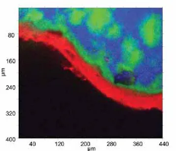
Raman hyperspectral imaging: a single tool to characterise pharmaceutical products
Texte intégral
Figure
Documents relatifs
The second disclosure profile concerns press releases issued by targets in hostile takeover bids and/or during the offer period (class 2), primarily used by the issuing
If some a priori knowledge of the noise statistics (e.g., K - distribution, t-distribution, etc.) is available, then M and µ should be estimated by the MLE ˆ M and µ ˆ of the
La pension moyenne des femmes faisant valoir un premier droit à la retraite dans l’année, tous régimes confondus (y compris la majoration de pension pour enfants), est inférieure
Electronic and Magnetic Communication in Mixed- Valent and Homovalent Ruthenium Complexes Containing Phenylcyanamide type Bridging Ligands.. Muriel Fabre and Jacques
Toutefois, ces pratiques idéales étant illusoires, seule la vaccination a permis le contrôle de la bronchite infectieuse dans les élevages intensifs de poulets de chair, de
Ainsi, le développement durable fait ressortir la perspective long terme ou court terme des acteurs « Si on fait du bio pour faire du blé à mon avis, cela peut marcher mais
Given a constraint mixing the rounding operator with non-linear real arithmetic terms, one can reduce it to an extension of non-linear real arithmetic with floor and ceiling
