HAL Id: hal-01737754
https://hal.archives-ouvertes.fr/hal-01737754
Submitted on 19 Mar 2018HAL is a multi-disciplinary open access archive for the deposit and dissemination of sci-entific research documents, whether they are pub-lished or not. The documents may come from teaching and research institutions in France or abroad, or from public or private research centers.
L’archive ouverte pluridisciplinaire HAL, est destinée au dépôt et à la diffusion de documents scientifiques de niveau recherche, publiés ou non, émanant des établissements d’enseignement et de recherche français ou étrangers, des laboratoires publics ou privés.
Electronic and Magnetic Communication in
Mixed-Valent and Homovalent Ruthenium Complexes
Containing Phenylcyanamide Type Bridging Ligands
Muriel Fabre, Jacques Bonvoisin
To cite this version:
Muriel Fabre, Jacques Bonvoisin. Electronic and Magnetic Communication in Mixed-Valent and Homovalent Ruthenium Complexes Containing Phenylcyanamide Type Bridging Ligands. Jour-nal of the American Chemical Society, American Chemical Society, 2007, 129 (5), pp.1434-1444. �10.1021/ja067255i�. �hal-01737754�
Electronic and Magnetic Communication in
Mixed-Valent and Homovalent Ruthenium Complexes
Containing Phenylcyanamide type Bridging Ligands
Muriel Fabre and Jacques Bonvoisin*
Contribution from the a CEMES/CNRS, NanoSciences Group, BP 94347, 29 rue Jeanne Marvig,
31055 Toulouse cedex 4, France
Keywords : Mixed-valence, Electron-transfer, Magnetic coupling, Exchange interaction, Ruthenium
complexes, dinuclear complex, cyanamide, Electrochemical properties, Spectroelectrochemistry, EPR
spectroscopy.
Corresponding author: CEMES/CNRS, NanoSciences Group, BP 94347, 29 rue Jeanne Marvig,
31055 Toulouse cedex 4, France Tel. : +33 5 62 25 78 52, fax: +33 5 62 25 79 99, jbonvoisin@cemes.fr
Abstract
Four new phenylcyanamido-bridge dinuclear ruthenium complexes [{Ru(tpy)(thd)}2(µ-L)] with tpy =
2,2’:6’,2”-terpyridine, thd = 2,2,6,6-tetramethyl-3,5-heptanedione and L = dcbp =
4,4’-dicyanamidobiphenyle; bcpa = cyanamidophenyl)acetylene; bcpda =
Bis(4-cyanamidophenyl)diacetylene; bcpea = 9,10,-bis(4-cyanamidophenylethynyl)anthracene have been
prepared and fully characterized. The mixed valent Ru(II)Ru(III) and homovalent paramagnetic
Ru(III)Ru(III) forms of all the complexes were electrochemically generated and studied by UV-VIS-nIR
and EPR Spectroscopy. Electronic communication was quantified by the electronic coupling parameter
Vab extracted from intervalence measurement in the near IR area and Magnetic communication was
quantified in term of the exchange coupling constant J, accessible from the intensity of the EPR signal
when varying the temperature. Exponential decay for both electronic and magnetic coupling versus
intermetallic distance were obtained and discussed.
1 Introduction
It is almost a truism now that, after the inception of the Creutz-Taube ion
[(H3N)5Ru(pyrazine)Ru(NH3)5]5+ in 1969,1 studies of mixed/intermediate valence complexes and their
role in intramolecular electro- and magnetocommunication have become vital parts of contemporary
research in coordination chemistry.2-6 Much efforts has been invested in the elucidation of the role that
the nature of the spacer between two interacting units plays in governing the redox splitting between
successive electron-transfer steps (electrocommunication) and the sign and magnitude of exchange
coupling of unpaired spins (magnetocommunication). The ability of the spacer to transmit electronic
effects has commonly been studied by electrochemistry, using the redox splitting, i.e. the potential
difference of consecutive redox steps as a measure of intramolecular intermetallic communication. One
has to remember that even in the unfavourable case where this potential difference is small, leading to a
single electrochemical wave a comfortable proportion of mixed valence species (> 50%) exists in
solution. 2 Once the mixed valence species obtained, from a practical point of view, the electron transfer
process is probed by the intensity of the intervalence transition (IT) in the NIR, according to the relation 2
devised by N. Hush in 1967. 7 This yields the electronic coupling parameter Vab describing the amount
of electronic interaction between remote sites. From this interaction, one can devise general rules for the
design of efficient bridging ligands allowing long-distance electron transfer. In addition, mixed-valence
compounds can be simple models for the expanding domain of nanojunctions, in which a single
molecule is bridging two nanoscale metallic conductors.8 However, as the size of the compounds
increase, synthetic problems become more acute, with in particular a decrease in free ligand solubility.
In addition, the electronic coupling decreases and its detection becomes problematic. Thus, there is a
need for new classes of compounds and/or new methods that would be more easily accessible and
would exhibit or allow reaching either stronger metal-metal couplings or longer metal-metal distance
than conventional systems studied so far. Some chemistry alternatives can be found by using
cyclometallated complexes. 9 With increasing intermetallic distance and/or diminishing transfer abilities
of the spacer, more sensitive detection methods must be applied. If the terminal groups at each
extremity of the bridging ligand are paramagnetic, determination of the exchange constant J provides
the information. This can be accomplished by magnetic susceptometry or EPR spectroscopy for J ≥ 1
cm-1 and by EPR spectroscopy for much smaller J values. At this point, an important question must be
addressed which is to know whether the more important phenomena of intramolecular long-distance
electron transfer and energy transfer are somehow related to electron-spin exchange-coupling, bearing
in mind that ET is a one-electron process, whereas exchange coupling is a two-electron process. In fact,
as Forbes10 has pointed out, both processes are facets of the more general donor-acceptor interaction
having in common the electron transfer matrix element Vab. The possibility of correlating J and Vab has
already been addressed by other authors. 5, 11-15 Our own contribution to the field is to find good
candidates which will allow to get both J (in the homovalent paramagnetic form) and Vab (in the mixed
valent form) in order then to correlate them. Bridging ligand containing cyanamide group seems to be
particularly well suited since strong interaction have been obtained in homovalent16 or mixed valence
ruthenium complexes. 17 Following this line, few years ago, we have reported two series of dinuclear
ruthenium complexes containing cyanamide derivatives as bridging ligands. 18, 19 For both series, Vab
and J could not be obtained because of stability and/or solubility problems. Here we present the
successful synthesis of four new dinuclear ruthenium complexes (See scheme 1) with terpyridine (tpy)
and bulky ‘acac’ type ancillary ligands named thd, with four cyanamide derivatives bridging ligands
with formula [{Ru(tpy)(thd)}2(µ-L)] with tpy = 2,2’:6’,2”-terpyridine, thd =
2,2,6,6-tetramethyl-3,5-heptanedione and L = dcbp = 4,4’-dicyanamidobiphenyle; bcpa = Bis(4-cyanamidophenyl)acetylene;
bcpda = Bis(4-cyanamidophenyl)diacetylene; bcpea = 9,10,-bis(4-cyanamidophenylethynyl)anthracene.
In addition, the work described here provides the first quantitative experimental comparison of the
decay law for both the electronic (in the mixed valent form) and the magnetic (in the homovalent
paramagnetic form) coupling parameters versus intermetallic distance using the same family of
compounds. N N N Ru O O N N N N N Ru O O N N = 3 4 5 6 Scheme 1
2 Results and discussion
Synthesis
The synthesis of [Ru(tpy)(thd)(TMSepcyd)] 2 from [Ru(tpy)(thd)(Ipcyd)] 1 was adapted from
literature procedures. 20 The Trimethylsilyl-protected alkyne complex [Ru(tpy)(thd)(TMSepcyd)] 2 was
prepared by a Sonogashira cross-coupling reaction between the iodoruthenium complex 1 and
Trimethylsilylacetylene under classic conditions ((Pd(PPh3)2Cl2, CuI, piperidine, DMF). Complex
[{Ru(tpy)(thd)}2(µ-dcbp)] 3 was synthesized using a method adapted from the Suzuki cross coupling
between an halogenaryl and an aryboronic ester. 21 It is a one-pot synthesis, the arylboronic ester being
generated in situ. Complex 1 was put in solution in DMF with half equivalent of bis(pinacolato)diboran
in the presence of PdCl2dppf catalyst and K2CO3 as basic agent. (Scheme 2) Complex
[{Ru(tpy)(thd)}2(µ-bcpea)] 6 was synthesized by a Sonogashira cross coupling reaction between the
iodoruthenium complex 1 and dietynylanthracene, which is formed in situ by deprotection of
9,10-bis(3-hydroxy-3-methylbutynyl)anthracene with potassium tert-butoxide (Scheme 2). Complex
[{Ru(tpy)(thd)}2(µ-bcpa)] 4 was synthesized by a Sonogashira cross-coupling reaction between the
iodoruthenium complex 1 and the ethynylated complex [Ru(tpy)(thd)(epcyd)] formed in situ by
deprotection of the Trimethylsilyl-protected alkyne complex 2 with Potassium carbonate, under classic
conditions (Pd(PPh3)2Cl2, CuI, piperidine and DMF) but in presence of a strong base as DBU
(1,8-diazabicyclo[5.4.0]undec-7-ene) given the weak reactivity of the alkyne function toward deprotonation
(scheme 3). Complex [{Ru(tpy)(thd)}2(µ-bcpda)] 5 was obtained by an homo-coupling reaction using
the ethynylated complex [Ru(tpy)(thd)(epcyd)] formed in situ in the same way as for complex 4. This
reaction was performed with DBU and copper(I)chloride in dry pyridine with oxygen bubling.
I N N N Ru O O N N N N N Ru O O N N N N N Ru O O N N O O B O O B OH O H N N N Ru O O N N N Ru O O N N N N K2CO3, PdCl2dppf DMF 13% tBuOK, CuI, Pd(PPh3)4 DMF/piperidine 35% 3 6 1
Scheme 2: Synthesis of [{Ru(tpy)(thd)} (µ-dcbp)] 3 and [{Ru(tpy)(thd)]} (µ-bcpea)] 62 2
I N N N Ru O O N N Si N N N Ru O O N N N N N Ru O O N N N N N Ru O O N N N N N Ru O O N N N N N Ru O O N N K2CO3, MeOH Pd(PPh3)2Cl2/CuI DMF/pipéridine 57% DBU 1) K2CO3, MeOH 2) DBU, CuCl, O2 pyridine 67% 4 5 2
Scheme 3: Synthesis of [{Ru(tpy)(thd)} (µ-bcpa)] 4 and [{Ru(tpy)(thd)} (µ-bcpda)] 5 2 2
Electrochemistry
Cyclic voltammograms (CV) of the complexes were recorded in dichloromethane under an argon
atmosphere with 0.1 M tetrabutylammonium hexafluorophosphate (TBAH) (See Figure 1 and Table 1).
The E1/2 potentials were determined from the average of the anodic and cathodic peak potentials for
reversible waves. For irreversible waves, only the anodic peak potentials are reported.
Insert Figure 1
Table 1: Electrochemical data, vs ECS, in CH2Cl2, 0.1M TBAH, 0.1 V/s
Complex Ru II/III E1/2, V(∆E, mV) Ligand Ea (V) Ref [Ru(tpy)(thd)Cl] 0.198 (83) - 22 1 0.200 (78) 1.064 22 3 0.137 (180) 0.725 (78) – 0.938 (73) [{Ru(NH3)5}2(µ-dcbp)][PF6]4a -0,215 (100) 0,775 (80) – 0,945 (85) 23 4 0.188 (108) 0.884 - 1.030 5 0.225 (88) 1.030 6 0.211 (83) 0.764 – 0.930 E1/2 = (Ea + Ec)/2; ∆E = │Ea - Ec│; Ea: anodic peak; ain CH3CN.
In reduction, no wave was observed at least down to -1.5V. In oxidation, several waves were
observed. The first reversible wave around 0.2V was attributed to the Ru(II/III) couple. For complexes
4-6, with the longest metal-metal distance (RMM= 18.5, 21.0 and 25.1 Å), the redox potentials of the
ruthenium centers are too close to be distinguished, a single bielectronic wave is then observed. One can
notice that as the intermetallic distance decreases, the difference ∆E between anodic and cathodic potentials increases (from 88mV for 6 to 108mv for 4). This can be explained by the increase of the
energy gap between the two individual ruthenium redox potentials when the metal-metal distance
decreases.
For complex 3, which presents the shortest metal-metal distance (RMM = 16 Å), ∆E is much bigger
(188mV) and one can observe a separation in two waves which corresponds to the successive oxidation
of the two ruthenium centers. Differential pulse voltammetry DPV (See Supplementary Materials) were
also performed in the range -0.1-0.4 V in order to measure the comproportionation constants KC for
complexes 3-6. The results are shown in Table 2, the KC constants were obtained by the method
described by Richardson and Taube. 24 The obtained values range from KC = 7 to KC = 145 from the
longest compound 6 to the shortest compound 3 and are quite usual for MV complexes with such
metal-metal distances.
Table 2 : Oxidation potentials and comproportionation constants from DPV for complexes 3, 4, 5 and 6
in DCM.
Complex E°1 (V/ECS) E°2 (V/ECS) KC
3 0.091 0.219 145 ± 5
4 0.184 0.268 26 ± 2
5 0.205 0.271 13 ± 1
6 0.184 0.234 7.0 ± 0.5
Electronic Absorption
The dinuclear compounds have been characterized by UV-Vis-Near-IR spectroscopy in DCM (See
Table 3). The spectra are comparable to the mononuclear and dinuclear compounds already studied with
the same phenylcyanamide type ligands. 18, 22
Table 3: UV-Vis-near IR absorption data of the investigated compounds in DCM.
Complex λmax in nm (ε × 10-3 en M-1.cm-1) 3 278 (73), 318 (69), 357sh (51), 578 (12) 4 278 (67), 318 (61), 386 (62), 574 (12) 5 278 (69), 318 (62), 408 (70), 570 (13) 6 270 (115), 318 (68), 344 (57), 434 (25), 552 (59) 32+ 272 (68), 312 (66), 1306 (37) 42+ 272 (69), 314 (71), 1160 (23) 8
52+ 272 (69), 314 (68), 1141 (29)
62+ 276 (121), 314 (65), 488 (58), 868 (17), 1234 (29)
The narrow and intense bands around 280 and 320 nm correspond to terpyridine ligand (π → π*). The larger and less intense band around 570 nm is attributable to dπ(RuII) → π*(tpy) MLCT transition. Its
position and intensity are about the same for 3 to 5 but for 6, an intra-ligand transition from bcpea
2-appears in this area (344 nm) as it has already been observed in the analogous complex
[{Ru(tpy)(acac)}2(µ-bcpea)] 18. Finally, the transitions at 357 nm for 3, 386 nm for 4 and 408 nm for 5
can be attributed to intra-ligands transitions from the bridging ligand according to the fact that they are
shifted to lower energy when the conjugation of the bridge increases. Spectra of electrogenerated
Ru(III)Ru(III) species are shown on Figure 2.
Insert Figure 2
As previously observed for other Ru(III) complexes containing phenylcyanamide type ligand, 22 one
can note the disappearance of the transition at 570 nm corresponding to the dπ(RuII) → π*(tpy) MLCT
transitions and the appearance of a broader and more intense band in the near infra-red area
(1000-1500nm) which is attributed to π(µ-L) → dπ(RuIII) LMCT type transition. The intensity and position of
these bands vary for each studied complex, which is consistent with the attribution proposed above:
LMCT transition from the bridge to the metal should be highly dependant on the structure of the
bridging ligand.
Mixed Valent Species - Intervalence Transitions and Electronic Coupling
Spectroelectrochemical studies of dinuclear complexes 3-6 were performed in DCM. Oxidation of
complexes 3-6 by electrolysis at controlled potential with coulommetry were followed by UV-Vis-near
IR spectroscopy. The electrolysis was performed at 0.46V, which allows the oxidation of the ruthenium
centers only. During the oxidation, for all four complexes, the dπ(RuII) → π*(tpy) MLCT bands
between 500 and 750 nm decrease in intensity and two new bands appear: - The first one between 1100
and 1300 nm corresponds to a π(µ-L) → dπ(RuIII) LMCT transition – The second one in the near
red area (between 1500 and 2000nm) corresponds to a Metal to Metal Charge Transfer MMCT or
Intervalence Transition IT. The intensity of this band reaches a maximum at half oxidation and then
decreases when the oxidation is prolonged. For the shortest complex 3, the IT is clearly visible
(λ=2078nm, see Figure 3). When the metal-metal distance increases (from 3 to 6), the intensity of this transition decreases (hypochromic effect) and the IT is shifted toward shorter wavelengths
(hypsochromic effect), where it is partly masked by the LMCT transition. (See Table 4). Consequently,
the IT becomes more and more difficult to detect and is almost undetectable for the longest complex 6
(See supplementary materials Figures S2-S4).
In order to get the electronic coupling Vab, it is necessary to have the spectrum of the pure mixed
valence state i.e. once corrected from the homovalent species.2 Because the IT appears sometimes as a
shoulder or extra absorption on the longer wavelengths side, it is important to perform a deconvolution
of the spectrum in order to have all the parameters (position, extinction coefficient, width) which are
required to calculate the parameter Vab using the Hush formula.7, 25 The deconvolutions for the four
complexes are available in the Supplementary Materials (Figure S5). The experimental parameters of
the Intervalence Transition for all the dinuclear complexes are gathered in Table 4
Insert Figure 3
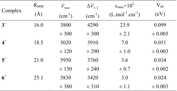
Table 4 : Intervalence Transition parameters and Experimental Vab Values for 3+, 4+, 5+ et 6+.
Complex RMM (Å) max ν (cm-1) 2 / 1 ν ∆ (cm-1) εmax×103 (L.mol-1.cm-1) Vab (eV) 3+ 16.0 3800 ± 300 4290 ± 300 23.9 ± 2.1 0.099 ± 0.005 4+ 18.5 5020 ± 120 3910 ± 290 7.0 ± 1.0 0.051 ± 0.003 5+ 21.0 5950 ± 150 3760 ± 240 3.6 ± 0.7 0.034 ± 0.002 6+ 25.1 5830 ± 380 3420 ± 310 3.0 ± 1.1 0.024 ± 0.003 10
The RMM values in Table 4 were calculated taking the average of the metal-metal distance in the syn
and anti conformation. (See scheme 4). In each of the conformations, the distances were evaluated using
a geometry optimized structure using Molecular Mecanics. (?)
N N N Ru O O N N N N N Ru O O N N N N N Ru O O N N N N N Ru O O N N Anti conformation Syn conformation
Scheme 4: Syn and Anti conformations of dinuclear compounds.
Insert Figure 4
In Figure 4 is illustrated the evolution of Vab with metal-metal distance. The decay law was obtained
using the first three values, which corresponds to the repetition of an alkyne unit:
[(tpy)(thd)Ru-NCN-Ph-(C≡C)n-Ph-NCN-Ru(tpy)(thd)] with n = 0, 1 or 2 for complexes 3, 4 et 5 respectively. The decay of
Vab follows an exponential law as given by equation (1), as it has already been observed and reported
elsewhere.2, 3 ) exp( MM ab ab V R V = ° −γ (1)
The decay slope was found to be γ = 0.21 ± 0.03 Å-1. This value is twice or three times bigger than the
one usually observed for various series (0.07 -0.12 Å-1).2 This could be explained by the nature of the
repeat units of the bridging ligand. By using a simple tight binding model, it has been shown that the
structure of the bridge plays a crucial role on the electronic coupling. 26 For all the compounds in
Launay’s review, 2 the bridging ligands show essentially vinylene or phenylene spacers. The difference
could thus be attributed to the alkyne nature of the spacer which may be less good π-connectors 27 than
polyene ones or which presents some bond-length alternation effect. 28 The value of Vab obtained for the
longest complex 5 appears to be noticeably higher than the one which would be extrapolated for the
same distance with the above decay law. The peculiar role of anthracene, inserted in a molecular bridge, 11
in mediating electronic effect and more precisely its amplification effect has already been noticed in
several cases. 9, 13, 29 An other explanation for the fact that compound 5, with the anthracene bridge, does
not follow the decay law would be to claim that for such a long compound, the energy gap is
comparable to the reorganization energy or electronic coupling and so, superexchange and sequential
mechanisms can compete. An increase in Vab for 5 could be due to the dominance of an incoherent
channel, namely a hopping process as it has already been evoked for charge and spin transport through
para-phenylene oligomers. 14, 15
Homovalent species – Magnetic Coupling
The RuIII-RuIII species were electrochemically generated. Total oxidation was checked by Linear
Voltammetry after oxidation (See Figure S6). LV allowed us to estimate that the dinuclear compounds
were oxidized up to 98%.
Insert Figure5
The EPR spectra of the oxidized species were then performed in frozen DCM solutions at 30K.
(Figure 5). The spectra are quite broad, with a peak to peak separation of about 500 G, and present an
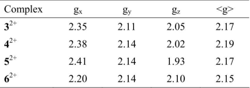
anisotropic shape. The Landé factors are gathered in Table 5.
Table 5: EPR parameters for 32+, 42+, 52+ and 62+ in frozen DCM solution (30 K).
Complex gx gy gz <g>
32+ 2.35 2.11 2.05 2.17
42+ 2.38 2.14 2.02 2.19
52+ 2.41 2.14 1.93 2.17
62+ 2.20 2.14 2.10 2.15
The g factors are in the range of 2.15 to 2.19, which is very comparable to the values obtained for 1+.
22 These signals are that of an effective S=1 spin state resulting from the magnetic interaction of two
S=1/2 spin states carried by each ruthenium(III). The spectra keep the characteristics of a mononuclear
ruthenium(III) S=1/2 spin state but with larger bandwidths. They also show a weak signal at half field,
which is typical of a spin triplet and corresponds to a ∆MS = 2 forbidden transition.
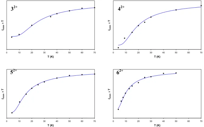
To measure the magnetic coupling for each complex, EPR measurements at several temperatures can
be performed.30 In Figure 6, are reported the product of the intensity of the EPR signal times the
temperature versus the temperature for the four complexes 3-5. Equation (2) was used in order to extract
the magnetic interaction J. The first term corresponds to the Bleaney Bowers equation, 4 which
describes the intramolecular interaction between two S=1/2 spins and the second term takes into
account the residual paramagnetism due to the presence of traces of the mixed valent species.
T C T k J T C I B totale 1 2 exp 3 1 1 1 + ⎟⎟ ⎠ ⎞ ⎜⎜ ⎝ ⎛ − + × = (2)
All the curves shown on Figure 6 present a drop at low temperature which is characteristic of an
antiferromagnetic interaction. Magnetic parameters are reported in Table 6.
Insert Figure 6
Table 6 : Magnetic parameters for 32+, 42+, 52+ et 62+.
Complex RMM (Å) C1 a C 2a J (cm-1) Rb 32+ 16.0 1.2×10¹¹ 3.3×10¹⁰ -33 ± 4 0.0004 42+ 18.5 5.8×10⁹ 4.2×10⁸ -25 ± 3 0.0043 52+ 21.0 1.1×10¹¹ 9.9×10⁹ -20 ± 2 0.0003 62+ 25.1 2.2×10¹⁰ 3.4×10⁹ -13 ± 2 0.0010 a Arbitrary units, b
∑
∑
− = 2 2 totale totale I Ifit I ROne has to notice that the magnetic couplings for 3-6 are quite strong, from J = -33(4) cm-1 for the
shortest to J = -13(2) cm-1 for the longest complex. No other examples of such couplings for such
metal-metal distances, at least with ruthenium complexes, are found in the literature. Figure 7 shows the decay
law of the magnetic coupling versus the metal-metal distance, it follows an exponential law, as given by equation (3). ) ' exp( 0 MM R J J = −γ (3)
The decay slope was found to be γ’ = 0.10 ± 0.01 Å-1. It has to be noted that this decay slope is the
lowest one obtained so far. In addition, one has to notice that the point corresponding to complex 6 with
the anthracene unit is aligned with the three other points contrary to what was observed above for the
electronic coupling. Anthracene does not seem to play a particular role for magnetic coupling.
Insert Figure 7
Very few studies have been reported concerning the dependency of the magnetic interaction with
intermetallic distances,31 probably due to the difficulties of finding the good objects to study.
Wasielewski et al. found a slope of -0.37 Å-1 in a study of magnetic interactions through p-phenylene
bridges in a series of radical pairs.14 The nearly same value was found by Journaux et al on dinuclear
copper(II) Metallacyclophanes with extended π-conjugated aromatic bridges. 32
Comparison of the Electronic and Magnetic Coupling Parameters
We obtained two decay laws versus the intermetallic distance for the electronic coupling Vab and the
magnetic coupling J for the same complex series. The slopes γ and γ’ are respectively -0.21 and -0.1 Å-1.
The question is to know how one can compare these two values ?
A relationship between the magnetic exchange coupling J and the electron transfer
super-exchange coupling V was first developed by Kramers in 1934, 33 and largely developed by Anderson in
considering the magnetic properties of solid insulators. 34 Considering antiferromagnetic exchange
interactions in π-stacked crystals, Soos pointed out an approximate relationship using the transition energy of charge transfer (CT) band (hνCT) : J ~ V2/ hνCT . 35 For an intramolecular ET system, the
stabilization of the adiabatic minima relative to parabolic diabatic (V=0) surfaces that interact by an
electronic coupling matrix element V is –V2/(λ+∆G°), where λ is the vertical reorganization energy and ∆G° is the free energy change for the reaction and hνCT = λ+∆G°.36 Okamura et al 37 gave eq. 4 which
describes the stabilization of a diradical pair where the singlet state is stabilized by ET and the triplet is not.
(
0) (
1)
(
2 0)
G V S E S E ∆ + = = − = λ (4)Making the same assumption as Okamura that only the singlet is stabilized, and extending the formula
toward three state system, Nelsen obtained the following equation (5): 38
(
0) (
1)
(
2 2 0)
G V S E S E ∆ + = = − = λ (5)Nelsen by using this equation provided the first quantitative experimental test of the relationship
between J, V, and (λ+∆G°) based upon the properties of the +1 and +2 oxidation states of a symmetrical bis-hydrazine compounds. The nearly same expression, equation (6) was given by Bertrand in studying
biological molecules coupled by an exchange interaction. 12, 39
U V J ab AF 2 2 = (6)
Where U represents the charge transfer energy difference between the initial and final state at the
same nuclear configuration.
In any case, one can see that J and V should follow the expression J ≈ V2, then according to equations
(1) and (3), the relationship between γ and γ’ should at first approximation be γ’ = 2γ, which is almost exactly the opposite of what we observed, i.e. γ’ ≈ γ/2 ! It could be due to the specific nature of the cyanamide bridge. We have recently shown that the dicyanamidobenzene ligand, because of the close
proximity in energy of its HOMO with the metal orbitals can be a noninnocent bridging ligand.22
Because of its ability to expend the frontier wave functions on the bridge, this would explain the
surprising behaviour of the magnetic exchange dependence with the intermetallic distance. Indeed,
looking at equation (6), the U term would vary with the length of the bridge and eventually would
decrease as soon as this bridge takes an important part in the HOMO. 40
This unexpected result is very encouraging since it makes cyanamide bridging ligand as a very good
candidate for mediating magnetic interaction over very long distance since the attenuation factor is 15
about four times lower than predicted! Work is in progress to consolidate these results by synthesizing
even bigger bridge.
Experimental Section
Materials
All chemicals and solvents were reagent grade or better. [Ru(tpy)Cl3],41 [Ru(tpy)(thd)Cl] 22 and
[Ru(tpy)(thd)Ipcyd] 1 22 were prepared according to literature procedures. Weakly acidic Brockmann I
type alumina (Aldrich) was used.
Physical Measurements
UV-visible spectra were recorded on a Shimadzu UV-3100 spectrophotometer. 1H and 13C NMR
spectra were recorded on a Bruker AMX-500 in CD2Cl2. IR spectra of samples in KBr pellets were
taken on a Perkin-Elmer 1725 FT-IR spectrophotometer. Mass spectra were recorded by the ‘Service de
Spectroscopie de Masse’ of Paul Sabatier University using ES (Perkin-Elmer Sciex System API 365).
Cyclic voltammograms were obtained with an Autolab system (PGSTAT 100) in dry dichloromethane
(DCM) (0.1 M tetrabutylammonium hexafluorophosphate, TBAH) at 25 °C with a three-electrode
system consisting of platinum-disk working (1mm diameter), platinum-wire counter and saturated
calomel reference electrodes. Electrochemical oxidations were performed by electrolysis with
coulometry in dry dichloromethane (0.1 M TBAH) at 25 °C at fixed potential with a three-electrode
system consisting of platinum-net working, platinum-wire counter and saturated calomel reference
electrodes. Frozen solution EPR experiments were performed in DCM with a typical concentration of
5x10⁻⁴ M on a Bruker Elexys 500 E X-band spectrometer (equiped with Bruker NMR Teslameter).
Synthesis of complexes
Synthesis of [Ru(tpy)(thd)(TMSepcyd)] 2.
16 In a Schlenk tube, [Ru(tpy)(thd)(Ipcyd)] 1 (612 mg, 0.805 mmol), CuI (26 mg, 0.14 mmol, 17 mol%)
and Pd(PPh3)2Cl2 (28 mg, 0.040 mmol, 5.0 mol%) were placed in solution in the solvent mixture
DMF/piperidine (4 :1, 12 mL) previously degassed with argon. Trimethylsilylacetylene was added
dryness. The crude was purified by column chromatography (weakly acidic alumina, solvent:
dichloromethane, eluent: dichloromethane/ethanol 99.5:0.5) to give a dark blue powder of 2 (530 mg,
0.725 mmol, 90%). ES mass spectrum (CH3CN) m/z: 732.6 [M+H]+ (calc. 732.2); 546.2
[Ru(tpy)(thd)(HCN)+H]+ (calc. 546.2); 518.3 [Ru(tpy)(thd)]+ (calc. 518.1). 1H NMR (CD2Cl2 δ = 5.35)
δ: 8.65 (2H, ddd, J1=5.5 Hz, J2=1.6 Hz, J3=0.8 Hz, H6); 8.17 (2H, ddd, J1=8.1 Hz, J2=1.3 Hz, J3=0.8 Hz, H3); 8.09 (2H, d, J=8.0 Hz, H3’); 7.88 (2H, ddd, J1=8.1 Hz, J2=7.6 Hz, J3=1.6 Hz, H4); 7.55 (1H, t, J=8.0 Hz, H4’); 7.52 (2H, ddd, J1=7.6 Hz, J2=5.5 Hz, J3=1.3 Hz, H5); 7.00 (2H, d, J=8.6 Hz, Hm); 6.21 (2H, d, J=8.6 Hz, Ho); 5.66 (1H, s, Hc); 1.59 (9H, s, He’); 0.52 (9H, s, Ha’) ; 0.24 (9H, s, Hi). 13C NMR (CD2Cl2 δ = 53.48) δ: 196.9 (Cb); 196.8 (Cd); 160.9 (C2’); 159.6 (C2); 154.8 (Cf); 150.7 (C6); 135.1 (C4); 132.3 (Cm); 126.7 (C4’); 126.0 (C5); 125.5 (CNCN); 121.3 (C3); 120.2 (C3’); 118.9 (Co); 109.7 (Cp); 107.3 (Cg);
90.3 (Ch); 88.9 (Cc); 41.6 (Ce); 40.2 (Ca); 28.7 (Ce’); 27.6 (Ca’); -0.1 (Ci). Anal. Calc. for
RuC38H43N5O2Si: C, 62.4; H, 5.9; N, 9.6. Found: C, 62.6; H, 5.7; N, 9.5. IR ν/cm-1 2179s (NCN); 2146s
(C≡C).
Synthesis of [{Ru(tpy)(thd)}2(µ-dcbp)] 3
A Schlenk tube was charged with [Ru(tpy)(thd)(Ipcyd)] (200 mg, 0.263 mmol), potassium carbonate
(109 mg, 0.789 mmol, 3.0 equ.), PdCl2dppf (8.9 mg, 0.011 mmol, 4.1 mol%) and bis(pinacolato)diboron
(33.4 mg, 0.132 mmol, 0.50 equ.). It was evacuated and backfilled with argon. DMF (10 mL) was added
under argon and the reaction mixture was stirred at 75°C for 15 hours. The solvent was removed under
vacuum. The crude was adsorbed on alumina and purified by column chromatography (weakly acidic
alumina, solvent: dichloromethane, eluent: dichloromethane/ethanol 99:1 then 98.5:1.5). The third band
(blue-green) was collected, evaporated to dryness and dissolved in the solvent mixture
dichloromethane/ethanol (4:1, 5 mL). Cyclohexane (20 mL) was added to precipitate a dark blue-green
powder of 3 (22.4 mg, 0.018 mmol, 13%). ES mass spectrum (CH3CN) m/z: 1269.3 [M+H]+ (calc.
1269.4); 752.4 [Ru(tpy)(thd)(dcbpH)+H]+ (calc. 752.2); 635.3 [M+2H]2+ (calc. 635.2); 546.2
[Ru(tpy)(thd)(HCN)+H]+ (calc. 546.2); 518.3 [Ru(tpy)(thd)]+ (calc. 518.1). 1H NMR (CD2Cl2/CD3OD,
4:1) δ: 8.63 (4H, d, J=5.4 Hz, H6); 8.20 (4H, d, J=8.0 Hz, H3); 8.11 (4H, d, J=8.0 Hz, H3’); 7.88 (4H,
ddd, J1=8.0 Hz, J2=7.7 Hz, J3=1.2 Hz, H4); 7.56 (2H, t, J=8.0 Hz, H4’); 7.52 (4H, ddd, J1=7.7 Hz, J2=5.4
Hz, J3=1.2 Hz, H5); 7.04 (4H, d, J=8.3 Hz, Hm); 6.33 (4H, d, J=8.3 Hz, Ho); 5.65 (2H, s, Hc); 1.55 (18H,
s, He’); 0.48 (18H, s, Ha’). 13C NMR (CD2Cl2/CD3OD, 4:1) δ: 197.1 (Cb); 197.0 (Cd); 161.1 (C2’); 159.8
(C2); 150.8 (C6); 149.6 (Cf); 135.4 (C4); 131.1 (Cp); 127.3 (C4’); 126.3 (Cm); 126.2 (C5); 124.6 (CNCN);
121.5 (C3); 120.4 (C3’); 118.7 (Co); 89.0 (Cc); 41.7 (Ce); 40.3 (Ca); 28.7 (Ce’); 27.6 (Ca’). Anal. Calc. for
Ru2C66H68N10O4(H2O): C, 61.7; H, 5.5; N, 10.9. Found: C, 61.5; H, 5.1; N, 11.2. IR ν/cm-1 2163s
(NCN). UV-Vis-near IR CH2Cl2, λ in nm (ε × 10-3 in L.mol-1.cm-1): 278 (73), 318 (69), 357sh (51), 578
(12). Cyclic voltammetry (CH2Cl2, 0.1 M TBAH, 0.1 V.s-1, vs. SCE): E1/2(RuII/RuIII) = 0.137 V.
Synthesis of [{Ru(tpy)(thd)}2(µ-bcpa)] 4.
Potassium carbonate (84 mg, 0.61 mmol, 2.1 equ.) was added to a solution of
[Ru(tpy)(thd)(TMSepcyd)] (211 mg, 0.289 mmol) in 35 mL of methanol previously degassed with
argon. The mixture was stirred under argon at 40°C for 2.5 hours and then evaporated to dryness. The
obtained blue residue of [Ru(tpy)(thd)(epcyd)] was used without further purification. It was dissolved in
the solvent mixture DMF/piperidine (4:1, 25 mL) and added under argon to a Schlenk tube charged with
[Ru(tpy)(thd)(Ipcyd)] (220 mg, 0.289 mmol), CuI (12 mg, 0.063 mmol, 22 mol%) and Pd(PPh3)2Cl2 (11
mg, 0.016 mmol, 5.4 mol%). DBU (91 µL, 0.608 mmol, 2.1 equ.) was added under argon to the
mixture, which was stirred at room temperature for 1.5 hours and then evaporated to dryness under
vacuum. The crude reaction residue was adsorbed on alumina and purified by column chromatography
(weakly acidic alumina, solvent: dichloromethane, eluent: dichloromethane/ethanol 99.2:0.8 then
98.5:1.5). The compound was dissolved in the solvent mixture dichloromethane/ethanol (4:1, 50mL)
and addition of 150 mL cyclohexane precipitated a dark green powder of 4 (213 mg, 0.165 mmol, 57%).
ES mass spectrum (CH3CN) m/z: 1293.6 [M+H]+ (calc. 1293.4); 776.6 [Ru(tpy)(thd)(bcpaH)+H]+ (calc.
776.2); 647.6 [M+2H]2+ (calc. 647.2); 546.5 [Ru(tpy)(thd)(HCN)+H]+ (calc. 546.2). 1H NMR
(CD2Cl2/CD3OD, 4:1) δ: 8.62 (4H, d, J=5.4 Hz, H6); 8.20 (4H, d, J=8.0 Hz, H3); 8.12 (4H, d, J=8.0 Hz,
H3’); 7.89 (4H, ddd, J1=8.0 Hz, J2=7.7 Hz, J3=1.5 Hz, H4); 7.58 (2H, t, J=8.0 Hz, H4’); 7.52 (4H, ddd,
J1=7.7 Hz, J2=5.4 Hz, J3=1.5 Hz, H5); 7.01 (4H, d, J=8.3 Hz, Hm); 6.28 (4H, d, J=8.3 Hz, Ho); 5.65 (2H,
s, Hc); 1.55 (18H, s, He’); 0.49 (18H, s, Ha’). 13C NMR (CD2Cl2/CD3OD, 4:1) δ: 197.1 (Cb); 197.0 (Cd);
161.0 (C2’); 159.8 (C2); 152.0 (Cf); 150.8 (C6); 135.5 (C4); 131.7 (Cm); 127.5 (C4’); 126.2 (C5); 125.2
(CNCN); 121.6 (C3); 120.4 (C3’); 118.5 (Co); 112.2 (Cp); 89.0 (Cc); 88.2 (Cg); 41.6 (Ce); 40.3 (Ca); 28.7
(Ce’); 27.6 (Ca’). Anal. Calc. for Ru2C68H68N10O4(H2O) : C, 62.4; H, 5.4; N, 10.7. Found: C, 62.4; H,
5.1; N, 10.9. IR ν/cm-1 2152s (NCN). UV-Vis-near IR CH
2Cl2, λ in nm (ε × 10-3 in L.mol-1.cm-1): 278
(67), 318 (61), 386 (62), 574 (12). Cyclic voltammetry (CH2Cl2, 0.1 M TBAH, 0.1 V.s-1, vs. SCE):
E1/2(RuII/RuIII) = 0.188 V.
Synthesis of [{Ru(tpy)(thd)}2(µ-bcpda)] 5.
To a solution of [Ru(tpy)(thd)(TMSepcyd)] (299 mg, 0.409 mmol) in 35 mL of methanol previously
degassed with argon was added potassium carbonate (125 mg, 0.904 mmol, 2.2 equ.). The mixture was
stirred under argon at 40°C for 2.5 hours and then evaporated to dryness to yield a blue residue of
[Ru(tpy)(thd)(epcyd)], which was kept under argon and used without further purification. A
three-necked round bottom flask equipped with an adaptor connected to an oxygen source was charged with
CuCl (18 mg, 0.18 mmol, 44 mol%). Pyridine (10 mL) and DBU (123 µL, 0.82 mmol, 2.0 equ.) were
added and the mixture was warmed to 40°C and vigorously stirred while bubbling oxygen. The initially
yellow solution turned green after several minutes and the blue residue of [Ru(tpy)(thd)(epcyd)],
previously prepared and dissolved in 15 mL pyridine, was added. The reaction mixture was stirred at
40°C with oxygen bubbling. Additional reactants were added after 2 hours (42 mol% of CuCl and 1.0
equ. of DBU) and after 3 hours (17 mol% of CuCl and 1.0 equ. of DBU). After 4 hours of stirring, the
mixture was evaporated to dryness under vacuum. The green residue was adsorbed on alumina and
purified by column chromatography (weakly acidic alumina, solvent: dichloromethane, eluent:
dichloromethane/ethanol 99.5:0.5 then 99:1). The second band (dark green) was collected, evaporated to
dryness and redissolved in the solvent mixture dichloromethane/ethanol (4:1, 50mL). Addition of 150
mL cyclohexane precipitated a dark green powder of 5 (180 mg, 0.137 mmol, 67%). ES mass spectrum
(CH3CN) m/z: 1317.7 [M+H]+ (calc. 1317.4); 800.5 [Ru(tpy)(thd)(bcpdaH)+H]+ (calc. 800.2); 659.2
[M+2H]2+ (calc. 659.2); 546.5 [Ru(tpy)(thd)(HCN)+H]+ (calc. 546.2). 1H NMR (CD2Cl2/CD3OD, 4:1)
δ: 8.61 (4H, d, J=5.4 Hz, H6); 8.21 (4H, d, J=8.1 Hz, H3); 8.13 (4H, d, J=8.0 Hz, H3’); 7.89 (4H, ddd,
J1=8.1 Hz, J2=7.5 Hz, J3=1.5 Hz, H4); 7.59 (2H, t, J=8.0 Hz, H4’); 7.52 (4H, ddd, J1=7.5 Hz, J2=5.4 Hz,
J3=1.2 Hz, H5); 7.05 (4H, d, J=8.4 Hz, Hm); 6.29 (4H, d, J= 8.4 Hz, Ho); 5.65 (2H, s, Hc); 1.54 (18H, s,
He’); 0.48 (18H, s, Ha’). 13C NMR (CD2Cl2/CD3OD, 4:1) δ: 197.1 (Cb); 197.0 (Cd); 161.0 (C2’); 159.8
(C2); 153.9 (Cf); 150.8 (C6); 135.5 (C4); 133.1 (Cm); 127.6 (C4’); 126.2 (C5); 124.6 (CNCN); 121.6 (C3);
120.4 (C3’); 118.6 (Co); 109.6 (Cp); 89.0 (Cc); 82.7 (Cg); 72.2 (Ch); 41.6 (Ce); 40.3 (Ca); 28.7 (Ce’); 27.6
(Ca’). Anal. Calc. for Ru2C70H68N10O4(H2O)0.7: C, 63.3; H, 5.3; N, 10.5. Found: C, 63.3; H, 5.3; N, 10.5.
IR ν/cm-1 2164s (NCN); 2135s (C≡C). UV-Vis-near IR CH
2Cl2, λ in nm (ε × 10-3 in L.mol-1.cm-1): 278
(69), 318 (62), 408 (70), 570 (13). Cyclic voltammetry (CH2Cl2, 0.1 M TBAH, 0.1 V.s-1, vs. SCE):
E1/2(RuII/RuIII) = 0.225 V.
Synthesis of [{Ru(tpy)(thd)]}2(µ-bcpea)] 6
N N N Ru O O N N N Ru O O N N N N 6 4 5 3 2 2' 3' 4' a b c d e a' e' NCN f o m p g h i j k l
A Schlenk tube was charged with [Ru(tpy)(thd)(Ipcyd)] (275 mg, 0.362 mmol),
9,10-bis(3-hydroxy-3-methylbutynyl)anthracene (50.8 mg, 0.148 mmol, 0.41 equ.), CuI (17 mg, 0.089 mmol, 24 mol%) and
Pd(PPh3)4 (29 mg, 0.025 mmol, 6.9 mol%). It was evacuated and backfilled with argon. The solvent
mixture DMF/piperidine (5:1, 12 mL) and potassium tert-butoxide (81 mg, 0.72 mmol, 2.0 equ.) were
added under argon. The reaction mixture was stirred under argon at 60°C for 1.5 hours, during which
the color of the mixture changed from blue-green to brown red and then to purple. The solvents were
removed under vacuum and the crude was purified by column chromatography (weakly acidic alumina,
solvent: dichloromethane, eluent: dichloromethane/ethanol 99.5:0.5 then 99:1). The obtained powder
was dissolved in the solvent mixture dichloromethane/ethanol (4:1, 50 mL) and addition of cyclohexane
(100 mL) gave a precipitate, which was filtered, washed with cyclohexane and diethylether and dried
under vacuum to yield a purple powder of 6 (93.1 mg, 0.062 mmol, 35 %). ES mass spectrum (CH3CN)
m/z: 1493.4 [M+H]+ (calc. 1493.4); 976.3 [Ru(tpy)(thd)(bcpeaH)+H]+ (calc. 976.3); 747.3 [M+2H]2+
(calc. 747.2); 546.4 [Ru(tpy)(thd)(HCN)+H]+ (calc. 546.2); 518.3 [Ru(tpy)(thd)]+ (calc. 518.1). 1H
NMR (CD2Cl2/CD3OD, 4:1) δ: 8.67 (4H, dd, J1=6.6 Hz, J2=3.3 Hz, Hk); 8.65 (4H, d, J=5.4 Hz, H6);
8.24 (4H, d, J=8.1 Hz, H3); 8.17 (4H, d, J=8.1 Hz, H3’); 7.92 (4H, ddd, J1=8.1 Hz, J2=7.5 Hz, J3=1.5 Hz,
H4); 7.64 (4H, dd, J1=6.6 Hz, J2=3.3 Hz, Hl); 7.62 (2H, t, J=8.1 Hz, H4’); 7.56 (4H, ddd, J1=7.5 Hz,
J2=5.4 Hz, J3=1.5 Hz, H5); 7.36 (4H, d, J=8.4 Hz, Hm); 6.44 (4H, d, J=8.4 Hz, Ho); 5.67 (2H, s, Hc); 1.57
(18H, s, He’); 0.50 (18H, s, Ha’). 13C NMR (CD2Cl2/CD3OD, 4:1) δ: 197.2 (Cb); 197.0 (Cd); 161.1 (C2’);
159.8 (C2); 153.7 (Cf); 150.8 (C6); 135.6 (C4); 132.4 (Cm); 131.7 (Cj); 127.6 (C4’); 127.3 (Ck); 126.5
(Cl); 126.3 (C5); 124.7 (CNCN); 121.6 (C3); 120.4 (C3’); 118.8 (Co); 118.3 (Ci); 111.2 (Cp); 104.6 (Cg);
89.1 (Cc); 84.6 (Ch); 41.8 (Ce); 40.3 (Ca); 28.7 (Ce’); 27.6 (Ca’). Anal. Calc. for Ru2C84H76N10O4(H2O):
C, 66.8; H, 5.2; N, 9.3. Found: C, 66.9; H, 5.1; N, 9.3. IR ν/cm-1 2161s (NCN). UV-Vis-near IR CH 2Cl2,
λ in nm (ε × 10-3 in L.mol-1.cm-1): 270 (115), 318 (68), 344 (57), 552 (59). Cyclic voltammetry (CH 2Cl2,
0.1 M TBAH, 0.1 V.s-1, vs. SCE): E1/2(RuII/RuIII) = 0.211 V.
Acknowledgment.
The authors thank CNRS and MENRS (M.F.) for financial support, Alain Mari (LCC, Toulouse) for
EPR measurements, Christophe Coudret (CEMES) for helpful discussion about synthesis, Jean-Pierre
Launay (CEMES) for his help in reading the manuscript.
Supporting Information Available.
S1 : Differential Pulse Voltammetry for complex 3, 4, 5 et 6 (CH2Cl2, 0.1 M TBAH, on rotating
platinum electrode); S2: Spectroelectrochemical oxidation of 4 in DCM, 0.1 M TBAH (electrolysis at
0.46 V vs. SCE); S3 : Spectroelectrochemical oxidation of 5 in DCM, 0.1 M TBAH (electrolysis at 0.5
V vs. SCE); S4 : Spectroelectrochemical oxidation of 6 in DCM, 0.1 M TBAH (electrolysis at 0.46V vs.
SCE); S5 : Déconvolution of the IV transitions in the NIR area for 3+, 4+, 5+ and 6+ in DCM; S6: Linear
Voltammetry for complex 32+, 42+, 52+ and 62+ electrochemically generated.
-0,3 0,0 0,3 0,6 -0,1 0,1 0,3 0,5 0,7 0,9 1,1 1,3 1,5 E (V/ECS) i ( µ A) -0,8 -0,4 0,0 0,4 0,8 1,2 1,6 -0,1 0,1 0,3 0,5 0,7 0,9 1,1 1,3 1,5 E (V/ECS) i ( µ A) 3 4 -0,2 -0,1 0,0 0,1 0,2 0,3 0,4 -0,1 0,1 0,3 0,5 0,7 0,9 1,1 1,3 1,5 E (V/ECS) i ( µ A) -0,1 0,0 0,1 0,2 -0,1 0,1 0,3 0,5 0,7 0,9 1,1 1,3 1,5 E (V/ECS) i ( µ A) 5 6
Figure 1 : Cyclic voltammetry of 3, 4, 5 and 6 (CH2Cl2, 0.1 M TBAH, 0.1 V.s-1).
0 2 4 6 8 250 500 750 1000 λ (nm) ε/10 4 (M -1 .cm -1 )
3
4
5
6
0 2 4 6 8 250 500 750 1000 1250 1500 1750 2000 λ (nm) ε/1 0 4 (M -1 .c m -1 )3
4
5
6
2+ 2+ 2+ 2+Figure 2 : - (top) UV-Vis-near IR spectra of 3, 4, 5 and 6 in DCM. - (bottom) UV-Vis-near IR spectra
of 32+, 42+, 52+ and 62+in DCM.
0 1 2 3 4 5 6 7 8 250 500 750 1000 1250 1500 1750 2000 2250 2500 λ (nm) ε/1 0 4 (M -1.c m -1) 0 1 2 3 4 5 6 7 8 250 500 750 1000 1250 1500 1750 2000 2250 2500 λ (nm) ε/1 0 4 (M -1.c m -1)
Figure 3 : Spectroelectrochemical oxidation of 3 in DCM, 0.1 M TBAH (electrolysis at 0.46 V vs.
SCE). left : 3 → 3 . right : 3 → 3 .+ + 2+
Figure 4 : Decay law for the electronic coupling parameter Vab in log scale vs the metal-metal distance
RMM.
42+ 32+
52+ 62+
Figure 5 : experimental and simulated EPR spectrum of 32+, 42+, 52+ et 62+ in frozen DCM solution (30
K).
0 10 20 30 40 50 60 7 T (K) Itotal e x T 0 0 10 20 30 40 50 60 7 T (K) Itota le x T 0 32+ 42+ 0 10 20 30 40 50 60 7 T (K) Itota le x T 0 0 10 20 30 40 50 60 7 T (K) Itota le x T 0 52+ 62+
Figure 6 : Double integrated EPR signal intensity times Temperature (Itotale×T) of 32+, 42+, 52+ and 62+
versus temperature : experimental (dots) simulated (lines).
Figure 7 : Decay law of the magnetic coupling versus the metal-metal distance RMM.
References
1. Creutz, C.; Taube, H. J. Am. Chem. Soc. 1969, 91, (14), 3988. 2. Launay, J.-P. Chem. Soc. Rev. 2001, 30, 386.
3. Launay, J.-P.; Coudret, C., Wires Based on Metal Complexes. In Electron Transfer in
Chemistry, Wiley-VCH ed.; 2001; Vol. 5, pp 3.
4. Kahn, O., Molecular Magnetism. Wiley-VCH ed.; 1993.
5. Crutchley, R. J. Adv. Inorg. Chem. 1994, 41, 273.
6. Ward, M. D. Chem. Soc. Rev. 1995, 24, (2), 121. Paul, F.; Lapinte, C. Coord. Chem. Rev. 1998, 178-180, 431. McCleverty, J. A.; Ward, M. D. Acc. Chem. Res. 1998, 31, (12), 842. Brunschwig, B. S.; Sutin, N. Coord. Chem. Rev. 1999, 187, 233. Kaim, W.; Klein, A.; Gloeckle, M. Acc. Chem. Res. 2000, 33, (11), 755. Demadis, K. D.; Hartshorn, C. M.; Meyer, T. J. Chem. Rev. 2001, 101, 2655. Ward, M. D.; McCleverty, J. A. J. Chem. Soc., Dalton Trans. 2002, (3), 275.
7. Hush, N. S. Prog. Inorg. Chem. 1967, 8, 391.
8. Joachim, C.; Gimzewski, J. Europhys. Lett. 1995, 30, 409. Langlais, V. J.; Schlittler, R. R.; Tang, H.; Gourdon, A.; Joachim, C.; Gimzewski, J. K. Phys. Rev. Letters 1999, 83, (14), 2809. Rousset, V.; Joachim, C.; Rousset, B.; Fabre, N. J. Phys. III 1995, 5, 1983.
9. Fraysse, S.; Coudret, C.; Launay, J.-P. J. Am. Chem. Soc. 2003, 125, (19), 5880. 10. Forbes, M. D. E.; Ball, J. D.; Avdievich, N. J. Am. Chem. Soc. 1996, 118, 4707.
11. Felthouse, T. R.; Hendrickson, D. N. Inorg. Chem. 1978, 17, (9), 2636. Brunold, T. C.; Gamelin, D. R.; Solomon, E. I. J. Am. Chem. Soc. 2000, 122, 8511. Bayly, S. R.; Humphrey, E. R.; de Chair, H.; Paredes, C. G.; Bell, Z. R.; Jeffery, J. C.; McCleverty, J. A.; Ward, M. D.; Totti, F.; Gatteschi, D.; Courric, S.; Steele, B. R.; Screttas, C. G. J. Chem. Soc., Dalton Trans. 2001, (9), 1401. Shultz, D. A.; Fico, R. M. J.; Bodnar, S. H.; Krishna Kumar, R.; Vostrikova, K. E.; Kampf, J. W.; Boyle, P. D. J. Am.
Chem. Soc. 2003, (125), 11761. Elschenbroich, C.; Plackmeyer, J.; Nowotny, M.; Harms, K.; Pebler, J.;
Burhgaus, O. Inorg. Chem. 2005, 44, 955. Elschenbroich, C.; Plackmeyer, J.; Nowotny, M.; Behrendt, A.; Harms, K.; Pebler, J.; Burghaus, O. Chem. Eur. J. 2005, 11, 7427.
12. Bertrand, P. Chem. Phys. Lett. 1985, 113, 104.
13. Nelsen, S. F.; Ismagilov, R. F.; Powell, D. R. J. Am. Chem. Soc. 1998, 120, (8), 1924. de Montigny, F.; Argouarch, G.; Costuas, K.; Halet, J.-F.; Toupet, L.; C., L. Organometallics 2005, 24, 4558.
14. Weiss, E. A.; Ahrens, M. J.; Sinks, L. E.; Gusev, A. V.; Ratner, M. A.; Wasielewski, M. R. J.
Am. Chem. Soc. 2004, 126, (17), 5577.
15. Weiss, E. A.; Wasielewski, M. R.; Ratner, M. A. Top. Curr. Chem. 2005, 257, 103.
16. Aquino, M. A. S.; Lee, F. L.; Gabe, E. J.; Bensimon, C.; Greedan, J. E.; Crutchley, R. J. J. Am.
Chem. Soc. 1992, 114, (13), 5130.
17. Rezvani, A. R.; Evans, C. E. B.; Crutchley, R. J. Inorg. Chem. 1995, 34, (18), 4600. Mosher, P. J.; Yap, G. P. A.; Crutchley, R. J. Inorg. Chem. 2001, 40, (6), 1189.
18. Fabre, M. A.; Jaud, J.; Bonvoisin, J. J. Inorg. Chim. Acta 2005, 358, (7), 2384. 19. Sondaz, E.; Jaud, J.; Launay, J.-P.; Bonvoisin, J. Eur. J. Inorg. Chem. 2002, 1924. 20. Sonogashira, K.; Tohda, Y.; Hagihara, N. Tetrahedron Lett. 1975, 16, (50), 4467. 21. Nising, C. F.; Schmid, U. K.; Nieger, M.; Bräse, S. J. Org. Chem. 2004, 69, 6830. 22. Fabre, M.; Jaud, J.; Hliwa, M.; Launay, J.-P.; Bonvoisin, J. Inorg. Chem. 2006, in press. 23. Aquino, M. A. S.; White, C. A.; Bensimon, C.; Greedan, J. E.; Crutchley, R. J. Can. J. Chem.
1996, 74, (11), 2201.
24. Richardson, D. E.; Taube, H. Inorg. Chem. 1981, 20, (4), 1278. 25. Hush, N. S. Coord. Chem. Rev. 1985, 64, 135.
26. McConnell, H. M. J. Chem. Phys. 1961, 35, 508. Joachim, C. Chem. Phys. 1987, 116, 339. Joachim, C.; Launay, J.-P.; Woitellier, S. Chem. Phys. 1990, 147, 131.
27. Falgarde, F.; Katz, N. E. Polyhedron 1995, 14, (9), 1213.
28. Kushmerick, J. G.; Holt, D. B.; Pollack, S. K.; Ratner, M. A.; Yang, J. C.; Schull, T. L.; Naciri, J.; Moore, M. H.; Shashidhar, R. J. Am. Chem. Soc. 2002, 124, 10654.
29. Piet, J. J.; Taylor, P. N.; Anderson, H. L.; Osuka, A.; Warman, J. M. J. Am. Chem. Soc. 2000, 122, 1749. Taylor, P. N.; Wylie, A. P.; Huuskonen, J.; Anderson, H. L. Angew. Chem. Int. Ed. 1998, 37, (7), 986. Hoshino, Y.; Suzuki, T.; Umeda, H. Inorg. Chim. Acta 1996, 245, 87.
30. Bencini, A.; Gatteschi, D., In EPR of Exchange Coupled Systems, Springer-Verlag: 1990; p 58. 31. Julve, M.; Verdaguer, M.; Faus, J.; Tinti, F.; Moratal, J.; Monge, A.; Gutierrez-Puebla, E. J. Am.
Chem. Soc. 1987, 26, 3520. Bürger, K.; Chaudhuri, P.; Wieghardt, K.; Nuber, B. Chem. Eur. J. 1995, 1,
(9), 583. Cano, J.; De Munno, G.; Sanz, J.-L.; Ruiz, R.; Faus, J.; Lloret, F.; Julve, M.; Caneschi, A. J.
Chem. Soc., Dalton Trans. 1997, 1915.
32. Pardo, E.; Faus, J.; Julve, M.; Lloret, F.; Munoz, M. C.; Cano, J.; Ottenwaelder, X.; Journaux, Y.; Carrasco, R.; Blay, G.; Fernandez, I.; Ruiz-Garcia, R. J. Am. Chem. Soc. 2003, 125, (36), 10770. 33. Kramers, H. A. Physica 1934, 1, 182.
34. Anderson, P. W. Phys. Rev. 1950, 79, (2), 350. Anderson, P. W. Phys. Rev. 1959, 115, (1), 2. Anderson, P. W., In Magnetism, Rado, G. T.; Suhl, H., Eds. Academic Press: New York, 1965; Vol. 1, p 25.
35. Soos, G. T. Ann. Rev. Phys. Chem. 1974, 25, 121. 36. Sutin, N. Prog. Inorg. Chem. 1983, 30, 441.
37. Okamura, M. Y.; Isaacson, R. A.; Feher, G. Biochim. Biophys. Acta 1979, 546, 394. 38. Nelsen, S. F.; Ismagilov, R. F.; Teki, Y. J. Am. Chem. Soc. 1998, 120, (9), 2200.
39. Bertrand, P., Application of Electron Transfer Theories to Biological Systems. In Structure and
Bonding, Spring-Verlag: Berlin Heidelberg, 1991; Vol. 75, pp 1.
40. Ruiz, E.; Rodriguez-Fortea, A.; Alvarez, S. Inorg. Chem. 2003, 42, (16), 4881. 41. Sullivan, B. P.; Calvert, J. M.; Meyer, T. J. Inorg. Chem. 1980, 19, (5), 1404.
For Table of Contents
Four phenylcyanamido-bridge diruthenium complexes [{Ru(tpy)(thd)}2(µ-L)] have been studied in their
mixed-valent Ru(II)Ru(III) and homovalent paramagnetic Ru(III)Ru(III) forms. Exponential decays of
the electronic Vab and magnetic exchange J parameters with intermetallic distance have been obtained
by optical and EPR spectroscopy respectively. Relationship between these two parameters is discussed.
Unpredictable low value for the slope of the magnetic exchange parameter makes this family of
compounds valuable for the study of long range magnetic coupling and molecular electronics.