U N IV E R S IT Y DE
SHERBROOKE
Faculty of Engineering
Department of Mechanical Engineering
THE EFFECT OF TISSUE MECHANICAL CHARACTERIZATION AND
STIMULATION PARAMETERS ON LIVE TISSUE MECHANOBIOLOGICAL
PROGRESSION W ITH REGARD TO VISCOELASTICITY AND
VISCOPLASTICITY
Master's thesis by:
Leila JAFARI
Jury:
Eve LANGELIER
Denis RANCOURT
Leonie ROULEAU
1+1
Library and Archives Canada Published Heritage Branch Bibliotheque et Archives Canada Direction du Patrimoine de I'edition 395 Wellington Street Ottawa ON K 1A0N 4 Canada 395, rue Wellington Ottawa ON K1A 0N4 CanadaYour file Votre reference ISBN: 978-0-494-93293-3 Our file Notre reference ISBN: 978-0-494-93293-3
NOTICE:
The author has granted a non
exclusive license allowing Library and Archives Canada to reproduce, publish, archive, preserve, conserve, communicate to the public by
telecomm unication or on the Internet, loan, distrbute and sell theses
worldwide, for commercial or non commercial purposes, in microform, paper, electronic and/or any other formats.
AVIS:
L'auteur a accorde une licence non exclusive permettant a la Bibliotheque et Archives Canada de reproduire, publier, archiver, sauvegarder, conserver, transmettre au public par telecomm unication ou par I'lnternet, preter, distribuer et vendre des theses partout dans le monde, a des fins com merciales ou autres, sur support microforme, papier, electronique et/ou autres formats.
The author retains copyright ownership and moral rights in this thesis. Neither the thesis nor substantial extracts from it may be printed or otherwise reproduced without the author's permission.
L'auteur conserve la propriete du droit d'auteur et des droits moraux qui protege cette these. Ni la these ni des extraits substantiels de celle-ci ne doivent etre imprimes ou autrement
reproduits sans son autorisation.
In compliance with the Canadian Privacy A ct some supporting forms may have been removed from this thesis.
W hile these forms may be included in the document page count, their removal does not represent any loss of content from the thesis.
Conform em ent a la loi canadienne sur la protection de la vie privee, quelques
form ulaires secondaires ont ete enleves de cette these.
Bien que ces form ulaires aient inclus dans la pagination, il n'y aura aucun contenu manquant.
T itre en fran^ais:
L'EFFET DE LA CARACTERISATION MECANIQUE ET DES PARAMETRES DE
STIMULATION DES TISSUS SUR LEUR EVOLUTION MECANOBIOLOGIQUE
Resume
La caracterisation des tissus est une etape majeure dans les etudes mecanobiologiques. En effet, a l'aide des methodes de caracterisation, la qualite des tissus, soit la combinaison des proprietes structurelles, compositionnelles et mecaniques, peut etre determinee. Ce p ro je t de maftrise focalise sur les methodes de caracterisation mecanique pour les etudes in vitro en bioreacteur. A travers toutes les methodes de caracterisation mecanique, nous proposons l’utilisa tio n de celles qui s o n t: 1) non destructives (i.e. qui o ffrent la possibility de realiser d'autres essais de caracterisation apres les essais de caracterisation mecaniques) et 2) en-ligne (i.e. qui perm ettent l’observation de la progression des tissus durant l’experimentation, e t ce, sans devoir deplacer les specimens d'une machine vers une autre). Toutefois, la caracterisation mecanique non-destructive en-ligne souleve la question a savoir si cette methode d'observation utilisee durant l'experim entation m odifie revolution des tissus dans le temps.
Ainsi, le b u t de ce p rojet de m aitrise etait d'approfondir nos connaissances sur les parametres qui pourraient affecter la qualite des tissus conjonctifs mous durant une experim entation in vitro en bioreacteur. Ceci passe par une meilleure comprehension de la viscoelasticite et viscoplasticite, deux comportements cles des tissus, qui affectent l’impact de ces parametres sur la reponse des tissus vivants a des stim uli biophysiques. Done, les deux objectifs de ce p ro je t y ta ie n t:
1. De re v o ir la litte ra tu re portant sur deux comportements mycaniques des tissus, soient la viscoelasticite et la viscoplasticite, et la fa^on avec laquelle ils affectent revolution des tissus sous stim u li biophysiques;
2. D'investiguer si l’utilisation d'essais diagnostiques d'am plitude physiologique pour quan tifie r les proprietes mecaniques des tissus peut affecter leur evolution dans le temps.
Dans ce memoire, nous expliquons que la viscoelasticite et la viscoplasticite des tissus proviennent de la structure et de la com position de la matrice extracellulaire. Nous decrivons egalement la fa^on avec laquelle ces com portem ents affectent la com petition dynamique entre la reparation, la degradation enzymatique et la degradation mecanique de la m atrice extracellulaire sous stim uli biophysiques. De plus, nous specifions des parametres de stim ulation, tels que le type de controle ou l'h isto ire des stim uli, qui p ourraient affecter revolution des tissus en reponse a des stim uli biophysiques a cause de la viscoplasticite et viscoyiasticite.
Aussi, nous relatons les resultats d'une experim entation de trois jours realisees sur des tendons fraichem ent extraits pour investiguer si l'application d’essais de relaxation de contrainte d'am plitude physiologique affecte re vo lu tio n des tissus sous stim uli mecaniques. Nous avons regroupe les tendons selon le protocole de caracterisation (0 ou 24 essais de relaxation d'am plitude physiologique chaque jo u r) et nous avons compare In v o lu tio n des groupes dans le temps. Les essais de relaxation de contraintes d'am plitude physiologique ont m odifie re vo lu tio n des tendons en reponse aux stim uli
mecaniques in vitro. De fa?on generate, le module pointe a augmente dans le temps pour le groupe de 0 essai de relaxation de contrainte alors q u 'il a d'abord dim inue puis legerement augmente pour le groupe de 24 essais de relaxation de contrainte chaque jour. La difference entre les deux groupes etait significative. Done, l'insertion d’essais de relaxation de contrainte d'am plitude physiologique pendant les periodes de repos entre les stim uli mecaniques peut influencer re vo lu tio n des tissus dans le temps.
Nous concluons qu’il im porte de te n ir compte de la viscoelasticite et de la viscoplasticite des tissus lors du developpement d’un protocole de stim ulation pour une etude en bioreacteur ou encore pour une application clinique.
Mots cles : tissus conjonctifs mous, mecanobiologie, evolution des tissus, proprietes des tissus, protocole de caracterisation, viscoelasticite, viscoplasticite, en-ligne, non- destructif
Abstract
Tissue characterization is a m ajor step in tissue mechanobiological studies. By characterization methods, tissue quality i.e. the combination o f tissue structural, compositional and mechanical properties, is determ ined. This research focuses on mechanical characterization methods. Among all mechanical characterization methods, we propose those ones w hich are: 1) Non-destructive, (i.e. that reserves the capability of doing other characterization tests at the end o f mechanical test; and, 2) In-line, (that enables tissue progression observation during experiment, and w ith o u t tra n sfe rring the specimen from one apparatus to another). However, in-line characterization raises the question o f w hether conducting tissue observation methods during experim entation modifies tissue progression over time.
Therefore, the purpose o f this study was to deepen ou r knowledge about the parameters which could affect tissue quality during mechanical testing. This requires a better understanding o f viscoelasticity and viscoplasticity, tw o key behaviors o f tissue, affecting the im pact o f these parameters (e.g. tissue quality, stim ulation parameters) on the response o f live tissue to biophysical stim uli. Thus, the objectives o f this study were:
1. To review the litera ture to find inform ation about tw o mechanical behaviors o f tissue i.e. viscoelasticity and viscoplasticity, and the way they affect tissue properties
2. To investigate w hether diagnostic tests, as mechanical characterization tests to observe tissue properties, affect tissue progression
We explain th a t viscoelasticity and viscoplasticity o f tissue originate fro m structure and components o f the extracellular m atrix. We also describe the w ay they affect tissue dynamic com petition between repair, enzymatic degradation and mechanical degradation o f the extracellular m atrix. Moreover, we specify some tissue stim ulation parameters, such as stim ulation control type o r stim ulus history, w hich could affect tissue progression in response to biophysical stim u li because o f viscoelasticity and viscoplasticity.
Moreover, by conducting a series o f 3-day experiments on freshly extracted tendons, we investigated w hether applying "stress relaxation" tests a t physiological am plitudes affects tissue response. We divided the tendons into tw o groups based on the characterization protocol (24 and 0 stress relaxation tests each day), and compared the progression o f these groups over time. The stress relaxation tests at physiological am plitude m odified tissue response to mechanical stim u li in vitro. In general, the modulus increased fo r 0 stress relaxation tests, w hile it first decreased and then increased slightly fo r 24 stress relaxation tests each day. The difference o f mechanical properties between the tw o groups was significant. Therefore, applying stress relaxation tests at physiological am plitude during the rest periods between mechanical stim uli can affect live tissue progression over time.
Therefore, it is essential to take into account the viscoelasticity and viscoplasticity of tissue w hile developing a stim ulation protocol fo r bioreactor studies o r clinical applications.
Keywords: mechanobiology, tissue progression, tissue properties, characterization protocol, mechanical characterization, viscoelasticity and viscoplasticity, in-line, non destructive.
Acknowledgements
I w ould like to express m y gratitude to Professor Eve Langelier fo r her guidance and kind support during this research p ro je c t Her encouragements m otivated me to overcome the challenges I faced during the project. Her involvem ent was beyond her responsibility and I cannot thank her enough.
In addition, I w ould like to thank the members o f ju ry Professor Denis Rancourt and Professor Leonie Rouleau fo r th e ir constructive comments.
Moreover, 1 w ould like to appreciate several people w ho helped me in this research project: Yoan Lemieux-LaNeuville, Charles Bertrand, Melina Narlis, Leonid Volkov, Caroline Bergeron, Amelie Caron-Laramee and CIRUS (Centre d’lnnovation Radicale de l'Universite de Sherbrooke).
I thank my dear friends: Fatemeh Mousavi, Hilda Harirforoush, Vahid Ikani, Azin Barsalani, Naser Pighon, Zari Khodadadi, Zahra Akbari and Mahmood Akbari.
Last but not least, I w ould like to express m y deepest gratitude to m y fam ily: Hossein, Mila, M itra and her fa m ily Parviz and Pooria, Pooyeh and her fa m ily Asghar and Amirreza and special thanks to Dr. Hossein Rouhani fo r his inspiration, encouragement and constructive comments during our scientific discussions. My profound appreciation goes to my m other and my beloved parents-in-law fo r their love, support and encouragement. 1 cannot thank them enough. My utm ost gratefulness goes to m y love, Hassan, to w hom 1 owe everything...
TABLE OF CONTENTS
1. In tro d u c tio n ...
1
2. State-O f-The-A rt...
5
2.1 Tendon compositional p roperties...5
2.2 Tendon structural properties... 6
2.2.1 Qualitative characterization... 8
2.2.2 Semi-quantitative characterization... 12
2.2.3 Quantitative characterization... 15
2.3 Tendon mechanical properties...15
2.4 Mechanobiology and m echanotransduction... 17
2.5 Literature review o f characterization m ethods... 18
2.6 In vitro experim entation on tendons w ith live cells... 19
2.7 In vitro experim entation on tendons w ith dead cells...28
2.8 In vivo experim entation...33
2.9 Summary and concluding rem arks...38
3. Viscoelasticity and viscoplasticity o f fibrous load-bearing tissues
influence tissue m echanobiological response...41
3.1 Avant-propos...41
3.3 Introduction
46
3.4 The composition and structure o f FLBT... 47
3.5 Viscoelasticity and viscoplasticity o f FLBT: Origin and m anifestations...48
3.5.1 Knowledge and assumptions behind the origin o f FLBT macro-mechanical behaviour... 48
3.5.2 Manifestations of viscoelasticity and viscoplasticity fo r ECM under biophysical s tim u li... 51
3.6 Live FLBT response to biophysical s tim u li... 54
3.6.1 ECM response under biophysical s tim u li... .’...54
3.6.2 Cellular response to biophysical s tim u li...56
3.6.3 Global FLBT response to biophysical s tim u li...58
3.7 Impact o f ECM viscoelasticity and viscoplasticity on live FLBT response to biophysical s tim u li... 59
3.8 Impact o f ECM viscoelasticity and viscoplasticity on in vitro mechanobiological research and in vivo clinical applications... 60
3.9 Concluding remarks and future perspectives...64
4.
Mechanical characterization tests of physiological am p litud e
conducted a t regu lar intervals can affect tissue response to
mechanobiological s tim u li...
70
4.2 Abstract... 73
4.3 In tro d u ctio n ... 74
4.4 Materials and M ethods... 76
4.5 Results...79
4.6 Discussion...80
4.7 Conclusion... 83
5. Unpublished microscopy results... 8 4
5.1 NI vision fo r tissue structural q u a lity ... 845.2 Bonar-movin fo r structural and cellular q u ality... 85
5.2.1 Using standard OM and TEM methods...85
5.2.2 A new alternative method fo r cellular q u a lity ...92
6. Conclusion...97
6.1 Sum m ary...97
6.2 Contributions...102
6.3 Lim itations... 103
TABLE OF FIGURES
Figure 2-1: Schematic structure o f a normal tendon (Liu, Ramanath et al. 2 0 0 8 )... 8 Figure 2-2: Light microscopy o f a ruptured Achilles tendon from a tw enty-nine-year-old woman. The arrow shows the th in and fragile collagen fibers, and the star shows the large vacuoles among the fibers. (Kannus P 1 9 9 1 )...10 Figure 2-3: Transmission electron microscopy o f a ru p tu re d extensor pollicis longus tendon from a sixty-four-year-old woman. The arrow shows the angulation o f the collagen fib rils (Kannus P 1991)...10 Figure 2-4: Transmission electron microscopy o f a ru p tu re d Achilles tendon from a thirty-four-year-old man. The arrow shows bubble form ation invo lving some fibrils. (Kannus P 1991)... 11 Figure 2-5: Transmission electron microscopy o f a ru p tu re d Achilles tendon from a thirty-three-year-old man. The image shows a high-level hypoxic degenerated tenocyte which includes lip id vacuoles (LV), enlarged lysosomes (L), and degranulated endoplasmic retinaculum (E) (Kannus P 1 9 9 1 )...11 Figure 2-6: Hematoxylin and eosin stain o f a control supraspinatus tendon in a 71-year- old man. Fiber structure, 0; fib e r arrangement, 0; rounding of the nuclei, 0; regional variations in cellularity, 1; increased vascularity, 0; decreased collagen stainability, 0; hyalinization, 0. Total score: 1 ... 13 Figure 2-7: Hematoxylin and eosin stain o f supraspinatus tendon harvested from the intact m iddle p o rtion o f the tendon between the lateral edge o f the tendon tear and the muscle-tendon junction in a 62-year-old woman. Fiber structure, 2; fib e r arrangement,
2; rounding o f the nuclei, 3; regional variations in cellularity, 2; increased vascularity, 0; decreased collagen stainability, 1; hyalinization, 0. Total score: 1 0 ...13 Figure 2-8: Hematoxylin and eosin stain o f supraspinatus tendon harvested from the intact middle portion o f the tendon between the lateral edge o f the tendon tear and the muscletendon junction in a 53-year-old man. Fiber structure, 2; fiber arrangement, 2; rounding o f the nuclei, 1; regional variations in cellularity, 1; increased vascularity, 1; decreased collagen stainability, 2; hyalinization, 0. Total score: 9 ... 13 Figure 2-9: Hematoxylin and eosin stain o f supraspinatus tendon harvested from the intact m iddle portion o f the tendon between the lateral edge o f the tendon tear and the muscletendon junction in a 59-year-old man. Fiber structure, 2; fiber arrangement, 2; rounding o f the nuclei, 1; regional variations in cellularity, 2; increased vascularity, 3; decreased collagen stainability, 2; hyalinization, O.Total score: 1 2 ...13 Figure 2-10: Stress-strain curve dem onstrating the mechanical properties o f normal tendon (Arnoczky, Lavagnino et al. 2 0 0 7 )... 16 Figure 2-12: Schematic o f the loading and assaying o f the te n d o n ... 22 Figure 2-11: Schematic o f the loading and assaying of the tendon, [ref: fig .l o f the a rtic le ]... 22 Figure 2-13: The changes o f strain and stiffness during different levels o f fatigue loading. As it can be viewed in the figure, strain always has an increasing pattern (at low, moderate and high fatigue levels), w hile stiffness increases at lo w level, remains almost constant at moderate level, and decreases at high level o f fatigue (Fung, Wang et al. 2009)...34
Figure 2-14: changes in stiffness and hysteresis do not show a m onotonic manner. Only at high-fatigue level th e ir changes are consistent to expected changes in damaged tendon (Fung, Wang et al. 2009)...35 Figure 3-1: Comparison o f the manifestations o f linear elasticity o f materials and viscoelasticity/viscoplasticity o f FLBT under static and dynamic stim u li. £ is strain; <7 is stress; t is tim e and A t is tim e d elay... 50 Figure 3-2: Dynamic stress relaxation during a strain-controlled dynamic test. Incomplete recovery o f tissue length and mechanical properties at the end o f the unloading phase leads to reduced peak stress a t the end o f the next loading phase. (Please note that changes were emphasized in the figure to facilitate conceptualization. However, in reality, changes may be more subtle, as they may occur microscopically, such as in m olecular rearrangem ent)... 52 Figure 3-3: Im pact o f rest periods on the m anifestation o f m aterial viscoelasticity/viscoplasticity under strain-controlled dynamic stim u li. When rest periods are too short (b and c), the overall stress level experienced by the ECM decreases, o is stress and t is time. Double-headed arrow s indicate rest periods. (Adapted from Viens e ta l. (2011) ASME Journal o f Medical Device w ith permission).... 53 Figure 3-4: Block diagram representation o f the mechanobiological response o f FLBT under biophysical stim uli including the im pact o f viscoelasticity/viscoplasticity. In blue: Under macroscopic biophysical stim uli, the ine rt extracellular m atrix (ECM) undergoes mechanical degradation (MD) w hich affects the tim e rate o f change o f tissue quality (X). In green: The ECM reduces the macroscopic stim u li applied to the tissue as a w hole into microscopic s tim u li detected by the cells. This process is called m echanotransduction.
The resulting biochemical signals instru ct the cells to re p a ir (R) o r use enzymatic degradation (ED) on the ECM, w hich again affects X. In red: As the tissue progresses in response to stim uli, its quality X changes. Thus, the microscopic stim uli, biochemical signals, R, ED and MD also progress, as illustrated by the tissue quality feedback. In orange: Because o f viscoelasticity and viscoplasticity, the microscopic s tim u li sensed by the cells change over time, even though the macroscopic biophysical s tim u li remain constant. The spring and dashpot model used to represent these macro-mechanical behaviours in the block diagram refers to the w idely used Zener model in linear viscoelasticity... 55 Figure 3-5 : M anifestation o f m aterial viscoelasticity/viscoplasticity under dynamic stim uli. (A) Under stress-controlled stim uli, mean strain fo llo w a triphasic pattern (A), compliance follows a U curve (B) w hile stiffness follow s a U-inverse curve (B). Under strain-controlled stim uli, the peak stress decreases nonlinearly over tim e (C)...57 Figure 4-1: Number and d istrib u tio n o f the tendons fo r each rat. For statistical analysis o f the peak-to-peak modulus between tw o groups, we used Wilcoxon matched-pairs signed rank t e s t ... 76 Figure 4-2: Integration o f stress relaxation tests between stim ulations... 78 Figure 4-3: Evaluation o f changes in peak modulus. The mean peak-to-peak modulus in the last 10 cycles o f the firs t stim ulation period was used as a reference. Every 6 hours, the mean peak-to-peak modulus in the last 10 cycles o f stim ulation was compared to
Figure 4-4: Changes in peak modulus o f each group (mean ± SD). A t the end o f day 3, changes in peak modulus were 93.5 ±35.1% fo r group 1, and 115 ± 20.5% fo r group 2. Stars indicate significant differences between the 2 groups...80 Figure 5-1: Impact o f contrast on density results, a. Longitudinal section o f H&E stained tendon under lig h t microscopy, b, c. black-red images w ith d iffe re n t contrasts of original image (a). Selected ROIs in images b and c are identical, bu t w ith different contrasts. The resulting fib e r densities are highly different: 78% vs. 97%. Bar = 200 pm. ... i. 86 Figure 5-2: lig h t m icrograph o f ra t ta il tendon from O-relaxation group. Cell m orphology:l; Cell aggregation^; Cell density: 1; Fiber waviness:3; Space between fiber: 1. Bar = 2 0 0 pm ... 88 Figure 5-3: lig h t micrograph o f ra t ta il tendon from 24-relaxation group. Cell m orphology:l; Cell aggregation: 1; Cell d e n s ity :l; Fiber w aviness:l; Space between fiber:2. Bar = 200 p m ... 88 Figure 5-4: M odified Bonar-Movin scores fo r cell aggregation on OM images. * shows the agreement o f tw o evaluations by the same author. ** shows the agreement o f all four evaluations... 88 Figure 5-5: M odified Bonar-Movin scores fo r cell density on OM images. * shows the agreement o f tw o evaluations by the same author. ** shows the agreement o f all fo u r evaluations... 88 Figure 5-6: M odified Bonar-Movin scores fo r cell morphology on 0M images. * shows the agreement o f tw o evaluations by the same author. ** shows the agreement o f all four evaluations... 89
Figure 5-7: M odified Bonar-Movin scores for space between fibers on OM images. * shows the agreement o f tw o evaluations by the same author. ** shows the agreement o f all four evaluations... 89 Figure 5-8: M odified Bonar-Movin scores fo r fib e r waves on OM images. * shows the agreement o f tw o evaluations by the same author. ** shows the agreement o f all four evaluations... 89 Figure 5-9: M odified Bonar-Movin scores fo r fib e r density on TEM images. * shows the agreement o f tw o evaluations by the same author. ** shows the agreement o f all four evaluations... 89 Figure 5-10: A fresh sample which was damaged during preparation. Bar = 200 pm.... 91 Figure 5-11 : Electron m icrograph o f ra t ta il tendon cross-section. 3000 x m agnification was used... 92 Figure 5-12 : Fluorescence micrograph o f rat ta il tendon section under lig h t microscopy. The sample is stained w ith D il...94 Figure 5-13: Fluorescence m icrograph o f ra t ta il tendon section under confocal microscopy. The picture is taken from very th in section o f the tendon, referred as 0- thickness, at 10 m icrom eter depth. The sample is stained w ith Dil and DAP1. In (a) solely the nuclei o f the cells are shown in blue. In (b) only membranes o f the cells are shown in red. In (c) both membrane and nuclei o f the cells are demonstrated... 95
TABLE OF TABLES
Table 2-1: Comparison o f norm al and tendinopathic tendon b y microscopy (Xu 2008)..9
Table 2-2: Sem i-quantitative scoring (Bonar scale) (Cook, Feller et al. 2 0 0 4 )...14
Table 2-3: Summary o f characterization tests conducted in this article... 20
Table 2-4: Summary o f characterization tests conducted in th is article... 23
Table 2-5: Summary o f characterization tests conducted in th is article... 24
Table 2-6: Summary o f characterization tests conducted in th is article... 26
Table 2-7: Summary o f characterization tests conducted in th is article... 28
Table 2-8: Summary o f characterization tests conducted in th is article... 30
Table 2-9: Summary o f characterization tests conducted in th is article... 32
Table 2-10: Summary o f characterization tests conducted in this a rtic le ...33
Table 2-11: Summary o f characterization tests conducted in this a rtic le ... 36
Table 2-12: Summary o f characterization tests conducted in this a rtic le ...37
Table 5-1: M odified Bonar-M ovin scoring scale in this research... 87
Table 5-2: ICC scores fo r each variable (1 indicates perfect agreement and 0 indicates no agreement. For this study the ICC was set at 0 .8 0 )... 90
1. In t r o d u c t io n
Mechanobiology is the science studying tissue remodeling in response to physical/mechanical environm ental stim ulation (van der Meulen and Huiskes 2002). The m ajor contributors to mechanobiology are: mechanical loading, the mechanisms by which cells could sense mechanical loading (mechanotransduction), cell response to received biophysical signals, and tissue progression based on mechanical loading and cell response.
Mechanobiology may play a m ajor role in preventing and healing mechanically based tissue disorders. In addition, im provem ent o f the function o f engineered tissues depends on progress in mechanobiology (van der Meulen and Huiskes 2002).
A m ajor step in mechanobiological studies is tissue characterization. Tissue characterization includes the methods which extract inform ation about tissue quality i.e. compositional, structural, and mechanical properties o f tissue. As it is observed in Chapter 2 (litera tu re review), different characterization methods exist and are used in different laboratories. Unfortunately, most laboratories use destructive methods fo r mechanical characterization at the end o f the experim ental protocol. Therefore, by the end o f experiment, no com plem entary characterization o f compositional and structural properties can be conducted on tissue.
In our view, among all available methods fo r tissue mechanical characterization, in-line non-destructive tests have more advantages. W ith in-line m onitoring, the data during experim entation are available at regular intervals thus tissue progression over tim e can be monitored. Moreover, since the stim ulation and characterization methods are conducted inside the same apparatus (fo r in v itro studies), the errors and damages which may occur w ith transferring the samples from one apparatus to another are eliminated. In addition, in non-destructive tests conducted a t regular intervals, samples can be self-compared, thus reducing the num ber o f samples and animals are needed. The data acquired from these self-compared samples are thus more reliable because
there is no intra-sample variability. Finally, at the end of non-destructive tests, other complementary characterization tests can be conducted.
All the bioreactor experim entations at Biometiss1 have been carried out based on in line non-destructive characterization protocols. For most o f them, tissue stim ulation protocols (a series o f operations applied on tissue during experiment including: preloading, preconditioning, cyclic loading-unloading, resting, etc.) have been designed based on the same standards. For example preconditioning, amplitude and duration of preloading and stress-relaxation tests and mechanical stim u li, duration o f resting between mechanical stim uli, etc are standardized.
Although it is very useful to have the inform ation o f tissue progression over tim e, it raises a concern. Does tissue react to our characterization method and does it alter its progression over time? In other words, does the method used to observe tissue during the experim ent affects experim ental results?
These concerns were questioned in the cell mechanics field by (Bao and Suresh 2003). The authors asked this paradox: "how can we measure the mechanical behaviour o f living cells if they react to our measurement tools? To our knowledge, this is the firs t time this topic was discussed at the tissue level. This issue is very im p o rta n t because the effect, o f methods used to characterize tissue, on tissue response, could make the experimental result un-reliable.
The objectives of this research project were:
1. To review the literature about two key behaviors of fibrous load bearing tissues (i.e. viscoelasticity and viscoplasticity) and explain how they affect live tissue response to mechanical characterization;
The effect o f viscoelasticity and viscoplasticity on tissue response is a very im po rta n t subject which m ust be taken into account fo r treating and preventing tissue disorders and im proving tissue quality based on mechanobiology. For example, since fibrous load
bearing tissues are viscoelastic and viscoplastic, the response of these tissues w ith tw o different qualities (e.g. healthy vs. damaged) to an identical mechanical stim ulation could be different (e.g. constructive vs. destructive). Moreover, because o f viscoelasticity and viscoplasticity o f tissues, changes in stim ulation parameters, (e.g. changes in nature o f loading: stress vs. strain o r static vs. cyclic) could make an essential difference in tissue responses.
2. To investigate if diagnostic tests conducted at regular intervals affect live tissue response or not.
Either "stim ulation protocol” o r "diagnostic test", i.e. mechanical tests interspersed at time intervals during the stim ulation protocol used to observe tissue progression over time, could be used as tissue mechanical characterization test. In e ith e r o f these methods, some mechanical variables are measured (e.g. load a n d/o r displacement) o r calculated (e.g. stiffness a n d /o r hysteresis). These variables represent the tissue mechanical quality. If we measure or calculate these variables at regular intervals, we w ill have tissue progression over time.
Using diagnostic tests to evaluate tissue progression over tim e has an advantage over using stim ulation protocols in w hich parameters such as frequency o r am plitude could change between different experiments, in different laboratories, in d iffe re n t days, and on different tissues. Using diagnostic tests (e.g. stress relaxation tests) makes it possible to define the "diagnostic test", in w hich parameters such as frequency o r am plitude remain constant between different experiments, as a "reference' standard" in all experiments. However, there is a concern w hether diagnostic tests affect tissue response o r not.
W ith these objectives in view, the thesis contains tw o articles, one fo r each objective, and is divided into six Chapters. In Chapter 2, compositional, structural, and mechanical tendon properties are b rie fly explained. I t is w o rth noting th a t the hypotheses and the discussions are not lim ite d to tendons b u t are a ttrib u te d to all fibrous-load- bearing tissues. Some methods used in litera ture fo r compositional, structural, and mechanical
characterization are then presented to have an overview o f the characterization methods used in tissue quality.
In Chapter 3, the origin o f viscoelasticity and viscoplasticity in tissues and the w ay they affect live tissue properties are explained. This chapter has been submitted as a review article.
Another article has been w ritte n to fu lfill objective 2 and is presented in Chapter 4. As reported in this manuscript, live healthy tendons w ere subjected to physical s tim u li at physiological am plitude in vitro. Stress-relaxation tests w ere conducted at regular intervals to observe tissue progression over time. We investigated i f stress-relaxation tests affect tissue progression o r not.
In Chapter 5, unpublished results are presented. These results include methods we used at Biometiss to characterize tendon structural ECM and cellular qu ality using microscopic images.
Finally, a discussion is presented in Chapter 6 (in both English and French), draw ing conclusions about this w o rk and proposing future studies.
2. State-Of-The-Art
This chapter reviews im po rta n t litera ture relative to the presented master's project. It is divided in tw o sections.
In the firs t section, we w ill introduce compositional, structural and mechanical properties o f tendons. A com bination o f these properties could be defined as tissue quality. In the study o f tendon physiology, pathology, o r healing an im p o rta n t step is determ ining tissue quality. One o f the m ost im portant fields o f tissue study w hich needs tissue quality inform ation is mechanobiology. We w ill therefore end the firs t section w ith a b rie f explanation o f mechanobiology b u t also o f mechanotransduction, the im portant mechanisms which are involved in mechanobiological rem odeling o f tissue.
In the second section, we w ill review some litera ture to highlight the methods of gathering inform ation regarding tissue quality, i.e. characterization methods. The mentioned characterization methods are the ones w hich have been m o stly used in the literature.
2.1 Tendon com positional properties
Tendons are those connective tissues w hich connect muscle to bone. Tendons generally consist o f the ECM and cells (tenocytes) which are, respectively, in e rt and active components o f tendons. Although these tw o components are in a closed and bidirectional interaction together, we can devote the mechanical behavior o f the tendon m ostly to the ECM, and consider cells as responsible for remodeling o f tissue (o r m ainly the ECM) (Kalson, Holmes et al.).
The ECM contains almost 70% w ate r and 30% solid (Margareta N ordin and L. 2001). Solid pa rt contains mostlycollagen fiber, some elastin, as well as ground substance (Margareta N ordin and L. 2001).
Collagen and elastin are structural proteins o f the ECM. In fact, the biomolecules in the ECM could be divided into three subgroups: 1) structural proteins like collagen and elastin, 2) specialized proteins like fibronectin, and 3) proteoglycans (Xu 2008).
Collagen is the most im po rta n t component and provides the strength o f tendons against applied tensile loads. There are 19 different kinds o f collagens o f w hich the most abundant type in tendons are type 1 collagens. T heir parallel alignm ent along the tendons let them resist tensile load in this direction.
Elastin fi ber s, the smallest representatives o f the ECM, represent 1-2% o f d ry w eight o f tendon. These proteins are associated w ith collagen fibers not only to w ithstand tensile loads, but to provide elasticity to tendons (Margareta N ordin and L. 2001; Sharma P 2006).
Ground substance constitutes the remainder. It consists m ainly o f proteoglycans, m atrix glycoproteins and w ater (Chun k 2003). Glycosaminoglycans, a major component of proteoglycans, are large negatively charged and hydrophilic molecules. Because o f the repulsive force between tw o negative charges, glycosaminoglycans offer tissue resistance to compression (Chun k 2003) and may play a role in the spacing o f collagen fibres (Hansen, Weiss et al. 2002). They also capture the m a jo rity o f the extracellular w ater (Margareta N ordin and L. 2001) and create a gel-like substance in the collagenous m atrix (Margareta N ordin and L. 2001). Finally, it is believed that molecules from the ground substance play an im po rta n t role in relative m otions of collagen fib rils in mechanically loaded tendons [(Mosler, Folkhard et al. 1985); (H R C Screen 2004)].
2.2 Tendon structural properties
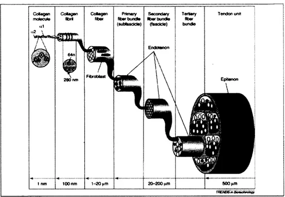
The hierarchical structure of a healthy tendon is shown in Figure 2-1. Tropocollagens (collagen molecules) unite into collagen fibrils, collagen fibers2, subfascicles (p rim a ry bundles), and fascicles (secondary bundles). Several fascicles constitute te rtia ry bundles (Liu, Ramanath et al. 2008).
2 There have been some misunderstandings in literature regarding using "fiber" and "fibril". In some texts, these two terms have been used interchangeably.
Primary, secondary and te rtia ry fiber bundles are covered by a th in layer called endotenon and the whole tendon is surrounded by another thin layer called epitenon (Sharma P 2006).
Tendon cells (tenocytes), w hich are responsible for production o f collagen fibers and of ground substance, are located between fibers. They have an elongated shape when observed in the tendon's longitudinal orientation (Margareta N ordin and L. 2001). Whereas in cross-section, they appear as star-shaped cells (C M McNeilly 1996).
Some structural crite ria to classify the quality include cell shape, collagen organization, cell-ECM interaction, cell density, etc. Methods could be divided in to three groups: qualitative, semi-quantitative, and quantitative w hich are introduced in the three follow ing sections.
We explain these methods, since they are used in clinical applications. Moreover, in our in vitro experimentations, we use these methods to compare the structu ra l qu ality o f different groups o f samples.
Collagen m olecule C otegen ftoer Prim ary ftoerbun cto (sufafasoda) 100 nm 1-20 pm Secondary fiberbun dte (fascicle) Tertiary fiber bundte EpHenon 280 run 20-200 am 500
Figure 2-1: Schematic structure of a normal tendon (Liu, Ramanath et al. 2008)
2.2.1 Qualitative characterization
One method fo r the characterizing tissue quality is histology, i.e. characterization o f tissue structure using microscopic images. Microscopic images are m a in ly from lig h t (optical) microscopy (OM), and electron microscopy (EM). They both have an objective lens to magnify the structures and are used in biology and m aterial science fields (Alberts B 1994). In OM, a photon beam is radiated to the objective lens to visualize the purpose structure, w hile in EM, the radiated beam is made up o f electrons (Keith Wilson 2005). This difference in the type o f radiated beams makes each microscope appropriate fo r special purposes. The electron microscope provides a much higher resolution and magnification than optical microscope. Therefore, to resolve very small objects, e.g. small molecules w ith approxim ate size o f 1 nm, EM should be used.
Table 2-1 demonstrates im po rta n t structural characteristics o f healthy and tendinopathic tendons (Xu 2008).
Table 2-1: Comparison of normal and tendinopathic tendon by microscopy (Xu 2008)
Findings Macroscopic Optical microscopy
(longitudinal sections) Electron microscopy (transversal sections) Normal tendon Brilliant white Fibroelastic Firm texture alignment
Organized parallel collagen bundles
Spindle shape tenocyte nuclei
Nuclei parallel alignment
Densely packed collagen fibers
Uniform in diameter and orientation of collagen fibers
Tendinopath ic tendon
Grey or brown
Tissue is thin, fragile and disorganized
Loose texture
Disorganized collagen bundle
Increased ground substance consisting of proteoglycan and glycosaminoglycan (GAG)
Large mucoid patches and vacuoles between fibers3 (Figure 2-2)
Round with darker-staining tenocyte nuclei
Markedly increased number of tenocyte nuclei with loss of parallel alignment
Increase of vascular and nerve ingrowths
Angulation (Figure 2-3), bubble formation (Figure 2-4) of collagen fibers
Variation in the diameters and orientation of collagen fibers
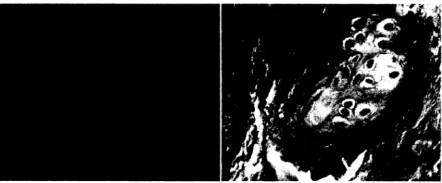
Hypoxic 4(Figure 2-5) changes in tenocyte (lipid vacuoles5, enlarge lysosomes 5and degranulated endoplasmic retinaculum7 (Figure 2 -5 ))
3 One type of tendon degeneration. Accumulation of large mucoid patches and vacuoles filled with GAGs and proteoglycans between collagen fibers Peter A. Huijbregts, M., MHSc, PT Scott E. Smith, MSc, OT (1999). "Tendon Injury: A Review." The lournal of Manual & Manipulative Therapy 7: 71-80.
4 One type of tendon degeneration which is deprivation of adequate oxygen. 5 Lipid accumulation
6 Lysosomes are one of subcellular components which contain waste-breaking enzymes.
7 The endoplasmic reticulum (ER) is a continuous membrane which has many different functions such as : translocation of proteins across the ER membrane; the integration of proteins into the membrane; etc. Gia K. Voeltz, M. M. R. (2002). "Structural organization of the endoplasmic reticulum." EM BO reports 3(10): 944-950.
Figure 2-2: Light microscopy of a ruptured Achilles tendon from a twenty-nine-year-old woman. The arrow shows the thin and fragile collagen fibers, and the star shows the large vacuoles among the fibers.
(Kannus P 1991)
Figure 2-3: Transmission electron microscopy of a ruptured extensor pollicis longus tendon from a sixty- four-year-old woman. The arrow shows the angulation of the collagen fibrils (Kannus P 1991)
Figure 2-4: Transmission electron microscopy of a ruptured Achilles tendon from a thirty-four-year-old man. The arrow shows bubble formation involving some fibrils. (Kannus P 1991)
Figure 2-5: Transmission electron microscopy of a ruptured Achilles tendon from a thirty-three-year-old man. The image shows a high-level hypoxic degenerated tenocyte which includes lipid vacuoles (LV),
2.2.2 Sem i-quantitative characterization
Most tissue histological characterization studies do n o t quan tify the properties, b u t use description o f the changes from healthy to damaged tissue histology (Nicola M a ffiilli 2008). The method o f description o f structural changes can lead to inadequacy in classifying the different levels o f tissue injury. This misunderstanding and uncertainty about tissue condition may result in inconsistent diagnosis between specialists (Nicola M affulli 2008).
To avoid this uncertainty in diagnoses by different specialists, some scoring methods have been suggested to be used to classify the tendinopathic tendons (Nicola M affulli 2008). These methods were developed fo r clinical applications so they score the level o f tendinopathy. There are tw o kinds o f such scoring systems: M ovin and Bonar systems. In each method they score specific variables which evaluate various aspects o f tissue quality. Both o f these methods were created fo r classifying OM images o f longitudinal tendon section.
The variables included in the M ovin scaling method are: (1) fib e r structure, (2) fib e r arrangement, (3) rounding o f the nuclei, (4) regional variations in cellularity, (5) increased vascularity, (6) decreased collagen stainability, and (7) hyalinization. For each variable, the score could be 0 (norm al tendon) to 3 (the m ost abnormal appearance detectable) (Longo, Franceschi et al. 2008). Therefore the total score of each sample could vary between 0 (norm al tendon) to 21 (the most severe abnorm ality detectable).
For example (Longo, Franceschi et al. 2008) used M ovin scoring method to investigate the histological changes o f Supraspinatus tendon in ro ta to r c u ff tears. They classified light micrographs o f norm al and injured tendons based on M ovin scoring scales (Figure 2-6 to Figure 2-9).
Figure 2-6: Hematoxylin and eosin stain of a control supraspinatus tendon in a 71-year-old
man. Fiber structure, 0; fiber arrangement, 0; rounding of the nuclei, 0; regional variations in
cellularity, 1; increased vascularity, 0; decreased collagen stainability, 0; hyalinization,
0. Total score: 1
Figure 2-7: Hematoxylin and eosin stain of supraspinatus tendon harvested from the intact middle portion of the tendon between
the lateral edge of the tendon tear and the muscle-tendon junction in a 62-year-old woman. Fiber structure, 2; fiber arrangement,
2; rounding of the nuclei, 3; regional variations in cellularity, 2; increased vascularity, 0; decreased collagen stainability,
1; hyalinization, 0. Total score: 10
^ « S t !
m i
Bi
H m
Figure 2-8: Hematoxylin and eosin stain of supraspinatus tendon harvested from the intact
middle portion of the tendon between the lateral edge of the tendon tear and the musdetendon junction in a 53-year-old man.
Fiber structure, 2; fiber arrangement, 2; rounding of the nuclei, 1; regional variations in
cellularity, 1; increased vascularity, 1; decreased collagen stainability, 2; hyalinization,
0. Total score: 9
Figure 2-9: Hematoxylin and eosin stain of supraspinatus tendon harvested from the intact middle portion of the tendon between
the lateral edge of the tendon tear and the muscletendon junction in a 59-year-old man.
Fiber structure, 2; fiber arrangement, 2; rounding of the nuclei, 1; regional variations
in cellularity, 2; increased vascularity, 3; decreased collagen stainability, 2;
The variables included in the Bonar scaling method are: (1) tenocytes; (2) ground
Table 2-2: Semi-quantitative scoring (Bonar scale) (Cook, Feller et al. 2004)
Grade 0 1 2 3
Tenocytes Inconspicuous elongated spindle shaped nuclei with no obvious cytoplasm at light microscopy Increased roundness: nucleus becomes more ovoid to round in shape without conspicuous cytoplasm Increased roundness and Size; the nucleus is round, slightly enlarged and a small amount of cytoplasm is visible Nucleus is round, large with abundant cytoplasm and lacuna formation (chondroid change) Ground substance
(alcian blue and colloidaliron
stains)
No stainable ground substance
Stainable mucin between fibers but bundles still discrete
Stainable mucin between fibers with loss of clear demarcation of bundles Abundant mucin throughout with inconspicuous collagen staining
Collagen (w ith and without polarized light) Collagen arranged in tightly cohesive well demarcated bundles with a smooth dense bright homogeneous polarization pattern with normal crimping Diminished fiber . polarization; separation of individual fibers with maintenance of demarcated bundles Bundle changes; separation of fibers with loss of demarcation of bundles giving rise to expansion of the tissue overall and clear loss of normal polarization Pattern Marked separation of fibers with complete loss of architecture Vascularity Inconspicuous blood vessels coursing between bundles Occasional cluster of capillaries, less than 1 per 10 high power fields
1-2 clusters of capillaries per 10 high power fields
Greater than 2 clusters per 10 high power fields
substance; (3) collagen; and (4) vascularity. For each variable the score could be 0 (normal tendon) to 3 (the most abnorm al tendon detectable)(Nicola M affulli 2008). Therefore, the total score o f each sample could vary between 0 (norm al tendon) and 12 (the most severe abnorm ality detectable) (Table 2-2).
Using either the Movin or Bonar method leads to sim ila r results (Nicola M affulli 2008). By using either o f these methods, one is capable of quantifying the appearance of normal and tendinopathic tendon.
2.2.3 Quantitative characterization
There are some methods to quantify tissue structural properties. Using image processing techniques, one could obtain various measurements in images. For example, (Parent G, Langelier et al. 2011) measured space between fibers using Vision assistant software, (Version 7.1 National Instrum ent, Austin, TX, USA). They chose three regions of interest (ROIs) fo r each o f th e ir microscopic images (OM) and found the spaces between the fibers by contrast d ividing the objects into 2 categories: fib ril (black), and space (red). Space density was calculated by dividing the number o f red pixels by the number o f pixels in the image. They also evaluated the mean area o f the spaces, i.e. average o f the num ber of connected red pixels using the same software
They have also calculated fib ril density through transmission electron microscopy (TEM) and scanning electron microscopy (SEM) images.by chosing three ROIs in each image, and finding the fib ril pixels from background pixels using b ottom -hat filte rin g in Matlab (Version 7.5, Mathworks, Natick, USA). F ib ril density was calculated by dividing the number o f fib ril pixels by the total num ber o f image pixels.
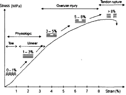
2.3 Tendon mechanical properties
The stress-strain diagram in Figure 2-10 shows the mechanical behavior o f ra t tail tendons from a study o f (Amoczky, Lavagnino et al. 2007). Although values are specified fo r ra t ta il tendons, the general trends are the same for all kinds o f tendons. As can be seen in this figure, the "physiologic range" includes a toe region and p a rt of linear region. In this range, collagen fib rils begin to un-crimp and then they are
stretched by increasing load. Near the end o f the lin e a r region, isolated collagen fib rils begin to fail (Arnoczky, Lavagnino et al. 2007), and stress-strain curve enters to the region specified as “overuse in ju ry" region which is the region o f isolated collagen fib ril microdamages. In this region, straightening o f collagens is continued and in te rfib rilla r sliding and shear between collagen fib rils produces a non-linear load-deform ation behavior o f the tendon (Arnoczky, Lavagnino et al. 2007). Some collagen fib rils are damaged before others u n til a complete "tendon rup tu re" occurs in the last region of stress-strain curve (Arnoczky, Lavagnino et al. 2007).
Therefore, collagen fibers properties, th e ir crim p structure and their failure level, play a significant role in biomechanical behavior o f tendon w hich are to support and tra n sm it tensional load.
Tendon rupture Stress ( M P a ) _ Overuse injury
5- 8% Physiologic Linear Toe 1- 3% 0- 1% 2 6 7 1 3 4 5 8 Strain (%)
Figure 2-10: Stress-strain curve demonstrating the mechanical properties of normal tendon (Arnoczky, Lavagnino et al. 2007)
A tendon is not a pure elastic m aterial and displays viscoelastic behaviors. It has the properties o f both elastic and viscous material, w hich means the rate o f loading has an effect on tendon behavior. Also, energy is lost during strain-loading, and thus the loading and unloading curves w ill not be identical, a phenomenon called "hysteresis".
Two other phenomena originating from viscoelasticity are stress relaxation and creep (Margareta Nordin and L. 2001). Stress relaxation is reducing (relaxing) stress under constant strain, whereas creep is increasing strain d u rin g a constant load. Related inform ation w ill be presented in Chapter 3.
Under certain conditions o f loading and tissue quality, tendons also show viscoplastic behavior. The tendon is not able to get back to its in itia l length after unloading since it undergoes some plastic/perm anent deformations. We w ill explain this in fu rth e r detail in Chapter 3.
Up to now, compositional, structural, and mechanical properties o f tendon have been introduced since a tendon is a live system w hich progresses w ith tim e, these properties can undergo some changes. Some factors which can affect tendon properties are aging, diseases, and changes in environm ental loading. As m entioned earlier, cells represent the active component o f tendons, and therefore tendon progression depends on cell response to these factors, among w hich only environm ental loading w ill be discussed in this thesis. It is im po rta n t to know how cells sense the environm ental loading and how they respond to it. In the follow ing section, this subject w ill be briefly described.
2.4 Mechanobiology and m echanotransduction
Since many tissue disorders result from mechanical overloading, i t is very im po rta n t to study the relationship between mechanobiological stim ulation and tissue progression. Mechanobiology discusses these issues. In other words, mechanobiology is the science which studies the rem odeling o f the tissues in response to physical loading (van der Meulen and Huiskes 2002). Tissues are constructed and remodeled by cells. Therefore, mechanotransduction is the mechanisms by w hich loading could be sensed by the cells.
Examples o f mechanotransduction mechanisms are: cell deform ation, nucleus deformation, cytoskeleton, stretch activated channels, and prim ary ciliu m (Wang 2006). Through these mechanisms, mechanical stim ulation is converted in to biochemical signals. Mechanical stim ulation, applied to the ECM can damage i t I t can fu rth e r undergo more damages o r can be repaired by cellular activity Biochemical signals,
resulted from converting mechanical stim ulation through mechanotransduction mechanisms, are detected by cells. Cells can respond differently depending on stim ulation, they can “ repair" tissue by producing and m aintaining the collagens (Devkota, Tsuzaki et al. 2007; Kjaer, Langberg et al. 2009) o r cause "degradation" by secreting o f collagen degradable enzymes i.e. proteases (Arnoczky, Lavagnino et al. 2007; Devkota, Tsuzaki et al. 2007; Cousineau-Pelletier and Langelier 2009). Therefore, mechanobiology plays a m ajor role in establishing tissue homeostasis.
Mechanobiology and mechanotransduction w ill be discussed in more depth in Chapter 3.
2.5 L iteratu re re vie w o f characterization m ethods
Although mechanical properties play an im p o rta n t role in tendon functio n ality (Duenwald-Kuehl, Lakes et al. 2012), compositional, and structural properties are also o f great value in providing complementary inform ation on tissue quality. In fact, compositional, structural, and mechanical properties are in a close relation, therefore, studying tendon biomechanical and mechanobiological behavior, not o n ly is im p o rta n t fo r characterizing mechanical properties, , b u t it is also im portant fo r characterizing compositional and structural properties.
Tissue quality can take different values depending on the properties o f the tissue. For example, tissue could be healthy vs. damaged. It should be noted th a t tissue quality affects the cellular response o f the tissue. This w ill be explained in Chapter 3.
Investigating tissue quality is im po rta n t to evaluate tissue progression over time. For example, to evaluate the efficiency o f a training protocol we need to compare the tissue quality before and after the training. Investigating tissue quality is therefore unavoidable for fu rth e r studies o f tendon mechanobiology.
In the follow ing section, litera ture introducing methods w hich have been used to characterize tissue properties w ill be reviewed.. Presented articles are divided into three categories corresponding to the type o f experim ent conducted: 1) in vitro w ith live cells, 2) in vitro w ith dead cells, and 3) in vivo. For each article, a table which
summarizes the tissue qu ality characterization techniques fo r tissue quality is presented. The tissue characterization table is divided into three categories: mechanical characterization, structural characterization, and com positional characterization. Each category, in turn, is divided into tw o subcategories, In the firs t subcategory, the conducted test w ill be explained (e.g. in-line, destructive). In the second subcategoiy, the inform ation related to data acquired from the conducted test w ill be presented. The hypothesis and results o f the article are also explained briefly. A t the end o f this section, a discussion about these techniques is provided.
Some o f the expressions used to describe mechanical tests which m igh t be less fa m ilia r to the reader, are defined here:
• In-line characterization: the mechanobiological experim entation (to examine the im pact o f a loading regime on tissue progression), and characterization testing (to determ ine tissue quality) are conducted in the same apparatus. Therefore, the inform ation about tissue mechanical properties are available at regular tim e intervals during the experim ent w ith o u t changing the tissue from one apparatus to another.
• Non-destructive characterization: the characterization does n o t lead to tissue damage or failure. Therefore, other characterization tests could be conducted after non-destructive characterization.
2.6 In vitro experim entation on tendons w ith liv e cells
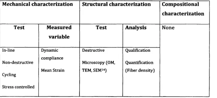
Article 1. Distributing a fixed amount o f cyclic loading to tendon explants over longer periods induces greater cellular and mechanical responses (Devkota, Tsuzaki etal. 2007)
Hypothesis/Objective:
1. Magnitude: High-magnitude cyclic loading w ould cause in ju ry, b u t not low - magnitude cyclic loading.
II. Duration: For a fixed num ber o f cyclic loading on tendon, the longer the period o f loading, the greater the mechanical and cellular responses o f tendon.
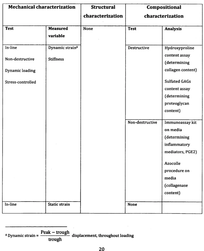
Table 2-3: Summary of characterization tests conducted in this article
Mechanical characterization Structural Compositional characterization characterization
Test Measured
variable
None Test Analysis
In-line Dynamic strain8 Destructive Hydroxyproline
Non-destructive Stiffness
content assay (determining
Dynamic loading collagen content)
Stress-controlled Sulfated GAGs
content assay (determining proteoglycan content)
Non-destructive Immunoassay kit on media (determining inflammatory mediators, PGE2) Azocolle procedure on media (collagenase content)
In-line Static strain None
Peak - trough ,
8 Dynamic strain = ---displacement, throughout loading
Non-destructive Creep Stress-controlled Off-line9 Destructive Failure Strain-controlled Strain at failure Energy density10 None Results
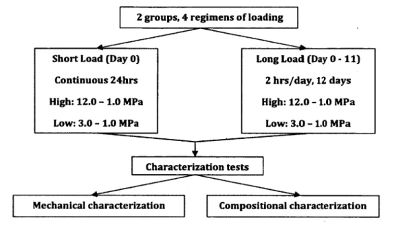
The loading was conducted on tw o groups and in four regimens. Figure 2-12 shows the schematic o f the loading regimens.
• Dynamic and static strain accumulations were larger in "High- m agnitude/Long-loading" compared to "High-m agnitude/Short-loading" groups.
• Static strain accumulation was greater in "Low -m agnitude/Long-loading" compared to "Low -m agnitude/Short-loading” groups. These results show the effect o f loading tim e on the tissue response.
9 Failure test was performed on dead tissues, since the tissues were first frozen (at -9"C) and then thawed. 10 The amount of energy tendons absorb before failing.
Characterization tests
Mechanical characterization Compositional characterization 2 groups, 4 regimens o f loading
High: 12.0 - 1 . 0 MPa Short Load (Day 0)
Low: 3.0 - 1.0 MPa Continuous 24hrs
Long Load (Day 0 - 1 1 )
High: 12.0 -1 .0 MPa 2 hrs/day, 12 days
Low: 3.0 -1 .0 MPa
Figure 2-12: Schematic of the loading and assaying of the tendon
• However fo r dynamic strain, there was no difference between "Low- m agnitude/Long-loading" and "Low-m agnitude/Short-loading" groups. Taking into account this result, along w ith the result from static strain, suggests that tim e is not the only factor affecting tissue response. As it is observed from mechanical and com positional analyses, loading magnitude also plays an im portant role in tissue response.
• The results from failure test did not show a definite effect of cellular response on tendon properties. The properties were either tim e dependent o r load- magnitude dependent but not both. The "High-m agnitude/Long-loading" did not consistently produce the most in fe rio r results expected. The authors suggest the effect o f the difference o f cross-sectional areas o f tendons as the reason o f these uncertain failure results.
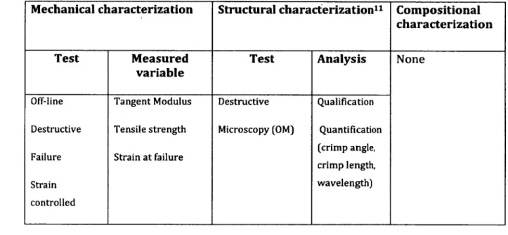
Article 2. Biomechanical response o f collagen fascicles to restressing after stress deprivation during culture. (Yamamoto, Kogawa et al. 2007)
H yp o th e sis/O b je ctive :
Restressing w ill im prove the decreased properties o f fascicles resulting from stress shielding.
Table 2-4: Summary of characterization tests conducted in this article
M echanical c h a ra c te riz a tio n S tru c tu ra l c h a ra c te riz a tio n 11 C o m p o s itio n a l c h a ra c te riz a tio n
Test M easured
v a ria b le
Test A n a lysis None
Off-line Destructive Failure Strain controlled Tangent Modulus Tensile strength Strain at failure Destructive Microscopy (OM) Qualification Quantification (crimp angle, crimp length, wavelength) Result:
• The decrease o f mechanical properties, represented by tangent m odulus and tensile strength, was stopped and, in m ost cases reversed by applying stress after stress deprivation b u t none o f them im proved to their norm al level.
• Structural characterization results were also consistent w ith mechanical characterization results. The crim p m orphology o f fascicles was not recovered to original levels, after restressing.
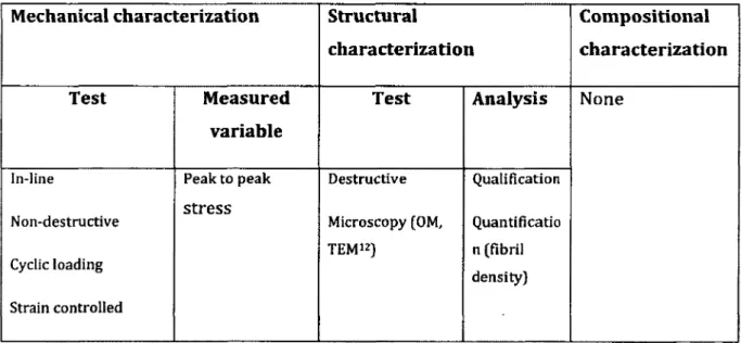
Article 3. Relative contribution o f mechanical degradation, enzymatic degradation, and repair o f the extracellular m atrix on the response of tendons when subjected to under- and over- mechanical stimulations in vitro. (Cousineau-Pelletier and Langelier 2009)
Hypothesis/Objective:
Investigating the contribu tio n of the three sub-processes o f tendon response (Repair, Mechanical degradation, and Enzymatic degradation) w hen subjected to cyclic mechanical loading.
Tendon mechanobiological response (TMR) could be approxim ated as:
T M R = R - M D - ED; where
R: Repair, MD: Mechanical degradation, ED: Enzymatic degradation.
Table 2-5: Summary of characterization tests conducted in this article
Mechanical characterization Structural
characterization
Compositional characterization
Test Measured
variable
Test Analysis None
In-line Non-destructive Cyclic loading Strain controlled Peak to peak stress Destructive Microscopy (OM, TEM12) Qualification Quantificatio n (fibril density)
Result:
• In the absence o f R and ED, i.e. when TMR is represented by -M D : the results from mechanical characterization, showed a fast decrease in peak stresses during experim entation tim e w ith o u t any increase indicating tendon damage. Structural analyses supported mechanical data since they showed loosely packed and w avy collagen structure.
• In the absence o f ED, i.e. when TMR is represented by R-MD, the results from mechanical characterization showed an overall increase in peak stresses during experim entation tim e indicating tendon im provem ent. Structural analyses are in w ell correspondence to mechanical results, since they show dense and w ell- oriented collagen structure.
• In the presence o f all three sub-processes, i.e. when TMR is represented by R- MD-ED, the results from mechanical characterization showed a decrease o f peak stresses after the in itia l increase. Structural analyses were consistent w ith these results since they showed disorganized collagen structure.
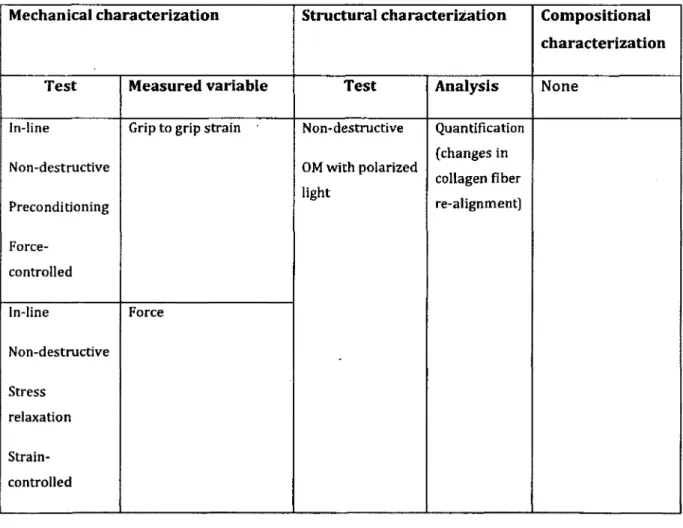
Article 4. Effect o f Preconditioning and Stress Relaxation on Local Collagen Fiber Re-Alignment- Inhomogeneous Properties o f Rat Supraspinatus Tendon13. (M iller KS 2012)
Hypothesis/Objective:
I. The greatest fiber re-alignm ent w ill occur in the toe-region at ram p-to-failure test bu t some fiber re-alignm ent w ill also occur during preconditioning.
II. Disorganization in collagen fib e r w ill occur during stress-relaxation test.
III. Mechanical properties and in itia l collagen fib e r alignm ent are greater at midsubstance o f tendon than tendon-to-bone insertion site.
Table 2-6: Summary of characterization tests conducted in this article
Mechanical characterization Structural characterisation Compositional characterization
Test Measured variable Test Analysis None
In-line
Non-destructive
Preconditioning
Force-controlled
Grip to grip strain Non-destructive
OM with polarized light Quantification (changes in collagen fiber re-alignment) In-line Non-destructive Stress relaxation Strain-controlled Force
In-line Both destructive and non destructive Ramp-to-Failure Strain-controlled Stiffness Strain at failure Stress Result:
• The greatest fiber re-alignm ent occurred during preconditioning and then at toe- and linear regions o f the ram p-to-failure te s t
• No collagen fib e r re-alignm ent observed during stress-relaxation test.
• Lower m oduli, more disorganizations and higher strains at insertion site than tendon midsubstance indicate th a t mechanics and structure o f the tissue d iffe r at different tissue locations, i.e. the tissue is n o t homogeneous.