R E S E A R C H A R T I C L E
Open Access
Central modulation in cluster headache patients
treated with occipital nerve stimulation: an
FDG-PET study
Delphine Magis
1†, Marie-Aurélie Bruno
2†, Arnaud Fumal
3, Pierre-Yves Gérardy
4, Roland Hustinx
5, Steven Laureys
6,
Jean Schoenen
7*Abstract
Background: Occipital nerve stimulation (ONS) has raised new hope for drug-resistant chronic cluster headache (drCCH), a devastating condition. However its mode of action remains elusive. Since the long delay to meaningful effect suggests that ONS induces slow neuromodulation, we have searched for changes in central pain-control areas using metabolic neuroimaging.
Methods: Ten drCCH patients underwent an18FDG-PET scan after ONS, at delays varying between 0 and 30 months. All were scanned with ongoing ONS (ON) and with the stimulator switched OFF.
Results: After 6-30 months of ONS, 3 patients were pain free and 4 had a≥ 90% reduction of attack frequency (responders). In all patients compared to controls, several areas of the pain matrix showed hypermetabolism: ipsilateral hypothalamus, midbrain and ipsilateral lower pons. All normalized after ONS, except for the hypothalamus. Switching the stimulator ON or OFF had little influence on brain glucose metabolism. The perigenual anterior cingulate cortex (PACC) was hyperactive in ONS responders compared to non-responders. Conclusions: Metabolic normalization in the pain neuromatrix and lack of short-term changes induced by the stimulation might support the hypothesis that ONS acts in drCCH through slow neuromodulatory processes. Selective activation in responders of PACC, a pivotal structure in the endogenous opioid system, suggests that ONS could restore balance within dysfunctioning pain control centres. That ONS is nothing but a symptomatic
treatment might be illustrated by the persistent hypothalamic hypermetabolism, which could explain why autonomic attacks may persist despite pain relief and why cluster attacks recur shortly after stimulator arrest. PET studies on larger samples are warranted to confirm these first results.
Background
Cluster headache (CH) is one of the most painful pri-mary headaches and is characterized by attacks of severe unilateral periorbital pain associated with ipsilateral autonomic features [1]. About 10% of patients have, or develop over time, a chronic form (CCH) [2] charac-terised by recurrent attacks for at least 1 year without remissions or with remissions of less than 1 month [1]. About 1% of CCH patients become drug-resistant
(drCCH) to most prophylactic drug treatments and fulfil published criteria for intractable headaches [3].
CH is the most prevalent member of the so-called trigeminal autonomic cephalalgias (TACs), which include paroxysmal hemicrania, SUNCT (Short-lasting Unilateral Neuralgiform headache with Conjunctival injection and Tearing) and probably hemicrania conti-nua [4]. Neuroimaging studies have provided new insight into the pathophysiology of these disorders. Besides non-specific changes in activity of brain areas belonging to the pain matrix like the anterior cingular cortex (ACC), insula(e), and thalamus, TACs are asso-ciated with ictal activation of ipsilateral posterior hypothalamus (CH, SUNCT) or dorsal pons
* Correspondence: jschoenen@ulg.ac.be † Contributed equally
7Headache Research Unit and Neurology Department, University of Liège,
CHR Citadelle, Boulevard du 12ème de Ligne 1, 4000 Liège and GIGA-Neurosciences, University of Liège, Belgium
Full list of author information is available at the end of the article
© 2011 Magis et al; licensee BioMed Central Ltd. This is an Open Access article distributed under the terms of the Creative Commons Attribution License (http://creativecommons.org/licenses/by/2.0), which permits unrestricted use, distribution, and reproduction in any medium, provided the original work is properly cited.
(hemicrania continua) or contralateral posterior hypothalamus (paroxysmal hemicrania) which may be more specific and disease-related [5].
As a consequence, deep brain stimulation (DBS) tar-geting the posterior hypothalamus was proposed for drCCH and was found to be more effective than any previously used invasive therapy [6,7]. However hypothalamic DBS is not a riskless procedure [7] and less invasive methods were thus explored. Among them, occipital nerve stimulation (ONS) had comparable effi-cacy to hypothalamic DBS, except for slower onset of action [8,9].
The mechanisms by which ONS improves drCCH remain unclear. In a study of ONS in drCCH we found no significant change in pain thresholds, which argues against a diffuse analgesic effect [8]. It was speculated that ONS might exert its action by decreasing excitabil-ity of second order nociceptors in trigeminal nucleus caudalis on which converge cervical, somatic trigeminal and visceral trigeminovascular afferents [10,11]. Yet, the nociception-specific blink reflex, mediated by spinal tri-geminal nucleus interneurons, was increased rather than decreased in our study of ONS in drCCH [8] and it remained unchanged in healthy subjects after short low frequency transcutaneous stimulation of the greater occipital nerve [12]. A more likely explanation for the therapeutic effect of ONS in headache including drCCH is the induction of slow neuromodulatory changes in brain regions belonging to the pain matrix or in centres more specifically involved in CCH pathophysiology. Hence, chronic migraine patients treated with ONS [13] show significant blood flow increases on H215O-PET in dorsal rostral pons, anterior cingulate cortex and cuneus, directly correlated to pain scores, and in left pulvinar, inversely correlated to such scores. Dorsal pons activation persisted after ONS, supporting the role of this structure in migraine pathophysiology [13].
So far, functional imaging studies have not been per-formed in ONS-treated drCCH patients. We perper-formed such a study focusing on the pain matrix, but also on hypothalamus and brainstem that seem more specifically involved in the pathophysiology of TACs. We enrolled patients from the published cohort [8] and newly implanted ones. We used 18-fluorodeoxyglucose-posi-tron emission tomography (18-FDG-PET) in order to detect long term activity modulation.
Methods
Subjects
We studied 10 patients with drCCH (9 males and 1 female, mean age at implantation 44.2 ± 9.9 years SD). Inclusion criteria were: CCH for at least 2 years, daily attacks by history, side-locked attacks from the begin-ning, resistance to drug treatment according to expert
consensus guidelines [3] and absence of associated dis-abling organic or psychiatric disorder. Five patients had left-sided, 5 right-sided attacks. At the time of the study, all patients were taking one or several of the following preventive drugs: verapamil (n = 9), lithium carbonate (n = 6), methylprednisolone (n = 2), methysergide (n = 2), melatonine (n = 1), gabapentine (n = 1). None took analgesics, in particular opioids.
Patients were recruited in two phases (1stand 2nd group), with written informed consent. Approval of the local Ethics Committee for ONS in drCCH was first obtained for 5 patients (EUDRACT-2004-004551-19). Because of the favourable results in ONS-treated patients after a 16 months follow-up, we requested Ethics Com-mittee approval for a protocol amendment allowing us to recruit 6 supplementary patients who were implanted and agreed to undergo PET before and after surgery. At the same time, patients of the 1stgroup were also asked to participate in the PET study and 4 of them accepted (the last patient of group 1 had been explanted [8]). Surgical and stimulation procedure
Surgical procedure and stimulation protocols have been described previously [8]. We used unilateral subcuta-neous implantation of paddle style stimulating leads with 4 electrode plots (Medtronic 3587A Resume II®; Medtronic Inc., Minneapolis, USA) via a retromastoid C1-2-3 approach [14] and Medtronic Itrel III® or Synergy®stimulators.
Stimulation protocols were adapted using a program-ming matrix [8] such as to induce paraesthesias in the largest possible occipital territory. The clinical evolution of patients was monitored with cluster headache paper diaries.
18-FDG-PET study design Study groups
In the 1stgroup, patients (n = 4), underwent a PET ses-sion after 24 to 30 months of ONS. All patients but one belonging to the 2ndgroup (n = 6) were scanned before implantation (baseline). In both groups, for each session after implantation, patients were scanned with the sti-mulator switched on (ON) and off for 3 days (OFF). This period was arbitrarily fixed in line with our pre-vious observation of headache recurrence within 2 days on average after switching off the stimulator [8]. They underwent 2 more pairs of scans after 1 and 6 months of ONS. None of the patients had a cluster attack dur-ing PET, nor within the 12 hours preceddur-ing or followdur-ing the procedure.
Data collected in patients were compared to a pool of 39 drug-free healthy volunteers (HV) (18 males, 21 females, mean age 45 ± 16 years SD) without headache history.
Data acquisition
PET data were obtained on a Siemens CTI 951 16/32® scanner (Siemens, Erlangen). Data were corrected for attenuation and background activity. Resting cerebral metabolism was studied after intravenous injection of 5-10 mCi (185-370 MBq) [18F]fluorodeoxyglucose (FDG). Subjects were scanned in a dark room, with minimal environmental noise.
Statistical analysis (Cyclotron Research Centre, Liège) Because we selected CH patients with side-locked uni-lateral attacks and as there was no side shift during the observational period, we flipped the PET scans of the patients with right-sided symptoms in the axial plane so that we could analyze all subjects together (all “left-sided”). We used a binary classification of patients responding or not to ONS with an arbitrary, but clini-cally relevant, cut-off point for responders set at 50% decrease in attack frequency. Given the data from pre-vious functional imaging studies, we conducted our ana-lysis with an a priori hypothesis towards regions known to be involved in CH and other TACs [5], areas which are modulated by ONS in chronic migraine [13] and areas belonging to the pain matrix.
Data were analysed using statistical parametric map-ping (SPM8 version; Wellcome Department of Cognitive Neurology Institute of Neurology, London, UK; http:// www.fil.ion.ucl.ac.uk/spm) implemented in MATLAB (version 7.1, MathworksInc., Sherborn, MA). Images were spatially normalized into a standard stereotactic space using a symmetrical MNI (Montreal Neurological Institute) PET template [15] and smoothed using a 14 mm full-width-half-maximum (FWHM) isotropic kernel [16]. T-test was used with a significance level set at p < 0.001 uncorrected (p < 0.05 FDR).
The first design matrix included the scans of the 39 HV, of the 4 patients of the 1stgroup performed 24 to 30 months post-ONS and of the 6 patients of the 2ndgroup, scanned before implantation (baseline), 1 month and 6 months post-ONS. Our first analysis identified brain regions with a significant hyper- or hypometabolism in drCCH patients as compared to HV independent on time of scanning or ONS stimulation. Then, we looked for brain regions showing short term ONS-induced (ON ver-sus OFF) increase or decrease in metabolism indepen-dent on delay since ONS implantation, in the early post-ONS phase (scans obtained after 1 month) and in the late post-ONS phase (scans obtained after 6 and 24-30 months). Finally, we compared metabolic activity mea-sured during baseline, early phase (1 month) and late phase (≥ 6 months) post-ONS (independent of ONS sti-mulator settings) searching for progressive increases and decreases in metabolism over time.
In a second design matrix we searched for differences between the subgroups of 7 responders and 3
non-responders. Here, we included only the PET data obtained in the late phase (≥ 6 months; both with sti-mulator ON and OFF) and the HV scans, searching for regions with metabolic differences between the two groups (i.e., increased metabolism - as compared to HV - present in responders but not in non-responders).
For all analyses, the resulting set of voxel values for each contrast, constituting an SPM of the t-statistics (SPM{t}), was transformed to the unit normal distribu-tion (SPM{Z}) and thresholded at p = 0.001. All group results were thresholded at false discovery-corrected p < 0.05, corrected for the whole brain volume. For differ-ences between responder and non-responder subgroups, results were corrected for multiple comparisons within the regions of interest identified during the previous whole group analyses by employing a small volume cor-rection (10 mm radius sphere).
Results
Clinical outcome
Clinical data of patients and changes in their attack fre-quency after various durations of ONS are summarized in table 1. All patients but one in the 1stgroup (N = 4, 24-30 months follow-up) improved after ONS: one patient was pain free; 2 patients had a 90% and 93% reduction in attack frequency; the “non-responder” patient had a 25% improvement. In the 2ndgroup (N = 6), at 6 months follow-up, 3 patients were pain free and one was improved by 90%; these 4 patients already reported significant improvement after 1 month of ONS. The 5th patient only had a 33% reduction in attack frequency while in the 6thpatient, attack frequency was slightly increased. According to the 50% cut-off criter-ion, 7 patients were thus considered responders and 3 non-responders to ONS for the binary analysis. During the 3-day period of ONS interruption, only one respon-der had recurrence of attacks. In the 2nd group, all patients kept the same preventive drug treatment during the follow-up scans except for one responder (8) who was able stop all medications after 6 months.
PET results
The main areas of peak voxels where a metabolic change was found for the various comparisons are shown in table 2.
We first pooled all scans performed in drCCH patients and compared them with those of the HV. In comparison to HV, a significant hypermetabolism was found in anterior cingulate cortex (ACC), left hypothalamus, left pulvinar, left visual cortex, cerebellum and brain stem (left lower pons and midbrain) (figure 1). By contrast, a significant hypometabolism appeared in both sensori-motor areas.
There was no significant difference between scans per-formed with stimulator ON or OFF, regardless of ONS
Table 1 Patients characteristics and clinical outcome
Patients Age Cluster & ONS side
Average number of attacks/ last 4 weeks % reduction in attack frequency ONS responder BEFORE ONS 24-30 mths ONS 24-30 mths ONS Group 1 1 48 R 131.6 98.0 26 N 2 46 L 107.6 7.6 93 Y 3 32 R 32.4 3.2 90 Y 4 53 L 28.0 0.0 100 Y BEFORE ONS 1 month ONS 6 months ONS 1 month ONS 6 months ONS Group 2 5 31 R 28.0 7.2 0.0 74 100 Y 6 60 R 28.0 7.2 2.8 74 90 Y 7 47 L 112.0 92.4 116.4 17.5 -4 N 8 50 R 42.0 4.4 0.0 80 100 Y 9 31 L 56.0 2.0 0.0 97 100 Y 10 44 L 16.4 13.2 11.2 22 33 N
ONS: occipital nerve stimulation, R: right, L: left, Y: yes, N: no.
Table 2 Main statistical results and localization of peak voxels where cerebral metabolism was activated (>) or deactivated (<)
Analysis Brain region Talairach coordinates Z score of peak p FDR corrected
x y z
drCCH > HV ACC 12 40 -4 5 < 0.001
Perigenual ACC -8 28 -8 5.29 < 0.001
Midcingulate 12 20 30 4.07 0.003
Left visual cortex -10 -98 -8 3.43 0.010
Left pulvinar -16 -36 8 4.49 0.001
Left hypothalamus -2 -12 -16 2.71 0.013
Cerebellum -16 -36 46 4.28 0.002
Midbrain 2 -34 -4 3.53 0.008
Left lower pons -8 -32 -46 4.13 0.003
drCCH < HV R/L Sensorimotor -58 -34 -18 4.43 0.012 58 -18 -26 4.42 0.012 -4 -22 70 4.26 0.012 -25 -22 66 3.73 0.015 -6 -24 68 3.37 0.025 Right prefrontal 42 26 24 3.76 0.015
Base > ONS ACC -8 28 -8 4.66 0.003
Midcingulate 12 20 30 3.75 0.013
Left visual cortex -10 -98 -10 3.25 0.031
Left pulvinar -18 -38 6 3.8 0.012
Cerebellum 2 -42 -16 3.67 0.015
Midbrain 2 -40 -12 3.63 0.016
Left lower pons -8 -30 -44 4.40 0.024
Base < ONS R/L Sensorimotor -58 -34 -18 4.59 0.013
58 -18 -26 4.26 0.037
42 26 24 3.75 0.026
-4 -22 70 3.78 0.029
Resp > non resp Perigenual ACC -8 28 -8 4.01 0.002*
Coordinates are in the standardized stereotactic Montreal Neurological Institute space (mm). FDR = False discovery rate corrected. drCCH: drug-resistant chronic cluster headache; HV: healthy volunteers; ACC: anterior cingulate cortex, R: right, L: left. * FDR corrected in region of interest identified in whole group analysis.
duration (1, 6 or 24-30 months). For scans obtained in the late phase (≥ 6 months) we observed a hypermeta-bolism in the left frontal lobe (BA 10, uncorrected p = 0.03, x = -12, y = 46, z = 4) and the left lower brainstem (uncorrected p = 0.014, x = -6, y = -30, z = -36) when the stimulator was turned ON, but these results, reported for the sake of completeness, did not survive correction for multiple comparisons.
Over time, ONS changed glucose uptake in several brain areas (independent of the stimulator settings ON or OFF). The anterior cingulate, mid cingulate, left pulvi-nar, midbrain, lower pons, visual cortex and cerebellum
had decreased metabolism over time, i.e. they became less hypermetabolic when comparing baseline to the early phase (1 month) or the late phase (≥ 6 months). By contrast, metabolism increased over time in sensorimotor cortices, i.e. they became less hypometabolic, and it was not modified in the left hypothalamus (figure 2).
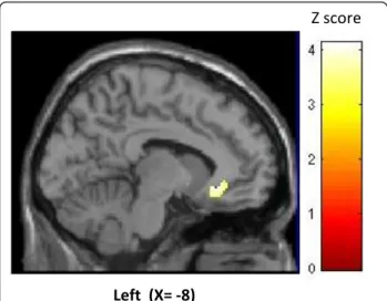
When finally comparing responders and non-respon-ders in the late phase, there was a significant hyperme-tabolism in previously identified perigenual ACC ipsilateral to the pain and stimulation side in responders (figure 3). No hypometabolic area differentiated respon-ders from non-responrespon-ders.
C
ACC
MB
LP
VC
ACC
C
P
H
Midline (X= 5)
Left (X= -12)
C
ACC
MB
LP
VC
ACC
C
P
H
Z score
Figure 1 Hypermetabolic areas in drCCH patients (all conditions: baseline - 1 month, 6 months, 24/30 months) compared with HV (p < 0.05 FDR corrected). Results are displayed on 2 sagittal sections of a normalized MRI template (through midline and left hemisphere). ACC: anterior cingulate cortex, C: cerebellum, MB: midbrain, LP: lower pons, VC: visual cortex, P: pulvinar, H: hypothalamus.
C ACC VC P ACC C B
Right (X= 12)
Left (X= -10)
C
ACC
VC
P
ACC
C
B
Z score
Figure 2 Areas progressively deactivated by ONS over time (p < 0.05 FDR corrected). Results are displayed on 2 sagittal sections of a normalized MRI template (right and left hemisphere). ACC: anterior cingulate cortex, C: cerebellum, B: brainstem, VC: visual cortex, P: pulvinar. White circle highlights hypothalamic area, which is not modified by the stimulation.
Discussion
The therapeutic outcome after ONS in 1stand 2ndgroups was overall similar, with good response as defined above in nearly 70% of patients [8,17]. A noticeable difference between the 2 groups was the latency to significant effi-cacy, which was on average shortened to 1 month in the 2nd group compared to 3 months in the 1stone. This remains compatible with a slow modulatory effect on the central nervous system. We ascribe faster efficacy in group 2 to a learning effect for investigators and to quicker optimisation of stimulator settings [8].
We will focus our discussion on the FDG-PET findings.
PET findings - interpretation
Metabolic pattern in drCCH compared to HV
The enhanced FDG uptake found ipsilaterally in hypotha-lamus of drCCH patients respective to HV is in line with the reports showing increased activity in this area with H215O-PET or fMRI during attacks [18-20]. However in our study all patients were scanned between attacks, i.e. at a time point when hypothalamic activation has not been reported yet. Another FDG-PET study of episodic CH comparing patients during and between bouts revealed no change in hypothalamic glucose uptake [21].
Areas belonging to the pain matrix like the cingulate gyrus or midbrain (periaqueductal grey - PAG) are clas-sically activated in various pain states including head-aches [5]. We also found activation in the cerebellum, in line with previous imaging studies showing consistent cerebellar activation across the spectrum of pain from visceral to somatic, acute to chronic [22]. Activation of cerebellar vermis and anterior lobe in drCCH may be
particularly strong due to dense somatotopically arranged trigemino-cerebellar connexions [22], but also because there are direct connections between the ven-tro-posterior hypothalamic area and the cerebellum as shown by MRI tractography in a drCCH patient treated with hypothalamic DBS [23].
The perigenual ACC (PACC) is of particular interest. Contrary to our results, it was found hypometabolic compared to HV in episodic CH [21]. However, when the authors compared the same patients during and between bouts, the PACC was hypermetabolic during the bout despite the absence of an attack at the time of scanning.
Lower pons activation has been described during attacks of hemicrania continua, but not of CH [5]. In hemicrania continua, dorsal pontine activation is ipsilat-eral, like in our study, while posterior hypothalamic acti-vation is observed on the opposite side, unlike in CH.
The pulvinar where we found ipsilateral activation in drCCH patients has not been a region of interest in CH before. Pulvinotomy and electrical stimulation of the pulvinar have been used successfully in the treatment of chronic pain in humans [24]. In functional neuroima-ging studies, pulvinar hypermetabolism is either asso-ciated with pain state [25] or with pain relief after various procedures [24], including ONS in chronic migraine [13].
We have no straightforward explanation for the increased metabolism of the ipsilateral visual cortex in our patients. Such activation has not been reported hitherto. Photophobia ipsilateral to the pain is a fre-quent attack-associated symptom in various TACs, in particular CH [26]. Like in migraineurs, the visual cortex of CH patients might thus be more sensitive to light sti-muli. This hypothesis seems unlikely, however, as all subjects were scanned in a dark room.
To summarize, the interictal FDG-PET hyperactive pattern of drCCH patients comprises areas reported to be hypermetabolic mainly during TAC attacks (ipsilat-eral hypothalamus and pons), but also during a bout of the disorder outside of an attack (perigenual ACC). Short-term changes associated with ONS
We found no significant differences between PET recordings performed with stimulator ON or OFF within a 72 hour delay. This finding is similar to Matharu et al.’s [13] observations in chronic migraine, except that they were not able to scan the patients OFF and pain-free because of an almost immediate recur-rence of pain after interrupting the stimulation. They concluded that central structures were not modulated in chronic migraine by ONS beyond the stimulation period.
Here, lack of short-term metabolic modification favours a slow neuromodulatory effect of ONS, as we Left (X= -8)
Z score
Figure 3 Activation of perigenual cortex in ONS responders vs. non responders after 6 to 30 months stimulation (p < 0.05 FDR corrected). Result is displayed on a left sagittal section of a normalized MRI template.
suspected before [8]. Interestingly, in CCH patients trea-ted with hypothalamic DBS, May et al. [27] found rapid metabolic changes with H215O-PET in various brain
structures involved in cluster headache and more gener-ally in the pain matrix within only 10 minutes of switch-ing the stimulator ON or OFF, but there were no clinical correlates suggesting that the therapeutic effect of DBS is also due to slow CNS changes.
Our subanalysis with a smaller significance level revealed a change in left lower brainstem metabolism. It might indicate a short term ONS effect in the trigemino-cervical complex. As expected from the neuro-anatomical connex-ions, the trigemino-cervical complex could relay stimula-tion to more rostral structures allowing for neuroplastic modulation of their activity. In comparison, a recent study of transcutaneous electrical nerve stimulation (TENS) in arthritic rats suggests that its antinociceptive effect is mediated by ascending activation of the opioid system ori-ginating in the ventrolateral PAG and projecting via the rostral ventro-medial medulla to the spinal cord [28]. Long-term changes associated with ONS
Hypermetabolism of most overactive areas in drCCH patients compared to HV decreased after ONS. This was particularly obvious for the anterior cingulate cortex, left pulvinar, left visual cortex, left lower pons, cerebellum and midbrain. Conversely, baseline hypometabolism of sensori-motor cortices increased after ONS. The note-worthy exception to these post-ONS metabolic changes is the ipsilateral hypothalamus. This is precisely the region activated during CH attacks on the side of the pain [18] and where increased gray matter density is found on voxel-based MRI between attacks [29]. Our findings in drCCH contrast with those by Sprenger et al [21] in episodic CH where no significant metabolic change was found in the hypothalamus, either outside or during the bout. If replicated, our data suggest that per-sistent hypothalamic activation is a hallmark of chronic CH. They may explain why attacks are non-remittent but also why some ONS-treated, pain-free patients still have autonomic attacks [8]. A similar conclusion was drawn for the dorsal rostral pons in chronic migraine following the finding of persistent activation in this area despite ONS-induced pain relief [13]. The persistence of an ipsi-lateral hypothalamic activation despite reduced attack frequency also confirms that ONS is no more than a symptomatic therapy, as already suggested by the recur-rence of attacks after interruption of the stimulation [8].
The metabolic changes observed after ONS could be due to the stimulation itself or to the reduction of attack frequency. Our protocol did not allow to favour either mechanism. However, despite the small number of non-responders in our study, some insight might be gained from the comparison of patients who clearly responded to ONS and those who did not.
Perigenual ACC (PACC) activation in responders
Comparison of ONS responders and non-responders revealed increased FDG uptake in the PACC of the for-mer. This area is of interest for several reasons. First, PACC plays a major role in central opioidergic pain con-trol system. It is selectively activated during analgesia induced by theμ-receptor agonists fentanyl and remifen-tanyl compared with placebo [30], providing evidence that opioidergic analgesia is mediated by activation of descending antinociceptive pathways. Second, PACC was found hypometabolic respective to HV in episodic CH, but its activity increased significantly during the bout [21]. Knowing the pivotal role of PACC in descending pain control, these authors hypothesized that deficient endogenous antinociceptive mechanisms between bouts might predispose CH patients to the disorder and to its recurrence. Concordantly, Sprenger et al [31], using PET with the opioidergic ligand [11C]diprenorphine, demon-strated an inverse linear relationship between the dura-tion of CH and opioid receptor availability in the rostral ACC (and ipsilateral hypothalamus). A recent case report by the same group of a drCCH patient in whom low dose levomethadone induced complete remission of attacks favours this hypothesis [32].
Compared to TENS that was shown to induce analge-sia through activation of a PAG-RVM-spinal cord path-way [28], ONS could activate this descending pain-control pathway even further up-stream at the level of PACC. Its therapeutic effect in drCCH patients could thus be due to progressive restoration of activity in defi-cient opioidergic antinociceptive pathways.
Study limitations
We are well aware of some methodological flaws that may limit the strength of our findings.
First, the number of patients included is rather small. This is the case in most similar studies as drCCH patients are rare and ONS is an emerging treatment modality for which only 38 cases, including ours, have been published. In a much commoner condition like chronic migraine, metabolic imaging studies have been limited to less than 10 patients [13].
A second shortcoming is the dichotomy of the PET design. PET studies were not planned in our initial pilot study of ONS in 5 patients as the outcome was uncer-tain and the sample considered too small. As clinical efficacy was encouraging, we were allowed to recruit 6 additional patients, for whom imaging studies were planned prospectively. For greater sample size, patients from the 1stgroup were also proposed to undergo PET. This explains why the latter had neither baseline nor 1 month scans and why long-term treatment periods vary between 6 and 30 months. We know since that there is no further clinical improvement after 6 months of ONS and that in most patients attacks recur after stimulation
interruption whatever the duration of ONS [8]. This is why we decide to merge scans obtained between 6 and 30 months of ONS, albeit statistically questionable.
Finally, one may argue that the prophylactic drugs taken by the patients may have influenced the PET results. This cannot be ruled out, as HV did not take any medication. However, pharmacotherapy remained stable in all patients of the 2nd group except one and was similar in ONS responders and non-responders.
Conclusions
We confirm that ONS is effective and safe in drCCH, reducing attack frequency by≥ 50% in more than 60% of patients, which is similar to the results obtained with hypothalamic DBS.
The FDG-PET results in our small sample appear con-sistent with the clinical impression that ONS exerts its beneficial effects via slow neuromodulatory processes in the central pain matrix. The finding of a possible selec-tive perigenual ACC activation in responders compared to non-responders might advocate that ONS activates descending pain control systems in a top-down manner and restores an equilibrium in anti-nociceptive opioider-gic pathways. We suggest for the first time that meta-bolic activity could be increased in ipsilateral posterior hypothalamus in chronic cluster headache patients out-side of an attack. That ONS, as suspected on clinical grounds, does not cure drCCH, but merely acts as a symptomatic treatment is underlined by its inability to reduce this ipsilateral hypothalamic hyperactivity which is typically found during attacks in episodic cluster headache. Persistent hypothalamic activation might also explain why ONS-treated pain-free drCCH patients may still have autonomic attacks and why attacks rapidly recur after interruption of ONS.
Abbreviations used in the text
18-FDG-PET: 18-Fluorodeoxyglucose-positron emission tomography; ACC: anterior cingular cortex; BA: Brodmann area; DBS: deep brain stimulation; drCCH: drug-resistant chronic cluster headache; HV: healthy volunteers; OFF: stimulator switched off; ON: stimulator switched on; ONS: occipital nerve stimulation; PACC: perigenual anterior cingular cortex; PAG: periaqueductal grey; TACs: trigeminal autonomic cephalalgias; TENS: transcutaneous electrical nerve stimulation
Acknowledgements and funding
The neurostimulators and leads used in this study were generously provided by Medtronic, Minneapolis, USA.
This study was supported by the National Fund for Scientific Research -Belgium (FNRS).
The authors are grateful to Jean-Michel Remacle for implanting the patients, and to Alain Maertens de Noordhout and Sarvy Shalchian for their advice during the manuscript revision.
Author details
1
Headache Research Unit and Neurology Department, University of Liège, CHR Citadelle, Boulevard du 12ème de Ligne 1, 4000 Liège, Belgium.
2
Cyclotron Research Centre and Neurology Department. Coma Science
Group, University of Liège, Belgium.3Headache Research Unit and Neurology
Department, University of Liège, CHR Citadelle, Boulevard du 12ème de Ligne 1, 4000 Liège, Belgium.4Headache Research Unit and Neurology Department, University of Liège, CHR Citadelle, Boulevard du 12ème de Ligne 1, 4000 Liège, Belgium.5Nuclear Medicine Department, University of Liège, CHU Sart-Tilman, 4000 Liège, Belgium.6Cyclotron Research Centre and
Neurology Department. Coma Science Group, University of Liège, Belgium.
7Headache Research Unit and Neurology Department, University of Liège,
CHR Citadelle, Boulevard du 12ème de Ligne 1, 4000 Liège and GIGA-Neurosciences, University of Liège, Belgium.
Authors’ contributions
All authors read and approved the final manuscript. DM participated to the clinical follow-up of patients, interpreted the PET results and drafted the manuscript. MAB analyzed the PET data and wrote the methodology. AF designed the PET study protocol. PYG participated to the clinical follow-up of patients. RH is head of the University Nuclear Medicine Department where patients underwent the PET scans. SL analyzed the PET data and did the matrix design. JS participated to the clinical follow-up of patients, interpreted the PET results and drafted the manuscript with DM. Authors’ information
DM, MD, is clinical chief associate at the University Neurology Department, Liège, Belgium.
MAB, MSc, is research fellow at the National Fund for Scientific Research (FNRS), Belgium.
AF, MD, PhD, is clinical chief at the University Neurology Department, Liège, Belgium.
PYG, MD, is research fellow at the University Neurology Department, Liège, Belgium.
RH, MD, PhD, is head of the University Nuclear Medicine Department, Liège, Belgium.
SL, MD, PhD, is senior research associate at the FNRS, Belgium. JS, MD, PhD, is head of the Headache Research Unit at the University of Liège, Belgium and Professor of Neuroanatomy.
Competing interests
The authors declare that they have no competing interests. Received: 10 November 2010 Accepted: 24 February 2011 Published: 24 February 2011
References
1. The International Classification of Headache Disorders. Cephalalgia , 2 2004, 24(Suppl 1):9-160.
2. Sjaastad O, Bakketeig LS: Cluster headache prevalence. Vaga study of headache epidemiology. Cephalalgia 2003, 23:528-533.
3. Goadsby PJ, Schoenen J, Ferrari MD, Silberstein SD, Dodick D: Towards a definition of intractable headache for use in clinical practice and trials. Cephalalgia 2006, 26:1168-1170.
4. Goadsby PJ, Lipton RB: A review of paroxysmal hemicranias, SUNCT syndrome and other short-lasting headaches with autonomic feature, including new cases. Brain 1997, 120(Pt 1):193-209.
5. May A: New insights into headache: an update on functional and structural imaging findings. Nat Rev Neurol 2009, 5:199-209.
6. Leone M, Franzini A, Broggi G, Bussone G: Hypothalamic stimulation for intractable cluster headache: long-term experience. Neurology 2006, 67:150-152.
7. Schoenen J, Di Clemente L, Vandenheede M, Fumal A, De Pasqua V, Mouchamps M, Remacle JM, de Noordhout AM: Hypothalamic stimulation in chronic cluster headache: a pilot study of efficacy and mode of action. Brain 2005, 128:940-947.
8. Magis D, Allena M, Bolla M, De Pasqua V, Remacle JM, Schoenen J: Occipital nerve stimulation for drug-resistant chronic cluster headache: a prospective pilot study. Lancet Neurol 2007, 6:314-321.
9. Burns B, Watkins L, Goadsby PJ: Treatment of intractable chronic cluster headache by occipital nerve stimulation in 14 patients. Neurology 2009, 72:341-345.
10. Bartsch T, Goadsby PJ: Increased responses in trigeminocervical nociceptive neurons to cervical input after stimulation of the dura mater. Brain 2003, 126:1801-1813.
11. Le Doare K, Akerman S, Holland PR, Lasalandra MP, Bergerot A, Classey JD, Knight YE, Goadsby PJ: Occipital afferent activation of second order neurons in the trigeminocervical complex in rat. Neurosci Lett 2006, 403:73-77.
12. Jurgens TP, Busch V, Opatz O, Schulte-Mattler WJ, May A: Low-frequency short-time nociceptive stimulation of the greater occipital nerve does not modulate the trigeminal system. Cephalalgia 2008, 28:842-846. 13. Matharu MS, Bartsch T, Ward N, Frackowiak RS, Weiner R, Goadsby PJ:
Central neuromodulation in chronic migraine patients with suboccipital stimulators: a PET study. Brain 2004, 127:220-230.
14. Oh MY, Ortega J, Bellotte JB, Whiting DM, Alo K: Peripheral nerve stimulation for the treatment of occipital neuralgia and transformed migraine using a C1-2-3 subcutaneous paddle style electrode: A technical report. Neuromodulation 2004, 7:103-112.
15. Laureys S, Faymonville ME, Degueldre C, Fiore GD, Damas P, Lambermont B, Janssens N, Aerts J, Franck G, Luxen A, et al: Auditory processing in the vegetative state. Brain 2000, 123(Pt 8):1589-1601. 16. Laureys S, Goldman S, Phillips C, Van Bogaert P, Aerts J, Luxen A, Franck G,
Maquet P: Impaired effective cortical connectivity in vegetative state: preliminary investigation using PET. Neuroimage 1999, 9:377-382. 17. Magis D, Schoenen J: Neurostimulation in chronic cluster headache. Curr
Pain Headache Rep 2008, 12:145-153.
18. May A, Bahra A, Buchel C, Frackowiak RS, Goadsby PJ: Hypothalamic activation in cluster headache attacks. Lancet 1998, 352:275-278. 19. Sprenger T, Boecker H, Tolle TR, Bussone G, May A, Leone M: Specific
hypothalamic activation during a spontaneous cluster headache attack. Neurology 2004, 62:516-517.
20. Morelli N, Pesaresi I, Cafforio G, Maluccio MR, Gori S, Di Salle F, Murri L: Functional magnetic resonance imaging in episodic cluster headache. J Headache Pain 2009, 10:11-14.
21. Sprenger T, Ruether KV, Boecker H, Valet M, Berthele A, Pfaffenrath V, Woller A, Tolle TR: Altered metabolism in frontal brain circuits in cluster headache. Cephalalgia 2007, 27:1033-1042.
22. Borsook D, Moulton EA, Tully S, Schmahmann JD, Becerra L: Human cerebellar responses to brush and heat stimuli in healthy and neuropathic pain subjects. Cerebellum 2008, 7:252-272.
23. Owen SL, Green AL, Davies P, Stein JF, Aziz TZ, Behrens T, Voets NL, Johansen-Berg H: Connectivity of an effective hypothalamic surgical target for cluster headache. J Clin Neurosci 2007, 14:955-960.
24. Kupers RC, Gybels JM, Gjedde A: Positron emission tomography study of a chronic pain patient successfully treated with somatosensory thalamic stimulation. Pain 2000, 87:295-302.
25. Buvanendran A, Ali A, Stoub TR, Berger RA, Kroin JS: The use of brain positron emission tomography to identify sites of postoperative pain processing with and without epidural analgesia. Anesth Analg 2007, 105:1784-1786, table of contents.
26. Irimia P, Cittadini E, Paemeleire K, Cohen AS, Goadsby PJ: Unilateral photophobia or phonophobia in migraine compared with trigeminal autonomic cephalalgias. Cephalalgia 2008, 28:626-630.
27. May A, Leone M, Boecker H, Sprenger T, Juergens T, Bussone G, Tolle TR: Hypothalamic deep brain stimulation in positron emission tomography. J Neurosci 2006, 26:3589-3593.
28. DeSantana JM, Da Silva LF, De Resende MA, Sluka KA: Transcutaneous electrical nerve stimulation at both high and low frequencies activates ventrolateral periaqueductal grey to decrease mechanical hyperalgesia in arthritic rats. Neuroscience 2009, 163:1233-1241.
29. May A, Ashburner J, Buchel C, McGonigle DJ, Friston KJ, Frackowiak RS, Goadsby PJ: Correlation between structural and functional changes in brain in an idiopathic headache syndrome. Nat Med 1999, 5:836-838. 30. Casey KL, Svensson P, Morrow TJ, Raz J, Jone C, Minoshima S: Selective
opiate modulation of nociceptive processing in the human brain. J Neurophysiol 2000, 84:525-533.
31. Sprenger T, Willoch F, Miederer M, Schindler F, Valet M, Berthele A, Spilker ME, Forderreuther S, Straube A, Stangier I, et al: Opioidergic changes in the pineal gland and hypothalamus in cluster headache: a ligand PET study. Neurology 2006, 66:1108-1110.
32. Sprenger T, Seifert CL, Miederer M, Valet M, Tolle TR: Successful prophylactic treatment of chronic cluster headache with low-dose levomethadone. J Neurol 2008, 255:1832-1833.
Pre-publication history
The pre-publication history for this paper can be accessed here: http://www.biomedcentral.com/1471-2377/11/25/prepub
doi:10.1186/1471-2377-11-25
Cite this article as: Magis et al.: Central modulation in cluster headache patients treated with occipital nerve stimulation: an FDG-PET study. BMC Neurology 2011 11:25.
Submit your next manuscript to BioMed Central and take full advantage of:
• Convenient online submission
• Thorough peer review
• No space constraints or color figure charges
• Immediate publication on acceptance
• Inclusion in PubMed, CAS, Scopus and Google Scholar
• Research which is freely available for redistribution
Submit your manuscript at www.biomedcentral.com/submit