UNIVERSITÉ DE SHERBROOKE Faculté de génie
Département de génie chimique et de génie biotechnologique
Développement et études comparatives de méthodes
pour améliorer la survie et les fonctions de cellules
productrices d’insuline et d’îlots pancréatiques
endocriniens porcins en conditions de culture in vitro
et de stress apoptotiques
Development and comparative studies of methods to
improve the survival and function of insulin-producing
cells and porcine endocrine pancreatic islets under
in vitro
culture conditions and apoptotic stress
Thèse de doctorat Spécialité: génie chimique
Carina Brigitte KUEHN
Jury: Professeur Patrick VERMETTE (directeur) Professeur Denis GROLEAU (Rapporteur)
Professeur Jean-François BEAULIEU Professeur Tamas FÜLÖP
Dr. Séverine SIGRIST
RÉSUMÉ
Durant les dernières années, l’encapsulation d’îlots pancréatiques endocriniens a reçu une grande attention parce qu’elle pourrait constituer une solution pour diminuer les taux d'échecs des transplantations. Dans le contexte de la perte de la matrice extracellulaire (MEC) native des îlots lors de leur isolation et le rejet de greffes par le système immunitaire du receveur, cette thèse vise à améliorer la compréhension des interactions entre la MEC et les cellules des îlots pancréatiques endocriniens ainsi qu’à étudier les effets de stress apoptotiques associés à des éléments du système immunitaire sur la survie et les fonctions des îlots. Ces études pourraient permettre de raffiner notre compréhension des mécanismes associés au rejet des greffes d'îlots de Langerhans.
Dans cette thèse, le premier chapitre constitue une revue de la littérature permettant de mettre en lumière les rôles réciproques de la MEC dans l'action des cellules immunitaires et l'influence de ces rôles sur le diabète de type 1 (DT1) et sur la transplantation d'îlots. Ce premier chapitre a été publié dans la revue Pathologie Biologie.
Le premier travail expérimental comprend la culture de cellules d'insulinomes de rat (INS-1) sur des surfaces composées de carboxyméthyl dextrane (CMD) recouvertes de fibronectine, RGD ou YIGSR, un peptide synthétique de la laminine. Dans cette étude, l'effet bénéfique d’éléments de la MEC sur ces cellules productrices d'insuline a été démontré. Les cellules INS-1 ont davantage proliféré sur ces surfaces et sécrétaient plus d’insuline que les cellules INS-1 cultivées sur les surfaces contrôle de CMD, CMD+RGE et dans les plaques à multi-puits de polystyrène vendues pour la culture tissulaire (TCPS). Cette première étude a été publiée dans Acta Biomaterialia.
La deuxième étude expérimentale avait pour objectif d’étudier l’effet protecteur de gels de fibrine pour enrober des îlots pancréatiques endocriniens isolés de jeunes porcs et exposés à deux concentrations de peroxyde d'hydrogène (H2O2). L’enrobage dans la fibrine a permis de
réduire l'apoptose chez les cellules des îlots et d’améliorer la sécrétion d'insuline par ceux-ci lorsque les résultats étaient comparés à ceux des îlots non-enrobés. Ce travail a été publié dans la revue Islets.
Dans la troisième étude expérimentale, des îlots porcins étaient enrobés dans des gels de fibrine et d'alginate et exposés à des monocytes humains pour comparer l’effet de l’enrobage par ces deux matériaux sur la survie et les fonctions des îlots. Les monocytes sécrétaient des
concentrations importantes de cytokines TNFα, IL-6, IL-1β en réponse à la fibrine seule et aux îlots. Les cellules des îlots enrobés dans les gels de fibrine et d'alginate étaient moins apoptotiques et sécrétaient plus d'insuline que leurs contrôles respectifs non-enrobés. Cette étude a été acceptée dans la revue Pathologie Biologie.
Mot clés: Modification de surface; Diabète; Sécrétion d’insuline; Transplantation d’îlots;
ABSTRACT
In recent years, the encapsulation of endocrine pancreatic islets has received enhanced attention as it might constitute a solution for islet transplantation failure. In the context of the loss of the native islet extracellular matrix (ECM) and graft rejection by the recipient’s immune system, this thesis aims to improve the understanding of ECM-islet cell interactions and immune system-related implications in islet survival and function in the context of type 1 diabetes mellitus (T1DM) and islet graft rejection.
In the first chapter, a literature review introduces the reciprocal roles of the ECM in immune cell action and the influence of these interactions on T1DM and islet transplantation. The most important ECM components are discussed followed by an overview of immune cells and their possible implication in diabetes. Immune cell integrins and cytokines and their communication with and influence on ECM are highlighted, concluding in a brief discussion of the significance of these interactions for islet transplantation and encapsulation. This review has been accepted for publication by Pathologie Biologie.
The first experimental work comprises the culture of rat insulinoma cells (INS-1) on well-defined low-fouling carboxymethyl-dextran (CMD) surfaces covalently grafted with fibronectin, RGD and YIGSR, a synthetic laminin peptide, resulting in higher cell proliferation and insulin secretion of INS-1 cells when compared to the controls CMD, CMD+RGE and tissue culture polystyrene (TCPS) plates. With this work, the beneficial effect of ECM cues on insulin-producing cells was proven. This study has been published in Acta
Biomaterialia.
The second experimental work aimed to study the effect of fibrin gels when used to embed endocrine pancreatic islets isolated from young pigs and exposed to hydrogen peroxide (H2O2). Fibrin-embedded islets showed less apoptosis and higher relative insulin secretion
than islets on TCPS, verifying the protective effect of fibrin towards islets. This study has been published in Islets.
In the third experimental study, porcine islets were encapsulated in fibrin and alginate gels and exposed to human monocytes to compare the two materials and to further investigate the immune protective properties of fibrin and alginate. Monocytes secreted high concentrations of TNFα, IL-6, and IL-1β in response to fibrin, but at the same time islets in both fibrin and
alginate gels were less apoptotic and secreted more insulin then their TCPS controls. This study has been submitted to Pathologie Biologie.
Keywords: Surface modification; Diabetes; Insulin secretion; Islet transplantation; Islet
ACKNOWLEDGEMENTS
First, I would like to thank my research director Prof. Patrick Vermette for the opportunity to work on this project and perform scientific research in a field I was always most interested in. His support and guidance encouraged me to advance my research and allowed me to grow as a research scientist.
I would like to thank the members of my jury for taking the time to read and evaluate this thesis. Their constructive criticism will make this work better.
I would also like to thank Prof. Tamàs Fülöp and his research group at the Université de Sherbrooke for providing me with their laboratory, donor blood and the technique to separate monocytes.
My thanks go out to Dr. Jonathan Lakey and his entire laboratory for the generous and unrestrictive supply of porcine islets.
I am grateful to my group members, Justin Dubois, Andriy Shkilnyy, Sylvain Vigier and Rajesh Damodaran, at the Université de Sherbrooke, for sharing the literature, pleasant lab hours, scientific discussions and invaluable assistance. Special thanks go to Georges Sabra and Dr. Evan A. Dubiel, who provided me with insights to the surface modification they developed and which I applied for my first paper. I want to express my gratitude to Jamie Sharp whose friendship and scientific companionship helped me through the hard times. I would like to thank my partner, Martin, for his love and support he has shown during the past years it took me to finalize this thesis. I am thankful to his family for welcoming me in their home and lives.
Words cannot express how grateful I am to my family. Especially my mother, Sabine Kuehn, and my brother, Thomas Kuehn, supported me immensely over those 4 years having been so far away from home. I am grateful to my father, Heinrich Kuehn, for his ongoing support. I want to dedicate this thesis to my family.
TABLE OF CONTENTS
RÉSUMÉ i
ABSTRACT iii
ACKNOWLEDGEMENTS v
LIST OF FIGURES xi
LIST OF TABLES xiv
LIST OF ABBREVIATIONS xv
Chapter 1 INTRODUCTION 1
Chapter 2 Cross Talk Between the Extracellular Matrix and the Immune System
in the Context of Endocrine Pancreatic Islet Transplantation 5
Foreword 6
2.1 Abstract 8
2.2 Introduction 9
2.3 The Extracellular Matrix 9
2.3.1 ECM Components 10 2.3.2 Glycosaminoglycans 10 2.3.3 Collagens 10 2.3.4 Fibronectin 11 2.3.5 Integrins 12 2.3.6 Laminins 12
2.3.8 ECM of the Human Endocrine Pancreatic Islets 13
2.4 The Immune System 14
2.4.1 Innate and Adaptive Immune Systems 14
2.4.2 Immune Cells 15
2.4.2.1 Neutrophils 15
2.4.2.2 Granulocytes: Eosinophils, Basophils and Mast cells 16
2.4.2.3 Natural Killer Cells 17
2.4.2.4 Dendritic Cells 17
2.4.2.5 Monocytes and Macrophages 17
2.4.2.6 T Lymphocytes 18
2.4.2.7 B Lymphocytes 19
2.4.2.8 Cytokines 19
2.4.3 Type 1 Diabetes Mellitus 20
2.4.4 Islet Transplantation 21
2.5 Interactions between the ECM and the Immune System 22
2.5.1 Integrins of Immune Cells 22
2.5.2 The ECM and Cytokines 25
2.5.3 Implications of ECM-Mediated Immune Response in Wound
Healing in the Context of Islet Transplantation 27
2.6 Conclusions 29
Chapter 3 Culturing INS-1 Cells on CDPGYIGSR-, RGD- and Fibronectin Surfaces
Improves Insulin Secretion and Cell Proliferation 31
Foreword 32
3.1 Abstract 34
3.3 Experimental Section 36
3.3.1 Surface Preparation 36
3.3.2 X-ray Photoelectron Spectroscopy (XPS) 37
3.3.3 Cell Culture 37
3.3.4 Glucose-Stimulated Insulin Secretion (GSIS) 37
3.3.5 Immunofluorescence 38
3.3.6 Ki-67 and Pdx1 Analysis 39
3.3.7 Assay for Assessing Cell Number 39
3.3.8 Statistical Analysis 39
3.4 Results and Discussion 39
3.5 Conclusions 50
3.6 Acknowledgements 50
Chapter 4 Young Porcine Endocrine Pancreatic Islets Cultured in Fibrin Show
Improved Resistance Towards Hydrogen Peroxide 52
Foreword 53
4.1 Abstract 55
4.2 Introduction 56
4.3 Results 57
4.3.1 Islets in Fibrin Maintained Their Typical Morphology Whereas
Islets Cultured in TCPS Plates Dispersed 57
4.3.2 Islets Cultured in Fibrin Maintained Their Responsiveness to Glucose Concentrations and Secreted More Insulin than Islets
in TCPS Plates 58
4.3.3 Insulin and Glucagon Expression is Diminished in Islets
Cultured in TCPS Plates 61
4.3.4 Integrin α5 is Strongly Expressed in Fibrin-Embedded Islets 62
4.3.5 Fibrin Embedding Protects Islets Against Apoptosis 63
4.4 Discussion 65
4.5 Materials and Methods 67
4.5.1 Isolation of Young Porcine Endocrine Pancreatic Islets 67
4.5.3 Islet Incubation with Hydrogen Peroxide (H2O2) 68
4.5.4 Glucose-stimulated Insulin Secretion (GSIS) and Insulin
Entrapment in Fibrin Gels 68
4.5.5 Immunofluorescence 69
4.5.6 TUNEL Apoptosis Assay 70
4.5.7 Statistical Analysis 70
4.6 Acknowledgements 71
Chapter 5 Young Porcine Endocrine Pancreatic Islets Cultured in Fibrin and
Alginate Gels Show Improved Resistance Towards Human Monocytes 72
Foreword 73
5.1 Abstract 75
5.2 Introduction 76
5.3 Materials and Methods 78
5.3.1 Isolation of Young Porcine Endocrine Pancreatic Islets 78
5.3.2 Young Endocrine Porcine Islet Culture 78
5.3.3 Separation of Monocytes from Human Whole Blood 78 5.3.4 Preparation of Fibrin and Alginate Gels Containing Young
Porcine Endocrine Islets 79
5.3.5 Islet Incubation with Human Monocytes 80
5.3.6 Cytokine Secretion 81
5.3.7 Insulin Secretion from Young Porcine Endocrine Pancreatic
Islets 81
5.3.8 Immunofluorescence 82
5.3.9 TUNEL Apoptosis Assay 82
5.3.10 Statistical Analysis 83
5.4 Results 83
5.4.1 Monocyte Migration Towards Young Porcine Pancreatic Islets 83 5.4.2 Levels of TNFα, IL-1β and IL-6 Secreted by Monocytes are the
Highest in Fibrin 85
5.4.3 Islets in Fibrin and Alginate are Less Apoptotic than Islets
5.4.4 Islets in Fibrin and Alginate Secrete Higher Levels of Insulin
than Islets in TCPS 90
5.4.5 Integrins αVβ3 and α5 Are Expressed More Profoundly in Fibrin
Islet Cultures 91
5.4.6 CD14 Is Expressed in Both Monocytes and Porcine Pancreatic
Islets 92 5.5 Discussion 95 5.6 Conclusions 98 5.7 Acknowledgements 99 Chapter 6 CONCLUSIONS 100 LIST OF REFERENCES 110 APPENDIX A 134 APPENDIX B 139
LIST OF FIGURES
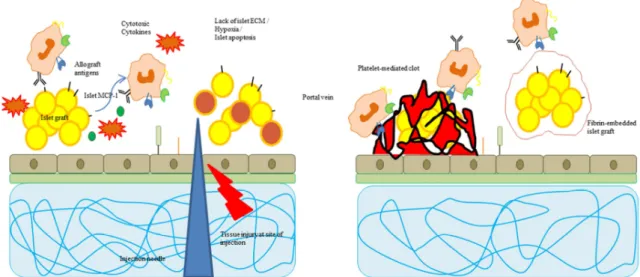
Figure 2.1: Schematic presentation of leukocyte recruitment to the site of inflammation (left) and immune cell response in tissue injury (right) 26 Figure 2.2: Schematic presentation of the immune response during islet grafting
(left), instant blood-mediated inflammatory reaction (IBMIR) and islet encapsulation
(right) 29
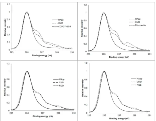
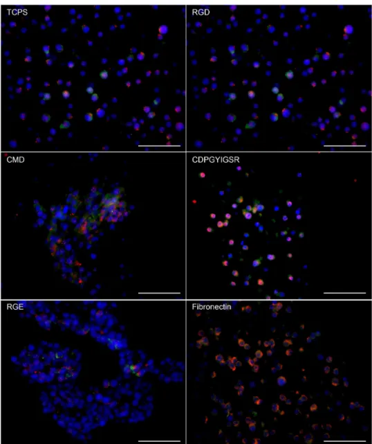
Figure 3.1: High resolution XPS C 1s spectra of CDPGYIGSR, fibronectin, RGD
and RGE surfaces and interlayers 41
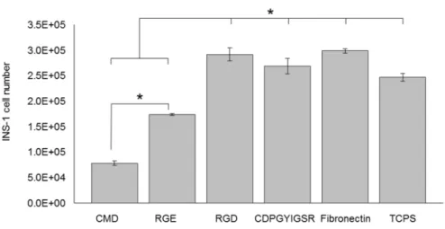
Figure 3.2: INS-1 cell number after 7 days of culture on the different surfaces 42 Figure 3.3: Percentage of INS-1 cells stained positively for Ki-67 after 7 days of culture
on the different surfaces 42
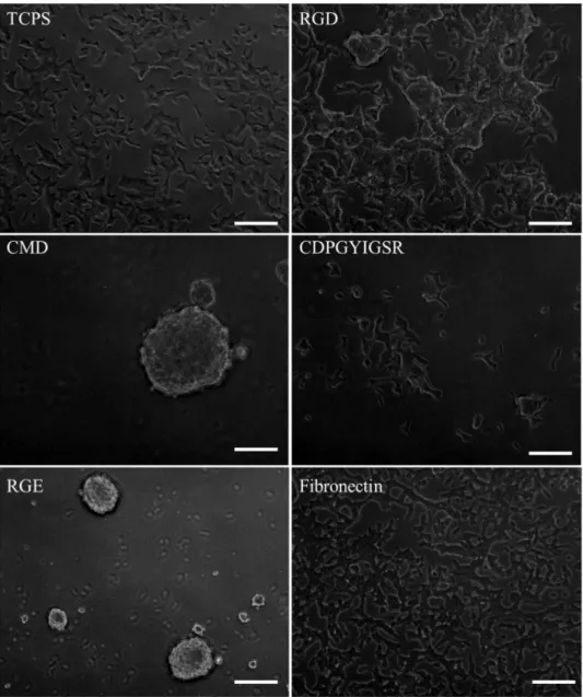
Figure 3.4: Phase contrast microscopy pictures of INS-1 cells grown on the different
surfaces after 72 h 44
Figure 3.5: Expression of integrins α5 (red) and αVβ3 (green) of INS-1 cells
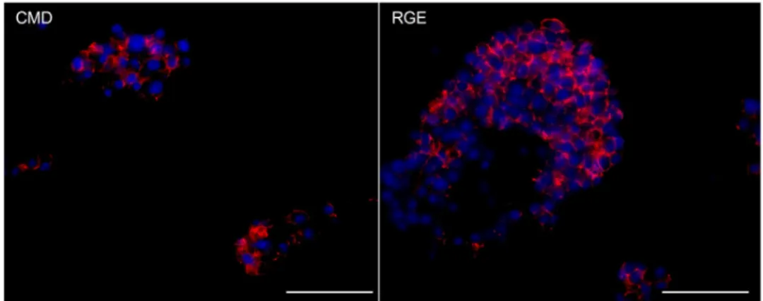
cultured for 7 days on the different surfaces 46
Figure 3.6: Immunofluorescence staining for E-cadherin of INS-1 cells cultured
for 7 days on CMD and RGE 47
Figure 3.7: Glucose-stimulated insulin secretion (GSIS) of INS-1 cells cultured
on different surfaces for 7 days 48
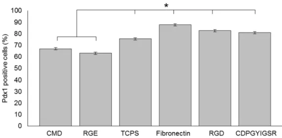
Figure 3.8: Percentage of INS-1 cells stained positively for Pdx1 after 7 days of
culture on the different surfaces 50
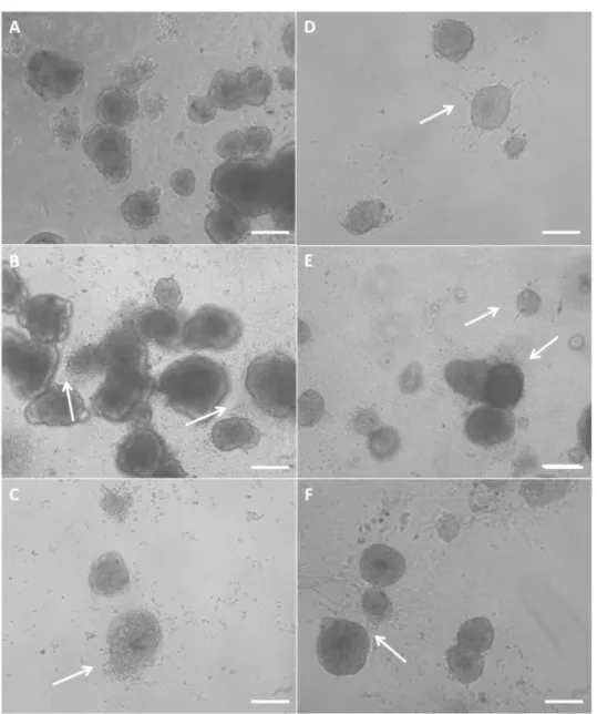
Figure 4.1: Morphology of young porcine endocrine pancreatic islets cultured for 12 h in TCPS plates with no H2O2 (A), 10 µM H2O2 (B) and 100 µM H2O2 (C).
Morphology of islets cultured for 12 h in fibrin with no H2O2 (D), 10 µM H2O2 (E)
and 100 µM H2O2 (F). 57
Figure 4.2: (A) Relative insulin secretion of young porcine endocrine pancreatic islets after 1h stimulation with low glucose concentration (LG, 2.8 mM), high glucose concentration (HG, 28 mM), high glucose concentration +
3-isobutyl-1-methylxanthine (IBMX) (HI, 28 mM + IBMX) and a second
low glucose concentration (LG2, 2.8 mM). (B) Three repeats of a glucose-stimulated insulin secretion assay with LG, HG, HI and a final LG. Shown are the insulin contents in the media (µg per L) of porcine pancreatic islets (200 IEQ) cultured on TCPS (T)
or in fibrin gels (F) with 0, 10 or 100 µM H2O2 59
Figure 4.3: Mass of insulin (in µg) secreted into the media by 200 IEQ embedded in fibrin and exposed to 0 (F0) and 100 µM H2O2 (F100) and the mass of insulin (in µg) in the
extract of fibrin gels rinsed with 500 µL PBS and centrifuged thereafter 60 Figure 4.4: Immunofluorescence staining for insulin (green) and glucagon (red) of young
porcine endocrine pancreatic islets 61
Figure 4.5: Immunofluorescence staining for integrin α5 (red) of young porcine endocrine
pancreatic islets 63
Figure 4.6: Light microscopy images of young porcine endocrine pancreatic islets treated with the APO-BrdU IHC staining kit. Islets cultured in TCPS plates with 0, 10 or
100 µM H2O2 compared to time zero (A) and islets embedded in fibrin with 0, 10 or
100 µM H2O2 compared to time zero (B) are shown. The percentage of apoptotic cells
(brown) in the total cell population (blue and brown) is shown in Fig. 4.6C 64
Figure 5.1: Schematic representation of the islet-monocyte co-culture systems with
non-encapsulated islets in a TCPS well and islets encapsulated in fibrin or alginate 80 Figure 5.2: Morphology of young porcine endocrine pancreatic islets and human
monocytes cultured for 8 h in TCPS plates, alginate, of fibrin 84 Figure 5.3: Morphology of young porcine endocrine pancreatic islets and human
monocytes cultured for 24 h in TCPS plates, alginate, or fibrin 85 Figure 5.4: Secretion of cytokines TNFα, IL-6, and IL-1β in the culture media of
young porcine endocrine pancreatic islets cultured alone or co-cultured with human monocytes, and human monocytes cultured alone for 24 h in TCPS plates,
alginate, or fibrin 87
Figure 5.5: Light microscopy images of young porcine endocrine pancreatic islets and human monocytes treated with the APO-BrdU IHC staining kit to reveal apoptotic cells. TCPS: Islets (A), islets+monocytes (B), and monocytes (C). Fibrin:
Islets (D), islets+monocytes (E), and monocytes (F). Alginate: Islets (G),
islets+monocytes (H), and monocytes (I) 89
Figure 5.6: Percentage of apoptotic cells in the indicated cell population of young porcine endocrine pancreatic islets and human monocytes either cultured alone or co-cultured in alginate, fibrin or TCPS plates and treated with the APO-BrdU IHC
staining kit to reveal apoptotic cells 89
Figure 5.7: Insulin secretion per IEQ from young porcine endocrine pancreatic islets
after a 24-h culture in TCPS, alginate, or fibrin with and with no human monocytes 90 Figure 5.8: Immunofluorescence staining for integrins αVβ3 (green) and α5 (red) of
young porcine endocrine pancreatic islets after a 24-h culture in TCPS plates, alginate,
or fibrin with and with no human monocytes 91
Figure 5.9: Immunofluorescence staining for CD14 (green) and E-cadherin (red) of young porcine endocrine pancreatic islets after a 24-h culture in TCPS plates, alginate,
or fibrin with and without human monocytes 93
Figure A1: Immunofluorescence staining for Ki-67 of INS-1 cells cultured for
7 days on TCPS and RGE 134
Figure A2: Expression of integrins α5 (red) and αVβ3 (green) of INS-1 cells cultured for
7 days on the different surfaces 135
Figure A3: Expression of integrins α5 (red) and αVβ3 (green) of INS-1 cells cultured for
3 days on the different surfaces 136
Figure A4: Immunofluorescence staining for E-cadherin of INS-1 cells cultured for
3 days 137
Figure A5: Immunofluorescence staining for insulin of INS-1 cells cultured for 7 days on
fibronectin 138
Figure A6: Immunofluorescence staining for Pdx1 of INS-1 cells cultured for
7 days on CDPGYIGSR and RGE 138
Figure B1: SEM images of fibrin and alginate gels 139
LIST OF TABLES
Table 3.1: XPS atomic concentration (%) and atomic ratio of HApp, HApp+CMD, HApp+CMD+CDPGYIGSR, HApp+CMD+Fibronectin,
HApp+CMD+RGD and HApp+CMD+RGE 40
Table 4.1: Number of cells stained positively for glucagon, insulin, and both
glucagon and insulin, counted on immunofluorescence images of porcine pancreatic islets at ‘Time Zero’, in fibrin with no (F0), 10 (F10), and 100 (F100) µM H2O2,
and on TCPS with no (T0), 10 (T10), and 100 (T100) µM H2O2 62
Table 4.2: Number of cells stained positively for integrin α5, counted on
immunofluorescence images of porcine pancreatic islets at ‘Time Zero’, in fibrin with no (F0), 10 (F10), and 100 (F100) µM H2O2, and on TCPS with no (T0), 10 (T10),
and 100 (T100) µM H2O2 63
Table 5.1: Manual counting of cells in all conditions stained positively for integrins αVβ3
and α5 92
LIST OF ABBREVIATIONS
3D three dimensional
ANOVA analysis of variance APC antigen-presenting cells bFGF basic fibroblast growth factor
BM basement membrane
BSA bovine serum albumin
CaCl2 calcium chloride
CDPGYIGSR synthetic laminin pentapeptide Tyr-Ile-Gly-Ser-Arg
CMD carboxymethyl-dextran
CO2 carbon dioxide
DAB 3,3'-diaminobenzidine
DAPI 4’, 6-diamidino-2-phenylindole, dihydrochloride
DC dendritic cells
DNA deoxyribonucleic acid
ECM extracellular matrix
ELISA enzyme-linked immunosorbent assay FACS fluorescence-activated cell sorting
FBS fetal bovine serum
FN fibronectin
GSIS glucose-stimulated insulin secretion H2O2 hydrogen peroxide
HApp n-heptylamine plasma polymer
HG high glucose
HI high glucose + IBMX
IBMIR instant blood-mediated inflammatory reaction IBMX 3-isobutyl-1-methylxanthine
IEQ islet equivalent
IFN interferon
IL interleukin
INS-1 rat insulinoma cells
KRBH Krebs-Ringer buffer with HEPES LFA leukocyte function-associated antigen
LG low glucose
LPS lipopolysaccharides
MHC major histocompatibility complex MIN-6 mouse insulinoma cells
MMP matrix metalloproteinase
NK natural killer cells
P probability
RGD Arg-Gly-Asp
RGE Arg-Gly-Glu
RT room temperature
RT-PCR real time polymerase chain reaction T1DM type 1 diabetes mellitus
TCPS tissue culture polystyrene
TGF transforming/tumor growth factor TNF tumor necrosis factor
TUNEL terminal deoxynucleotidyl transferase dUTP nick end labeling VCAM vascular cell adhesion molecule
Chapter 1
INTRODUCTION
Islet transplantation is considered to be a promising treatment if not a cure for Type 1 Diabetes mellitus (T1DM). With more than 346 million people worldwide being affected by diabetes, the impact on the international health system and the life quality of patients is apparent [1]. Between 5 and 7 % of diabetes patients suffer from the autoimmune form, T1DM, characterized by the loss of endogenous insulin production. A combination of genetics and environmental factors leads to the onset of T1DM through an immune system-mediated destruction of insulin-producing β-cells within the endocrine Langerhans’ islets in the pancreas. Lifelong insulin therapy comprising constant blood glucose monitoring and insulin injections is required for T1DM treatment. Even though advances have been made in this field, such as automatic monitoring and insulin pumps, in some cases the regulation of blood glucose levels is extremely difficult. The once fatal disease has become treatable but T1DM diagnosis still implicates reduced life expectancy and a wide range of acute and chronic secondary complications such as hypo- and hyperglycemia, ketoacidosis, diabetic coma, and macro- and microvascular diseases [2].
For patients with uncontrolled blood glucose levels, pancreatic islet transplantation has become a promising alternative [3]. The idea of islet transplantation, as opposed to whole pancreas transplantation, is to replace the tissue affected by the autoimmune destruction. Therefore, islets are enzymatically, mechanically and chemically disrupted from the surrounding exocrine tissue [4,5]. In most cases, the purified islet graft is then injected into the patient’s portal vein or else, co-transplanted with a donor kidney [6]. Since organ donor shortage is one of the primary limitations of islet transplantation, xenotransplantation (i.e. porcine islet transplantation into human patients) has moved into the focus of research. Other limitations are based on islet graft function and cell loss due to multiple factors [7]. Among those is the loss of islet extracellular matrix (ECM) during islet isolation and the response of the host’s immune system to the allograft [8]. In recent years, both islet-ECM interactions and graft-immune response dynamics have been studied widely and islet encapsulation approaches have emerged as potential solution for both problems [9]. Encapsulation materials can be altered with ECM cues to support islet structure and function and could simultaneously protect the islet graft from immune cell invasion and consequent destruction.
This thesis aims to help understand underlying implications of the immune system in islet cell destruction in both T1DM and islet transplantation and the postulated beneficial effect of
ECM-related cell interactions. It was hoped to determine the feasibility of islet encapsulation in the context of graft rejection, ECM and structural support, and maintenance of islet cell viability and function.
To study the interaction of insulin-producing cells with the ECM, rat insulinoma cells (INS-1) were used [10]. INS-1 cells were cultured on fibronectin, RGD (Arg-Gly-Asp) and a synthetic laminin pentapeptide (YIGSR), specifically grafted on carboxymethyl-dextran (CMD) surfaces, to assess their morphology, viability, and function i.e., insulin secretion in response to different glucose concentrations. The objective was to find conditions that would improve the insulin secretion and proliferation of insulin-producing cells. To address the ongoing research in xenotransplantation and islet encapsulation, young porcine endocrine pancreatic islets were embedded in fibrin and later alginate gels and either exposed to two hydrogen peroxide (H2O2) concentrations or later, to human monocytes. It was hypothesized that fibrin-
and/or alginate-embedded islets would show improved integrity after isolation, maintained glucose-responsiveness and insulin secretion, and resistance towards mediators of the immune system.
Chapter 2 of this thesis titled, “Cross Talk Between the Extracellular Matrix and the Immune System in the Context of Endocrine Pancreatic Islet Transplantation”, is a review of most important ECM components, immune cells, the interactions of ECM and immune cells, and their implication in T1DM and islet transplantation. As islet transplantation research is trying to exploit the beneficial potential of ECM-islet cell interactions, this review highlights now known reciprocal action of immune cells, their specific integrins, and the ECM with a focus on islet transplantation and T1DM development.
In Chapter 3 titled, “Culturing INS-1 Cells on CDPGYIGSR-, RGD- and Fibronectin Surfaces Improves Insulin Secretion and Cell Proliferation”, INS-1 cells were cultured on specifically modified surfaces with well-defined properties, grafted with various ECM cues to improve cell proliferation and insulin secretion. In comparison with the controls – tissue culture polystyrene (TCPS) plates, CMD surfaces and RGE surfaces – YIGSR-, RGD- and fibronectin surface-cultured INS-1 cells showed higher net cell numbers after 7 days of culture and enhanced insulin secretion in response to glucose. Interestingly, INS-1 cells on the low-fouling
surfaces CMD and RGE stayed in suspension and remained functional, forming so called “pseudo-islet” structures.
In Chapter 4 titled, “Young Porcine Endocrine Pancreatic Islets Cultured in Fibrin Show Improved Resistance Towards Hydrogen Peroxide”, islets were embedded in fibrin gels or cultured on TCPS and exposed to 10 and 100 µM H2O2 for 12 h. Consequent analysis revealed
maintained insulin secretion of fibrin-embedded islets whereas TCPS-cultured islets lost their glucose-responsiveness with increasing H2O2 concentrations. Islets in fibrin were also less
apoptotic and expressed higher levels of α5 integrin as well as insulin and glucagon.
Chapter 5 titled, “Young Porcine Endocrine Pancreatic Islets Cultured in Fibrin and Alginate Gels Show Improved Resistance Towards Human Monocytes”, included alginate gels as another promising encapsulation material. Human monocytes were used to further highlight the xenotransplantation approach and to possibly establish an in vitro model for T1DM and immune responses to islet grafts. Fibrin alone evoked a strong cytokine secretion of TNFα, IL-6 and IL-1β, but islets in fibrin and alginate were less apoptotic and secreted more insulin than islets on TCPS. Integrin expression was higher in fibrin-embedded islets and monocyte-presence enhanced αVβ3 expression in all conditions.
In Chapter 6, titled “Conclusion”, the outcome of this thesis is discussed and future directions are suggested.
Chapter 2
CROSS TALK BETWEEN THE EXTRACELLULAR
MATRIX AND THE IMMUNE SYSTEM IN THE
CONTEXT OF ENDOCRINE PANCREATIC ISLET
Foreword
Authors and Affiliations:
Carina Kuehn: Ph.D. Candidate, Université de Sherbrooke, Département de génie chimique et de génie biotechnologique.
Patrick Vermette: Professor, ingénieur, Université de Sherbrooke, Département de génie chimique et de génie biotechnologique.
Tamàs Fülöp: Professor, MD, Université de Sherbrooke, Institut universitaire de gériatrie de Sherbrooke, Research Centre of Aging.
Date of Submission: December 13th, 2013
State of Acceptance: Final Version Published. Journal: Pathologie Biologie
Reference: [Kuehn, C., Vermette P., Fülöp T. (2014) Cross talk between the extracellular
matrix and the immune system in the context of endocrine pancreatic islet transplantation. A review article. Pathologie Biologie (Paris), volume 62 (2), p. 67-78]
Contribution:
This article is the literature review of this thesis performed by Carina Kuehn. All work was performed under the direction and supervision of Patrick Vermette and Tamàs Fülöp.
Titre en français :
Interactions entre la Matrice Extracellulaire et le Système Immunitaire dans le Contexte de la Transplantation d’Îlots Pancréatiques Endocriniens
Résumé
Cette revue de la littérature a pour but de mettre en évidence l’importance des interactions bidirectionnelles de la matrice extracellulaire (MEC) et des cellules du système immunitaire dans le contexte du diabète de type 1 (DT1) et de la transplantation d'îlots pancréatiques endocriniens. Nous faisons un survol des grandes classes de molécules qui composent la MEC ainsi que les protéines, cellules et cytokines impliquées dans le système immunitaire. Cet article examine aussi les rôles de la MEC et du système immunitaire dans la transplantation d'îlots pancréatiques et plus particulièrement les effets des intégrines exprimées par les cellules du système immunitaire et leurs fonctions. Finalement, les cytokines liées à la MEC et leur influence sur les cellules immunitaires et la MEC sont abordées.
2.1 Abstract
This review aims to highlight the importance of the bidirectional influence of the extracellular matrix (ECM) and immune cells in the context of type 1 diabetes mellitus (T1DM) and endocrine pancreatic islet transplantation. We introduced the main classes of molecules and proteins constituting the ECM as well as cells and cytokines of the immune system with the aim to further examine their roles in T1DM and islet transplantation. Integrins expressed by immune cells and their functions are detailed. Finally, this article reviews the roles of the ECM and the immune system in islet transplantation as well as ECM-related cytokines and their influence on the ECM and immune cells.
2.2 Introduction
The extracellular matrix (ECM) was long considered to be solely the non-cellular connective tissue and served mainly as mechanical support for the surrounding cells. Now it is known that apart from a structural support, the ECM is fulfilling functions such as tissue segregation, regulation of intercellular communication, storage and activation of growth factors and cytokines, as well as mediation of cell development, growth, survival, adhesion and migration [11]. The immune system was long believed to act on its own but it emerges now that interactions with other cells, integrins, growth factors, cytokines and the ECM play a pivotal role in immune cell development, maturation, and function [12]. In this review, the specific role of the ECM in the immune system is investigated. A highlight will be set on type 1 diabetes mellitus (T1DM) and pancreatic islet transplantation.
2.3 The Extracellular Matrix
There are two major categories of ECM, the basement membrane (BM) and the interstitial or stromal ECM. The BM is a thin layer, between 50- and 100-nm thick, of a specialized ECM made up of collagen, laminin, and fibrillin (microfibrils), separating the epithelial or endothelial tissue from the connective tissue. Besides providing a structural support to cells, the BM modifies the cellular behavior via outside-in signalling. The structure, composition, and function of the BM is organ-specific [13].
The interstitial or stromal ECM is a tissue of high complexity and variety. It constitutes a network of multiple proteins and polysaccharides. Those are secreted by local cells and stay closely associated to the surface of those cells that produced them. After secretion, the ECM proteins and polysaccharides are assembled into a highly organized meshwork [14]. There is a great diversity in ECM organization and different types of matrix proteins and polysaccharides. This diversity results in different types of ECM, each formed depending on the function of the particular tissue e.g., hard, mineralized structures for bone or teeth, soft ECM in internal organs, transparent layers of the cornea, and strung ECM in tendons [14].
2.3.1 ECM Components
The two main classes of extracellular macromolecules that make up the matrix are namely: • polysaccharide chains of glycosaminoglycans (GAGs), which are usually found
covalently linked to proteins in the form of proteoglycans;
• fibrous proteins, including collagens, elastin, fibronectin (FN), and laminins.
Whereas proteoglycans constitute the basic substance of ECM by forming a gel-like hydrated network, the fibrous proteins embedded within provide the structural and functional cues for cells [14,15].
2.3.2 Glycosaminoglycans
GAGs are long, unbranched, mostly high molecular weight polysaccharides (also mucopolysaccharides) with a backbone of repeating disaccharide units incorporating an aminosugar and an uronic acid. There are two main types of GAGs, heparan sulphate and chondroitin sulphate. Proteoglycans occur when one or more GAGs are attached to a core protein at specific sites. Proteoglycans are found throughout the extracellular matrix and attached to the cell membrane. Chondroitin sulphate chains are mostly found on matrix proteoglycans (aggrecan) whereas membrane proteoglycans contain mostly heparan sulphate chains (syndecan, glypican) [16]. Proteoglycans form a gel-like ground substance of the connective tissue and fill most of the extracellular space because of their hydrophilic nature. Other functions are to interact with signaling molecules, to modulate ligand-receptor interactions, and to play a role in development, migration, and enzyme activity. Proteoglycans are furthermore involved in several signaling pathways like TGFß, FGF, Hedgehog, etc. Additionally, proteoglycans form networks by linking to collagen fibers [17].
2.3.3 Collagens
Collagens represent the most abundant fibrous proteins in vertebrates, constituting 30 percent of the total body protein. To date, 28 types of collagen have been identified and described with
the five most common types of collagens being collagen I (skin, tendon, vascular ligature, organs, bone), collagen II (main component of cartilage), collagen III (main component of reticular fibers, commonly found alongside collagen I), collagen IV (basal lamina) and collagen V (cell surfaces, hair and placenta) [18].
Ubiquitously expressed in all mammals, type I collagen is the most abundant type, constituting more than 90 percent of all collagens. It is one of the largest and most complex macromolecules. Some of the proposed functions of collagen I include mediation of cell adhesion, binding to other matrix molecules, interactions with tissue calcification factors, but also glycation. Glycation occurs when reducing sugars bind to protein, forming adducts. Those glycation products are believed to contribute to diabetes development and pathologies of aging [19].
2.3.4 Fibronectin
Another essential ECM molecule is the ubiquitously cell-mediated expressed glycoprotein fibronectin (FN), found in blood as soluble FN and in interstitial connective tissue as insoluble FN [20]. The dimeric protein, consisting of two nearly identical monomers linked by a pair of disulfide bonds, is organized into a fibrillar network through direct interactions with cell surface receptors called integrins [20–22]. FN plays a pivotal role during embryogenesis, guiding cell migration and adhesion. Being part of the ECM network, FN is essential for cell growth, attachment, migration, and development [21]. As soluble FN, it is also very important in wound healing, contributing to the initial blood clot formed at the site of injury together with fibrin [23]. FN can bind and interact with other ECM components, such as collagens and proteoglycans, especially heparin, and fibrin. Within the cell-binding domain of FN a specific adhesion sequence called RGD is found [24]. RGD is a tripeptide composed of L-arginine, glycine, and L-aspartic acid (Arg-Gly-Asp). This sequence constitutes the best known integrin-binding region in FN. Other integrin-binding minimal sequences can be found in the cell-binding domain of FN such as LDV (Leu-Asp-Val), REDV (Arg-Glu-Asp-Val), IDAPS (Ile-Asp-Ala-Pro-Ser) and KLDAPT (Lys-Leu-Asp-Ala-Pro-Thr). Those binding sites recognize the α4β1 integrin and α4β7 integrin (except for IDAPS).
2.3.5 Integrins
Integrins are a family of 24 identified trans-membrane receptors, mediating cell adhesion to the ECM, activating intracellular signaling pathways, and supporting cell-cell contacts [25]. Integrins are heterodimers, composed of two distinct polypeptide chains, called α and β subunits. To date, 18 α and 8 β subunits have been identified. Both subunits breach the cell membrane, with the major part being in the extracellular space and a minor domain in the cytoplasm [25,26]. Integrin receptors bind to and interact with several ligands such as collagen, RGD, leukocytes, and laminin.
Some α subunits can only bind to one specific β subunit e.g., α5 with β1 or α1 with β1, whereas others can bind to several β subunits e.g., αV with β1, 3, 5, 6, or 8 [25]. Integrins are
known for their ability of bidirectional signalling. “Outside-in” is conceived through binding of extracellular ligands found in the ECM or to integrins on the cell surface, leading to the initiation of intracellular signalling pathways associated with cytoskeleton proteins. “Inside-out” signalling is achieved by a reciprocal interaction of integrins with the extracellular domains. This occurs after cells have received signals through other surface receptors. These signals are then translated by integrins to enhance adhesiveness and migration [25].
2.3.6 Laminins
Laminins are a family of major proteins in the basal lamina of the basement membrane. They are cross-shaped heterotrimers, composed of one α, one β and one γ chain [27]. As glycoproteins in the BM, laminins function as structural support for cells, promote cell adhesion, migration, differentiation, and morphology, and prevent cell death via anoikis. Anoikis is a special form of apoptosis, the programmed cell death, caused by detachment or loss of anchorage of cells from their ECM [28]. Laminins exhibit their actions through various cell receptors such as integrins [29] but also through non-integrin receptors such as the 67kDa laminin receptor [30,31].
2.3.7 Fibrin as a Transient Component of the ECM
Additionally to collagens, laminins, proteoglycans, and FN, fibrin can be considered being part of the ECM as cells interact with fibrin through integrin receptors. Fibrin is naturally formed from fibrinogen, a soluble liver-derived protein found in blood plasma, when tissue damage results in bleeding [32]. Thrombin, a clotting enzyme, converts circulating fibrinogen into fibrin at the site of injury [33,34]. Fibrin molecules are arranged in long fibrous chains [35] which are combined to form thread which trap platelets within, forming a spongeous clot to stop the bleeding. The fibrin mesh provides anchorage sites for platelets, fibroblasts, neutrophils and monocytes supporting wound healing in the process [36–38]. While the clot in the vessel helps to re-establish normal blood flow, part of the clot in the injured tissue provides a transient ECM for cell migration and tissue reformation.
In vitro biodegradable fibrin gels are quite similar to in vivo fibrin clots [34,39]. In the context
of biomaterials for tissue engineering and cell encapsulation in transplantation, fibrin matrices can provide structural support for cells and cues from the ECM [32,37,40]. Fibrin has been already widely used in clinical applications. In the context of pancreatic islet transplantation, fibrin has shown potential for improving islet viability, function, and preservation of native islet morphology [40–45]. Another advantage of the usage of fibrin matrices in transplantation medicine could be the possibility to use the graft recipient’s blood fibrinogen to obtain a fibrin scaffold which would probably lower the intensity of the initial immune response to the transplant. However, in vivo fibrin clots undergo fibrinolysis eventually in the course of an inflammatory and wound healing setting, consequently, impairing the feasibility of fibrin as sole component to constitute a device to encapsulate islets for transplantation purpose [46,47].
2.3.8 ECM of the Human Endocrine Pancreatic Islets
The structure and composition of the native ECM of islets are still under investigation. Islets are among the most vascularized structures in the human body. Communication between islet cells and the endothelial cells is achieved through their common vascular basement membrane. Islets have a second basement membrane of unknown origin [48,49]. FN, RGD, and laminins have been studied widely; the interactions between endocrine pancreatic islets
and FN, RGD, and laminins have been investigated with the aim to enhance islet survival and function as well as insulin secretion [50–53]. Certain integrins have been found to be
expressed within the islet and by beta cells. Integrin β1 appears to be the most prominent islet integrin and has been widely studied in terms of islet cell adhesion, proliferation,
differentiation, and survival [53–55]. The islet basement membrane contains various ECM proteins such as laminins and collagens, providing binding and interaction sites with (β1) integrins [48,56]. In vitro, a matrix enriched with laminin enabled α6β1 integrin-mediated beta cell spreading, decreased apoptosis and improved insulin secretion [52,53,57]. FN, RGD, and the laminin peptide YIGSR, covalently bound to low-fouling carboxymethyldextran, improved INS-1 (rat insulinoma) cell proliferation, integrin expression, and glucose-stimulated insulin secretion [58]. As those beneficial ECM cues are absent during islet transplantation, due to the islet isolation procedure, provision of a ECM-enriched encapsulation matrix such as fibrin or alginate could constitute a valuable protection from the immune response to an allogenic or xenogenic islet graft [9,59–62].
2.4 The Immune System
The complex immune system has developed to protect the human body from various pathogens such as parasites, viruses and bacteria. Its complexity is becoming clear when regarding the cells and soluble mediators involved in the immune response and the fact that, besides an innate immune system, an adaptive immune system evolved in the human body.
2.4.1 Innate and Adaptive Immune Systems
The immune system in its entity is comprised of two distinct types of immune responses, the innate and the adaptive immune responses, each functioning apart from each other but interacting with each other intensely. The main difference between the innate and the adaptive immune response is the respective speed of the immune response and its specificity towards the pathogen resulting in immunological memory by the adaptive immune response [63–65].
The innate immune system has been highly conserved. A fast but unspecific immediate defence reaction is provided without any memory by elements like neutrophils, monocytes, macrophages, natural killer cells, complement, cytokines, and acute phase proteins but also through physical, chemical, and microbiological barriers. Sometimes, the unspecific response of the innate immune system can cause tissue damage via an uncontrolled inflammatory reaction [64,66]. In contrast, the response of the adaptive immune system is exhibited through antigen-specific T and B lymphocytes and may take days or even weeks to develop. After successful immune response to a foreign antigen, the adaptive immune system produces long lasting memory cells [64,65].
2.4.2 Immune Cells
Leucocytes (“white blood cells”) are the cells without whom any immune response would not be possible. They are divided into lymphocytes, phagocytes and auxiliary cells but normal tissue cells also contribute to the immune response by expressing foreign DNA fragments on their surface, thus signalling to lymphocytes. In addition, normal tissue cells respond to secreted cytokines, and even secret certain cytokines themselves. Lymphocytes are subdivided into B cells, T cells, and large granular lymphocytes, namely natural killer cells (NK). Phagocytes are neutrophils, eosinophils, and mononuclear phagocytes. Basophils, mast cells and platelets are part of the auxiliary cell compartment [63].
2.4.2.1 Neutrophils
Neutrophils are central in the innate immune system’s response as they are the first to arrive at the site of aggression. They are recruited and activated by cytokines released from activated macrophages at the site of tissue injury or inflammation, namely granulocyte- and granulocyte-macrophage-colony stimulating factors (Figure 2.1). Bone-marrow derived neutrophils circulate in the blood and follow a cascade of pro-inflammatory mediators, adhesion molecules, and chemokines to reach a site of infection [67]. On site, neutrophils phagocytize pathogens and then kill the organism through the release of reactive oxygen
species and highly toxic cationic proteins and enzymes. If pathogenic organisms are coated (opsonized) first with specific antibodies or complement, the neutrophils ability to ingest and destroy those pathogens, is highly elevated [63].
The complement system is a complex entity of proteins and glycoproteins, mainly produced in the liver, circulating in blood as pro-proteins. Among the several functions of the complement system are opsonisation, cell lysis, and chemotaxis. Once activated, the complement system amplifies the immune response, both innate and adaptive, and therefore, is an important mechanism for removing a foreign organism from the host [68,69].
2.4.2.2 Granulocytes: Eosinophils, Basophils and Mast cells
Eosinophils main function is the removal of parasites via binding to the antibody-coated organism. Eosinophils destroy the parasite by releasing cytotoxins onto the parasite’s cell surface [63,70]. They are involved in the removal of fibrin in inflammation and play a role in asthma and allergy control.
Basophils circulate in blood and are involved in allergic reactions [71]. They are thought to contribute to the severity of these reactions acting as pro-inflammatory effector cell. Since basophils express major histocompatibility complex (MHC) proteins, class II, they are potentially antigen-presenting cells (APC) and could act in T cell activation and regulation [72].
Mast cells are quite similar to basophils as both cell types contain basophilic granules. As opposed to basophils, which mature in the bone marrow and are then released into the blood stream, mast cells are released from bone marrow immaturely and mature later in the tissue where they reside. Mast cells, not unlike basophils, are involved in anaphylactic and other allergic reactions. They also play a role in wound healing and pathogen defence [73]. Mast cells, as well as basophils, have granules which contain the vasodilator histamine and the anticoagulant heparin. The granules can also contain various effector molecules such as serine proteases and serotonin. Recent evidence suggests that mast cells play a role in the onset and progression of T1DM [74].
2.4.2.3 Natural Killer Cells
NK cells share the same lineage as T and B lymphocytes but are part of the innate immune system and they should not be mistaken with natural killer T cells. NK cells are essential for the innate immune response as they act fast and recognize cells in distress i.e., infected cells or cancer cells, even in the absence of antibodies or MHC expressed on the cell surface [66,75,76]. In fact, the absence of MHC proteins could trigger recognition by NK cells and direct cell susceptibility to be lysed [77]. Besides their cytotoxic functions, NK cells secrete a wide variety of cytokines, especially interferon gamma (IFNγ), of which they are recognized to be the main producer [78,79].
2.4.2.4 Dendritic Cells
Antigen-presenting cells (APC) link the adaptive immune response of B and T cells with the innate immune response. Dendritic cells (DC), macrophages, certain B lymphocytes, and activated epithelial cells comprise the pool of professional APC alongside with unprofessional APC, such as Langerhans beta cells, fibroblasts, glial cells etc.
DCs primary function is to internalize and present antigens through their dense levels of MHC proteins on their surface. Even though B lymphocytes present antigen on their surface as well, DC activate and regulate both B and T lymphocytes. DC are the only cells known to be able to activate resting naïve T cells [80].
2.4.2.5 Monocytes and Macrophages
Bone marrow-derived monocytes are the largest leukocytes which are released into blood circulation after maturation and differentiate into macrophages upon tissue infiltration [81]. Monocytes were once characterized by strong expression of CD14, a surface receptor interacting with the toll-like receptor in the recognition of bacterial lipopolysaccharides (LPS). Now it is known that some subpopulations of monocytes have differential expression profiles of CD14 and CD16, such as CD14++/CD16- (classical monocytes), CD14+/CD16++ (non-classical monocytes), and CD14++/CD16+ (intermediate monocytes), resulting in different
functions [81–83]. For example, non-classical monocytes secrete more pro-inflammatory cytokines upon activation. Amongst the multiple functions of monocytes is the provision of myeloid precursor cells for the replenishment of tissue macrophages and DC. Although large numbers of monocytes are residing in the spleen, blood-circulating monocytes can reach a site of injury or inflammation within 8 to 12 hours. When entering the tissue, monocytes differentiate into macrophages or DC. Macrophages exist in two forms, M1 and M2. M1 macrophages, the “killer” phenotype, are the classically activated macrophages. M1 express inducible nitric oxide synthase (iNOS) and a multitude of pro-inflammatory cytokines, such as TNFα, IL-1β, IL-6, etc. [84]. M2, on the contrary, function in a more regulatory manner, removing debris and promoting tissue repair. By secreting IL-10 and TGFβ1, M2 macrophages contribute to the regulation of the immune response to prevent an overreaction [85]. As APC, macrophages internalize pathogens, digest them and then present antigens linked to MHC proteins on their surface which consequently activates T lymphocytes [63].
2.4.2.6 T Lymphocytes
T cells are part of the adaptive immune system [63]. T cells are distinguished from other lymphocytes by the T cell receptor expressed on their surface [86]. As B cells, T cells originate from the bone marrow but mature in the thymus gland, hence their name. T cells are not antigen-presenting immune cells but require APC such as monocytes/macrophages, dendritic cells (DC) and also B cells or natural killer cells for activation [87]. There are multiple subsets of T cells: helper T cells (TH), cytotoxic T cells (TC), memory T cells, natural
killer T cells, and regulatory T cells (Treg) [63].
T cells recognize antigens which are associated with so called major histocompatibility complex (MHC) proteins on the cell surface of APC [86,88,89]. TH cells recognize MHC class
II associated antigens with their CD4 receptor whereas TC cells are activated by MHC class I
associated antigens which are recognized by their CD8 receptor [88,90]. Treg cells suppress T
cell activity towards the end of an immune response and help identifying and cleaning auto-reactive T cells, which escape negative selection in the thymus, from the system. Memory T cells are comparable in their function to B cell-derived memory cells and can be either CD4+
or CD8+. Natural killer T cells, other than TH and TC, recognize glycolipid antigens presented
by CD1d. Upon activation, natural killer T cells can function as TH or TC [63,91].
In T1DM, T cells were previously believed to be the main acting immune cell, mediating pancreatic beta cell death through recognition of self-beta cell-antigens as foreign, probably following an initial viral infection. Another theory suggested that the viral antigen resembled the beta cell antigen, thus, inducing an immune response to beta cells. Several facts contradict the supposed T cell-mediated beta cell destruction, as the course of the disease is a slow process over years whereas a T cell immune response is very specific and effective, lasting for several days or weeks only [92].
2.4.2.7 B Lymphocytes
B cells are considered being effector cells of the humoral arm of the adaptive immune response [63]. B cells are antigen-specific antibody-producing lymphocytes, expressing a specific B cell receptor on their surface. This receptor, comprised of immunoglobulins, only recognizes one specific antigen [91]. Produced in the bone marrow, mature B cells circulate in the blood system until activation through antigen recognition and an additional T cell signal [93]. B cells will then differentiate into either B memory cells or plasma B cells. The immunoglobulin (antibody) which recognizes the foreign antigen is released abundantly by plasma cells, whilst memory cells are responsible for the subsequent recognition of the same antigen in case of repeated exposure, ensuring facilitated clearance [94].
B cells play a role in the development of T1DM autoimmunity. Islet-autoantibodies to insulin, glutamic acid decarboxylase, tyrosine phosphatase IA-2, etc. found in T1DM patients or even before the onset of T1DM indicate that B cells recognized, bound and internalized islet antigens and produced antibodies in response [95].
2.4.2.8 Cytokines
Cytokines serve a multitude of purposes and show a great variety in their size, structure and function, and they are synthesized by almost every cell in the body. They are considered to be a group of soluble proteins, glycoproteins and peptides, secreted in response to a trigger i.e., a
stress signal. Cells secrete cytokines to either change their own behaviour or influence other cells. Cytokines interact with cell-surface receptors and induce cytokine-specific intracellular pathways [91]. Cytokines can exhibit pro- and anti-inflammatory characteristics of sometimes contradicting nature [96]. For example, interleukins (IL)-10 are known to activate B lymphocytes but they are anti-inflammatory cytokines as they act in the impairment of pro-inflammatory cytokine expression [97]. In the context of T1DM, TNFα, 1β, IFNγ, and IL-6 are the most prominent pro-inflammatory cytokines contributing to beta cell apoptosis, B- and T- lymphocyte activation, and excessive autoimmune reaction [98].
2.4.3 Type 1 Diabetes Mellitus
Type 1 diabetes mellitus (T1DM) is an auto-immune disease defined by the loss of endogenous insulin due to the destruction of insulin-producing β cells by the immune system [99]. Autoimmunity occurs when immune cells recognize self-antigens as foreign antigens and start reacting to them. In T1DM, autoimmunity is most probably based on a combination of genetic susceptibility and its clinical expression through the influence of various environmental factors such as viruses, toxins, stress, and diet [100–103]. Islet auto-antigens are presented by APC to helper T cells (Th cells) associated with major histocompatibility complex (MHC) class II molecules [104]. In the thymus, T cells are matured and selected for their reactivity. Normally, T cells reacting to self-antigens undergo negative selection and are destroyed [63]. T1DM develops over the course of several months up to several years and is the product of an immune reaction cascade leading to beta cell destruction. It is now believed that T cells are not the main effector immune cells in T1DM as their misled response to islet- or beta cell-auto-antigens would lead to a specific and efficient, relatively fast removal of all cells expressing these antigens [92]. Nonetheless, T cells must play a role in T1DM as islet-auto-antibodies produced by T cell activating B cells are found before T1DM onset and in patients. These auto-antibodies contribute to the development of autoimmunity and exacerbate the ongoing immune response [105].
The T cell-mediated beta cell destruction occurs after CD4+ T cell activation via APC-derived IL-12. This leads to an imbalance of regulatory and effector immune cells. In turn, CD4+ T cells activate CD8+ cytotoxic T cells through IL-2 secretion. IFNγ, produced by CD4+ T cells,
activates cytotoxic macrophages which secrete pro-inflammatory TNFα, IL-1β, and IFNγ, as well as reactive oxygen species i.e., O2*, H2O2, and NO [98,106].
Cytotoxic T cells, in response to antigen presentation, migrate towards the source i.e., the islets, and start secreting apoptotic and pro-inflammatory cytokines. Additionally, cytotoxic T cells can induce islet cell apoptosis via the Fas ligand pathway [107,108]. Interplay of T cell activation, antigen presentation, antibody secretion and circulation, formation of memory cells, further immune cell recruitment, and the development of auto-immunity, consequently result in the onset of T1DM when more than 90 percent of beta cells have been killed and have lost their functionality.
2.4.4 Islet Transplantation
Islet transplantation research aims to improve the outcomes and longevity of the islet graft to eventually provide a long-term treatment of T1DM patients or even a cure of this life threatening disease [4]. The basic idea of islet transplantation as opposed to whole pancreas transplantation is the circumstance of insulin deprivation in the patient which could be restored by just transplanting the functional unit of insulin production, the pancreatic islets [8]. A whole pancreas transplant comes with the limitations of organ donors, major surgery complications, life-long immune suppression and the sustenance of diabetes-related secondary complications. In contrast, islet transplantation is performed through injection of a suspension of isolated islets in the portal vein with lower risks associated with the surgery. Co-transplantation with a kidney transplant is also administered. But the success of islet transplantation is impaired by the requirement of multiple donor organs for a sufficient islet graft, graft loss due to immune rejection and the immediate blood-mediated immune response, damages to islets caused by isolation procedures, lack of efficient re-vascularisation, as well as loss of function over time [109–111].
The introduction of the Edmonton’s protocol resulted in improved islet isolation procedures, islet yield, transplantation, immune suppressive treatments, and consequently with an improved success rate [112]. In spite of this, the majority of patients reverse from temporary insulin independence to exogenously applied insulin [113].
Multiple factors are responsible for the loss of islets and the loss of function of the islet graft. In the context of this review, the most prominent reason is the rejection of the graft by the recipient’s immune system in combination with loss or damage of the native islet ECM. Recent studies have shown that the islets ECM, and more specifically, cell-integrin interactions, mediate apoptosis resistance and support viability and function of pancreatic beta cells. Over the years, various islet encapsulation attempts using (bio)materials have been made to either protect the islets from immune cell invasion or to provide ECM cues for better engraftment [9,59–61,114].
But how does the ECM actually interact with the immune system and are those interactions beneficial or detrimental for an islet graft are questions that are not completely elucidated.
2.5 Interactions between the ECM and the Immune System
The ECM is essential for immune system response in various aspects. Here, the role and function of leukocyte integrins will be discussed as well as the interactions between the ECM and cytokines. Furthermore, the involvement of the ECM in wound healing and islet transplantation will be investigated.
2.5.1 Integrins of Immune Cells
ECM-immune cell interactions play a pivotal role in immune cell development and function. Integrins are the main adhesion receptors for those interactions. Of the 24 integrins known to date, 13 are expressed by leukocytes, but only β2 and β7 integrins are exclusively expressed by leukocytes [115,116]. Integrin β2 plays an especially important role in leukocyte biology. Of β2, four αβ heterodimers exist, namely leukocyte function-associated antigen 1 (LFA-1; CD11a/CD18, αLβ2 integrin, ITAL antigen), Mac-1 (CD11b/CD18, αMβ2 integrin, ITAM antigen), p150,95 (CD11c/CD18, αXβ2 integrin, CR4, ITAX antigen), and αdβ2 (CD11d/ CD18, ITAD antigen) [117]. Of the β1 integrins, pairings with α1, 2, 3, 4, 5 and 6 are found in leukocytes. Integrins α4β7 and αEβ7 are found to be expressed on leukocytes. Integrin β2 is the only integrin found to be expressed on all leukocytes. Apart from integrin β2, the integrin
expression profile differs between the different immune cells. Integrins β1, 2 and 7 are variably expressed by T lymphocytes e.g., integrins α4β7 and α4β1 are expressed in immature and activated T cells [118]. The integrin expression of T lymphocytes depends on the T cell subtype and the state of inflammation [119]. Monocytes express β1 and β2 integrins but also express αVβ3 after differentiation into macrophages [120–123]. Granulocytes (neutrophils, eosinophils, basophils and mast cells) express β1, 2 and 3 heterodimers for migration and motility in tissues [124–128].
Leukocyte integrins are inactive during a non-inflammatory state and are only activated when membrane receptors involved in an immune response are stimulated. In a healthy individual, leukocytes are circulating in the blood and need integrins to attach to blood vessel walls and migrate into inflamed tissues or lymph nodes. When chemokine-, B cells- or T cell- receptors bind to antigens, cytokines, chemokines, etc., the inside-out signalling of integrins on leukocytes is activated. For leukocytes to migrate to the site of inflammation or into lymph nodes, binding to endothelial cells is of great importance for initiating the adhesion and migration cascade. Selectins allow the leukocytes to bind weakly to endothelial cells. The stimulation with chemokines or chemotactic molecules activates leukocyte integrins which are then able to engage with counter-receptors of endothelial cells. Both β1- and β2-integrins are activated by signals that are elicited by chemokines [129].
With the blood circulating in the vessels, creating shear flow on cells, cell binding to endothelium has to occur in a step-by-step process. Leukocytes that interact with endothelial cells first roll along the vessel walls. This first weak adhesion step is supported by L-selectin and α4 integrins [130–133]. During the rolling step, chemokine-induced activation of leukocyte integrins follows to allow firm adhesion [132].
The sources of the chemokines responsible for activating leukocyte integrins are endothelial cells, leukocytes or stromal cells. ECM proteins are known to be capable of binding and storing cytokines and chemokines [134]. Transmembrane proteoglycans i.e., heparan sulphates, on the luminal surface of endothelial cells can bind some chemokines, such as CC-chemokine ligand 5 (CCL5; also known as regulated upon activation, normal T-cell expressed and secreted, RANTES) and CXC-chemokine ligand 8 (CXCL8; also known as interleukin-8, IL-8) [135,136]. The main heparan sulphates binding chemokines are syndecan 1 and syndecan 4 (which bind CCL5) and syndecan 2 (which interacts with CXCL8) [135–137]. To
be able to activate leukocyte integrins, the chemokine receptor binding site of the chemokines remains available even when bound to proteoglycans [138]. The stimulation of the leukocytes with those membrane-bound chemokines leads to firm adhesion to the endothelium [119,132]. Together with integrin α4β1, integrin αLβ (LFA-1) is one of the most important leukocyte integrins contributing to leukocyte-endothelium adherence [139,140]. Arrest of leukocytes on the vessel wall is followed by their migration through the endothelium and underlying basement membrane. This process is also called diapedesis, transmigration or extravasation [141]. It was shown that LFA-1 and integrin α4β1 are the mediators of leukocyte migration depending on chemokine activation and ligand expression [142,143]. Interestingly, in the case of monocytes, binding to FN at the site of inflammation is necessary for their differentiation into tissue macrophages [144,145]. After extravasation, ECM molecules serve as an anchor for immune cell migration [146].
T lymphocytes migrate into lymph nodes where they are regularly exposed to APC to be possibly activated by foreign antigens. It is of great importance to spend sufficient time in the lymph nodes to establish a stable bond with APC through integrin interactions to ensure appropriate delivery of antigens to T lymphocytes for their activation and consequent immune response [147].
Integrins have been shown to accumulate on one part of the cell to increase local adhesiveness and avidity. This occurs on T lymphocytes, especially for integrin LFA-1, when T cells are activated with antigens. LFA-1 accumulation on the binding site of T cells with APC results in enhanced ligand binding and, therefore, facilitates antigen presentation and T cell activation [148]. LFA-1 is also rearranged with the TCR and other T cell receptors to form a so-called immunological synapse (supramolecular activation cluster). Even though questions about the exact function of this synapse remain, it is believed that it supports cytotoxic granule release by T lymphocytes or NK cells [149–151]. In addition, integrin signalling in T cells supports T cell activation, proliferation and cytokine secretion [152–154]. In macrophages, integrin activation supports phagocytic activity, cell differentiation and also cytokine secretion [155,156]. In neutrophils, degranulation and release of ROS through NADPH oxidase activation is facilitated through integrin signalling [157].
It is clear that integrins play an integral role in leukocyte migration, function and interaction with cells and tissues [156,158]. They also contribute to leukocyte development and