Université de Montréal
Cell wall composition regulates cell shape and growth
behaviour in pollen tubes
par Youssef Chebli
Institut de recherche en biologie végétale. Département de sciences biologiques Faculté des Arts et des Sciences
Thèse présentée à la Faculté dess Arts et des Sciences en vue de l’obtention du grade de Ph.D
en Sciences Biologiques
option biologie moléculaire et cellulaire
Août 2012
Cette thèse intitulée :
Cell wall composition regulates cell shape and growth behaviour in pollen tubes
Présentée par : Youssef Chebli
a été évaluée par un jury composé des personnes suivantes :
David Morse, président-rapporteur Anja Geitmann, directeur de recherche
Tamara Western, membre du jury Kris Vissenberg, examinateur externe David Morse, représentant du doyen de la FES
Résumé
L’une des particularités fondamentales caractérisant les cellules végétales des cellules animales est la présence de la paroi cellulaire entourant le protoplaste. La paroi cellulaire joue un rôle primordial dans (1) la protection du protoplaste, (2) est impliquée dans les mécanismes de filtration et (3) est le lieu de maintes réactions biochimiques nécessaires à la régulation du métabolisme et des propriétés mécaniques de la cellule. Les propriétés locales d’élasticité, d’extensibilité, de plasticité et de dureté des composants pariétaux déterminent la géométrie et la forme des cellules lors des processus de différentiation et de morphogenèse. Le but de ma thèse est de comprendre les rôles que jouent les différents composants pariétaux dans le modelage de la géométrie et le contrôle de la croissance des cellules végétales. Pour atteindre cet objectif, le modèle cellulaire sur lequel je me suis basé est le tube pollinique ou gamétophyte mâle. Le tube pollinique est une protubérance cellulaire qui se forme à partir du grain de pollen à la suite de son contact avec le stigmate. Sa fonction est la livraison des cellules spermatiques à l’ovaire pour effectuer la double fécondation. Le tube pollinique est une cellule à croissance apicale, caractérisée par la simple composition de sa paroi et par sa vitesse de croissance qui est la plus rapide du règne végétal. Ces propriétés uniques font du tube pollinique le modèle idéal pour l’étude des effets à courts termes du stress sur la croissance et le métabolisme cellulaire ainsi que sur les propriétés mécaniques de la paroi. La paroi du tube pollinique est composée de trois composantes polysaccharidiques : pectines, cellulose et callose et d’une multitude de protéines. Pour comprendre les effets que jouent ces différents composants dans la régulation de la croissance du tube pollinique, j’ai étudié les effets de mutations, de traitements enzymatiques, de l’hyper-gravité et de la gravité omni-directionnelle sur la paroi du tube pollinique. En utilisant des méthodes de modélisation mathématiques combinées à de la biologie moléculaire et de la microscopie à fluorescence et électronique à haute résolution, j’ai montré que (1) la régulation de la chimie des pectines est primordiale pour le contrôle du taux de croissance et de la forme du tube et que (2) la cellulose détermine le diamètre du tube pollinique en partie sub-apicale. De plus, j’ai examiné le rôle d’un groupe d’enzymes digestives de pectines exprimées durant le développement du tube
pollinique : les pectate lyases. J’ai montré que ces enzymes sont requises lors de l’initiation de la germination du pollen. J’ai notamment directement prouvé que les pectate lyases sont sécrétées par le tube pollinique dans le but de faciliter sa pénétration au travers du style.
Mots-clés : Tube pollinique, paroi cellulaire, pectines, cellulose, pectate lyases, hyper-gravité, prorpiétés mécaniques
Abstract
One of the most important features characterizing plant cells and differentiating them from animal cells is the cell wall that surrounds them. The cell wall plays a critical role in providing protection to the protoplast; it acts as a filtering mechanism and is the location of many biochemical reactions implicated in the regulation of the cell metabolism and the mechanical properties of the cell. The local stiffness, extensibility, plasticity and elasticity of the different cell wall components determine the shape and geometry of the cell during differentiation and morphogenesis. The goal of my thesis is to understand the role played by the different cell wall components in shaping the plant cell and controlling its growth behaviour. To achieve this goal, I studied the pollen tube, or male gametophyte, as a cellular model system. The pollen tube is a cellular protuberance formed by the pollen grain upon its contact with the stigma. Its main purpose is to deliver the sperm cells to the female gametophyte to ensure double fertilization. The pollen tube is a tip-growing cell characterized by its simple cell wall composition and by the fact that it is the fastest growing cell of the plant kingdom. This makes it the ideal model to study the effects of drugs, mutations or stresses on cellular growth behaviour, metabolism and cell wall mechanics. The pollen tube cell wall consists mainly of proteins and three major polysaccharidic components: pectins, cellulose and callose. To understand the role played by these components in regulating pollen tube growth, I investigated the effects of mutations, enzymatic treatments, hyper-gravity and omni-directional gravity on the pollen tube cell wall. Using mathematical modeling combined with molecular biology and high-resolution electron and fluorescent microscopy I was able to show that the regulation of pectin chemistry is required for the regulation of the growth rate and pollen tube shape and that cellulose is crucial for determining the pollen tube diameter in the sup-apical region. Moreover, I investigated the role of the pectate lyases, a group of pectin digesting enzymes expressed during pollen tube development, and I showed that this enzyme activity is required for the initiation of pollen germination. More importantly, I directly showed for the first time that the pollen tube secretes cell wall loosening enzymes to facilitate its penetration through the style.
Keywords : Pollen tube, cell wall, pectins, cellulose, pectate lyase, hyper-gravity, mechanical properties.
Table of content
Résumé ... iii
Abstract ... v
Table of content ... vii
List of tables ... xviii
List of figures ... xix
List of movies ... xxiv
Abbreviation list ... xxv
Acknowledgments ... xxix
1 Introduction ... 1
1.1 The plant cell wall: Role and importance of its mechanical properties ... 2
1.2 Primary and secondary cell walls ... 2
1.3 State of the art ... 4
1.4 Pectins in the primary cell wall ... 4
1.4.1 Pectin-methyl-esterases and pectins ... 7
1.4.2 Pectate lyases (PLs) and pectins ... 9
1.4.3 Polygalacturonases and pectins ... 10
1.5 Cellulose in the primary cell wall ... 12
1.5.1 Cellulose synthesis ... 12
1.5.2 Regulation of cellulose synthesis ... 13
1.6 Hemicelluloses in the primary cell wall ... 14
1.6.1 Xyloglucans... 14
1.6.2 Xylans ... 16
1.6.3 Mannans ... 16
1.7 Cell growth and cell wall expansion ... 17
1.7.1 The orientation of cellulose microfibrils ... 17
1.7.3 The role of abiotic factors like gravity ... 22
1.8 Pollination and importance of pollen ... 22
1.8.1 Microsporogenesis in angiosperms ... 22
1.8.2 Pollen tube germination ... 25
1.8.3 Significance of pollen grains ... 27
1.8.4 The pollen tube as a model system for plant cell growth ... 28
1.9 Mechanical principles governing pollen tube growth ... 29
1.9.1 Abstract ... 29
1.9.2 Introduction ... 30
1.9.3 Polarity is reflected in the cytoarchitecture... 31
1.9.4 Mechanics of anisotropic growth ... 34
1.9.5 Theoretical models for unidirectional growth ... 36
1.9.6 Construction of the apical cell wall: exo-/endocytosis ... 37
1.9.7 Mechanics of Oscillatory growth ... 40
1.9.8 Oscillatory growth - Converging the models ... 48
1.9.9 Ion-based parameters influencing - Oscillatory growth ... 50
1.9.10 Other oscillating parameters ... 55
1.9.11 Pollen tube growth in planta - Guidance and invasion... 57
1.9.12 Conclusions ... 59
1.9.13 Acknowledgements ... 59
1.9.14 Figures ... 61
1.10 Goals and objectives ... 64
2 Optimization of conditions for germination of cold stored Arabidopsis thaliana pollen . 66 2.1 Abstract ... 67
2.2 Introduction ... 68
2.3 Materials and methods ... 70
2.3.1 Arabidopsis thaliana growth and pollen harvest ... 70
2.3.2 Storage of pollen grains ... 71
2.3.4 Germination media ... 71
2.3.5 Experimental setups ... 72
2.3.6 Viability test ... 72
2.3.7 Microscopy ... 73
2.3.8 Determination of the germination, growth rate and pollen tube length ... 73
2.4 Results and Discussion ... 73
2.4.1 Influence of storage conditions on pollen grain viability ... 73
2.4.2 Effect of storage conditions on germination ... 74
2.4.3 Optimization of medium composition for the germination of frozen stored pollen in various experimental setups ... 75
2.4.4 Comparison with other media ... 80
2.5 Conclusions ... 81
2.6 Acknowledgements ... 82
2.7 Table ... 83
2.8 Figures ... 84
3 Microwave assisted processing of plant cells for optical and electron microscopy ... 96
3.1 Introduction ... 97
3.2 Optical microscopy ... 98
3.2.1 Methodology ... 99
3.3 Electron microscopy ... 100
3.3.1 Transmission electron microscopy ... 100
3.3.2 Scanning electron microscopy ... 101
3.4 Results and Discussion ... 101
3.5 Conclusion ... 102
3.6 Figures ... 103
4 The cell wall of the Arabidopsis thaliana pollen tube - spatial distribution, recycling and network formation of polysaccharides ... 107
4.1 Abstract ... 108
4.3 Results... 111
4.3.1 Cytoarchitecture of the Arabidopsis pollen tube ... 111
4.3.2 High and low esterified pectins show steep, opposite gradients at the same distance from the tube pole ... 111
4.3.3 Callose is only detected in the distal part of the tube ... 112
4.3.4 Crystalline cellulose is more abundant in the apical region of the tube ... 113
4.3.5 Microfibrils and CESA6 complexes are arranged near parallel to the longitudinal axis of the tube ... 114
4.3.6 Fucosylated xyloglucans are uniformly deposited in the cell wall ... 115
4.3.7 Highly esterified pectins are tightly embedded into the cellulose network ... 115
4.3.8 The cellulose layer is removed when pectin and callose are digested... 117
4.3.9 The mechanical properties of the pollen tube display a longitudinal gradient 117 4.4 Discussion ... 118
4.4.1 Pectin deposition in Arabidopsis pollen tubes takes place at the first 5 µm .... 118
4.4.2 The spatial distribution of pectin de-esterification determines the pollen tube diameter ... 118
4.4.3 Callose distribution is consistent with its role in resisting tension stresses ... 119
4.4.4 Fucosylated xyloglucans are secreted in their final form ... 120
4.4.5 Cellulose synthesis might be initiated in vesicles ... 121
4.4.6 Spatial distribution and orientation of cellulose microfibrils suggest particular mechanical functions ... 121
4.4.7 The cellulosic network is stabilized by the pectic gel and callose ... 123
4.5 Conclusion ... 124
4.6 Material and methods ... 125
4.6.1 Plant material... 125
4.6.2 Pollen culture... 125
4.6.3 CESA6 localization in pollen tubes ... 125
4.6.4 Immunohistochemistry ... 126
4.6.5 Selective digestion of cell wall components ... 127
4.6.7 Image processing and fluorescence quantification ... 128
4.6.8 Sample preparation for transmission electron microscopy ... 128
4.6.9 Rapid freeze fixation and freeze substitution ... 128
4.6.10 Conventional sample preparation for transmission electron microscopy ... 129
4.6.11 Immunogold label ... 129
4.6.12 Sample preparation for scanning electron microscopy ... 130
4.6.13 Micro-indentation ... 130
4.7 Figures ... 131
4.8 Supplemental data ... 142
5 Morphogenesis of complex plant cell shapes - the mechanical role of crystalline cellulose in growing pollen tubes ... 143
5.1 Abstract ... 144
5.2 Introduction ... 145
5.3 Materials and Methods ... 148
5.3.1 Plant material... 148
5.3.2 Pollen culture... 148
5.3.3 Brightfield observations ... 148
5.3.4 Fluorescence label ... 148
5.3.5 Fluorescence microscopy ... 149
5.3.6 Analysis of cell wall anisotropy ... 149
5.4 Results... 150
5.4.1 The net orientation of cellulose microfibrils is not transverse ... 150
5.4.2 Cellulase affects pollen tube germination and growth differently in Lilium and Solanum ... 151
5.4.3 Cellulose is implicated in determining the pollen tube diameter ... 152
5.4.4 The effects of cellulase addition on pre-germinated pollen tubes ... 152
5.4.5 Cellulase treatment leads to an increased deposition of other cell wall components ... 153 5.4.6 Inhibition of cellulose crystal formation affects pollen tube cell wall mechanics154
5.5 Discussion ... 155
5.5.1 Despite low abundance cellulose plays a role in pollen tube cell wall mechanics ... 155
5.5.2 Pollen tube sensibility to cellulose affecting drugs and enzymes differs between Lilium and Solanum ... 155
5.5.3 Growing pollen tubes can compensate for the lack of cellulose with the overproduction of pectin... 157
5.5.4 Cell wall anisotropy and the mechanics of cellular growth ... 158
5.5.5 The pollen tube diameter is determined in the subapical region ... 159
5.6 Conclusions ... 160
5.7 Acknowledgements ... 161
5.8 Figures ... 162
6 Finite element model of polar growth in walled cells ... 172
6.1 Abstract ... 173
6.2 Introduction ... 174
6.2.1 Construction of the finite element model and validation methods ... 177
6.2.2 Geometry and meshing ... 177
6.2.3 Mechanical Properties... 178
6.2.4 Boundary conditions ... 179
6.2.5 Loading parameters ... 179
6.2.6 Simulation of pollen tube growth ... 180
6.2.7 Validation of the model ... 180
6.2.8 Degree of self-similarity ... 180
6.2.9 Pattern of surface deformation ... 181
6.3 Results... 182
6.3.1 Identification of crucial parameters ... 182
6.3.2 Modulation of mL ... 183
6.3.3 Modulation of mT ... 183
6.3.5 Quantitative validation of selected parameter combinations ... 184
6.3.6 Effect of geometry and turgor pressure ... 185
6.3.7 Comparison with experimentally determined spatial distribution of cell wall components ... 186
6.3.8 Removal of pectin disturbs cell shape determination ... 187
6.4 Discussion ... 188
6.4.1 Mechanics of growth in walled cells ... 188
6.4.2 Information gained from the finite element approach ... 189
6.4.3 Impact of a mechanical model of tip growth ... 190
6.4.4 Limitations of the finite element approach ... 191
6.4.5 Potential of the finite element approach for modeling complex geometries ... 192
6.5 Experimental procedures ... 193
6.5.1 Plant material and pollen culture ... 193
6.5.2 Fluorescence label ... 194
6.5.3 Fluorescence microscopy ... 194
6.5.4 Image processing and fluorescence quantification ... 195
6.6 Acknowledgements ... 195
6.7 Figures ... 196
6.8 Supplemental table ... 204
7 Pectate lyases promote pollen germination and lubricate the path of the pollen tube in Arabidopsis thaliana ... 205
7.1 Abstract ... 206
7.2 Introduction ... 207
7.3 Material and Methods ... 209
7.3.1 Plant material... 209
7.3.2 In vitro and semi in vivo pollen tube growth... 210
7.3.3 Fluorescent tagging of pectate lyases ... 210
7.3.4 Agrobacterium mediated transformation ... 211
7.3.6 Semi-quantitative RT-PCR ... 212
7.3.7 Microscopic observations ... 212
7.3.8 Detection of PL2-GFP in the growth medium ... 213
7.4 Results... 214
7.4.1 PL expression in pollen grains ... 214
7.4.2 Pollen grains from PL knock-out mutants display lower germination rate and shorter pollen tubes ... 214
7.4.3 PLs are located at the collar region of the pollen grain aperture and at the tip of the pollen tube ... 215
7.4.4 PLs are secreted by the pollen tube at the apex... 217
7.5 Discussion ... 218
7.5.1 Pectate lyase in required during the initiation of pollen grain germination ... 218
7.5.2 Pectate lyase is secreted by the pollen tube to digest the transmitting tissue .. 220
7.5.3 Digested pectins could act as signaling molecules ... 222
7.6 Conclusion ... 223
7.7 Acknowledgment... 223
7.8 Figures ... 225
7.9 Supplemental data ... 233
8 Gravity research on plants: use of single cell experimental models ... 237
8.1 Abstract ... 238
Key words ... 238
8.2 Introduction ... 239
8.3 Concepts of cellular gravisensing in plants ... 242
8.3.1 Statolith-based gravisensing... 242
8.3.2 The gravitational pressure model ... 243
8.3.3 Tensegrity model ... 244
8.4 Single cell systems useful for understanding statolith independent graviperception ... 245
8.4.2 Calcium fluxes in polar fern spores ... 247
8.4.3 Microtubule cytoskeleton in BY-2 cells ... 248
8.4.4 Endomembrane trafficking in the pollen tube ... 249
8.5 Conclusion ... 252
8.6 Acknowledgments ... 253
8.7 Glossary ... 254
8.8 Figures ... 255
9 Cell wall assembly and intracellular trafficking in plant cells are directly affected by changes in the magnitude of the gravitational force ... 258
9.1 Abstract ... 259
9.2 Introduction ... 260
9.3 Material and Methods ... 262
9.3.1 Plant material... 262
9.3.2 Pollen culture... 263
9.3.3 Viability test ... 263
9.3.4 Omnidirectional gravity conditions ... 263
9.3.5 Hyper-gravity conditions for pollen tube cell wall labeling ... 264
9.3.6 Fixation and fluorescence label ... 264
9.3.7 Fluorescence microscopy ... 265
9.3.8 Image processing and fluorescence quantification ... 265
9.3.9 Live imaging of vesicle trafficking in pollen tubes grown in hyper-gravity ... 265
9.4 Results... 266
9.4.1 A change in gravity levels affects pollen germination, pollen tube diameter, growth rate and volume ... 266
9.4.2 A change in gravity value does not affect the spatial profile of pectin distribution ... 267
9.4.3 A change in gravity conditions causes the relocalization of cellulose towards the apex of the tube ... 268
9.4.5 Vesicle trafficking is reduced at hyper-gravity levels ... 269
9.5 Discussion ... 271
9.5.1 Altered gravity affects pollen performance ... 271
9.5.2 Gravity stress affects cell wall assembly and morphogenesis ... 272
9.5.3 Intracellular trafficking is reduced at hyper-gravity ... 273
9.5.4 Hyper-gravity affects cellulose assembly ... 274
9.5.5 Omnidirectional gravity disrupts callose deposition ... 275
9.6 Conclusion ... 276
9.7 Acknowledgment... 276
9.8 Figures ... 277
9.9 Supplemental data ... 281
10Conclusion and perspectives ... 283
10.1 Optimizing the protocols ... 284
10.2 Cellulose: the unexpected roles during pollen growth ... 285
10.3 Tight regulation of pectin chemistry, a sine qua non condition for functional growth 287 10.4 Pectin, cellulose and callose: the three vertices of the pollen tube cell wall ... 290
References ... 291 11Curriculum vitæ ... xlviii 11.1 Languages ... xlviii 11.2 Expertise and skills ... xlviii 11.3 Informatic skills... xlviii 11.4 Education ... xlviii 11.5 Lab experience ... xlix 11.6 Working and teaching experience ... li 11.7 Conferences and presentations ... lii 11.8 Publications ... liv 11.9 Scholarships and awards ... lv
11.10 Activities and Hobbies ... lvi 11.11 References ... lvii
List of tables
Table 2.1: Optimized conditions for in vitro Arabidopsis pollen germination in four different experimental setups. ... 83 Supplemental Table 6.1: Parameter settings used for second set of simulations. ... 204
List of figures
Figure 1.1: Structure of some cell wall components. ... 6
Figure 1.2: Structure of the primary cell wall. ... 11
Figure 1.3: Membrane topology of a CesA protein. ... 13
Figure 1.4: Schematic representation of microsporogenesis in dicots... 24
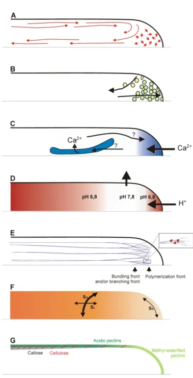
Figure 1.5: Schematic representation of the polar arrangement of cellular structures and processes in the apical and subapical regions of a growing pollen tube. ... 62
Figure 1.6: Compilation of cellular features and processes that have been observed to undergo changes during oscillatory pollen tube growth. ... 63
Figure 2.1: Arabidopsis thaliana pollen viability test using fluorescein diacetate (FDA). .. 84
Figure 2.2: Change of Arabidopsis pollen viability and germination percentage with duration of cold storage. ... 85
Figure 2.3: Effect of calcium concentration on the percentage of germination of Arabidopsis thaliana pollen. ... 86
Figure 2.4: Effect of calcium concentration on percentage germination of Arabidopsis thaliana pollen. ... 87
Figure 2.5: Effect of boron concentration on the percentage germination of Arabidopsis thaliana pollen. ... 88
Figure 2.6: Effect of potassium concentration on the percentage germination of Arabidopsis thaliana pollen. ... 89
Figure 2.7: Effect of the sucrose concentration on the percentage germination of Arabidopsis thaliana pollen. ... 90
Figure 2.8: Arabidopsis pollen germination on agarose medium. ... 91
Figure 2.10: Effect of the temperature on the percentage germination of Arabidopsis
thaliana pollen. ... 93
Figure 2.11: Micrographs of Arabidopsis pollen tubes. ... 94
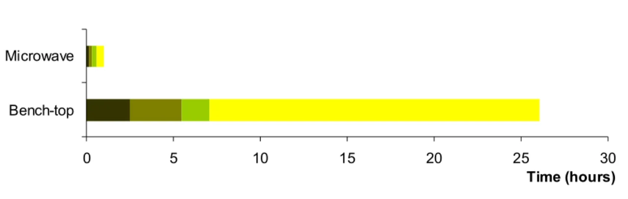
Figure 2.12: Comparaison of Arabidopsis pollen percentage of germination and pollen tube length after five hours of growth on different agarose stiffened media. ... 95 Figure 3.1: Immunofluorescence label of lily pollen tubes... 103
Figure 3.2: Callose rings observed with aniline blue staining in Camellia pollen tubes. ... 103
Figure 3.3: Camellia pollen tube actin cytoskeleton as visualized by rhodamine-phalloidin label. ... 104 Figure 3.4: Transmission electron micrograph of a cross-section of a Camellia pollen tube.104
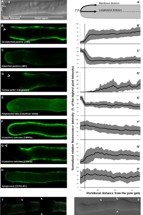
Figure 3.5: Scanning electron micrograph of an Arabidopsis leaf trichome and germinated lily pollen grains. ... 105 Figure 3.6: Comparison of experimentation time between bench-top (orange) and microwave assisted (red) methods of sample preparation for optical microscopy. ... 106 Figure 3.7: Comparison of experimentation time between conventional bench-top and microwave assisted methods of sample preparation for transmission electron microscopy.106 Figure 4.1: Organization of the cytoarchitecture and relative spatial distribution of cell wall components in the Arabidopsis pollen tube. ... 132 Figure 4.2: Transmission electron micrographs of immunogold label for cell wall polysaccharides. ... 132 Figure 4.3: Transmission electron micrographs of immunogold label for crystalline cellulose in freeze fixed Arabidopsis pollen tubes. ... 134 Figure 4.4: Fluorescence micrographs of pollen tubes labeled with the styryl dye FM4-64, CBM3a and VAEM image of a pollen tube expressing GFP-CESA6. ... 135 Figure 4.5: Scanning electron micrograph of the surface of Arabidopsis pollen tubes and VAEM images of pollen tube expressing GFP-CESA6... 136
Figure 4.6: Relative fluorescence intensity after enzymatic treatments. ... 137
Figure 4.7: Distribution of pectins and cellulose after enzymatic treatments. ... 139
Figure 4.8: Label for crystalline cellulose (CBM3a) in pollen tubes digested with pectinase and lyticase. ... 139 Figure 4.9: Spatial profile of cellular stiffness along the longitudinal axis of the
Arabidopsis pollen tube... 140
Figure 4.10: Conceptual model of the assembly and structure of the pollen tube cell wall.142
Figure 5.1: Mechanical anisotropy of cell wall mechanics in Lilium pollen tubes. ... 162
Figure 5.2: Mechanical anisotropy of cell wall mechanics in Solanum pollen tubes. ... 163
Figure 5.3: Effect of cellulase on pollen tube length and diameter measured 2h after germination for Lilium and 4h for Solanum. ... 164 Figure 5.4: Fluorescence label of Lilium orientalis pollen tubes for pectins. ... 165
Figure 5.5: Fluorescence label for cellulose and callose in Lilium orientalis pollen tubes. 166
Figure 5.6: Fluorescence label for cellulose in Solanum chacoense pollen tubes... 167
Figure 5.7: Effect of CGA on the diameter of Solanum chacoense and Lilium orientalis pollen tubes germinated in the presence of this agent. ... 168 Figure 5.8: Effect of CGA administration on the morphology of pre-germinated Lilium tubes. ... 169 Figure 5.9: Schematic drawing of strain rates and hypothetical configurations of microfibrils in the pollen tube cell wall. ... 170 Figure 5.10: Schematic drawing of the mechanical properties and chemical composition characterizing the pollen tube cell wall. ... 171 Figure 6.1: Differential interference contrast micrographs of in vitro growing lily pollen tubes. ... 196 Figure 6.2: Finite element structure of a tip growing cell. ... 197
Figure 6.3: Subset of simulations showing the deformation of the cell wall structure after 50 load cycles. ... 198 Figure 6.4: Spatial distribution of the Young's modulus in meridional direction of a representative subset of simulations. ... 199 Figure 6.5: Validation of simulations. ... 200
Figure 6.6: Effect of cell wall thickness, turgor pressure, tube radius, and structure on the growth pattern. ... 201 Figure 6.7: Spatial distribution of cell wall components in in vitro growing lily pollen tubes. ... 202 Figure 6.8: Effect of pectin digestion on pollen tube shape. ... 203
Figure 7.1: Expression of pectate lyases in pollen tubes and hydrated pollen grains. ... 225
Figure 7.2: Expression of pectate lyases in wild type and knock out lines. ... 226
Figure 7.3: Percentage of germination and pollen tube length in knock out lines, complemented lines and wild type grown in in vitro conditions. ... 227 Figure 7.4: Localization of PLs in pollen grains and tubes... 228
Figure 7.5: Periplasmic localization of PL2-YFP in plasmolysed pollen tubes. ... 229
Figure 7.6: FRAP analysis of PL1-CFP dynamics in the apex of a growing pollen tube. . 230
Figure 7.7: Immuno-detection of PL2-GFP in the pollen tube growth medium... 231
Figure 7.8: Conceptual model for the localization and dynamics of PLs during pollen tube development. ... 233 Supplemental Figure 7.1 : Relative expression levels of endogenes and transgenes assessed by semi-quantitative RT-PCR in whole Arabidopsis flowers. ... 235 Figure 8.1: General principles used in gravity research. ... 255
Figure 8.3: Spatial profile of mechanical properties and biochemical processes in the cell wall of growing pollen tubes ... 257 Figure 9.1: Large Diameter Centrifuge at the research facilities of the European Space Research and Technology Centre of the European Space Agency in Noordwijk, The Netherlands. ... 277 Figure 9.2: Response of Camellia pollen tube morphology to altered gravity conditions. 278
Figure 9.3: Relative spatial distribution of the cell wall components in Camellia pollen tubes grown in omnidirectional-g, at 1g and in 5g conditions ... 279 Figure 9.4: Geometry and fluorescence intensity of the apical vesicle cone. ... 280
Supplemental Figure 9.1: Relative spatial distribution of cellulose and callose in Camellia pollen tubes grown in omnidirectional-g, at 1g, and hyper-g. ... 281 Supplemental Figure 9.2: Confocal laser scanning micrographs of a pollen tube grown in omnidirectional-g labelled with (1→3)-β-glucan against callose. ... 282
List of movies
Supplemental Movie 4.1: VAEM of Arabidopsis pollen tube expressing GFP-CESA6. .. 142
Supplemental Movie 7.1: Localisation of pPL2:PL2-YFP expressed in Arabidopsis pollen tube. 3D reconstruction from stacks taken at 0.5 µm interval in false colors where higher fluorescence intensity in green and lower intensity in purple. Width of the frame equals 55 µm. ... 236 Supplemental Movie 7.2: Arabidopsis pollen grain expressing pPL2:PL2-YFP during the first stages of germination and during pollen tube growth. Images were acquired at a rate of 60 frames.s-1. The video was reconstructed from 4 separate videos of the same tube. Width of the frame equals 55 µm. ... 236 Supplemental Movie 9.1: Immuno-localization of callose in Camellia pollen tubes grown in omnidirectional-g conditions. 3D reconstruction from z-stack of confocal laser scanning micrographs acquired at 0.5 µm interval. Width of the frame equals 225 µm. ... 282
Abbreviation list
ABP Actin binding protein ADF Actin depolymerizing factor ATP Adenosine triphosphate
BK Brewbaker and Kwack
BP Band pass
BSA Bovine serum albumin CBM3a Cellulose Binding Module 3a CCD Charged coupled device
cDNA Complementary DNA
CESA Cellulose synthases CFP Cyan fluorescent protein
CGA 1-cyclohexyl-5-(2,3,4,5,6-pentafluorophenoxy)-1λ4,2,4,6-thiatriazin-3-amine DCB 2,6-dichlorobenzonitrile
DIC Differential interference contrast DNA Deoxyribonucleic acid
EGTA Ethylene glycol tetraacetic acid ER Endoplasmic reticulum
ESTEC-ESA
European Space Research and Technology Centre – European Space Agency
FDA Fluorescein diacetate
FE Finite element
FP Fluorescent protein
FRAP Fluorescence recovery after photobleaching FTFLP Fluorescent tagging of full length protein
g Earth gravity level GalA Galacturonic acid GAX Glucoarabinoxylan GFP Green fluorescent protein GTP Guanosine triphosphate
HG Homogalcturonan
Ig Immunoglobulin
LDC Large diameter centrifuge
LP Long pass
MW Microwave
NAD Nicotinamide adenine dinucleotide
NAD(P)H Nicotinamide adenine dinucleotide phosphate-oxidase PB Phosphate buffer
PBS Phosphate buffer saline PCR Polymerase chain reaction
PG Polygalacturonase
PL Pectate lyase
PME Pectin methylesterases
PMEI Pectin Methylesterase Inhibitor PtdInsP2 Phosphatidylinositol 4,5-bisphosphate
RGI Rhamnogalacturonan I RGII Rhamnogalacturonan II RNA Ribonucleic acid
ROS Reactive oxygen species RPM Random position machine
Rpm Revolution per minute RT Reverse transcription
RT-PCR Real time polymerase chain reaction
SDS-PAGE
Sodium dodecyl sulfate polyacrylamide gel electrophoresis SEM Scanning electron microscopy
SI Self incompatibility
SNARE Soluble N-ethyl-maleimide-sensitive fusion protein attachment protein receptors
TBS Tris-buffered saline T-DNA Transfer DNA
TEM Trransmission electron microscopy TIRF Total internal reflection fluorescence UDP Uracil di-phosophate
UTR Untranslated region
UV Utlra-violet
VAEM Variable angle epifluorescence microscopy XGA Xylogalacturonans
XTH Xyloglucan endotransglycosylase/hydrolases
XyG Xyloglucans
Nothing shocks me. I'm a scientist
Indiana Jones,
To the best parents one could wish for who always taught me to pursue
my dreams till the end and never surrender
in front of any obstacle…
Acknowledgments
I would first like to thank Prof Anja Geitmann for believing in me. It is an honour and a privilege to have her as a mentor. I am most grateful for all the scientific opportunities, support, guidance, tutoring and help she offered me. I would also like to thank her for her humanity, care, generosity and friendship she continuously shows. I am touched and deeply thankful for everything, Anja.
Without the constant and indefectible support, counsel, blessings and prayers of my parents and my family, none of this would have been possible. Thank you Mom, Dad, Ziz, Naaman, Dalal, Nay, Nad and Léyou for being who you are and for your perpetual encouragements. I would also like to thank all my family in Lebanon, Belgium and Canada for their kindliness, I love you all!
Their encouragements, counsel, help and presence at all times were primordial throughout the evolution of this thesis. I am profoundly grateful to Simon Desharnais, Leila Aouar, Firas Bou Daher, Chloe van Oostende Triplet and Thomas Triplet for everything they did and are still doing!
I would like to thank my friends Monisha Sanyal, Mahsa Naghavi, Minako Kaneda and Samuel Juillot, as well as all my colleagues Marie Glavier, Pascal Arpin, Amir Jafari, Robert Palin, Xudong Sun, Olivier Gossot, Rabah Zerzour, Jérôme Bove, Pierre Fayant and Orlando Girlanda for all the help they provided.
During these years, I had the opportunity to supervise several internship students. Thank you Maxime Grare, Aude Kouma, Lauranne Pujol, Anahid Shojaeifard and Marius Andre for your hard work, commitment and for the excellent times we shared together.
A very special thank to my friend Louise Pelletier for her kindness, her invaluable help in microscopy, her precious advices and for all the care she shows.
I thank all the staff members, professors and students of the Institut de Recherche en Biologie Végétale for all the help and support they provided.
I would like to thank the European Space Agency for giving us the opportunity and providing the technical expertise, the funding and the logistics to study the effect of hypergravity on pollen tube development. I am particularly thankful to Dr. Jack van Loon and to the Spin Your Thesis! staff: Dr. Natacha Callens, Tim Setterfield, Carlos Gomez-Calero, Jutta Krause and Alan Dowson.
I wish to thank all the agencies who funded me throughout my Ph.D: The Ann Oaks doctoral scholarship of the Canadian Society of Plant Biologists / La Société canadienne de biologie végétale. The Canadian Space Agency / L’Agence spatiale canadienne. The Marie-Victorin doctoral scholarship of the Institut de Recherche en Biologie Végétale. And various Université de Montréal scholarship programs.
I would like to thank all the members of the jury for critically reading this thesis and providing edifying comments.
Finally I would like to thank God for putting on my way such incredible people and opportunities.
1
Introduction
The first plants probably appeared on Earth around 1 200 million years ago (during the mezoproterozoic era) in form of algal scum, although the first fossil evidence of land plants dates from 470 million years ago (Wellman and Gray 2000), and it is generally thought that plants started to colonize the land between 630 and 510 million years ago (Raven and Edwards 2001; Clarke et al. 2011). During evolution, different groups of land plants appeared successively. The first to appear were non-vascular bryophytes (liverworts, worts and mosses), while seedless vascular plants made their appearance along with lycophytes and pterophytes around 425 million years ago. Gymnosperms (vascular seed plants) appeared roughly 325 ± 50 million years ago followed by angiosperms (or flowering vascular seed plants) 200 ± 50 million years ago (Jiao et al. 2011).
Plants have contributed to the development of mankind in several ways. More generally, due to their capacity for oxygenic photosynthesis, they have contributed to the richly oxygenated atmosphere that has allowed animal life to develop extensively. Human life, appearing only around 4.5 million years ago, has depended on plants as the foundation for development since the dawn of civilization. Not only are plants essential for food, nutrient supplies and medication, they have also been used as day-to-day tools to tie silex stones together, to set traps for animals and to construct shelters. With time, the importance of plants became more pronounced in fulfilling daily needs, being used for clothing, paper, furniture, housing construction, isolators, and biofuels. Interestingly, most of these applications rely on the composition and structure of the plants in general and in particular, on one major component of the plant cell. This component, called the cell wall, is an outer envelope rich in carbohydrates and polymers that confers to the cell its characteristic mechanical properties.
1.1
The plant cell wall: Role and importance of its mechanical
properties
The plant cell wall is a tough outer structure surrounding plant cells. It is characterized by a heterogeneous chemical composition that confers, to each different cell and tissue, specific mechanical properties defined by tensile strength, rigidity, elasticity, and plasticity. These characteristics crucially influence most industrial applications of plant material ranging from the use of wood for construction to the texture of plant-derived food products.
On a cellular level, the plant cell wall provides the cell with protection from pathogen infections, acts as a filtering mechanism and most importantly, plays a structural role (Essau 1977). Because of the cell wall, plant cells are able to build up a high internal pressure called turgor. If the cell wall mechanical properties are not well balanced with the turgor pressure, the cell risks bursting. The cell wall also allows plants to stand upright, as without it, plants would only be a pile of cells collapsed by gravity and without a defined structure. The cell wall thus has a function analogous to the extracellular matrix of animal cells. During plant development, the balance and spatio-temporal regulation of the mechanical properties of the cell wall are therefore vital: while cell growth is ongoing, the cell wall is characterized by higher elastic and malleable properties than the already differentiated cells that have stopped growing and are more rigid.
1.2
Primary and secondary cell walls
The layers of the plant cell walls can be distinguished into two types, a primary cell wall that is formed during cell morphogenesis and that is highly flexible, and a secondary cell wall, which is formed of more complex molecules and appears when the cell has stopped growing. The secondary cell wall is not formed in all cell types but is restricted to cells requiring structural reinforcements such as xylem fibers, tracheids, or sclereids. The secondary cell wall accounts for most of the carbohydrate biomass of the plant and provides additional protection and strength to the whole organism. The secondary wall is deposited between the plasma membrane and the primary cell wall of cells that have stopped
expanding and need mechanical reinforcement. For example, wood consists mostly of secondary cell wall; its mechanical role is to hold the plant up against the gravity.
While the primary cell wall consists mainly of a cellulose-hemicellulose network embedded in a matrix of pectins and proteins and is generally flexible and extensible, the secondary cell wall is more rigid and is mainly formed of cellulose and xylan and contains many glycoproteins and polysaccharides as well as other compounds such as lignin and cutin. Contrary to the primary cell wall which is basically formed of one layer, the secondary cell wall is usually formed of different laminated sheaths where the cellulosic fiber orientation is parallel within an individual layer but can differ between the layers. The importance of the cellulose fiber orientation is detailed in paragraph 1.7. The subsequent deposition of hydrophobic components such as lignin drastically reduces the permeability of the secondary cell wall. Hence, water transport across the secondary cell wall is severely limited and is accomplished through specialized openings in the cell wall called pits. Pits can be found mostly between vascular cells (tracheids and vessel elements) and between parenchyma cells (Essau 1977; Raven et al. 1986; Cosgrove 2005; Obel et al. 2007).
During morphogenesis, the undifferentiated somatic cell has to undergo several differentiation steps to acquire its final function, size and shape. During this process, the cell undergoes drastic morphological changes. From a relatively simple spherical shape, the cell will develop into a body with more complex geometry such as in jigsaw puzzle-shaped pavement cells, in trichomes, in fibres, in sclereids or in pollen grains. Because the cell wall is the most important architectural element of the plant cell, it plays a central role throughout the morphogenetic processes. The regulation of the local cell wall mechanical properties (reflected by the local chemical composition) is decisive in shaping individual cells, organs and organisms.
1.3
State of the art
The goal of this thesis is to understand the role played by the different cell wall components in shaping the cell and controlling the growth behaviour.
To achieve this goal, I rely on a specific experimental model system, the male gametophyte or pollen tube. The reasons for the use of this system are detailed in the paragraph 1.7.4 (importance of the pollen and the pollen tube) below.
In the following sections of the introduction, I review first what is known about the primary cell wall components in plant cells from a metabolic, regulatory and mechanical point of view. I also review the factors that influence the biological and mechanical regulation of plant cell growth. In a second part, I review what is known about pollen tube growth and finally I expose the goals and objectives of the present thesis. The thesis will be constructed from 8 chapters exploring the roles of the different cell wall components (mainly cellulose and pectins) and the role of several biotic (pectate lyases and pectinase) and abiotic (omni-directional gravity and hyper-gravity) factors in regulating pollen tube shape and growth behaviour. The last part of the thesis consists in a general discussion and elaborates on the perspectives for future research.
1.4
Pectins in the primary cell wall
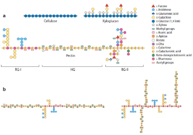
Pectins are the major component of primary cell walls and of the middle lamella in dicotyledonous plants where they can amount to around 35% of the cell wall dry weight (Smith and Harris 1999). They are rare in non-extensible secondary cell walls. Pectins are polysaccharides rich in galacturonic acid (GalA) with an extremely complex composition as they may contain up to 17 different monosaccharides (Willats et al. 2001). Pectins form a highly heterogeneous group. Three different types of polysaccharides can be distinguished depending on the diversity of both their backbones and their side chains: Homogalacturonan (HG), Rhamnogalacturonan I (RGI) and Rhamnogalacturonan II (RGII) (Figs 1.1 and 1.2).
HG and RGII have the same backbone formed by a linear polymer of (1,4)-linked-α-d-GalA. HG do not have any side chains and can be methyl-esterified at C-6 or carry acetyl groups at O-2 and O-3. The GalA may be substituted at C-2 and C-3 with an apiose thus forming apiogalacturonans found in certain species such as Lemna minor and
Spirodela polyrrhiza (Hart and Kindel 1970; Longland et al. 1989). The substitution at C-3
with a xylose leads to the formation of xylogalacturonans (XGA) that can be found in the seed coats of pea, apple, watermelon fruit and carrot cells (Kikuchi et al. 1996; Schols and Voragen 1996; Renard et al. 1997; Le Goff et al. 2001). In RGII, four heteropolymeric side chains containing more than eleven different sugars can be attached to the main backbone (Vidal et al. 2000). The structures of RGII are very well conserved in plants. RGII can also be cross-linked by borate ester links through their apiosyl residues (Ishii et al. 1999; Pelloux et al. 2007). The backbone structure of RGI differs from HG and RGII as it consists of repeated [(1→2)-α-l-rhamnose-(1→4)-α-d-galacturonate]n disaccharide units,
where n can be greater than 100 (McNeil et al. 1980; Vincken et al. 2003). Galacturonyl residues can be acetylated at their O-2 or O-3 positions and rhamnosyl residues can be substituted at O-4 with neutral sugars (Vincken et al. 2003). The diversity of pectins is summarized in figure 1.1.
Pectins are synthesized from nucleotide sugars in the Golgi apparatus. They are polymerized in the cis-Golgi by at least 53 different glycosyl-transferases. Pectins (mainly HG) are then methylesterified in the medial-Golgi by methyl-esterases at the C-6 carboxyl of GalA. Side chains are then attached to the backbone in the trans-Golgi. Pectins are secreted in a highly esterified form (70 to 80% of all carboxyl groups are esterified) into the apoplast through vesicle exocytosis (Willats et al. 2001; Vincken et al. 2003).
Figure 1.1:Structure of some cell wall components.
(A) Schematic representation of cellulose, xyloglucan, and the pectic rhamnogalacturonan I (RG-I), homogalacturonan (HG), and RG-II. Adapted from (Burton
et al. 2010) with permission. (B) Alternative models for pectin domain organization. A
linear, contiguous arrangement of HG interspersed with RG-I is shown on the left, whereas on the right, HG is drawn as side chains linked to Rha residues of the RG-I scaffold. A combination of both models may also be possible. Adapted from (Round et al. 2010) with permission. This figure and legends were taken from (Wolf et al. 2012).
Pectins are the major adhesive material between cells; its network serves as a matrix for the deposition, slippage and extension of the cellulose-glucan network and is involved in the regulation of cell wall porosity and modulation of the cell wall pH and ion balance (Cosgrove 1999; Willats et al. 2001). Pectins also play the role of signalling molecules during plant defence reaction to pathogen and insect attacks (Collmer and Keen 1986). Because of their stabilizing properties, pectins are wildely used in the food industry as a gelling and thickening agent and many studies have investigated their rheological and
gel-forming properties depending on their pattern of methyl-esterification (Daas et al. 2000; Osborne 2004).
The degree of pectin esterification plays an important role in determining the mechanical properties of the cell wall. Pectins are secreted at the cell wall in a highly esterified form and subsequently subjected to the action of different enzymes such as pectin methylesterases (PME), pectate lyases (PL) and polygalacturonases (PG) and different ions such as boron or calcium. PME is known to lead to the de-esterification of pectins in muro and render them susceptible either to gelation by divalent ions such as Ca2+ or to the action of pectin degrading enzymes such as PL and PG, giving the cell wall different mechanical properties. PMEs, PLs and PGs are known to be implicated in many physiological processes in plant cells such as cell differentiation, cell wall expansion, cellular adhesion and maturation and senescence of fruits (Bosch and Hepler 2005).
1.4.1 Pectin-methyl-esterases and pectins
PMEs catalyze the specific demethylesterification of HG in muro at the C-6 position of GalA, releasing methanol and generating a negatively charged carboxyl group. PMEs have two modes of action; they can either act in a block-wise or in a random fashion. PME isoforms working in a block-wise fashion create long blocks of contiguous free carboxyl groups susceptible to interact with Ca2+, thus forming the ‘egg-box’ model structure resulting in a stiff gellified three dimensional pectate network (Willats et al. 2001). At least 9 contiguous carboxyl residues are required to favour calcium cross-linking (Liners et al. 1992). PME isoforms acting randomly lead to a random demethylesterification of the GalA, thus promoting the action of pectinases such as PL and PG, leading to cell wall loosening (Willats et al. 2001; Pelloux et al. 2007).
Different PME isoforms were identified and isolated in different plant species and tissues. In Arabidopsis, 66 ORFs have been annotated as putative full-length enzymes (CAZy, http://www.cazy.org)(Cantarel et al. 2009) and transcriptomic studies showed that more than 22 PMEs are expressed in the pollen and leaves (Honys and Twell 2003; Pina et
in the development and differentiation (Pina et al. 2005) of organs such as mucilage release from seeds, during pollen and pollen tube development, and in hypocotyl and apical meristem differentiation (Micheli 2001; Jiang et al. 2005; Francis et al. 2006; Peaucelle et
al. 2008; Rautengarten et al. 2008). In many other plant species PMEs have also been
isolated from various organs such as the poplar and mung bean cambium, Vigna radiata hypocotyls, the pea root border cells, the yellow cedar seeds, Solanum tuberosum stems, carrot roots, Peunia inflata pollen, and in the development of a plethora of fruits like apple, kiwi, water melon, fig tomato, strawberries, berries, bananas (Versteeg et al. 1978; Kozo et
al. 1990; Bordenave et al. 1996; Louvet et al. 2006). The activity of several PME isoforms
was found to be temporally and spatially regulated during cell wall formation and maturation in seeds, hypocotyls, root hairs and cambium. Collectively, these findings suggest that during the first developmental stages of the above mentioned tissues, PME isoforms working in a non-blockwise fashion seem to be activated to loosen the cell wall by facilitating the action of PL and PG and thus allowing cell wall expansion. In later stages, during cell wall maturation, other isoforms working in a blockwise manner seem to be activated, leading to stiffening the cell walls via the formation of calcium bridges (Bordenave and Goldberg 1994; Bordenave et al. 1996; Ebbelaar et al. 1996; Guglielmino
et al. 1997; Micheli et al. 2000; Ren and Kermode 2000; Micheli 2001; Louvet et al. 2006;
Western 2006; Derbyshire et al. 2007; Rautengarten et al. 2008; Arsovski et al. 2010). PMEs can be divided into two groups (group I and group II) depending on the absence or presence of a PRO region in the N-terminus of the mature protein. The PRO region shares sequence similarities with a PME inhibitor (PMEI) domain (Camardella et al. 2000) and exerts an inhibitory effect on PME activity (Giovane et al. 2004; Röckel et al. 2008) as shown in tobacco pollen tubes by inhibiting premature deesterification of the pectins in the Golgi apparatus (Bosch and Hepler 2005). In Arabidopsis, the PMEs of group I do not have PRO regions; they are characterized by five or six introns and have a molecular weight under 45 kDa, whereas the PME of group II have one to three PMEI-like domains, two to three introns and a molecular weight comprised between 50 and 105 kDa (Micheli 2001; Tian et al. 2006). Both groups of PMEs are targeted to the cell wall and the presence or absence of the PRO region does not influence the targeting to the plasma
membrane since the PMEs of group I contain a N-terminus rich in hydrophobic amino acids which could play the role of a membrane target (Camardella et al. 2000; Micheli 2001; Al-Qsous et al. 2004; Bosch et al. 2005; Bosch and Hepler 2005; Tian et al. 2006; Pelloux et
al. 2007).
1.4.2 Pectate lyases (PLs) and pectins
Pectate lyases (also known as pectate trans-eliminases) are known in plants and fungi to hydrolyse the cell wall by transforming non-esterified pectins into unsaturated oligogalacturonades, leading to an increase in the elasticity and/or plasticity of the cell wall. PL activity has been shown for the first time in 1962 in pathogeneous cultures of Bacillus
sp. (Starr and Moran 1962). The structures and roles of bacterial PLs have been well
studied in Erwinia chrysanthemi (Wing et al. 1990): the bacterial PLs hydrolyse the plant cell walls which leads to the necrosis and death of the infected tissues (Yoder et al. 1993; Lietzke et al. 1994; Pickersgill et al. 1994; Scavetta et al. 1999; Herron et al. 2000; Herron
et al. 2003).
In plants, the majority of the studies related to PLs were conducted in the context of fruit maturation. In tomato (Marin-Rodriguez et al. 2002), banana (Pua et al. 2001) and strawberry (Jimenez-Bermudez et al. 2002) PLs play a role during fruit maturation and studies showed that when PL expression was knocked-out, fruit senescence was retarded. Studies conducted on the pollen of tomato (Kulikauskas and McCormick 1997) showed the presence of highly conserved homologous PL coding sequences with Erwinia. These sequences were also found to be highly expressed in anthers and pistils of tomato (Kulikauskas and McCormick 1997). PLs are also known to pose a problem of public health as many of them are thought to be implicated in human allergic reactions to pollen (Taniguchi et al. 1995; Shreffler 2011; Augustin et al. 2012; Suzuki et al. 2012).
A number of studies have shown that PL-like genes are highly expressed in pollen grains from tobacco, Arabidopsis (Kulikauskas and McCormick 1997), alfalfa (Wu et al. 1996) and Japanese cedar (Taniguchi et al. 1995), leading to speculation that they might be implicated in the early stages of pollen germination or are employed by the pollen tube to
digest the transmitting tract. Several PLs and PL-like genes have been shown to be expressed in Nicotiana tabacum pollen (Wing et al. 1990; Kulikauskas and McCormick 1997), and in Arabidopsis more than 20 PL-like genes (Pina et al. 2005) are expressed in pollen grains. However PLs are known to be synthesized in pollen, it is unknown what role they play. They could potentially be involved in initiating pollen tube growth by softening the aperture wall or could be involved in regulating the cell wall mechanical properties of the elongating tube. Alternatively, they might not act on the pollen itself, but on the apoplast of the pistillar tissues with the purpose of loosening it to facilitate pollen tube invasive growth. These functions are not mutually exclusive. In chapter 6 I show and discuss in more detail the role of PLs during pollen germination and pollen tube growth.
1.4.3 Polygalacturonases and pectins
Other genes coding pectin-degrading enzymes such as pectin-esterases, β-galactosidase and polygalacturonases are expressed during cell differentiation. Their major roles, just like PLs, are to cleave pectins and increase the elasticity of the cell walls. These enzymes are known to be expressed in different organs like leaves, seeds, roots, flowers and in pollen grains as shown by transciptomic studies (Torki et al. 2000; Pua et al. 2001; Pina et al. 2005). They were shown to be required during senescence and abscission of certain organs and fruits (Torki et al. 2000). They are also known to be secreted by fungi and bacteria to facilitate their penetration into the plant tissues.
Many of these enzymes are specifically expressed in pollen grains and pollen tubes of
Brassica napus (Robert et al. 1993; Daas et al. 2000), maize (Niogret et al. 1991) and
tobacco (Tebbutt et al. 1994), but despite the fact that they are studied in more detail than PLs, their roles in pollen development are still not clear. Just like PLs, their putative role during pollen and pollen tube development could be to facilitate pollen tube elongation by hydrolyzing the middle lamella of the transmitting tissue.
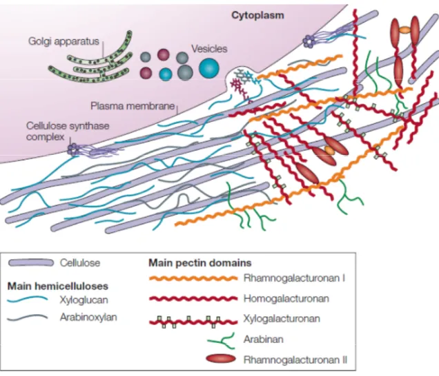
Figure 1.2: Structure of the primary cell wall.
Cellulose microfibrils (purple rods) are synthesized by large hexameric complexes in the plasma membrane, whereas hemicelluloses and pectins, which compose the matrix polysaccharides, are synthesized in the Golgi apparatus and are deposited to the wall surface by vesicles. For clarity, the hemicellulose–cellulose network is shown on the left part of the cell wall without pectins, which are emphasized on the right part of the figure. In most plant species the main hemicellulose is xyloglucan (blue), while hemicelluloses such as arabinoxylans (grey) and mannans (not shown) are found in lesser amounts. The main pectin polysaccharides include rhamnogalacturonan I and homogalacturonan, with smaller amounts of xylogalacturonan, arabinan, arabinogalactan I (not shown) and rhamnogalacturonan II. Pectin domains are believed to be covalently linked together and to bind to xyloglucan by covalent and non-covalent bonds. Neutral pectin polysaccharides (green) are also able to bind to cellulose surfaces. This figure and legend were taken from (Cosgrove 2005).
1.5
Cellulose in the primary cell wall
Cellulose (1,4-β-D-glucan) is the most abundant biopolymer on Earth. It accounts for more than a third of the total dry weight in plants. It is most abundant in secondary cell walls (Fig 1.2), but the primary cell wall contains a significant amount as well. Cellulose microfibrils are formed by parallel chains of 500 to 14 000 β-1,4-glucose linked by hydrogen bonds and by xyloglucans chains (Somerville 2006). Cellulose microfibrils are 3 to 5 nm wide and can reach many micrometers in length (Cosgrove 2005).
1.5.1 Cellulose synthesis
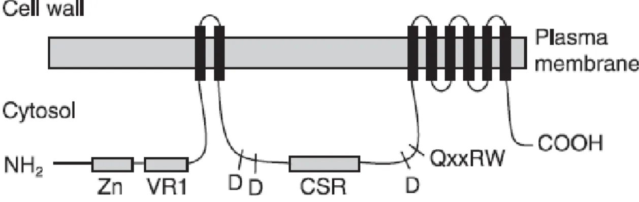
Cellulose is synthesized at the plasma membrane by large enzymatic complexes formed by cellulose synthases (CESA) identified in the late 1990s by molecular and genetic studies (Pear et al. 1996; Arioli et al. 1998). In angiosperms, cellulose synthases are organized in hexameric arrays called rosettes which were revealed by freeze fracture (Kimura et al. 1999). The rosettes are located at the plasma membrane and contain six rosette subunits each of which is formed from six CESA subunits encoded by three different CESA genes. The dimerization of different CESAs is thought to be mediated by two adjacent zinc fingers in the N-terminal region of each CESA protein (Kurek et al. 2002; Cosgrove 2005). Each species is characterized by a defined number of CESA genes (10 for Arabidopsis, 12 for maize, 9 for rice, 8 for barley, 7 for aspen ...) with the sequences of the respective orthologues being closer than that of the paralogues (Somerville 2006; Taylor 2008) suggesting that from an evolutionary point of view, the different CESA families were established in the common ancestors of vascular plants. Different sets of genes are required for the synthesis of cellulose in the primary (AtCESA1, AtCESA2,
AtCESA3, AtCESA5, AtCESA6, AtCESA9) and secondary walls (AtCESA4, AtCESA7, AtCESA8) (Arioli et al. 1998; Fagard et al. 2000; Taylor et al. 2003; Cosgrove 2005;
Taylor 2008). Because of the structure of the rosette, it is thought that cellulose microfibrils are made of 36 glucan chains (Taylor 2008). However, recent studies have shown that because of geometrical and mechanical constraints it is more likely that 18 to 30 glucan chains form the cellulosic microfibrils (Emons et al. 2007). Proteomic analysis coupled to computational methods showed that CESA are characterized by eight trans-membrane
domains (Nühse et al. 2004). The structure of CESA is summarized in figure 1.3. The cellulose synthases are not fixed at one position, but are in constant movement in the plane of the plasma membrane (Paredez et al. 2006). This movement seems to be guided by cortical microtubules, but can also occur independently of cytoskeletal guidance. It is thought to be driven in self-generated fashion by the forces created by polymerization and crystallization of the cellulose microfibrils (Emons et al. 2007).
Figure 1.3: Membrane topology of a CesA protein.
Black bars indicate trans-membrane domains. Zn, zinc-binding region; VR1, variable region 1; CSR, class-specific region. Approximate positions of D, D, D, QXXRW processive glycosyl transferase motifs are shown. This figure was taken from (Taylor 2008).
1.5.2 Regulation of cellulose synthesis
Other proteins are known to be important for cellulose synthesis regulation: KORRIGAN, COBRA and KOBITO. KORRIGAN encodes for a endo-β-1,4-glucanase and was shown to be required during normal cellulose synthesis in primary and secondary cell walls (Nicol et al. 1998; Lane et al. 2001). Mutations in KORRIGAN result in improper crystallization of cellulose microfibrils and reduced amount of cellulose in the primary and secondary cell walls. This lead to a defect in cytokinesis and cell elongation, dwarfism (Nicol et al. 1998), radial swelling of root tips (Lane et al. 2001) and collapse of xylem
vessels (Szyjanowicz et al. 2004). KORRIGAN is not directly associated with CESA complexes, its roles during cellulose synthesis are not clearly defined and attempts to characterize the subcellular localization of the protein have been inconclusive and inconsistent (various studies proposed association with Golgi, endosomes, primary and secondary cell walls). COBRA encodes a glycosyl-phosphatidylinositol (GPI)-anchored protein targeted to the plasma membrane. Knock-out COBRA mutants show a reduction in the amount of synthesized cellulose and a disorganized cellulosic net orientation. This results in deficiency of polarized cell growth of most developing organs in Arabidopsis (Roudier et al. 2005). KOBITO encodes a protein of unknown function but mutants for this gene were characterized by a dwarf phenotype and randomized microfibrils orientation (Pagant et al. 2002).
1.6
Hemicelluloses in the primary cell wall
Hemicelluloses are defined as being “wall polysaccharides that are not solubilized
from wall materials with buffers, hot water, or chelating agents but only with more or less strong chaotropic agents such as alkali’’ (Obel et al. 2007). They comprise several families
of polysaccharides that have in common a β-1,4-d-pyranosyl backbone but that differ from each other and from cellulose by their respective side chains. Depending on the chemical nature of these side chains, three major families of hemicelluloses can be distinguished, the xyloglucans, the mannans and the xylans. These families can be subdivided in sub-families depending on the nature of all their side chains (Fig 1.2).
1.6.1 Xyloglucans
Xyloglucans (XyG) consist of a backbone of 1,4-linked β-d-Glc with several α-d-Xyl substituents at O6. They are characterized by several side chains made of galactose, fucose, xylose or glucose. The nature of the side chains varies depending on the tissue and on the local physiological requirements. They are the most abundant hemicelluloses present in the cell walls of dicotyledon plants (Obel et al. 2007). The XyG backbone is synthesized in the Golgi cis-cisternea whereas the addition of the side chains by fucosylation or galactosylation occurs in the trans-cisternea and in the transport vesicles (Brummell et al.
1990). Once exported to the apoplast by exocytosis, XyG are directly bound to cellulose in the cell wall (Edelmann and Fry 1992) in a non-covalent way (Acebes et al. 1993; Hayashi
et al. 1994a), probably with hydrogen bonds, since their binding to cellulose is
pH-dependent (Hayashi et al. 1987). Several enzymes such as glucosyl- and xylosyltransferases (Gibeaut and Carpita 1994), XyG-glucan synthase, α-Xylp-transferase (AtXT1) (Faik et al. 2002), α-Fucp-transferase (Faik et al. 2000; Vanzin et al. 2002; Madson et al. 2003), GDP-D-mannose 4,6-dehydratase (Bonin et al. 1997), fucosidase, β-galactosidase, α-xylosidase, and β-glucosidase (Iglesias et al. 2006; Obel et al. 2007) as well as a multitude of nucleotide sugar interconversion enzymes (Reiter and Vanzin 2001) are implicated in the different stages of XyG synthesis and regulation. In Arabidopsis, eight allelic mutants were identified as being implicated in the de novo synthesis of xyloglucans and more specifically of l-fucose. These genes were named the mur mutants in reference to the wall (murus in Latin) (Reiter et al. 1993; Reiter et al. 1997). The mutant for the mur1-1 gene which encodes a GDP-D-mannose 4,6-dehydratase isoform (Bonin et al. 1997), was found to have a 40% deficiency in l-fucose in root and 1% in aerial parts of the plant when compared to the wild type. This resulted in slightly dwarfed plants with a two-fold reduction in the strength of the elongating inflorescence stems when compared to the wild type (Reiter et al. 1993). These phenotypes were later attributed to an l-fucose deficiency affecting not only XyG, but also glycoproteins and pectins (RGI and RGII) (Bonin et al. 1997; O'Neill et al. 2001). Leaky mutants mur1-3 and mur1-7 also show a reduced amount of fucose in leaves (respectively 30% and 10%) but were normal in regard to the wall mechanical strength and growth behavior (Reiter et al. 1993; Reiter and Vanzin 2001). In the mur2 mutant, the absence of fucosyl residues does not have any effect on the plant phenotype or the cell wall mechanical properties, whereas the absence of the α-l-Fucp-β-GalA side chain in the mur3 mutant leads to a 50% reduction in the etiolated hypocotyls tensile strength and in the swelling of the hypocotyl base (Vanzin et al. 2002; Peña et al. 2004). All mur1,mur2 and