HAL Id: hal-01392663
https://hal.archives-ouvertes.fr/hal-01392663
Submitted on 4 Nov 2016
HAL is a multi-disciplinary open access
archive for the deposit and dissemination of
sci-entific research documents, whether they are
pub-lished or not. The documents may come from
teaching and research institutions in France or
abroad, or from public or private research centers.
L’archive ouverte pluridisciplinaire HAL, est
destinée au dépôt et à la diffusion de documents
scientifiques de niveau recherche, publiés ou non,
émanant des établissements d’enseignement et de
recherche français ou étrangers, des laboratoires
publics ou privés.
Racemic compound and conglomerate of anhydrous
sibutramine hydrochloride: a rare case of relative
stability
F Rosa, P Négrier, P Espeau
To cite this version:
F Rosa, P Négrier, P Espeau. Racemic compound and conglomerate of anhydrous sibutramine
hy-drochloride: a rare case of relative stability. CrystEngComm, Royal Society of Chemistry, 2016, 18
(36), pp.6903 - 6907. �10.1039/C6CE01123C�. �hal-01392663�
Racemic compound and conglomerate of
anhydrous sibutramine hydrochloride: a rare case
of relative stability
†
F. Rosa,
aP. Négrier
band P. Espeau*
aThe thermal behaviours of racemic sibutramine hydrochloride monohydrate as well as that of the anhydrous state were investigated by DSC. A new anhydrous solid was then put in evidence and its crystal structure was solved from XRPD data. This new phase was found to be the conglomerate. Depending on the heat treat-ment, the initial anhydrous racemic compound may be completely resolved. DSC analyses have shown that the anhydrous conglomerate melts at a higher temperature than the anhydrous racemic compound.
Introduction
Sibutramine hydrochloride monohydrate, N-(1-(4-chloro phe-nyl cyclobutyl)-3-methylbutyl)-N-N-dimethyl amine hydrochlo-ride monohydrate, is an active substance used for the treat-ment of obesity. Indeed, a decrease in appetite and a satiety effect have been observed.1 Nevertheless, secondary effects such as cardiovascular risk, fibrosis, a decline in fertility or serious adverse effects on the thyroid gland and liver func-tions have been reported.2–5In 2010, in view of the risks to patients, the European Medicines Agency decided to prohibit the marketing of this product in Europe. As a consequence, the number of counterfeit products containing sibutramine hydrochloride monohydrate has been steadily increasing.
Sibutramine hydrochloride monohydrate (rac-Sibut·HCl·H2O)
is a crystalline racemic compound the chirality of which is due to the presence of a sp3-hybridized carbon with four different moieties, including a secondary amine group (Scheme 1). Hence, two enantiomers, the R-configuration and the S-configu-ration, as well as the equimolar mixture between the two enantiomers, are encountered. The racemic compound is the only product commercially available.
Upon heating, rac-Sibut·HCl·H2O dehydrates at around
100°C.6–10The melting temperatures of the so-obtained race-mic anhydrous compound vary, according to the authors, from 188 to 197°C, with melting enthalpies ranging from 91 to 97 J g 1. The crystal structure of the anhydrous phase, obtained from single crystal X-ray diffraction, was also reported in the literature.10
The present study focuses on the anhydrous state of sibutramine hydrochloride. We show that, starting from the commercial hydrated form, the anhydrous solid state of S·HCl may exhibit two different phases, depending both on the heating rates applied by differential scanning calo-rimetry and the annealing conditions. These two phases are then characterised combining differential scanning cal-orimetry (DSC), thermogravimetric analysis (TGA) and X-ray powder diffraction (XRPD). Then, a thermodynamic relationship between those two anhydrous crystalline forms is proposed, based on the binary phase diagram in-volving the two enantiomers R and S.
Experimental
ChemicalsSibutramine hydrochloride monohydrate (rac-Sibut·HCl·H2O)
was purchased from Molekula, with purity higher than 98%. The compound was used without further purification.
a Unité de Technologies Chimiques et Biologiques pour la Santé, Inserm U 1022
CNRS UMR 8258, Faculté des Sciences Pharmaceutiques et Biologiques, Université Paris Descartes, Sorbonne Paris Cité, 4 avenue de l'Observatoire, 75006 Paris, France. E mail: philippe.espeau@parisdescartes.fr
b Laboratoire Ondes et Matière d'Aquitaine, UMR CNRS 5798, Université de
Bordeaux, 351 cours de la Libération, 33405 Talence Cedex, France
Scheme 1 Molecular structure of sibutramine hydrochloride monohydrate (*: asymmetric carbon).
X-ray powder diffraction
XRPD experiments were performed with a horizontally mounted cylindrical position-sensitive detector CPS-120 (Debye–Scherrer geometry, transmission mode) from INEL, using monochro-matic Cu Kα1radiation (λ = 1.5406 Å), selected with an
asym-metric focusing incident-beam curved quartz monochromator. The generator power was set to 1.0 kW (40 kV and 25 mA). The detector consisted of 4096 channels providing angular step of 0.029° (2) between 4° and 120°. External calibration using the Na2Ca2Al2F14 (NAC) cubic phase mixed with silver behenate
was performed by means of cubic spline fittings. From that, each channel was converted into a diffraction angle.
The samples were gently crushed before being introduced into Lindemann glass capillaries with 0.5 mm inner diame-ter, which were then rotated perpendicular to the X-ray beam direction in order to decrease as much as possible the effects of preferred orientations.
Crystal structure was determined with the reflex plus mod-ule of Materials Studio Modeling 5.5.11First, the pattern was indexed by means of the peak picking option of the software package. Potential solutions for cell parameters and space group were found using the X-cell algorithm.12 Then, a Pawley profile-fitting procedure13 was applied, including re-fined cell parameters, experimental profile fitting with pseudo-Voigt function and zero shift. Distances, angles and torsions in the molecule were obtained via energy-minimization calculations with the forcite module using the Dreiding force field.14 Then, a Monte-Carlo approach, in-cluded in the reflex plus module, was carried out in the di-rect space to solve the structure moving the molecule as a rigid-body and allow the change of the chain torsion angles.15 The structure refinement was achieved using the Rietveld method combined with energy minimization, including Pawley refined parameters, rigid-body molecular units, torsions, overall isotropic factor and March–Dollase preferred orientations.16 Thermal analysis
Differential scanning calorimetry and thermo-gravimetric analysis experiments were performed using an 822e thermal analyser, equipped with an FRS5 sensor, and a TGA 850 from Mettler-Toledo (Switzerland). The DSC and TGA experiments were carried out in the 25 to 250°C temperature range, at dif-ferent scan rates from 1 to 20°C min1and under a constant nitrogen flow. Temperature and enthalpy calibration of the apparatus were carried out at 10°C min 1. Indium and zinc were used for temperature and enthalpy calibration. Regard-ing the enthalpy values, a relative standard uncertainty on the values was estimated at 5%. For all the experiments, an empty aluminium pan was used as a reference. The melting temperatures were determined at the onset of the corre-sponding endotherms. Samples were weighed with a micro-balance sensitive to 1μg and then introduced into a crucible with a perforated cover. The standard uncertainty on the tem-peratures was determined from the standard deviation of three independent measurements.
Results and discussion
The XRPD pattern of the starting material, i.e. the commer-cial product rac-Sibut·HCl·H2O, was recorded at room
tem-perature and was found to be the same as the profile calcu-lated from the crystal structure.17The Pawley refinement and the comparison of the unit cell parameters can be found as part of the ESI† (Table S1 and Fig. S1).
Then, a first thermal characterisation of rac-Sibut·HCl·H2O
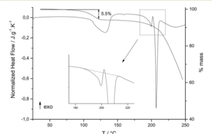
was carried out by DSC and TGA at 5 °C min1. The DSC curve (Fig. 1) reveals a first endothermic peak at ∼100 °C, corresponding to the dehydration of the compound. A weight loss of approximately 5.5% was determined by TGA (Fig. 1), which corresponds to the loss of one molecule of water. The enthalpy of dehydration was found to be 41.2 ± 2.8 kJ per mole of water, comparable to previously reported values for similar molecules presenting internal cavities: 45.5 kJ per mole of water for quinacrine dihydrochloride dehydrate18 and between 50 and 63 kJ per mole of water for β-cyclodextrin hydrates19and hydrated cucurbituril.20
Still on heating after dehydration, two endothermic peaks were then observed, approximately at 194°C and 205 °C. This re-sult disagrees with previous DSC analyses,6–10in which only one endothermic peak was observed for the melting of the anhydrous phase, at a temperature comprised between 188 and 197°C.
As can be seen on the zoom of Fig. 1, the first endothermic peak at 194°C was followed by a recrystallization process. This suggests that this first endothermic peak has to be associated to a melting process. Then, still on heating, the second endother-mic peak appears at 205°C, and should correspond to the melt-ing point of another solid phase. The TGA curve (Fig. 1) re-vealed a weight loss of approximately 4% just before and during melting, i.e. during the melting–crystallization–melting sequence. This weight loss could be explained as sublimation and evaporation of the sample. This was confirmed from experi-ments carried out at high scan rates, up to 100°C min1 (re-sults not shown), where no more mass loss was then observed. Moreover, this phenomenon could not be attributed to degrada-tion since the onset of the first endothermic signal did not vary
Fig. 1 DSC and TGA curves ofrac Sibut·HCl·H2O recorded at 5 °C min−1. The inset corresponds to the zoom of the melting region between 190 and 220°C, showing the melting crystallization melting sequence.
with the heating rate. When degradation occurs upon melting, it is well known that the “apparent” melting temperature in-creases with the DSC heating rate.21
After melting, one can observe on the DSC and TGA curves (Fig. 1) that the molten anhydrous sibutramine (Sibut·HCl) degrades. Indeed, the broad DSC endothermic peak following melting is accompanied with an important weight loss.
Depending on the heating rate, the relative intensities of both melting peaks changed (Fig. 2). More precisely, when creasing the heating rate, the intensity of the second peak in-creased compared to the first one. This suggests that each peak may correspond to the melting of one different anhy-drous form of Sibut·HCl.
To confirm this assumption and to be able to characterise each anhydrous solid, different methods of preparation were used to try and isolate each form.
First, the commercial product was dehydrated at 120 °C and the so-obtained anhydrous compound was annealed for several days at this temperature. After that, a DSC experiment was performed on heating of the annealed solid and a single endothermic peak was observed at 198.8 ± 0.2°C (curve 1 in Fig. 3), with an associated melting enthalpy of 38.8 ± 1.6 kJ mol1. The same behaviour was observed when rac-Sibut ·HCl·H2O was ground before introducing it into the DSC
pan (curve 2 in Fig. 3).
Interestingly, when dehydrated sibutramine hydrochloride was recrystallised in anhydrous methanol, and rehydrated in ambient conditions, a single melting peak was also observed by DSC but, this time, at a higher temperature equal to 205.0 ± 0.3°C, with an associated melting enthalpy of 27.8 ± 0.6 kJ mol1(curve 3 in Fig. 3). The same melting point was noticed when the anhydrous form was obtained from ground rac-Sibut·HCl·H2O dehydrated at 180°C (result not shown).
These results showed that the two peaks observed in Fig. 1 were not an experimental artefact, but consistent with the existence of two distinct anhydrous phases. To confirm this finding, the two solids obtained by the two preceding methods were characterised by XRPD.
The pattern matching of the anhydrous form obtained by annealing at 120°C (called in the following anhydrous A) was
determined at room temperature. The space group and cell parameters were found to match with the previously reported crystal structure determined on single crystal obtained by sublimation/decomposition of the monohydrate form at 190 °C.10The Pawley refinement and the unit cell parameters are
given in Fig. S2 and Table S2† respectively. The structure showed that the anhydrous compound is a racemic com-pound with a unit cell consisting of two molecules having the R configuration and two having the S configuration.
The unknown crystal structure of the anhydrous form whose melting point was found at 205 °C (called anhydrous B in the following) was determined from an XRPD pattern recorded at room temperature during eight hours. The diffraction pattern was reproduced with a monoclinic space group P21 and with
cell parameters: a = 12.0282IJ19) Å, b = 9.0364(15) Å, c = 8.1578(11) Å,β = 90.601IJ9)°. The final Rietveld refinement (Ta-ble S3†) converged to a final Rwpvalue of 4.36% (Fig. 4).
The crystal structure reveals that only one enantiomer is present in the unit cell, as shown in Fig. 5. This indicates that the initial anhydrous racemic compound (anhydrous A) was transformed into conglomerate. This explains why one
Fig. 2 Normalized DSC curves ofrac Sibut·HCl·H2O recorded at 1, 2, 5 and 20°C min−1(from top to bottom). The inset corresponds to the zoom of the melting region between 190 and 220°C.
Fig. 3 Normalized DSC curves recorded at 10 °C min−1 of rac Sibut·HCl·H2O and Sibut·HCl. (1) Anhydrous solid annealed at 120 °C, (2) ground rac Sibut·HCl·H2O, (3) recrystallised anhydrous solid rehydrated in ambient conditions.
Fig. 4 Final Rietveld refinement of X ray diffraction pattern of sibutramine obtained at 294 K (anhydrous B). Blue line: Experimental pattern, empty red circles: calculated pattern, green vertical bars: peak positions, black line: residual XRPD patterns. The inset corresponds to the scale for the data between 46° and 70° magnified 15 times.
parameter, in this case b, is divided approximately by 2 com-pared to anhydrous A, formed by an alternating stack of R-and S-enantiomers. However, both structures have nearly the same density in the solid state: 1.185 g cm3for the conglom-erate instead of 1.180 g cm3for the racemic compound.10
The structure is maintained by seven inter-molecular H-bonds mainly between the secondary amine group and the counter ion Cl2 (Table 1). The other chlorine atom Cl1, com-ing from the chlorobenzene group, also initiates H-bond in-teractions with C6 and C12. However these inin-teractions re-main weaker (Table 1).
By comparison, six H-bonds were reported for anhydrous A (Table S4†). As for anhydrous B, S·HCl interacts with Cl2 through an H-bond interaction with N1 but also with C10 and C11. The distances and the angles are almost identical in both structures. The C17 methyl group interacts with Cl1, while it interacts with Cl2 in anhydrous B. This is explained by the fact that the methyl groups are not positioned in the same manner in both anhydrous, as discussed below.
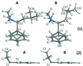
As observed in Fig. 6 and reported in Table 2, the confor-mation differences between the molecules of A and B are mainly noticeable for the isobutyl group. As can be seen in Table 2 and Fig. 6-1, the C16 and C17 methyl groups rotated of about 107° and C15 by 78.4°.
The cyclobutane group was found to be less planar in hydrous A than in anhydrous B (Fig. 6-2), as stated by the an-gle between C2–C3–C4 and C1–C2–C4 planes which equals 13.8° for anhydrous B and 30.3° for A, as well as the angle be-tween C1–C2–C3 and C1–C3–C4 planes equal to 14.6° for an-hydrous B and 30.4° for A.
Regarding the phenyl group, it has practically undergone no major changes (Fig. 6-2, Table 2) and remained almost planar in both conformations.
Taking these new findings into account, the endothermic peak observed in the DSC curve 3 in Fig. 3 at 139.6 ± 3.6°C, with an associated heat of 2.6 ± 1.0 kJ mol 1, can be ascribed to a peritectoid transformation of the anhydrous racemic compound into conglomerate.22 Above this point, the con-glomerate becomes more stable than the racemic compound. This explains why the conglomerate (in a stable state) melts at a higher temperature than the racemic compound (in a metastable state), i.e. 205°C instead of 198.8 °C. From an ergetic point of view, the conglomerate melts with a lower en-thalpy than the racemic compound (27.8 instead of 38.8 kJ mol 1). Such behaviour has been previously reported for race-mic 1,1′-binaphtyl23or for hydantoin derivative.24
The anhydrous racemic compound is always obtained af-ter dehydration of rac-Sibut·HCl·H2O whatever the
Fig. 5 Crystal structure of anhydrous sibutamine B viewed alongc.
Table 1 Inter molecular H bond distances and angles for anhydrous B
D H A D H (Å) H⋯A (Å) D⋯A (Å) D H⋯A (deg.) Symmetry
N1 H1 Cl2 1.041(5) 2.098(7) 3.060(7) 152.6(5) i C10 H10 Cl2 1.025(5) 2.803(7) 3.749(7) 153.5(4) i C11 H11 Cl2 1.098(9) 2.763(8) 3.800(8) 157.4(4) ii C13 H13A Cl2 1.099(11) 2.610(9) 3.593(9) 148.5(4) ii C17 H17C Cl2 1.088(8) 2.93(6) 3.885(9) 146.9(5) v C12 H12B Cl1 1.092(11) 2.91(8) 3.852(6) 144.9(6) iii C6 H6 Cl1 1.018(7) 2.97(7) 3.854(8) 145.8(7) iv
(i)−x, 1/2 + y, 1 − z; (ii) x, 1 + y, z; (iii) x, y, z + 1; (iv) 1 − x, 1/2 + y, −z; (v) x, 1 + y, z.
Fig. 6 Molecular conformations of the anhydrous A and B of sibutramine. (1) Part of the molecule including the secondary amine and isobutyl groups, (2) part of the molecule including the phenyl and cyclobutane groups.
experimental conditions. However, no analogy was found be-tween the crystal structures of rac-Sibut·HCl·H2O and
rac-Sibut·HCl (Fig. S3†). Indeed, the molecular arrangements seen in two different directions do not show any possible structural filiation between both structures.25Then, one can assume that the hydrated material undergoes a destructive process when the water molecules are released followed by a reconstructive process since the resulting phase is the anhy-drous crystalline racemic phase rac-Sibut·HCl.
Conclusion
By coupling thermal analyses and XRPD experiments, it was shown that, depending on experimental conditions, the dehy-dration of rac-Sibut·HCl·H2O led to either the anhydrous
ra-cemic form or the conglomerate. When the rara-cemic com-pound is stabilized, it transformed into conglomerate through a peritectoid reaction that occurs at around 140°C. Then, it was shown by DSC that the anhydrous conglomerate melts at higher temperatures than the racemic compound. When the racemic compound persists above this point, it be-comes metastable and its melting point necessarily takes place before that of the stable conglomerate, but with a higher melting enthalpy.
Acknowledgements
This work was financially supported by the ANR project “AlyPOTEC”. Ms. K. Debbasch is kindly thanked for her ad-vice on the manuscript.
References
1 S. Higgs, A. J. Cooper and N. M. Barnes, Psychopharmacology, 2011, 214, 941.
2 Y. H. Zhou, X.-Q. Ma, C. Wu, J. Lu, S.-S. Zhang, J. Guo, S.-Q. Wu, X.-F. Ye, J.-F. Xu and J. He, PLoS One, 2012, 7, e39062. 3 H. M. Oberholzer, C. van der Schoor and M. J. Bester,
Environ. Toxicol. Pharmacol., 2015, 40, 71.
4 A. Saleh Bin Bisher, Indian J. Sci. Technol., 2010, 3, 1129. 5 C. S. Borges, G. Missassi, E. S. A. Pacini, L. Ri, A. Kiguti, M.
Sanabria, R. F. Silva, T. P. Banzato, J. E. Perobelli, A. S. Pupo and W. G. Kempinas, PLoS One, 2013, 8, e66091.
6 I. Nicolás-Vázquez, J. Hinojosa Torres, J. Cruz Borbolla, R. Miranda Ruvalcaba and J. M. Aceves-Hernández, J. Mol. Struct., 2014, 1062, 1.
7 A. Ravikiran, M. Arthanareeswari, P. Kamaraj, C. Praveen and K. V. Pavan, Chem. Sci. Trans., 2013, 2, S288.
8 P. R. Oliveira, H. K. Stulzer, L. S. Bernardi, S. H. M. Borgmann, S. G. Cardoso and M. A. S. Silva, J. Therm. Anal. Calorim., 2010, 100, 277.
9 A. Pajzderska, D. M. Chudoba, J. Mielcarek and J. Wasicki, J. Pharm. Sci., 2012, 101, 3799.
10 E. Maccaroni, E. Alberti, L. Malpezzi, N. Masciocchi and C. Pellegatta, J. Pharm. Sci., 2008, 97, 5229.
11 Materials Studio Modeling 5.5., (http://accelrys.com/products/ collaborative-science/biovia-materials-studio/).
12 M. A. Neumann, J. Appl. Crystallogr., 2003, 36, 356. 13 G. S. Pawley, J. Appl. Crystallogr., 1981, 14, 357.
14 S. L. Mayo, B. D. Olafson and W. A. Goddard III, J. Phys. Chem., 1990, 94, 8897.
15 G. E. Engel, S. Wilke, O. König, K. D. M. Harris and F. J. J. Leusen, J. Appl. Crystallogr., 1999, 32, 1169.
16 W. A. Dollase, J. Appl. Crystallogr., 1986, 19, 267.
17 Y. Chen, H. W. Zhou and J. Z. Chen, Z. Kristallogr., 2005, 220, 513.
18 R. Rotival, P. Espeau, Y. Corvis, F. Guyon and B. Do, J. Pharm. Sci., 2011, 100, 3223.
19 M. Bilal, C. de Brauer, P. Claudy, P. Germain and J. M. Létoffé, Thermochim. Acta, 1995, 249, 63.
20 P. Germain, J. M. Létoffé, M. P. Merlin and H. J. Buschmann, Thermochim. Acta, 1998, 315, 87.
21 Y. Corvis, M.-C. Menet, P. Négrier, M. Lazerges and P. Espeau, New J. Chem., 2013, 37, 761.
22 G. Coquerel, Enantiomer, 2000, 5, 481–498.
23 K. R. Wilson and R. E. Pincock, J. Am. Chem. Soc., 1975, 97, 1474.
24 Y. Amharar, S. Petit, M. Sanselme, Y. Cartigny, M.-N. Petit and G. Coquerel, Cryst. Growth Des., 2011, 11, 2453–2462. 25 S. Petit and G. Coquerel, Chem. Mater., 1996, 8, 2247–2258.
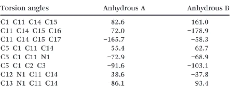
Table 2 Torsion angles for anhydrous forms of sibutramine
Torsion angles Anhydrous A Anhydrous B
C1 C11 C14 C15 82.6 161.0 C11 C14 C15 C16 72.0 −178.9 C11 C14 C15 C17 −165.7 −58.3 C5 C1 C11 C14 55.4 62.7 C5 C1 C11 N1 −72.9 −68.9 C5 C1 C2 C3 −91.6 −103.1 C12 N1 C11 C14 38.6 −37.8 C13 N1 C11 C14 −86.1 93.4