Pépite | Fonctionnalisation et transformation de nanoparticules et leur application biomédicale
127
0
0
Texte intégral
(2) Thèse de Volodymyr Turcheniuk, Lille 1, 2016. ACKNOWLEDGEMENT Over the past two years, I have received support and encouragement from great number of individuals to make my academic stay in France fruitful. First of all, I would like to give my warmest gratitude to my Ph.D. supervisor Prof. Sabine Szunerits for her continued support, guidance, scientific discussion and encouragement throughout my study period and le I was fortunate to find her not only as a mentor but also as a guardian thousand miles from my country, Ukraine. Her guidance has made this journey thoughtful and rewarding. I am also very grateful to her for providing me opportunities to explore researches and to learn different techniques in other labs outside France. Of course deepest thanks to Prof. Vladimir Zaitsev, he always supported me and when I was in panic I could ask him and then everything started to be fine. My deepest thanks go to Dr. Rabah Boukherroub for his suggestions, sharpening my scientific thinking which is essential for research field and for organizing lab meeting every week which helped me to learn different techniques and to broaden my knowledge in material science research. I would like to thank Dr. Aloysius Siriwardena for suggestions and scientific discussions that he shared to us via email and phone and being available even after his busy schedule whenever I need to discuss my work. It was big pleasure to work with him. Sadly, he passed away recently and this work is thus dedicated to him, as his input was of high importance. I would like to thank Dr. Alexandre Barras for his valuable suggestions during my research work. He always helped me when I didn’t know something Also, I would like to thanks Amer, Pawan, Roxane, Flori, Nadia, Wang Qi, and all the lab members for maintaining the ambiance and peaceful working environment in lab and I truly appreciate the nature of sharing the ideas among lab mates. Special for Amer and Pawan for making me company outside the laboratory) 2|Page. © 2016 Tous droits réservés.. lilliad.univ-lille.fr.
(3) Thèse de Volodymyr Turcheniuk, Lille 1, 2016. I would like to thank all the Ukrainian friends in Lille and in Ukraine specially; Ugeen and Svitlana for care, giving me company during my hard and good times, Alexandre and polish guy Artour for a lot of fun days. I will definitely miss the gathering and fun with you all. I am always grateful to my mother, grandmother, brother who has supported my decision to study miles away from hometown, kept patience for years, and provided continuous emotional support and inspiration. And the biggest gratitude for my brother Dr. Kostiantyn Turcheniuk, without him there would be no PhD) Sometime he was angry on me and forced me to work; but I very much appreciate it at the end. Another biggest gratitude for my first teacher. Without her I’ve never started to study chemistry. I have no words to explain my gratitude to my beloved girlfriend for continuous support, encouragement and keeping faith on me during my academic carrier. She has taught me to stay positive even in the worst condition. I am indebted to his unconditional love and understanding nature despite of long distance relationship.. 3|Page. © 2016 Tous droits réservés.. lilliad.univ-lille.fr.
(4) Thèse de Volodymyr Turcheniuk, Lille 1, 2016. ABSTRACT Functionalized nanoparticles continue to attract interest in biomedical applications and bioassays and have become a key focus in nanobiotechnology research. The use of nanomaterials in biomedical applications is of great interest since their size scale is similar to biological molecules and structures. One of the primary focus of the research work was the development of versatile surface functionalization strategies for different nanoparticles ranging from diamond nanostructures to gold nanorods and nanocomposites. One particular aim was the use of reduced graphene oxide (rGO) and silica coated gold nanorods for the photothermal and photodynamic ablation of pathogens. Embedding of verteporfin, a clinically approved photosensitizer, into silica-coated gold nanorods allowed an efficient eradication of a virulent strain of E. coli associated with urinary tract infection. The great heating effect of graphene-coated gold nanorods when illuminated with a near-infrared laser allowed for a photothermal destruction of the same pathogenic strain. In parallel, we have shown the interest of using diamond nanoparticles (NDs) modified with menthol as well as different sugars as antibacterial agent against Gram-positive (Staphylococcus aureus) and Gram-negative (Escherichia Coli) bacteria.. We developed a strategy for the. covalent attachment of sugars by taking advantage of the photochemistry of arylazides, which upon light activation convert to reactive nitrenes. The highly reactive nitrene intermediate formed is believed to interact with glycans through C-H and N-H insertion reactions, creating highly robust covalent linkages. The resulting glyco-NDs maintained their expected binding affinity and specificity towards their partner lectins. Through a fluorescent based agglutination assay, we showed that mannan-NDs display E. coli agglutination at concentrations of ≈10 µg mL-1, being much lower than free mannan and mannose-NDs.. 4|Page. © 2016 Tous droits réservés.. lilliad.univ-lille.fr.
(5) Thèse de Volodymyr Turcheniuk, Lille 1, 2016. TABLE OF CONTENT ACRONYMS OBJECTIVES. 10 12. CHAPTER I: Nanoparticles for biological applications 1.1. Introduction 1.2. Gold nanoparticles (GNPs) 1.2.1. Introduction 1.2.2. Plasmonic photothermal therapy 1.2.3. Photodynamic treating with GNPs 1.3. Nanodiamonds (NDs) 1.4. References. 16 17 17 20 22 23 28. CHAPTER II: Glycans-Modified Nanodiamonds 2.1. Introduction 32 2.2. Synthesis of glyco-NDs via photoactivation of perfluorophenylazide-modified NDs 35 2.3. Lectin binding assays 42 2.4. Effect of mannose-NDs and mannan-NDs on agglutination of E. coli UTI89 strains 44 2.4.1. Fluorescence-based agglutination assay in the presence of mannose and mannose-NDs 44 2.4.2. Fluorescence-based agglutination assay in the presence of mannan and mannan-NDs 45 2.5. Conclusions 46 2.6. References 47. CHAPTER III: Menthol Modified Nanodiamonds and their antibacterial properties 3.1. Introduction 3.2. Synthesis and physico-chemical properties of ND-Menthol particles 3.3 Antimicrobial activity of the ND-menthol particles 3.4. Conclusions 3.5. References. 49 50 53 58 58. CHAPTER IV: Highly effective photodynamic eradication of Escherichia Coli pathogenic organisms by using golden nanorods/SiO2 core-shell nanostructures with included verteporfin 4.1 Introduction 4.2 Synthesis and photodynamic properties of AuNRs@SiO2VP 4.3. Conclusions 4.4. References. 63 64 70 70. 5|Page. © 2016 Tous droits réservés.. lilliad.univ-lille.fr.
(6) Thèse de Volodymyr Turcheniuk, Lille 1, 2016. CHAPTER V: Plasmonic photothermal cancer therapy nanorods/reduced graphene oxide core/shell nanocomposites. with. 5.1. Introduction 5.2. Synthesis and physic-chemical properties of AuNrs@rGO particles 5.3. Photothermal features of Au NRs@rGO-PEG and photothermal eradication cells of UM87MG in vitro. 5.4. NIR fluorescence imaging and biodistribution 5.5. In vivo photothermal therapy of mice with implanted cells of U87MG tumors. 5.6. Conclusions 5.7. References. gold 74 75 78 83 86 88 89. CHAPTER VI: Reduced graphene oxide nanosheets decorated with AuPd bimetallic nanoparticles : a multioperational material for the Photothermal Treatment of Cancer Cells 6.1. Introduction 6.2. Synthesis and characteristics of AuPd NPs-rGO-Peg nanocomposites 6.3. Photothermal efficacy and photothermal removing of cancer cells 6.4. Conclusions 6.5. References. 93 94 99 102 102. CHAPTER VII: CONCLUSIONS AND PERSPECTIVES. 106. APPENDIX Experimental Part. 109. 7.1. Materials 109 7.2. Synthesis of organic compounds 110 7.2.1. Synthesis of menthol derivative (2) 110 7.2.2. Synthesis of 4-azido-N-(3,4-dihydroxyphenethyl)-2,3,5,6-tetrafluorobenzamide (1) 111 7.2.2.1. 4-Azidotetrafluorobenzoic acid 111 7.2.2.2. N-Succinimidyl-4-azidotetrafluorobenzoate 111 7.2.2.3 4-azido-N-(3,4-dihydroxyphenethyl)-2,3,5,6-tetrafluorobenzamide 112 7.3. Synthesis of nanoparticles 112 7.3.1 Nanodiamonds (NPs) 112 7.3.1.1. Formation of ND-PFPA 112 7.3.1.2. Photochemical linkage of glycans to ND-PFPA 112 7.3.1.3. Preparation of menthol-modified NDs (ND-menthol) 112 7.3.2. Gold nanoparticles (Au NPs) 113 7.3.2.1. Synthesis of gold nanorods (AuNRs) 113 7.3.2.2. Preparation of AuNRs@SiO2 particles 113 7.3.2.3. Loading of AuNRs@SiO2 particles with verteporfin (VP) 114 7.3.2.4. Synthesis of pegilated rGO-wrapped gold nanorods (AuNRs@rGO-PEG) 114 6|Page. © 2016 Tous droits réservés.. lilliad.univ-lille.fr.
(7) Thèse de Volodymyr Turcheniuk, Lille 1, 2016. 7.3.2.5. Conjugation of AuNRs@rGo-PEG with Cy7 or FITC and Tat protein vector 115 7.3.2.6. Synthesis of AuPd NPs-rGO nanocomposite materials 115 7.3.2.7. Functionalization of AuPd NPs0rGO with polyethylene glycol (PEG) 115 7.4 Determination of carbohydrate loading on the NDs 117 7.5 Lectin binding assay 117 7.6. Fluorescence-based agglutination assay 118 7.7. Biological assay 118 7.7.1. Antimicrobial assays 118 7.7.2. Cell culture and cellular uptake of Au NRs@rGO-PEG-FITC and Au NRs@rGO-PEGTat/FITC 120 7.7.3. Cellular toxicity of AuNrs@rGO-PEG 120 7.8. Photothermal experiments 121 7.8.1. 1O2 quantum yield estimation 121 7.8.2. Single oxygen detection 121 7.8.3. Photodynamic ablation of bacteria solutions 121 7.8.4. Photothermal in vitro experiment 122 7.8.5. In vivo photothermal therapy and histology 122 7.9. Instrumentations 123 7.9.1. Fourier transformed infrared spectroscopy 123 7.9.2. Micro-Raman spectroscopy 124 7.9.3. X-ray photoelectron spectroscopy 124 7.9.4. Absorption spectra 124 7.9.5. Transmission electron microscope (TEM) 124 7.9.6. Scanning electron microscope (SEM) 125 7.9.7. Confocal microscopy 125 7.9.8. Thermogravimetric analysis (TGA) 125 7.9.9. Particle size measurements 125 7.9.10. Measurements of the photothermal effect 126. List of publications. 127. 7|Page. © 2016 Tous droits réservés.. lilliad.univ-lille.fr.
(8) Thèse de Volodymyr Turcheniuk, Lille 1, 2016. ACRONYMS ACN APTMS ATP Au NRs Au NRs@rGO-PEG BSA CDCl3 CMC Cu (I) CuI(PPh3) DAPI DCC DCM DMAEMA DMAP DMEM DMF DMSO DNA DOP-N3 DOP-PEG DSPE E.coli EDTA EDC ER EPR ESI EtoAc FCS FDA FT-IR GFP GNPs G-Ins HCC HCV HCVcc HCVpp Hex HIV. Acetonitrile 4-aminopropyltrimethoxysilane Adenosine triphosphate Gold nanorods Pegylated reduced graphene oxide wrapped gold nanorods Bovine serum albumin Deuterated chloroform Critical micellar concentration Copper iodide Copper iodide triphenylphospine 4’-6-diamidino-2-phenylindole N,N’-dicyclohexylcarbodiimide Dichloromethane 2-(Dimethylamino)ethyl methacrylate 4-Dimethylaminopyridine Dulbecco modified Eagle medium Dimethylformamide Dimethyl sulfoxide Deoxyribonucleic acid Azide-terminated dopamine derivative Polyethylene glycol-terminated dopamine derivative 1,2-distearoyl-sn-glycero-3-phosphoethanolamine- N-[carboxylic acid (polyethylene-glycol)] Echerichia coli (Katushka) Ethylenediaminetetraacetic acid N-(3-Dimethylaminopropyl)-N′-ethylcarbodiimide hydrochloride Endoplasmic reticulum Enhanced permeability and retention Electron spray ionization Ethyl acetate Fetal calf serum Food and Drug Administration Fourier transformed-infrared spectroscopy Green fluorescence protein Gold nanoparticles Gluconic acid-modified insulin Hepatocellular carcinoma Hepatitis C Virus HCV cell culture systems HCV pseudotyped particles Hexane Human Immunodeficiency Virus 8|Page. © 2016 Tous droits réservés.. lilliad.univ-lille.fr.
(9) Thèse de Volodymyr Turcheniuk, Lille 1, 2016. HLB HNO3 H2SO4 HS HTS Huh-7 IFNα IC50 IRES JAM JFH KBr LDLR LiAlH4 LNCs MAb Man-9 MLV MPS MPS MRI MTS nAb NaCl NaOH NC NDs ND-PFPA NHS NH2-PEG-NH2 NIH NMR NPs NPC NS (N-V)PBS pDNA PEG PEI PET PDI PIT pKa. Hydrophilic lipophilic balance Nitric acid Sulfuric acid Heparin sulfate High throughput screening Hepatocyte derived cellular carcinoma cell line Interferon-alpha Half maximal inhibitory concentration Internal ribosome entry site Junction-associated adhesion molecule Japenese Fulminant Hepatitis Potassium bromide Low-density lipoprotein receptor Lithium aluminium hydride Lipid nanocapsules Monoclonal antibody Mannonanose-di-(N-acetyl-D-glucosamine) Murin leukemia virus Mesoporous silica Mononuclear phagocyte system Magnetic resonance imaging Cell Proliferation Assay Neutralizing antibody Sodium chloride Sodium hydroxide Non-coding Nanodiamonds Pentafluorozidobenzoate modified nanodiamond N-Hydroxysuccinimide Poly(ethylene glycol)bis(3-aminopropyl) National Institute of Health Nuclear magnetic resonance Nanoparticles Nuclear pore complexes Non-structural Nitrogen-vacancy center Phosphate buffer saline Plasmid deoxyribonucleic acid Polyethylene glycol Polyethylenimine Positron emission tomography Polydispersity index Phase-inversion temperature Ionization constant 9|Page. © 2016 Tous droits réservés.. lilliad.univ-lille.fr.
(10) Thèse de Volodymyr Turcheniuk, Lille 1, 2016. PLGA PLL PS-ON Rcf RISC RNA ROS SEM siRNA SOCl2 SRB1 SPIO TEA TEM TEOS TGA THF THP-1 TNF UTR VP VSVg XPS ZO. Poly(lactic-co-glycolic acid) Poly-L-lysine Phosphorothioate oligonucleotides Relative centrifugal force RNA-induced silencing complex Ribonucleic acid Reactive oxygen species Scanning electron microscope Silencer RNA Thionyl chloride Scavenger receptor class B type 1 Superparamagnetic iron oxide nanoparticle Triethylamine Transmission electron microscope Tetraethyl orthosilicate Termogravimetric analysis Tetrahydrofuran Human macrophage-like cells Tumor necrosis factor 3’Untranslated region Verteporphin Vesicular stomatitis virus glycoprotein G X-ray photoelectron spectroscopy Zona occludens. 10 | P a g e. © 2016 Tous droits réservés.. lilliad.univ-lille.fr.
(11) Thèse de Volodymyr Turcheniuk, Lille 1, 2016. OBJECTIVES The past decade has observed significant advancement in the field of nanobiotechnology. Functionalized nanoparticles continue to attract interest in biomedical applications and bioassays and have become a key focus in nanobiotechnology research. The use of nanomaterials in biomedical applications is of great interest since their size scale is similar to biological molecules and structures. Recent studies have shown that integrating biological components with nanomaterials can revolutionize the field of pharmacology and help to tackle diseases at a molecular level. In this thesis, different surface modification strategies have been developed. An effort was put mainly on the surface modification of nanodiamonds (NDs), gold NPs (Au-NPs), Au-Pd bimetallic nanocomposites, graphene. These particles were investigated for their possibility to inhibit Gram-positive (Staphylococcus aureus) and Gram-negative (Escherichia coli) bacteria and destruct of human glioblastoma astrocytoma (U87MG). After a general overview on nanoparticles for biological applications (Chapter 1), a brief description of nanoparticles, their synthesis, modification and perspectives in nanomedicine. Chapter 2 outlines the synthesis and characterizations of the azido-modified nanodiamonds and their ability of photochemical reaction with sugars. We described the affinity of glycans-modified NDs toward lectins and Echerichia coli. In Chapter 3, we elucidate the effect of menthol modified nanodiamond particles (ND-menthol) on bacterial viability against Gram-positive (Staphylococcus aureus) and Gram-negative (Escherichia coli) bacteria. We show that while NDmenthol particles are non-toxic to both pathogens, they show significant antibiofilm activity We described the development of novel nanostructures for photodynamic therapy comprising silica-encased gold nanorods modified with verteporfin as photosensitizer and the efficiency of these nanostructures for killing of pathogens in Chapter 4. From another site gold nanorods are known for their efficient conversion of photon energy into heat, resulting in hyperthermia and suppression of tumor growth in vitro and in vivo. Au NRs are thus of great promise for photothermal therapy (PTT) of different cancers. In Chapter 5 we investigate the potential of polyethylene glycol functionalized reduced graphene oxide (rGO-PEG) enrobed Au NRs for the 11 | P a g e. © 2016 Tous droits réservés.. lilliad.univ-lille.fr.
(12) Thèse de Volodymyr Turcheniuk, Lille 1, 2016. photothermal destruction of human glioblastoma astrocytoma (U87MG) cells in mice. Based on these results we showed in Chapter 6 a simple and green solution phase synthetic approach for the formation of bimetallic AuPd NPs on rGO nanosheets for photothermal therapy. Chapter 7 summarizes the results and gives some perspectives of the work.. 12 | P a g e. © 2016 Tous droits réservés.. lilliad.univ-lille.fr.
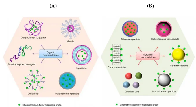
(13) Thèse de Volodymyr Turcheniuk, Lille 1, 2016. CHAPTER I Nanoparticles for biological applications 1.1. Introduction Nanoparticles, widely used over more than 35 years now, have made important contributions to a variety of different domains, including the biomedical field. The nanometric size of nanoparticles change the physico-chemical properties when compared to the bulk substantially, thus opening new possibilities in nanomedicine. The use of nanomaterials in medicine has several advantages. Nanomaterials have the characteristics of high surface-to-volume ratios enabling packaging multiple agents onto the same nanoparticles. Using these nanomaterials it might be possible to overcome problems associated with the use of high doses of drugs. Moreover, this type of approach provides also the possibility of targeting specific biological sites actively or passively. Because. of. their. unique. features. such. as. nanometric. size. and. controllable. hydrophobicity/lipophilicity, such nanocarriers can target drugs to specific tissues or organs, while modification of nanocarrier surfaces enables them to reach particular therapeutics with the results of minimizing side effects onto healthy cells and tissue. With respect to intravenous administration, due to their small size, nanoparticles can circulate in the bloodstream without being retained by the pulmonary capillaries or up taken by the reticuloendothelial system (RES). Indeed, the most frequently used approach to increase the longevity of nanocarriers to avoid the RES uptake it so modify their surface with hydrophilic polymers such as polyethylene glycol (PEG) units. Various nanocarriers have been proposed over the years and some of the most currently considered are listed in Figure 1.1. Next to dendrimers, liposomes and polymeric nanostructures (Figure 1.1A), inorganic nanoparticles (Figure 1.1B) such as gold, silver, iron oxide, etc have found their application in nanomedicine (2, 3). In this work, we focused on the use of gold nanoparticles (GNPs) as well as nanodiamonds (NDS) and the interest in these nanostructures will be outlined below.. 13 | P a g e. © 2016 Tous droits réservés.. lilliad.univ-lille.fr.
(14) Thèse de Volodymyr Turcheniuk, Lille 1, 2016. (A). (B). Figure 1.1: Classification of nanomaterials that are commonly used as nanomedicine. Some examples of widely used organic (A) and inorganic nanomedicines (B) (1).. 1.2. Gold nanoparticles (GNPs) 1.2.1. Introduction Gold has been among the first metals, which were discovered by people, and the history of its research and use is at least several thousand years old approximately. The first mention about colloid gold is able to be discovered in tracts by Indian, Chinese and Arabic researchers, who got colloid gold as long ago as in the fourth and fifth centuries B.C. and used it, particularly, in medical purposes (the Indian ‘‘liquid gold’’ and the Chinese ‘‘golden solution’’).In the European Middle Ages, colloid gold was researched and used in laboratories of alchemists. Namely, Paracelsus informed about the medicinal features of quinta essentia auri, which he had got by mixing auric chloride with oil plant extracts or alcohol. He employed ‘‘potable gold’’ in order to heal syphilis and certain mental disorders. Also, Paracelsus stated that chemistry is for producing 14 | P a g e. © 2016 Tous droits réservés.. lilliad.univ-lille.fr.
(15) Thèse de Volodymyr Turcheniuk, Lille 1, 2016. drugs, but not for getting gold out of metals. Another researcher of his age, Giovanni Andrea used aurum potabile for the treatment of ulcer, leprosy, diarrhea and epilepsy. In 1583, David de Planis-Campy, who was a surgeon of Louis XIII, the King of France, recommended an ‘‘elixir of longevity’’, which was an aqueous colloid gold solution, as a tool of prolongation of life. The first book on colloid gold, which has preserved to these days, was published in 1618 by doctor of medicine and philosopher Francisco Antonii (4). It includes data on the preparation of colloid gold and its healing applications. This book also includes practical suggestions. In 1880, in order to treat alcoholism through intravenous injection of colloid gold solution, a way was brought forward (5). This method was called ‘‘golden cure’’. In 1927, the application of colloid gold was offered in order to alleviate the suffering of inoperable patients with cancer (6). Since the first half of the 20th century, colloid gold in color reactions to blood-serum and spinalfluid proteins has been taken advantage of (7). Colloid solutions of the Au golden isotope (halflife time, 65 h) were medically successful at oncological institutions (8). More recent cases of using of colloid gold contain transport of substances into cells by endocytosis, catalytic processes and electron transport in biomacromolecules, improvement of PCR effectiveness and researching of cell motility. In spite of the centuries-long history, a ‘‘immuno-chemical revolution”, which was associated with the application of golden particles in biological studies, occurred in 1971 (9). That year, two British scientists, G. M. Taylor and W. P. Faulk, issued the article, which was named ‘‘An immunocolloid method for the electron microscope.” This article described technology of conjugating antibodies with colloid gold for immediate electron microscope view of Salmonella surface antigens, which was presenting for the first time when colloid gold conjugation served as an immunochemical marker (10). After this, the application of colloid-gold bio-specific conjugates in different biological and medical areas has become very frequent. There is a lot of information, which relates to the use of functionalized golden nanoparticles (GNPs; conjugates with recognizing biomacromolecules, for example, antibodies, aptamers, enzymes or lectins) (11) in the researches of morphologists, microbiologists, biochemists, cytologists, immunologists, physiologists, and other specialists. 15 | P a g e. © 2016 Tous droits réservés.. lilliad.univ-lille.fr.
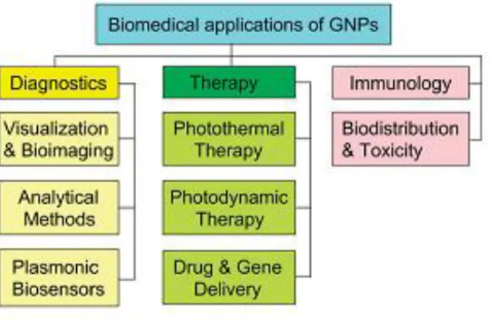
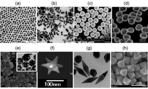
(16) Thèse de Volodymyr Turcheniuk, Lille 1, 2016. The scope of applications of GNPs in modern biological and medical studies is very large, ranging from biosensorics, optical bio-imaging, targeted delivery of medicines to the photothermolysis of cancer cells and microorganisms (Figure 1.2).. Figure 1.2: Generalized scheme for the biomedical application of GNPs. Along with basic applications in diagnostics and therapy, this review briefly discusses the immunological properties of GNPs (12). This large scope of application is founded on the unique chemical and physical features of GNPs. The exceptional optical properties of GNSs, including large optical field enhancements and their addressability via spectroscopic techniques have made them of particular interest as labels and for biosensing. The optical properties are dominated by the excitation of collective oscillations of the nanoparticles’ conduction band electrons, called localized surface plasmon resonances (LSPR), by the incoming electromagnetic waves. At the LSPR, the incoming light is absorbed or scattered by the GNSs, and concurrently, there is an electromagnetic field enhancement close to the surface of the particle. The position of the plasmonic resonance depends on the particle size, shape, composition, interparticle distance as well as the dielectric environment. However, the use of as-synthesized Au NPs (Figure 1.3) in biological systems is limited due by the particle instability and nonspecific binding. These limitations are even increasingly pronounced in high ionic strength media, such as those used for biological assays. The particle aggregation depends strongly on the type of ligand used to passivate the nanoparticles’ surface. 16 | P a g e. © 2016 Tous droits réservés.. lilliad.univ-lille.fr.
(17) Thèse de Volodymyr Turcheniuk, Lille 1, 2016. The ligands can be displaced or even cross-linked due to the multitude of ionic species present in solution. Care has, in addition, to be paid to the biocompatibility of the particle ligand. For instance, cetyltrimethylammonium bromide (CTAB) modified gold nanorods can be routinely prepared and display good colloidal dispersibility and stability. However, one of the major concerns is related to the cytotoxicity of the surfactant, CTAB, used to stabilize the particles. CTAB molecules can induce cytotoxicity by disrupting cellular membranes CTAB is not only toxic to cells, but also very difficult to remove completely.. Figure 1.3: TEM images of 15-nm nanospheres (a), 15 50-nm GNRs (b), 160(core)/17(shell)-nm silica/gold nanoshells (c, SEM), 250-nm Au nanobowls with 55-nm Au seed inside (d), silver cubes and gold nanocages (inset) (e), nanostars (f), bipyramids (g), and octahedrals (h) (12).. 1.2.2. GNPs based photothermal therapy (PTT) While the high surface area is primordial for efficient drug and gene loading, the possible strong optical absorption in the near infrared light (NIR) region makes gold nanocomposites suitable for photothermal therapy (PTT). The first report of GNPs used it in photothermal therapy (13) of cancer calls. Indeed, local heating of cancer cells, known as hyperthermia (warming to 41°–47° C for 1 h) leads to unalterable damage to the cells, caused by destruction of cell membrane permeability and denaturation of proteins. In the case of GNPs based PTT, the generation of heat 17 | P a g e. © 2016 Tous droits réservés.. lilliad.univ-lille.fr.
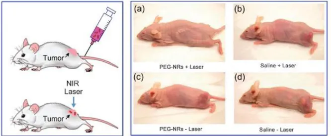
(18) Thèse de Volodymyr Turcheniuk, Lille 1, 2016. is based on laser irradiation of the tumor cells. Pitsillides et al. (14) described in this sense a new technology for selective injuring of target cells, which is based on the using of 20 and 30 nm gold nanospheres that irradiated with 20 ns laser radiation (532 nm) in order to create local heating. Figure 1.4 displays the example of successful treating of an implanted tumor in mice (15). One of the current modalities for PTT is the use of NIR light absorbing agents. The use of NIR has the advantage that water, melanin and hemoglobin have absorption minima in this region and light between 700-900 nm is most likely to pass directly through tissues without significant absorption and heat generation. (16) GNP nanomaterials exhibit strong visible to NIR absorption owing to the localized surface plasmon resonance effects, rendering them the most power agents for PTT cancer treatment and bacteria killing.. Figure 1.4: Scheme and the results of an experiment on the photothermal destruction of an implanted tumor in a mouse (2–3 weeks after injection of MDA-MB-435 human cancer cells). Laser irradiation (a, b; 810 nm, 2 W cm-2 , 5 min) was performed at 72 h after injection of gold nanorods functionalized with poly(ethylene glycol) (PEG) (a, c; 20 mg Au per kg) or of buffer (b, d). It can be seen that the tumor continued developing after particle-free irradiation (control b), as it did after particle or buffer administration without irradiation (controls c and d), and that complete destruction was obtained only in the experiment (a). Designations: NIR, near-IR region; NRs, nanorods. 18 | P a g e. © 2016 Tous droits réservés.. lilliad.univ-lille.fr.
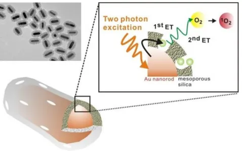
(19) Thèse de Volodymyr Turcheniuk, Lille 1, 2016. The interest of using GNPs is also linked to the possibility of targeted therapy, which has two significant aspects: increasing of the particle concentration in the target and decreasing of side effects, which are caused by GNPs gathering in other parts of a body, mainly in the spleen and liver). In vivo, PEGylated nanoparticles primarily gather in tumor tissue, thanks to the enhanced permeability of the vessel of tumor and are held in it thanks to the reduced lymph outflow (17). Moreover, PEGylated nanoparticles are less available to the immune system (stealth methods). This delivery technology is named passive, unlike the active variant, which applies antibodies (Figure 1.5). The active technology of delivery is more relevant and efficient, using antibodies to certain tumor markers, most often to receptor of epidermal growth factor (EGFR) and its variations (for example, Her2) (18), and to tumor necrosis factor (TNF) (19). Certain prospects are offered by the synchronous using of GNP–antibody conjugates for PPTT and diagnosis (this technology is called theranostics). Besides antibodies, active delivery can as well apply folic acid, which acts as a ligand for the many folate receptors of hormones and tumor cells (20).. Figure 1.5: Scheme for a PPTT employing active delivery of GNPs to cancer cells (21).. 1.2.3 Photodynamic treating with GNPs The photodynamic way of treating of certain skin or infectious diseases and oncological diseases is based on the using of light-sensitizing substances, which are called photosensitizers (including dyes) and, commonly, of visible lights at a specific wavelengths (21). As a rule, sensitizers are injected intravenously, but oral and contact using is also possible. The agents, which are used in 19 | P a g e. © 2016 Tous droits réservés.. lilliad.univ-lille.fr.
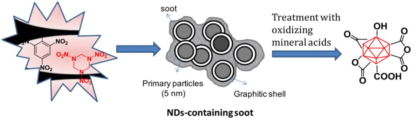
(20) Thèse de Volodymyr Turcheniuk, Lille 1, 2016. photodynamic treating (PDT) are able to selectively gather in tumors or other target objects (cells). The infected tissues are irradiated by laser light at a wavelenths, which corresponds to the maximum of dye absorption (21). At the same time, besides the usual warm emission by absorption, a significant role is played by another mechanism, which is associated with the photochemical generation of singlet oxygen (Figure 1.6.) and the forming of highly active radicals, which bring apoptosis and necrosis in tumor cells.. Figure 1.6: two-photon-activated photodynamic therapy (TPA-PDT) using mesoporous silicaencased gold nanorods (21). PDT also undermined the tumor nutrition and brings to its death by injuring its micro-vessels. The main PDT shortcoming is that the photosensitizer stays for a long time in the organism, leaving tissues of a patient highly sensitive to light. Although, the efficiency of dye application for selective tissue warming is low because of the little cross section of chromophore absorption (21).. 1.3. Nanodiamonds The first record of the production of ND particles dates back to the 1960s, when a group of Soviet scientists discovered single crystals of cubic diamond particles in soot produced by detonating an 20 | P a g e. © 2016 Tous droits réservés.. lilliad.univ-lille.fr.
(21) Thèse de Volodymyr Turcheniuk, Lille 1, 2016. oxygen-deficient TNT/hexogen composition in inert media without using any extra carbon source (22) (Figure 1.7.) This man-made diamond did not attract much attention at the beginning due to a continued failure in disintegrating ND particles into primary particles (23). While the particle size of its primary crystallites is ~5 nm with a very narrow size distribution (23, 24), due to harsh conditions in the reaction chamber, detonated ND particles exist mainly in the form of strongly bound agglomerates. The particles are not only linked by the usual electrostatic interactions by also via covalent bonds between surface functional groups as well as by soot structures surrounding each primary particle (25, 26).. The detonation soot can be purified using oxidizing mineral acids (HNO3, mixtures of H2SO4 and HNO3, K2Cr2O7 in H2SO4, KOH/KNO3, HNO3/H2O2 under pressure, etc) (27), as the reactivity of disordered sp2 carbon is higher than that of diamond, thus removing non-diamond impurities. During the cleaning step, the surface of NDs is covered with a variety of functional groups such as hydroxyl, carbonyl, carboxyl, anhydride and lactone moieties.. These purified ND particles show still a strong tendency to agglomerate, which can withstand ultrasonic treatment. To overcome this limitation, de-aggregation process in suspension by milling detonated NDs with ceramic microbeads (ZrO2, SiO2) or by microbead-assisted ultrasonic disintegration can be applied to NDs (28-30), leading to stable colloidal solutions of individual ND particles of 4-5 nm in diameter. However, contamination of NDs with difficult-to-remove zirconia, the high cost of zirconia microbeads, and NDs amorphization (or even graphitization) in the course of milling are major drawbacks of the microbeads-assisted milling. Retreatment of the de-agglomerated particles with oxidizing chemicals is therefore needed, which results in a partial re-aggregation of the primary particles. Other attempts have been undertaken to limit the NDs’ agglomeration. ND aggregates disintegration using mechano-chemical treatment, that is, milling in the presence of chemicals such as surfactants and electrolytes (e.g., sodium oleate) was investigated (31, 32). Mechanical action was applied in the form of grinding or sonication in the 21 | P a g e. © 2016 Tous droits réservés.. lilliad.univ-lille.fr.
(22) Thèse de Volodymyr Turcheniuk, Lille 1, 2016. presence of sodium oleate and resulted in a reduction of the aggregate size of the treated ND in water from 2 μm down to 40-60 nm. However, no particles smaller than 20 nm were observed. More recently, dry salt- or sucrose-assisted milling was successfully used as simple, inexpensive, and efficient alternative method for the disaggregation of NDs (33). The technique uses non toxic and non-contaminating compounds such as sodium chloride and sucrose, which can be easily removed at the end of the milling process. By using the dry media assisted milling with subsequent pH adjustment, it is possible to produce stable aqueous NDs colloidal solutions with particles <10 nm in diameter. High-temperature hydrogen treatment resulted, moreover, in stable single particle aqueous NDs suspension of 4-5 nm (34). Independent of the method chosen, one concern that needs to be addressed in any case is the possibility of re-aggregation over time, during drying and/or after surface functionalization due to capillary forces or attractive van der Waals forces, pulling the particles together.. soot. Primary particles (5 nm). Treatment with oxidizing mineral acids. Graphitic shell. NDs-containing soot. Figure 1.7: Formation of detonation NDs with well-defined surface termination: A mixture of TNT (60 wt%) and hexogen (40 wt %) is detonated in a closed metallic chamber in an atmosphere of N2, CO2 and liquid or solid water forming a diamond-containing soot. The soot is purified with oxidizing mineral acids to produce oxidized NDs (23, 27, 35).. Independent on their treatment, such detonation ND particles have been rapidly gaining popularity for biological applications. This is related to the presence of various functional groups 22 | P a g e. © 2016 Tous droits réservés.. lilliad.univ-lille.fr.
(23) Thèse de Volodymyr Turcheniuk, Lille 1, 2016. enabling quite complicated surface functionalizations without compromising of the helpful features of the diamond center Reactive C–Cl and C–F surface species, which are created by halogen hardening and photochemical chlorination have been applied in many wet chemical reactions (36). Nanodiamond with O–H terminations was used in esterification with acetyl chlorides, which then were terminated by long alkyl chain (37), and in silanization/deaggregation (38). Other reactions were also applied (39, 40). They include those ones, which are especially convenient for biomedical using (41). Diazonium chemistry was also employed with hydrogenated nanodiamond (42) in order to form C–C bindings between the adhered part and diamond center, and with hydroxylated nanodiamond (38) in order to form C–O–C bindings. Therefore, nanodiamond gives many variants for surface functionalization, but the result sharply depends on the clearing and uniformity of the surface chemistry of the starting substance. As already noted, functionalization also impacts the stability of diamond surfaces. One task in the area is to discover methods for quantitative analysis of different surface groups of nanodiamond. By now, most of the data are qualitative.. Thanks to the various clearing procedures, which are used by different producers, and the multiple variants for surface change, the toxicity of nanodiamonds is of legitimate doubt (43). In vivo and in vitro studies have been carried out in order to examine features as diverse as cell vitality, activity of gene program, and in vivo physiological and mechanistic behavior (43, 44). Nanodiamonds, which are instilled onto trachea, were noted to be of low pulmonary toxicity, with the number of nanodiamonds in the alveolar area reducing with time, and macrophages bore by nanodiamonds were monitored in the bronchia for 28 days after subjecting (45). Intravenously delivered nanodiamond complexes in high dosages did not modify serum indicators of systemic and liver (46). In order to assess the fate of nanodiamonds and their influence on stress response activities and worm reproduction, nanodiamond aggregates with average size of ~120 nm were fed and microinjected in the semitransparent Caenorhabditis elegans worm, and then monitored for some days (47). Bald nanodiamonds usually stayed in the worm lumen, where nanodiamonds were covered with bovine or dextran serum albumin (BSA), were absorbed by intestinal cells. 23 | P a g e. © 2016 Tous droits réservés.. lilliad.univ-lille.fr.
(24) Thèse de Volodymyr Turcheniuk, Lille 1, 2016. Nanodiamonds, which were injected into worm gonads, were moved into the larvae and offspring, but this had no influence on the reproductive abilities or survival of these worms. Subsequent experiments, which involved DAF-16:GFP (DAF-16 is a group of genes that controls the stress and immune cell responses; GFP is green fluorescent protein), proved that fluorescent nanodiamond is not toxic and does not bring stress in the worm model, therefore ensuring support for its application in in vivo imaging. However, given the number of surface modifications, which are probable, it is essential to be certain that the functionalized nanodiamonds, which are intended for biomedical applications, stay safe. Thus, we have lately compared the cytotoxicity and osteoblast proliferation and gene expression impacts of octadecylamine modified nanodiamond (ND–ODA), carboxylated nanodiamond, and compounds of poly(l-lactic acid) with ND–ODA (48). Despite no harmful impacts were found in these materials, toxicity and biocompatibility testing of new nanodiamond-based materials must be continued. In this thesis we took advantage of the interesting surface properties of gold nanorods and diamond nanoparticle and investigated their use for biomedical applications. In particular the design and development of new antibacterial treatments could be based on novel nanoparticles. We will show in more details the use of particles for the treatment of uropethogenic E. coli. Complications related to infectious diseases have significantly reduced, particularly in the developed countries, due to the availability and use of a wide variety of antibiotics and antimicrobial agents. However, excessive use of antibiotics and antimicrobial agents increased the number of drug resistant pathogens, and this has resulted in a significant threat to public health. The inexorable rise in the incidence of antibiotic resistance in bacterial pathogens, coupled with the low rate of emergence of new clinically useful antibiotics, has refocused attention on finding alternatives to overcome antimicrobial resistance. Among the various approaches, the use of engineered nanoparticles is currently the most promising strategy to overcome microbial drug resistance by improving the remedial efficiency due to their high surface-to-volume ratio and their intrinsic or chemically incorporated antibacterial activity. We show in this thesis that diamond nanoparticles (NDs) as well as gold nanorods are revealing themselves to have great promise as useful materials for combating microbial infections, in particular 24 | P a g e. © 2016 Tous droits réservés.. lilliad.univ-lille.fr.
(25) Thèse de Volodymyr Turcheniuk, Lille 1, 2016. 1.4. References 1.. Tang L, Cheng J. Nonporous silica nanoparticles for nanomedicine application. Nano. today. 2013 ;8(3):290-312. 2.. Jana NR, Chen Y, Peng X. Size-and shape-controlled magnetic (Cr, Mn, Fe, Co, Ni). oxide nanocrystals via a simple and general approach. Chemistry of materials. 2004; 16(20):3931-5. 3.. Kass DA, Champion HC, Beavo JA. Phosphodiesterase type 5 expanding roles in. cardiovascular regulation. Circulation research. 2007;101(11):1084-95. 4.. Antonii F. Panacea aurea-auro potabile. Hamburg: Ex Bibliopolio Frobeniano.1618:250.. 5.. Keeley LE. The So-Called Gold Treatment of Inebriety. British medical journal. 1892;. 2(1647):182. 6.. Ocshner D. The use of colloidal gold in inoperable cancer. J Med & Surg. 1927.. 7.. Green F. The colloidal gold reaction of the cerebrospinal fluid. Canadian Medical. Association journal. 1925; 15(11):1139. 8.. Rogoff EE, Romano R, Hahn EW. The Prevention of Ehrlich Ascites Tumor Using. Intraperitoneal Colloidal 198Au: Dose vs. Size of Inoculum 1. Radiology. 1975; 114(1):225-6. 9.. Beesley J, editor Colloidal gold: a new revolution in marking cytochemistry. Proc R. Microsc Soc; 1985. 10.. Faulk WP, Taylor GM. Communication to the editors: an immunocolloid method for the. electron microscope. Immunochemistry. 1971; 8(11):1081-3. 11.. Hermanson G. Bioconjugate Techniques Academic Press, san Diego. CA; 1996.. 12.. Khlebtsov NG, Dykman LA. Optical properties and biomedical applications of plasmonic. nanoparticles. Journal of Quantitative Spectroscopy and Radiative Transfer. 2010; 111(1):1-35. 13.. Hirsch LR, Stafford R, Bankson J, Sershen S, Rivera B, Price R, et al. Nanoshell-. mediated near-infrared thermal therapy of tumors under magnetic resonance guidance. Proceedings of the National Academy of Sciences. 2003;100(23):13549-54. 14.. Pitsillides CM, Joe EK, Wei X, Anderson RR, Lin CP. Selective cell targeting with light-. absorbing microparticles and nanoparticles. Biophysical journal. 2003; 84(6):4023-32. 25 | P a g e. © 2016 Tous droits réservés.. lilliad.univ-lille.fr.
(26) Thèse de Volodymyr Turcheniuk, Lille 1, 2016. 15.. von Maltzahn G, Park J-H, Agrawal A, Bandaru NK, Das SK, Sailor MJ, et al.. Computationally guided photothermal tumor therapy using long-circulating gold nanorod antennas. Cancer research. 2009; 69(9):3892-900. 16.. Weissleder RA. A clearer vision for in vivo imaging. Nat Biotechnol. 2001; 19(4):316.. 17.. Jain RK, Booth MF. What brings pericytes to tumor vessels? The Journal of clinical. investigation. 2003; 112(8):1134-6. 18.. Loo C, Lowery A, Halas N, West J, Drezek R. Immunotargeted nanoshells for integrated. cancer imaging and therapy. Nano letters. 2005; 5(4):709-11. 19.. Visaria RK, Griffin RJ, Williams BW, Ebbini ES, Paciotti GF, Song CW, et al.. Enhancement of tumor thermal therapy using gold nanoparticle–assisted tumor necrosis factor-α delivery. Molecular cancer therapeutics. 2006; 5(4):1014-20. 20.. Lu W, Xiong C, Zhang G, Huang Q, Zhang R, Zhang JZ, et al. Targeted photothermal. ablation of murine melanomas with melanocyte-stimulating hormone analog–conjugated hollow gold nanospheres. Clinical cancer research. 2009; 15(3):876-86. 21.. Wilson R. The use of gold nanoparticles in diagnostics and detection. Chemical Society. Reviews. 2008; 37(9):2028-45. 22.. Danilenko VV. Phys Solid State. 2004; 46:595.. 23.. Krüger A, Katoako F, Ozawa M, Fujino T, Suzuki Y, Aleksesnkii AE, et al. Carbon.. 2005; 43:1722. 24.. Shenderova OA, Zhirnov VV, Brenner DW. Crit Rev Sol State Mater Sci. 2007; 27:38.. 25.. Krüger A, Ozawa M, Jarre G, Liand Y, Stegk J, Lu L. Phys Stat Sol (a) 2007;204:2881.. 26.. Osswald S, Yushin G, Mochalin V, Kucheyev SO, Gogotski Y. J Am Chem Soc. 2006;. 128:11635. 27.. Ushizawa K, Sato Y, Mitsumori T, Machinami T, Ueda T, Ando T. Chem Phys Lett.. 2002; 351:105. 28.. Ozawa E. Diamond Rel Mater. 2007; 16:2018.. 29.. Ozawa E. Single-nano buckydiamond particles: Synthesis strategies, characteriztion. methodologies and emerging applications. Ho D, editor. New York: Springer; 2010. 1-33 p.. 26 | P a g e. © 2016 Tous droits réservés.. lilliad.univ-lille.fr.
(27) Thèse de Volodymyr Turcheniuk, Lille 1, 2016. 30.. Ozawa M, Inaguma M, Takahashi M, Kataoka F, Krüger A, Ōsawa E. Preparation and. Behavior of Brownish, Clear Nanodiamond Colloids. Adv Mater. 2007; 19:1201-6. 31.. Xu X, Yu Z, Zhu Y, Wang B. Effect of sodium oleate adsorption on the colloidal stability. and zeta potential of detonation synthesized diamond particles in aqueous solutions Diamond Relat Mater. 2005; 14:206-12. 32.. Xu X, Zhu Y, Wang B, Yu Z, Xie S. Mechanochemical dispersion of nanodiamond. aggregates in aqueous media. J Mater Sci Technol. 2005; 21:109. 33.. Pentecost A, Gour S, Mochalin V, Knoke I, Gogotsi Y. ACS Appl Mater Interfaces. 2010;. 2:3289. 34.. Williams OA, Hees J, Dieker C, Jager W, Kirste L, Nebel CE. Size-dependent reactivity. of diamond nanoparticles. ACSNano. 2010; 4:4824-30. 35.. Krüger A. New Carbon Materials: Biological Applications of Functionalized. Nanodiamond Materials. Chem-Eur J. 2008;14:1382-90. 36.. Liu Y, Gu Z, Margrave JL, Khabashesku VN. Functionalization of nanoscale diamond. powder: fluoro-, alkyl-, amino-, and amino acid-nanodiamond derivatives. Chemistry of materials. 2004;16(20):3924-30. 37.. Krueger A, Boedeker T. Deagglomeration and functionalisation of detonation. nanodiamond with long alkyl chains. Diamond and related materials. 2008; 17(7):1367-70. 38.. Liang Y, Ozawa M, Krueger A. A general procedure to functionalize agglomerating. nanoparticles demonstrated on nanodiamond. ACS nano. 2009; 3(8):2288-96. 39.. Krueger A. Diamond nanoparticles: jewels for chemistry and physics. Advanced. Materials. 2008; 20(12):2445-9. 40.. Krueger A. The structure and reactivity of nanoscale diamond. Journal of Materials. Chemistry. 2008; 18(13):1485-92. 41.. Krueger A, Stegk J, Liang Y, Lu L, Jarre G. Biotinylated nanodiamond: simple and. efficient functionalization of detonation diamond. Langmuir. 2008; 24(8):4200-4. 42.. Yeap WS, Chen S, Loh KP. Detonation nanodiamond: an organic platform for the suzuki. coupling of organic molecules. Langmuir. 2008; 25(1):185-91.. 27 | P a g e. © 2016 Tous droits réservés.. lilliad.univ-lille.fr.
(28) Thèse de Volodymyr Turcheniuk, Lille 1, 2016. 43.. Schrand A, Johnson J, Dai L, Hussain S, Schlager J, Zhu L, et al. Safety of Nanoparticles.. Nanostructure Science and Technology Springer. 2009:159-87. 44.. Schrand AM, Huang H, Carlson C, Schlager JJ, Osawa E, Hussain SM, et al. Are. diamond nanoparticles cytotoxic? The journal of physical chemistry B. 2007; 111(1):2-7. 45.. Yuan Y, Wang X, Jia G, Liu J-H, Wang T, Gu Y, et al. Pulmonary toxicity and. translocation of nanodiamonds in mice. Diamond and Related Materials. 2010; 19(4):291-9. 46.. Chow EK, Zhang X-Q, Chen M, Lam R, Robinson E, Huang H, et al. Nanodiamond. therapeutic delivery agents mediate enhanced chemoresistant tumor treatment. Science translational medicine. 2011; 3(73):73ra21-73ra21. 47.. Mohan N, Chen C-S, Hsieh H-H, Wu Y-C, Chang H-C. In vivo imaging and toxicity. assessments. of. fluorescent. nanodiamonds. in. Caenorhabditis. elegans.. Nano. letters.. 2010;10(9):3692-9. 48.. Zhang J, Su DS, Blume R, Schlögl R, Wang R, Yang X, et al. Surface Chemistry and. Catalytic Reactivity of a Nanodiamond in the Steam‐Free Dehydrogenation of Ethylbenzene. Angewandte Chemie International Edition. 2010; 49(46):8640-4.. 28 | P a g e. © 2016 Tous droits réservés.. lilliad.univ-lille.fr.
(29) Thèse de Volodymyr Turcheniuk, Lille 1, 2016. CHAPTER II Affinity of Glycans-Modified Nanodiamonds towards Lectins and Uropathogenic Echerichia Coli 2.1. Introduction Gram-negative bacteria are usually responsible for the majority of the urinary infections and Uropathogenic E.coli (UPEC) is one of them and may cause up to 90% of the infections of the urinary tract. Despite of the presence of different defense mechanisms, such as cytokine triggered recruitment of neutrophils that is followed by the superficial infected tissue exfoliation, production of specific antimicrobial factors, bacteria may remain unaffected even with the constant presence of the antibiotic treatment. Basing on that, new methods of treatment of the diseases that are caused by these bacteria need to be created (1).. One of the most efficient practices in terms of the treatment was considered the usage of nanoparticles (NP) since it allows sensing the presence of microbes and in addition to it, requires less costs for equipment and consumes less time. A protocol that is based on functionalized gold NPs (Au-NPs) and carries quaternary ammonium salts was developed by Rotello and colleagues, and it allows to trigger the detection, based on enzyme-enhanced pathogen. Other Au-NPs reports also are capable of detection of bacteria, viruses and proteins. Besides that, Au-NPs may be used for the detection of other elements such as gadolinium sulfide, silver, iron oxides and nanomaterials of carbons that also may be used for the detection of the bacteria that are considered to be pathogenic (2).. Nevertheless, it should be noted that most of the current methods are based on the electrostatic interactions of the NPs that respond to the viruses, proteins and microbes that are detected (3). 29 | P a g e. © 2016 Tous droits réservés.. lilliad.univ-lille.fr.
(30) Thèse de Volodymyr Turcheniuk, Lille 1, 2016. This interaction is characterized by non-selectiveness and it means that the process of selectivity cannot be applied to specific type of bacteria. Selectivity problem can be resolved in terms of the conjugation with specific molecules that should be able to remove bacteria on a NP-based platform. Since the majority of bacteria show specificity to certain types of saccharides, carbohydrates can be useful for the indicated aim. That is why it is necessary to design specific nanoparticles that are capable of the selective detection of the specific saccharide motifs and will be able to remove various bacterial strains.. With the usage of the indicated approach, several aspects need to be fulfilled: availability should be broad, surface functionalization should be stable, dispersibility on aqueous solutions should be stable as well, medical and environmental nanoparticles should be benign. The material that is able to maintain indicated requirements is nanoscale diamond (ND). It is available in rather big amounts, it is hydrophilic, can be used (functionalized) in different ways and it is also safe and biocompatible. It has some advantages in comparison with other nanoparticles and objects, for example, carbon nanotubes and polymer NP, since it is an inert particle and will not well in any other solvents, it can be used on labeling applications, can stand high temperatures (it is thermal, up to 4508 C), has very low toxicity and shows stability in terms of mechanics.. Glyco-nanodiamonds (glyco-NDs) are only described in few reports (4-7) . Multistep reaction was presented by Krueger and his colleagues, specifically in Diels–Alder cycloaddition of 1,2dimethylbromide phenol to the ND surface, followed by a classical aromatic sulfonation and reduction to thiol (5). For allyl-modified glycans in a “thiol-ene” type reaction, thiol-modified NDs were used as anchors and it allowed efficient detection and removal of bacteria that are considered to be pathogenic. Inhibition of bacterial adhesion was presented more recently and it occurs due to the potential stored in thiourea-bridged ND glycoconjugates (6). Glyco-NDs were constructed via the propargyl-terminated sugar components to azide-functionalized NDs conjugation (4), together with azide-terminated sugars to propargyl-terminated NDs conjugation (4, 8). As result, we were able to show that glyco-NDs inhibit type 1 fimbriae-mediated yeastagglutination bind in a sugar-selective manner with human bladder-cell adherence. Consequently, 30 | P a g e. © 2016 Tous droits réservés.. lilliad.univ-lille.fr.
(31) Thèse de Volodymyr Turcheniuk, Lille 1, 2016. indicated inhibitory efficiency of the ND-mannose was higher than other glycan-modified particles and nanostructures directed against E. coli.. This passage reports and presents the application and synthesis of a new, saccharide-modified ND material, which is capable of detection of bacteria. The construct itself, which is considered to be synthetic, consists of a nanoscale diamond core (Figure 2.1a) and is adjusted with the linker moiety that allows photoinduction of covalent attachment to unmodified sugars (Figure 2.1b). Interaction of glycosylated ND is possible with the bacterial cells, for example, fluorescing E. coli (Figure 2.1c, d), which occurs because of the certain interactions of protein–carbohydrate. It is a typical bacterial function to bind with the carbohydrates that are usually utilized in order to attach to their target cells via glycosylated surface. It can eventually lead to the infection of the entire host organism and also lead to the formation of biofilms.. In order to perform adhesion effectively, the majority of bacteria have hairy protein appendages that are usually named pili or fimbriae. Indicated parts of the bacteria expose protein domains and are also functioning as lectins and are able to mediate interaction of molecules with the certain carbohydrates for which their selectiveness is usually shown. Type 1 fimbriae are organized the best and are able to recognize terminal α-D-mannoside units with the high specify in highmannose type glycoproteins on the host-cell surface. The unit responsible for the mannoside binding, is a lectin domain and is located on the fimbral tips and is called FimH. Throughout the family of Enterobacteriaceae, Type 1 fimbriae are very common and include highly pathogenic strains of EHEC and UPEC. As a result, we have chosen mannose specificity of the bacterial binding as an object of investigation and research the role of glycosylated ND in removal and detection of the bacteria.. The aim of the work is to create photochemical strategy that will be useful for the formation of different glyco-NDs. In case when created glyco-NDs will maintain their affinities to lectins, cross-selective and binding studies were performed with the usage of fluorescently labeled lectins in order to determine the validity. Indicated study may have one drawback that is presented in the 31 | P a g e. © 2016 Tous droits réservés.. lilliad.univ-lille.fr.
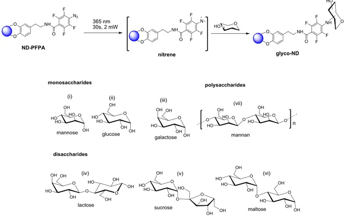
(32) Thèse de Volodymyr Turcheniuk, Lille 1, 2016. non-specificity of the coupling reaction, when the glycans to NDs’ attachment is not happening with the usage of the reducing ends. We will also show that when monosaccharides such as mannose are linked, binding affinities of glyco-NDs to lectins are partially sacrificed, while it is fully preserved for binding of disaccharides and oligosaccharides. Moreover, it was checked whether glyco-NDs are capable of presenting affinity to uropathogenic Escherichia coli strain (E. coli UTI89). Type 1 fimbriae have been identified as the main target, which constitute major virulence factors produced by E. coli UTI89. Type 1 fimbriae have tubular structure in the size of 0.2-2.0 nm in length and 5-7 nm in diameter and they are located all over the surface of the bacterium. On the extremity of the type 1 fimbriae, lectin is located, FimH, it contributes to the colonization of tissue through the specific recognition of The α-D-mannopyranosyl terminal units that are located on the glycoproteins surface of the cell. Mannose-modified NDs exhibit antiadhesive activity for E. coli UTI89 that marked and cell-based assays do not display toxicity for the eukaryotic cells (4, 7). CuI-catalyzed Huisgen cycloaddition reaction (“click” reaction) between NDs decorated with surface azidophenyl or propargyl functions and the corresponding synthetic sugar analogue was used as a base for the coupling strategy. Fluorescence-based agglutination assay was used in order to investigate potential of photochemically grafted native mannan and mannose in the interaction with E. coli UTI89 (6).. 2.2. Synthesis of glycol-NDs via photoactication of perfluorophenyl- azidemodified NDs We have indicated previously that dopamine is known for having significant interactions with various oxides of metal and structures that are graphene-like and it is an agent that is viable for the nanodiamonds’ functionalization (8, 9). Synthesis of a dopamine derivative of perfluorophenylazide (1) was performed through the reaction of the N-succinimidyl 4azidotetrafluorobenzoate with the amine groups of dopamine (Figure 2.1a). Functionalization of hydroxylated nanodiamonds (ND-OH) was performed via ligand (1) (Figure 2.1b). ND particles that were functionalized could be characterized with the Fourier transform infrared (FTIR) spectroscopy (Figure 2.2a) and X-ray photoelectron spectroscopy (XPS) (Figure 2.2b). The spectrum of the FTIR of the as-received ND-OH (Figure 2.2a) presents a high measure at 3447 32 | P a g e. © 2016 Tous droits réservés.. lilliad.univ-lille.fr.
(33) Thèse de Volodymyr Turcheniuk, Lille 1, 2016. cm-1 with the reference to the vibration of surface hydroxyl groups or/and adsorbed water molecules, and an additional sharper one at 1633 cm-1 due to the bending mode d(OH) of surface hydroxyl groups on the NDs. In addition, the band at 1107 cm-1 shows the presence of C-O-C functional groups of cyclic ethers. After ND-OH particles reacted with ligand (1), another vibration peak at 2125 cm-1 common for the νas(N3) stretching appears. The C-H stretching vibration modes are detected at 2850–2970 cm-1 and are partially masked with the large band at 3447 cm-1. The band at 1546 cm-1 must be linked to the NH-C=O bond that is present in ligand (1). The azide group presence on ND-PFPA is additionally confirmed by the N1s high resolution XPS spectrum (Figure 2.2b).. (a). (b). (c). 33 | P a g e. © 2016 Tous droits réservés.. lilliad.univ-lille.fr.
(34) Thèse de Volodymyr Turcheniuk, Lille 1, 2016. Figure 2.1: a) Synthesis of perfluorophenyl azide modified dopamine (1): (i) NaN3, acetone/water, 90C, 2h, 85%; (ii) NaOH, water, 3h, RT, 90%, (iii) NHS, DCM, RT, overnight, 95%, (iv) dopamine hydrochloride, TEA, DMF, RT, argon, 91.4%. b) Modification of ND-OH with ligand (1) forming NF-PFPA. c) Formation of glycol-NDs through the photochemical linking of mono-, di-, and a polysaccharide onto NDs. Bands at 405.2 (Ar N=N+ = N) and 401.9 eV (Ar N= N+ = N), characteristic for the N contribution of the NH C=O linkage is seen at 400.6 eV. Bands at 402.7 and 399.2 eV are also present in the initial ND-OH. It most likely corresponds with the nitrogen functions, for instance, N-O and C-N were probably generated during the process of detonation of trinitrotoluene with the NDs’ formation. Table 2.1 presents the results where nitrogen content has 1.5 at % and possible may be in charge of the positive surface potential of ND-OH (Table 2.1), which was also reported in different studies. In ND-PFPA particles, the presence of N 1s was increased to 8.4 at % with a F/(N-1.5) ratio of 1.28, which is near the theoretical measure of 1.33. In order to make sure that conjugates are stable and dopamine ligand will not be detached after certain 34 | P a g e. © 2016 Tous droits réservés.. lilliad.univ-lille.fr.
(35) Thèse de Volodymyr Turcheniuk, Lille 1, 2016. period of time, ND-PFPA particles were immersed for 24 h at various pH (3, 7, and 9) as well as in biological medium such as Dulbecco’s modified Eagle medium (DMEM) and the FTIR spectra were recorded.. Table 2.1: Physical properties of the NDs modified with ligand (1). As it is indicated in the (Figure 2.2c), no strong lowering of the νas(N3) band at 2125 cm-1 was observed due to immersion into solutions of different pH, and only a slight decrease was observed when incubated in biological medium. (a). (b) C-O-C. OH. ND-PFPA. 3500. 3000. 2500. 2000. Wavenumber / cm. NH. N. C-H. 3. OH. OH OH. Transmittance / a.u.. N 1s. ND-OH ND-PFPA 1500 1000. -1. ND-OH 408. 406. 404. 402. 400. 398. 396. binding energy / eV. (c). 35 | P a g e. © 2016 Tous droits réservés.. lilliad.univ-lille.fr.
(36) Transmittance / a.u.. Thèse de Volodymyr Turcheniuk, Lille 1, 2016. pH=3 pH=7 pH=9 DMEM 3500. 3000. 2500. Wavenumber / cm. 2000. 1500. -1. Figure 2.2: a) FTIR spectra of ND-OH (black) and ND-PFPA (red). b) N 1s high-resolution XPS spectra of ND-OH (black) and ND-PFPA (red). c) FTIR spectra of ND-PFPA after immersion for 24 h in pH 3, pH 7, pH 9 buffer, and DMEM.. The ether bond between the diamond surface and the aromatic ring of dopamine can be considered as stable in comparison with the organic molecules that take place within the indicated period of time with no hydrolysis. Photochemical linkage of glycans to NDs is better than the photochemistry of arylazides that are converted to the reactive nitrenes because of the light activation (Figure 2.2c). Nitrenes that are highly reactive are capable of the direct interaction with any type of glycan due to the C-H and/or N-H reactions of insertion and create highly robust covalent linkage. Mono-, di- and a polysaccharide were photochemically integrated into ND-PFPA particles (Figure 2.2c) using the mixture of the solution of ND-PFPA in acetonitrile with aqueous solutions of the respective glycan, and irradiating the mixtures at 365 nm for 30 s at 2 mW.. Representative FTIR spectrum of mannan-modified NDs and mannose are presented in Figure 2.3 A and show that characteristic N3 vibration band at 2128 cm-1 completely disappears, which in its turn suggest consummation of all azido groups in the photochemical process. The band at 1633 cm-1 due to the bending mode ∂OH increased significantly in the case of ND-mannan. The N 1s XPS spectra after photochemical linking of mannose also changed significantly. The 36 | P a g e. © 2016 Tous droits réservés.. lilliad.univ-lille.fr.
(37) Thèse de Volodymyr Turcheniuk, Lille 1, 2016. conversion of the azide group into C-N bonds is proved by the disappearance of the bands at 405.2 (Ar N=N+ = N) and 401.9 eV (Ar N=N+ = N) and the appearance of a band at 400.6 eV due to the formation of C-N bonds. The presence of F 1s signal at 686 eV is an additional indication of the formation of ND-mannose (Figure 2.3c).. To validate further the covalent linking of the glycan to ND-PFDA, a solution of ND-PFPA in acetonitrile was mixed with an aqueous solution of mannan and left for 24 h. The characteristic N3 vibration band at 2128 cm-1 was still present on ND-PFPA and that confirmed the glycan interaction with the dopamine ligand on the NDs. Representative transmission electron microscopy (TEM) images of ND-mannose and ND-mannan (Figure 2.3d) show the presence of spherical particles with a mean diameter of 24 nm for all structures, regardless of the glycan presence on their surface.. Transmission / a.u. (a). ND-mannose ND-mannan 3500. 3000. 2500. 2000. Wavenumber / cm. (b). 1500. 1000. -1. (c). 37 | P a g e. © 2016 Tous droits réservés.. lilliad.univ-lille.fr.
(38) Thèse de Volodymyr Turcheniuk, Lille 1, 2016. N 1s. 406. ND-mannose. 404. 402. 400. 398. 396. F1s. 695. ND-mannose. 690. 685. 680. binding energy / eV. binding energy / eV. (d). ND-mannose. ND-mannan. 20 nm. 20 nm. Figure 2.3: a) FTIR spectra of ND-mannose (black) and ND-mannan (grey); b) N 1s spectrum of ND-mannose, c) F 1s spectrum of ND-mannose, d) TEM images of ND-mannose and NDmannan.. The amount of glycans that were gradually integrated into the ND-PFPA was analyzed as well with the usage of the well-established phenol–sulfuric acid assay and that was proved to be glycan dependent (Table 2.2).. Table 2.2: Physical properties of different glycan-NDs. 38 | P a g e. © 2016 Tous droits réservés.. lilliad.univ-lille.fr.
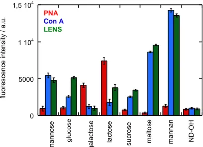
(39) Thèse de Volodymyr Turcheniuk, Lille 1, 2016. Talking about mannose, it should be noted that the loading of sugar can be compared to the mannose-ND that were formed with the usage of “click”. Disaccharide-modified NDs has the amount of incorporated saccharides which is almost twice higher than in mannose and the highest loading of sugar was common for mannan-NDs.. 2.3. Lectin binding assays The affinity binding study was focused on the glyco-NDs that has a range of fluorescently labeled lectins (Figure 2.4). FITC-labeled Concanavalin A from Canavalia ensiformis (Con A) and FTIR-labeled Lens culinaris (LENS) are actually specific to mannopyranoside and were used in terms of the positive controlling. For the negative control for glucose-, mannose-, sucrose-, maltose- and mannan-modified ND particles, the α-D-galactopyranose specific Arachis hypogaea (PNA) was used, while Con A and LENS were negative controls for galactosemodified NDs. It is expected for Lactose-NDs to interact with all lectins since indicated disaccharide is retrieved from the condensation of galactose and glucose-forming ß(1→4)glycosidic linkage. With the usage of the fluorescence intensities that were recorded after the interaction of glycoNDs solutions within 30 min with different lectins confirm that the binding affinities of the photolinked sugars are congruent with the expected binding characteristics for each lectin. 39 | P a g e. © 2016 Tous droits réservés.. lilliad.univ-lille.fr.
(40) Thèse de Volodymyr Turcheniuk, Lille 1, 2016. Several times larger fluorescence signals were shown by mannose-modified NDs during the incubation with the Con A and LENS in comparison with the incubation with PNA because of the higher binding affinity. 4. PNA Con A LENS 1 10. 4. ND-OH. mannan. maltose. sucrose. lactose. mannose. 0. galactose. 5000. glucose. fluorescence intensity / a.u.. 1,5 10. Figure 2.4: Fluorescence intensities evaluated using FTIC-labeled lectins after reaction with glycan-terminated NDs in a 1/1 ratio (1 mg mL-1 ) in tris buffer solution (pH 7.4 containing Mg2+, Ca2+, NaCl): Fluorescent measurments were performed using an excitation wavelength of 485 nm and an emission wavelength of 520 nm; the results are derived from the data of 4 independent experiments. However, the results were identical for the fluorescence intensity recorded for mannose-NDs in the presence of FITC-labeled PNA that was measured with the ND-OH. Indicated level of fluorescence tends to be most likely linked to non-specific protein interaction with the surface of the particle. Opposite behavior is shown by galactose-modified NDs: strong fluorescence due to the incubation with PNA and weak with Con A and LENS lectins. The fluorescence signal upon incubation is strongest for LENS, lower for Con A, and weak for PNA when we are talking about glucose-NDs. The tetrameric lectin Con A was next to mannose and reported binding site specific to glucose, with a lesser extent. Correlation to the indicated difference in affinity of the lowered fluorescence signal upon incubation of glucose-ND is found due to the comparison with Con A binding to glucose-ND specific LENS. 40 | P a g e. © 2016 Tous droits réservés.. lilliad.univ-lille.fr.
(41) Thèse de Volodymyr Turcheniuk, Lille 1, 2016. 4. PNA Con A LENS 1 10. 4. ND-OH. mannan. maltose. sucrose. lactose. mannose. 0. galactose. 5000. glucose. fluorescence intensity / a.u.. 1,5 10. Figure 2.4: Fluorescence intensities evaluated using FTIC-labeled lectins after reaction with glycan-terminated NDs in a 1/1 ratio (1 mg mL-1) in tris buffer solution (pH 7.4 containing Mg2+, Ca2+, NaCl): Fluorescent measurments were performed using an excitation wavelength of 485 nm and an emission wavelength of 520 nm; the results are derived from the data of 4 independent experiments. For further investigation of the indicated coupling method, together with the specificity of the glycans, disaccharide and polysaccharide that are surface-bonding, they were photochemically linked to ND-PFPA in order to determine their affinity to the three lectins. Lactose-NDs with PNA or LENS interaction resulted into the twice more of the fluorescence signal for PNA. It means that this disaccharide tends to be coupled with the NDs through its glucose end. For sucrose-NDS (a disaccharide that is composed of glucose and fructose) no interaction with PNA was noticed, while the interaction with glucose-specific Con A and LENS was higher, meaning that some of the sucrose is linked via the fructose end to ND-PFPA. With the usage of maltoseND, two-unit glucose disaccharide formed with an α-(1→4) bond, strongly binds with the Con A and LENS, which is stronger than the bind that was observed with mannose. The strongest interaction with Con A and LENS are observed with mannan-modified NDs: these particles showed a three times higher sugar loading than mannose-NDs, which correlates to an approximately three times larger fluorescent signal. 41 | P a g e. © 2016 Tous droits réservés.. lilliad.univ-lille.fr.
(42) Thèse de Volodymyr Turcheniuk, Lille 1, 2016. 2.4. Effect of mannose-NDs and mannan-NDs on agglutination of E. coli UTI89 strains 2.4.1. Fluorescence-based agglutination assay in the presence of mannose and mannose-NDs The next step was the examination of mannose-NDs and mannan-NDs affinity to uropathogenic E. coli UTI89. As it was already mentioned, FimH, a lectin located at the extremity of type 1 fimbriae, is a major virulence factor produced by E. coli UTI89, which contributes to the colonization of tissue due to its specific recognition of the terminal α-D-mannopyranosyl units present on cell-surface glycoproteins. Interference of the interaction became possible due to the mannose-modified NDs that was formed by “click” as a chemistry of propargyl and/or azidemodified mannose derivatives. In order to research the potential of the mannose-NDs that were formed photochemically, it was necessary to check whether indicated particles show increased agglutination effects to E. coli UTI89 in comparison with the free mannose in solution. For the mentioned purpose, fluorescence-based agglutination assay was used (34). It is mainly grounded on the mixture of different concentrations of mannose and mannose-NDs with fluorescently labeled E. coli UTI89 formed through genetic modification to express turbo FP635 (Katushka) fluorescent proteins, emitting at 635 nm (upon excitation at 580 nm). Diverse fluorescence images of Katushka expressing E. coli after interaction for 4 h at 4 °C with different concentrations of mannose-NDs (1–300 mg mL-1) are presented on (Figure 2.5).. For this purpose, the influence of ND-OH and mannose in solution was researched as a controlling aspect. These agglutination tests show that mannose, as expected, shows no E. coli UTI89 agglutination in the tested concentration range. In comparison with the free mannose in solution, mannose-NDs display a concentration dependent agglutination behavior at a minimal concentration of 50 mg mL-1.. 42 | P a g e. © 2016 Tous droits réservés.. lilliad.univ-lille.fr.
(43) Thèse de Volodymyr Turcheniuk, Lille 1, 2016. mannose. 10 µg mL-1. 10 µm. 50 µg mL-1. 10 µm. 300 µg mL-1. 10 µm µm 10. mannose-ND No particles. 10 µm. ND-OH. 10 µm. 10 µg mL-1. 10 10µm µm. 50 µg mL-1. 10 µm. 300 µg mL-1. 10 10µm µm. Figure 2.5: Fluorescence-based agglutination assay for: Fluorescence images of Turbo FP635 (Katushka protein) expressing E. coli UTI89 (1 108 cfu ml-1 ) in the presence of different concentrations of mannose, in the absence of particles (no particles) and in the presence of NDOH (100 mg mL-1 ) and different mannose-NDs (the values correspond to total mannose concentration in the solution and is directly comparable with the results of free mannose).. 2.4.2. Fluorescence-based agglutination assay in the presence of mannan and mannan-NDs Furthermore, in a comparable manner, the agglutination behavior of mannan and mannan-NDs were investigated. A cell-wall component of microorganisms is called Mannan, which consists of d-mannose residues expanded by α-(1→6)-, α -(1→3)-, α -(1→2)- linkages. Park with the colleagues described the formation of carboxylic mannan-coated iron oxide nanoparticles to target antigen-presenting cells (APCs), including macrophages, by the specific interaction between the mannose ligand and the mannose receptors on APCs. Mannan was chosen as an 43 | P a g e. © 2016 Tous droits réservés.. lilliad.univ-lille.fr.
(44) Thèse de Volodymyr Turcheniuk, Lille 1, 2016. integral component in NDs in order to take advantage of the predicted high binding affinity towards E. coli UTI89 because of the presence of multiple mannose ligands in this polysaccharide.. (Figure 2.6) presents the results of mannan and mannan-NDs addition to a solution of E. coli UTI89. In contrast to mannose only (Figure5), addition of 100 mgmL-1 mannan reveals partial E. coli UTI89 agglutination. Moreover, mannan-ND particles have an ability to agglutinate E. coli with an onset at concentrations as low as 10 mg mL-1. Indicated statement corresponds with the usually presented multivalence of mannan on nanoparticles. mannan 10 µg mL-1. 10 µm. 50 µg mL-1. 10 10µm µm. 10 µm. 300 µg mL-1. 10 µm. 10 µm. 10 µm. 10 µm. mannan-ND No particles. 10 µm. ND-OH. 10 µm. 10 µg mL-1. 10 µm. 10 µm. 50 µg mL-1. 10 µm. 300 µg mL-1. 10 µm. Figure 2.6: Fluorescence-based agglutination assay for: Fluorescence images of Turbo FP635 (Katushka protein) expressing E. coli UTI89 (1 108 cfu ml-1) in the presence of different concentrations of mannan, in the absence of particles (no particles) and in the presence of NDOH (100 mg mL-1) and different mannan-NDs (the values correspond to total mannan concentration in the solution and is directly comparable with the results of free mannan). 44 | P a g e. © 2016 Tous droits réservés.. lilliad.univ-lille.fr.
Figure
+7


Documents relatifs