Université de Montréal
Sirni function in the developing and aduit brain
Par Chun Yang
Département de Biochimie Faculté de médecine
Thèse présentée à la faculté des études supérieures En vue de l’obtention du grade dc
Philosophie Doctoral (Ph.D.) en biochimie
Avril, 2006
Université
de Montré al
Direction des bibliothèques
AVIS
L’auteur a autorisé l’Université de Montréal à reproduire et diffuser, en totalité ou en partie, par quelque moyen que ce soit et sur quelque support que ce soit, et exclusivement à des fins non lucratives d’enseignement et de recherche, des copies de ce mémoire ou de cette thèse.
L’auteur et les coauteurs le cas échéant conservent la propriété du droit d’auteur et des droits moraux qui protègent ce document. Ni la thèse ou le mémoire, ni des extraits substantiels de ce document, ne doivent être imprimés ou autrement reproduits sans l’autorisation de l’auteur.
Afin de se conformer à la Loi canadienne sur la protection des renseignements personnels, quelques formulaires secondaires, coordonnées ou signatures intégrées au texte ont pu être enlevés de ce document. Bien que cela ait pu affecter la pagination, il n’y a aucun contenu manquant.
NOTICE
The author of this thesis or dissertation has granted a nonexciusive license allowing Université de Montréal to reproduce and publish the document, in part or in whole, and in any format, solely for noncommercial educational and research purposes.
The author and co-authors if applicable retain copyright ownership and moral rights in this document. Neither the whole thesis or dissertation, nor substantial extracts from it, may be printed or otherwise reproduced without the author’s permission.
In compliance with the Canadian Privacy Act some supporting forms, contact information or signatures may have been removed from the document. While this may affect the document page count, t does not represent any Ioss cf content from the document.
Université de Montréal Faculté des études supérieures
Cette thèse intitulée:
Simi fuction in the developing and aduit brain
présentée par: Chun Yang
a été évaluée par un jury composé des personnes suivantes
Dr. Edgard Delvin, président-rapporteur Dr. Jacques L Michaud, directeur de recherche
Dr. André Tremblay, co-directeur
Dr. Andréa Richter. membre du jury
‘Il
SUMMARY
Sim], which codes for a bHLH-PAS transcription factor, is expressed in various areas of the brain, including the developing and postnatal paraventricular nucleus (PVN), a region of the hypothalamus that regulates food intake. Haploinsufficency of Sim] causes hyperphagia in mice and humans without any decrease of energy expenditure. Isolated hyperphagia is suggestive of PVN dysfunction. Indeed, the number of PVN ceils is decreased by 24% in Sim]‘
mice, suggesting that their hyperphagia is caused by a developmental mechanism. However, the possibility that Sim] functions in the postnatal PVN to regulate food intake cannot be ruled out. In order to explore this hypothesis, two strategies were used.
f irst, in an attempt to identif a PVN specific element that could be used to modulate Sim] expression levels in mice, we studied the mechanism of regulation of Sim] gene expression. We characterized the promoter and regulatory elements in an 8. 1kb 5’-sequence of$im] gene. We found an AHR ARNT/2 binding site which positively regulates prornoter activity in the context of transfection experiments in Neuro-2A ceils. The bHLH-PAS complex AHR-ARNT/2 can bind to this consensus site in gel shifi assay. Overexpression of Arnt. Arnt2, or Ahr increased the activity of a reporter construct containing the Sim] prornoter and the AHR-ARNT/2 binding site by 2-fold, but deletion of 4hp of its core sequence abolished this induction. S imilarly, addition of 2,3,7, $ -tetrachlorodibenzo-p-dioxin (TCDD), a ligand of AHR, increased the activity of the reporter construct by 1. $-fold but flot that of its rnutated version. f inally, we found that TCDD increased Simi expression in Neuro-2A celis and in mouse kidney and hypothalamus by more than two fold. We conclude that Sim] expression is regulated by AHR-ARNT/2, pointing to complex regulatoiy interactions between bHLH-PAS proteins. However, our transgenic studies suggest that the elements required for Sim] expression in the PVN are out ofthe 5’-region was studied.
iv
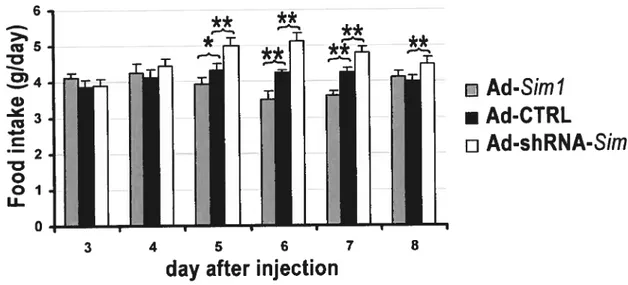
Second, we used adenoviral vectors to modulate Sim] expression in the postnatal PVN of wild-type mice. Unilateral stereotaxic injection into the PVN of an adenoviral vector producing a short hairpin RNA directed against Sim] (Ad-shRNA-81m1) resulted in a significant increase of food intake. The maximal effect was about 22% which was reached on the 6th
day afier injection, when compared to the injection of a control virus. In contrast, injection of an adenovirus that expresses 81m] (Ad-Siml) induced a decrease of food intake that was maximal on the 7th day afier injection, reaching 20%. In order to explore the impact of more extensive changes in 81m] expression levels on food intake, we performed a set ofbilateral injection of these viruses. Again, we found the Ad-shRNA-Sim] virus increased whereas Ad-Sim] virus decreased food intake. Although the impact of bilateral injections of these vectors into die PVN xvas not greater than that of unilateral injections, it further validates the resuits obtained from the unilateral injections. Taken together, our resuits strongly suggest that Sim] functions physiologically to regulate food intake, and support the existence of a pathway in the PVN that regulates food intake independently energy expenditures. SIMÏ can be added to a growing list of PAS dornain proteins that are involved in the regulation of physiological processes acting at the interface between the environment and the internai milieu.
Key Words: $im], paraventricular nucleus (PVN), bHLH-PAS protein, promoter, gene regulation, food intake, hyperphagia, adenovinis, arylbydrocarbon receptor (AHR)
V
RÉSUMÉ
Le facteur de transcription SIM1, qui est caractérisé par des domaines bHLH et PAS, est exprimé dans des régions spécifiques du cerveau, incluant le noyau paraventriculaire (PVN) de l’hypothalamus, une région qui participe à la régulation de la prise alimentaire. L’haploinsuffisance de Simi engendre une hyperphagie chez la souris comme chez l’homme, conduisant à l’obésité. Une hyperphagie isolée est compatible avec une dysfonction du PVN. De fait, le nombre de cellules constituant le PVN est réduit de 24% chez les souris Sirn]. suggérant que leur hyperphagie est d’origine développernentale. Cependant, comme Simi est exprimé de façon continue dans le PVN adulte, la possibilité que ce gène agisse le long d’une voie physiologique pour réguler la prise alimentaire n’est pas exclue. Afin d’explorer cette hypothèse, nous avons
mis
à profit deux approches.Dans un premier temps, nous avons voulu utiliser la transgénèse chez la souris pour moduler l’expression de Sim] dans le PVN. Afin d’identifier un élément régulateur spécifiquement actif dans le PVN. nous avons étudié un fragment de
8J kb de séquence 5’ qui contient le promoteur basal. Nous avons identifié au sein de ce fragment un site de liaison pour le complexe bHLH-PAS AHR
ARNT/2 qui contrôle positivement l’activité promotrice d’un
fragment génomique de Si,nl dans des cellules Neuro-2A. Des expériences de migration retardée sur gel ont confirmé que ce complexe lie ce site. L’expression de Arnt, Arnt2 ou d’Aur augmente l’activité du promoteur de Sim] par un facteur de deux lorsque ce site est présent en amont. De plus, l’addition de 2,3,7,8-tetrachÏorodibenzo-p-dioxin (TCDD). un ligand de AHR, augmente l’activité du promoteur par un facteur de 1,8. mais n’a aucun effet si le site de liaison n’est pas présent. Finalement, l’administration de TCDD augmente l’expression de Simi dans les cellules Neuro2A ainsi que dans le rein et l’hypothalamus de souris.
vi Nous concluons que l’expression de Sïm] est régulée par le complexe AHR ARNT/2. suggérant l’existence d’interactions régulatrices complexes entre les protéines bHLH-PAS. Par contre, des études de transgénèse indiquent que le fragment de séquence 5’ ne contient pas d’éléments régulateurs spécifiquement actifs dans le PVN.
Nous avons utilisé une approche différente pour moduler Fexpression de 57m] dans le PVN qui met à profit des vecteurs adénoviraux. L’injection stéréotaxique unilatérale d’un virus produisant un petit RNA en épingle dirigé contre Sim] (shRNA-Sim]) dans le PVN induit une augmentation de la prise alimentaire. L’effet maximal est atteint au sixième jour après l’injection: à ce stade, la prise alimentaire des souris injectées avec le virus shRNA-Sim] est accrue de 22% en comparaison avec celle de souris infectées avec un virus contrôle. Par contre. l’injection d’un adénovirus qui produit SIM1 (Ad-Sim]) induit une diminution de la prise alimentaire qui était maximale au 7 jour suivant la procédure. atteignant 20%. Afin d’évaluer l’impact de changements plus extensifs de l’expression de Sim] sur la prise alimentaire, nous avons effectué des injections bilatérales de ces virus. Malgré le fait que les injections unilatérales et bilatérales ont un impact similaire sur la prise alimentaire, ces expériences ont permis de confirmer l’effet de ces virus avec une deuxième série d’injections. Globalement, ces résultats suggèrent fortement que Sim] fonctionne le long d’une voie physiologique qui contrôle la prise alimentaire. SMl peut être ajouté à une liste croissante de protéines à domaines PAS qui agissent à l’interface de l’environnement et du milieu interne.
Mots clés: Sim], proteine bHLH-PAS. AHR, noyau paraventriculaire (PVN). promoteur, régulation géniqueie. prise ah mentaire. hyperphagie, adénovirus. réceptor arylhydrocarbon (AHR)
v i Table of contents Summary Résumé V Table of contents vii List of figures xi List ofabbreviations (general)
xii Neuroanatomical abbreviations xiv Acknowledgements xv Dedication
Chapter 1. General introduction
1
1. The bHLH-PAS proteins
2
1.1. Highly conserved structures of bHLH-PAS proteins 4 1.2. Regu]ation physiological processes by bHLH-PAS proteins 5
1.2.1. AHR mediating xenobiotic metabolism 5
1.2.2. Regulation of hypoxia responsiveness by HIF $ 1.2.3. Regulation ofcircadian rhythm by bHLH-PAS proteins 10 1.2.4. ARNTand ARNT2: functionat partners of bHLH-PAS proteins 12
1.2.5. Crosstalk between bHLH-PAS protein-rnediated signalling pathways
15
2. SIM proteins
16 2.1. Drosophila sim, a regulator ofmidline developrnent
16 2.2. Sequence similarity ofthe mammalian SIM proteins
17
2.3. Transcriptional properties ofmammalian SIM proteins 17 2.4. Expression patterns ofmammalian Sim genes in the brain 18
3. The paraventricular nucleus ofthe hypothalamus 20
3.1. Structure ofparaventricular nucleus 20
3.2. Regulation ofenetgy expenditure by the paraventricular nucleus 23 4. function ofSirnÏ and Sini2 in the paraventricular nucleus and in other
parts of the brain
26 4.1. Rotes ofSim] in the controlling ofneuron differentiation in
PVN/SON/aPV
26
4.2. Role ofSim] in energy balance 27
4.3. Sim2, a paralogofSim]
vut
4.4. SimI andSirn2 function during mammiÏlary body developrnent .30 4.4.1. Structure and function ofthe rnamrnillary body 30 4.4.2. Transcription factors controlling mamrnillary body
development
32 5. Hypothesis and objectives
34 Chapter Ii: Regulatory interaction between arylhydrocarbon receptor and
51M 1, two basic helix-Ioop-helix pas proteins involved in the control offood intake
35 Abstract 36 introduction 37 Experimental procedures 3$ Constructs 3$ Chemical 39 RACE 39
Ccli transfection and luciferase assay 39
Electrophoretic mobiÏity shifi assay (EMSA) 40
Reverse transcriptase-PCR analysis
41
Resuits
42 Discussion
45
TCDD and the control ofappetite 45
Regulation interaction between bHLH-PAS proteins 47 Acknowledgements
47 References
4$ Chapter 111: Adenoviral-mediated modulation ofSim] expression in the
paraventricular nucleus affects food intake 57 Abstract
59 introduction
60 Material and methods
61 Production ofadenoviruses 61 Protein analysis 62 Stereotaxic injection 62 Validation of injection site
63 Resuits
65 Discussion
ix Acknowledgements
.70 Refcrenccs
.71
Chapter 1V: General discussion
7$
1. Characterization ofregulatory elements driving81m] expression in thebrain
79
2. Interaction between SIMI and AHR $0
3. Modulation ofSim] expression levels in postnatal PVN using adenoviral vectors
82 3.1. Technical considerations: use ofadenovirus to modulate
81m] expression
$2
3.2. Regulation offood intake by Simi 84 3.3. Diverging pathway controlling energy balance 85 4. Investigation ofrnechanisms ofSi,n] pathway controlling food Intake $6 4.1. Meclianisms involved in the fecding bchaviour of8im]rnicc $6 4.2. Developrnent of the hypothalamus and the search for obesity
Genes
87
4.3. Generation ofa conditional allele ofSiin] 8$
4.4. Characterization ofsmall molecule regulating Sim] 90
5. Redundant roles of $1m] and Sim2 in developing MB 90 Conclusion
91
References
92 Chapter V: Annex I:81m] and Si,,,2 are required for correct targeting
ofmammitlarybody axons 11$ Abstract 120 Introduction 121
Material and methods
123
Generation ofthe81m!tiz allele 123
Genotyping ofmice
124 Histology, in situ hybridization, 3-galactosidase staining and Du
labeling
125 Resuits
125 Development ofmammiÏlaiy body projections requires 81m] and Sim2 125
ASim1”allele allows staining ofmammillaiy body axons
127
Siln]/Sim2 mutants axons are directed towards the midline
X
SimJ/Sim2 mutant neurons are generated and survive until E 18.5
129
Sim] and Sim2reprcss Rigl/Robo3 expression in the deveÏoping
mamrnillarybody
130 Discussion
132 Requirement ofSirn] andSim2 for MB axonal developrncnt 133 Cascade of transcription factors controlling MB developmcnt 134 Respective offtinctions ofbHLH-PAS proteins during MB development 135 AcknowÏedgements
137 References
138 Annex II: Characterization of regulatory elernents drivingSinilexpression in
the brain using transgenesis 154
xi
List of figures
Figure I-1. Schematic representation ofthe domain structure ofsome bHLH-PAS familv members
3
f igure I-2. Mechanisms oftranscriptional activation by AHR and ofnegative
feedback regulation of AHR by AURR 6
Figure I-3. ARNT fonns both homodimers and heterodimers with AHR, HIf-ΠandSIM
14
Figure I-4. Amino acid identity ofthree STM proteins 17 figure I-5. Expression pattem of 81m] and 81m2 in the developing brain 19 figure I-6. Structural and fiinctional relationship between hypothalamic
nuclei
22
Figure I-7. Interaction between the hypothalamus, the brainstem and the periphery....23
Figure I-8. Interaction between the arcuate nucleus (ARC) and the PVN 25
Figure I-9. Transcriptional regulation of anterior hypothalarnic developrnent 28 Figure I-10. Axonal projection originating from the mammillary body 31 Figure II-1. Identification of 81m] transcription start site by 5’-RACE analysis 52 figure II-2. Identification of a functional DRE in 81m] promoter region 53 figure II-3. Binding activity ofAHR-ARNT2 to the 81m] DRE 54 Figure II-4. Effect of Ahr, Arnt and Arnt2 overexpression or of TCDD
supplementation on 81m] promoter activity 55
figure II-5. Effect ofTCDD on weight gain, appetite, and Sim] mRNA levels 56 Figure III-Ï. Western blot detection ofSIMl production in cultured 293A celis
infected with adenoviruses
74
Figure III-2. Unilateral infection ofthe PVN with the Ad-shRNA-$im] virus
increases food intake 75
Figure III-3. Unilateral infection of the PVN with the Ad-81m] virus decreases food intake
76
Figure III-4. Bilaterai infection ofthe PVN with adenoviruses that modulate
81m] expression levels affects food intake 77
xii
Figure AI-1. Coexpression ofSirnÏ and Sii,z2 in the developing rnammillary body....144
Figure AI-2. Organization of the mammilÏary body projections 145 Figure AI-3. MTEG and MTT development affected by Siml/Sim2 gene dosage 146 f igure AI-4. Creation ofa Sim] allele expressing Tau-lacZ 147
figure AI-5. LacZ staining ofmammillary body axonal projections in E14.5
SirnJ/Sirn2 mutant embryos 148
figure AI-6. Abnorrnal targeting ofmammilïary body axons as revealed by Dii labelling
149
figure AI-7. LacZ staining ofrnamrnillary body axonal projections in El 1.5
$irnl/Sirn2 mutant embryos 150
figure AI-8. Loss ofFoxbl expression in E 12.5 Sim]/S1m2 double mutants 151
Figure AI-9. Mammillary body neurons survive until the end of gestation in
Sim]/$im2 double mutants 152
Figure III-10. Sim] and Sirn2 repress Rig-1 expression in the developing
marnmillary body 153
f igure Ail-1. Charaterization ofSirnJ regulatory elements in cultured ceils 157
XIII
List of abbreviations (general)
AAV, adeno-associated viral; AgRP, agouti-related protein; AHR. arylhydrocarbon receptor; AhRR, aryl hydrocarbon receptor repressor; ARNT. aryl hydrocarbon receptor nuclear transiocator: BAC, bacterial artificial chromosome: BAT. brown adipose tissue; BAT. brown adipose tissue; bHLH, basic helix-loop-helix; BMAL1, brain and muscle Amt-like protein 1; BRN2, brain 2; CARI, cocaine- and amphetamine-regulated transcript; CBP, CREB binding protein; CCX, cholecystokinin;
CME, central midiine elernent; CMV, cytornegalovirus; CREB, cAMP-repsonse elernent binding: CR11. corticotropin-releasing hormone; CRY, cryptochrome; CTL, cytotoxic T lymphocyte; Cyplal, cytochrome P450, family 1, subfamily A, polypeptide 1; DRE, dioxin-responsive element; DS, Down syndrome; EMSA, electrophoretic mobility-shifi assay;
EPO, erythropoietin; ESTs, expressed sequence tags; GFP, green fluorescent protein; GI, gastrointestinal; GRJP 1, glucocorticoid receptor interacting
protein ; GRIPI, glutamate receptor interactirg protein 1; 111f, hypoxia inducihle factor; HIV-l. immunodeficiency virus type 1; HRE, hypoxia response element: Hsp9O, heat shock protein 90; IPAS, inhihitoiy PAS domain protein; LPL, lipoprotein lipase; MC4R, melanocortin receptor 4; MSH, melanocyte stimulating hormone; Nf-H, neurofilament H regulatory element; NLS, nuclear localisation sequence; NPAS. neuronal bHLH-PAS protein; NPY, neuropeptide Y; NTX. naltrexone; ODD, oxygen dependent degradation domain; 01, oxytocin; OTP, orthopedia protein; PAS, Per.
ARNT, 51M homoÏogy domain; Per, period gene; POMC, pro
opiomelanocortin; PS, protease; rAAV, Recombinant AAV; RACE, rapid amplification of 5’-cDNA ends; RFP, red fluorescent protein; RSV, Rous sarcoma virus; SIM, single minded; SRC-l, steroid receptor coactivator-1;
2,3,7,8-xiv
tetrachlorodibenzo-p-dioxin; Tif2, transcriptional interrnediaiy factor2; TRI-I, thyrotropin-releasing hormone: TSS, transcription start site; UCP, uncoupling protein; VEGf, vascu]ar endothelial growth factor; VHL, von
Hippel—Lindau protein; VP, vasopressin; WAT, white adipose tissue; WT,
wild-type; XAP2. hepatitis B virus X-associated protein 2; XRE, xenobiotic response elcment;
Neuroanatomical abbreviations
aPV, anterior periventricular nucleus; ARC, arcuate nucleus; BS: brain stem; CNS, central nervous system; DMV, dorsal motor nucleus of vagus nerve; LHA. lateral hypothalamic area; MB, mammillary body; ME, medial eminence; MTEG. mammillotegmental tract; MTH, mammillothalamic tract; NLOT, nucleus of the lateral olfactorv tracr; NTS. nucleus of soÏitary tract: PMT. principal mammillary tract; PVN, paraventricular nucleus; rNST, rostral nucleus of solitary tract; SC: spinal cord: SCN. suprachiasmatics nucleus; SON, supraoptic nucleus; ZLI, zona limitans intrathalarnica;
xv
ACKNOWLEDGEMENTS
I wish to acknowledge rny sincere gratitude to ail who have contributed to this
work, especially to:
Dr. Jacques Michaud, my research supervisor, for providing me with the
opportunity to work in his laboratoiy. for his support and guidance.
Dr. Andre Tremblay, my co-supervisor, for his constructive advice and
support.
To the people in our group:
I thank Francine Boucher for her heip with the maintenance of mice. I thank
Aurore Caqueret and Sabine Duplan for sharing their technical expertise. I greatly enjoyed working with them; their friendship and relationship are unforgettable.
To my collaborators:
I thank Dr Bernard Massie for providing us with the adenovirus vectors and for his insiglit. I thank David Gagnon for teaching me the techniques of
manipulating adenovirus. I thank Laurent Knafo for his technical assistance with the real-time PCR and DNA sequencing service. I thank Dr Pascal Vachon for teaching us the stereotaxic microinjection technique. I thank ail collaborators for their kind support and advice.
I deeply appreciated the studentships from the Fondation de l’hôpital Sainte
Justine, and from the l’Université de Montréal.
I wouid like to thank the members of my jury, for reading, correcting and
xvi
DEDICATION:
To rny parents, for teaching me to be a nice person.
To my husband, for bis profound support and love.
Studies of development pathways, ceil growth and celi death have in part become studies of transcriptional regulators. Efforts to identify the nature of these regulators have revealed families of proteins that are characterized by highly conserved DNA binding, dimerization, transactivation and repression motifs. An emerging farnily of transcriptional regulators is characterized by the presence of basic-helix-loop-helix (bHLH) and PAS dornain. These proteins control a variety of developrnental and physiological processes including neural developrnent, toxin rnetabolisrn, respond to hypoxia, circadian rhythms, tracheal formation, and hormone receptor function.
1. The bHLH-PAS proteins
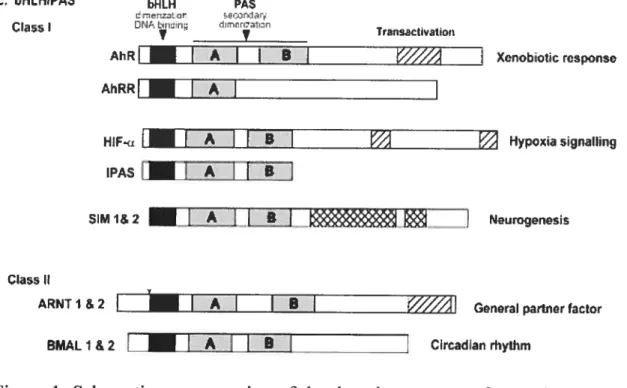
The bHLH-PAS proteins usually function as dimeric DNA-binding protein complexes. Most bHLH-PAS farnily members can be grouped into two classes (fig. 1; Kewley et al., 2004). Class I bHLH/PAS factors neither homodimerize nor heterodirnerize with other class I factors. This group incÏudes the aryl hydrocarbon receptor (AHR), aryl hydrocarbon receptor repressor (AHRR), the hypoxia inducible factors (HIF; HIF-ici, HIF-2u, and HIF-3u), CLOCK, neuronal bHLH-PAS protein (NPAS2), and single minded proteins ($1M 1 and SIM2) (Ema et al., 1996; Lindebro et al., 1995; Wang et al., 1995). They must dirnerize with a class II bHLH/PAS factor to form active transcription factor complexes. The best characterized class II protein is the aryl hydrocarbon receptor transiocator (ARNT) (Hoffman et al., 1991), which is ubiquitously expressed in mesodermal derived tissues. Other members of this class include ARNT2, which is predorninantly expressed in the central nervous system (CNS), and the circadian rhythm proteins, brain and muscle ARNT-like protein (BMALÏ/MOP3 and BMAL2/MOP9), whose expression is more restricted (Hirose et al., 1996; Okano et al., 2001). Typically, a class II protein can bind different class I proteins. The combinatorial and interactive properties of bHLH-PAS proteins
3
provide a variety of potential rnechanisms to control their function as transcriptional regulators, which may heÏp explain their widesprcad use in complex bioÏogical events.
c, bHL.I-IiVAS bHLH PAS e’z]or .JrI.Cj
Çlass t DNAIiLIn.
Transsctkation
MiR
I
A B Xenobotic responseAhRRI
I
AIHIF.
I
AI I
BI
Hypoxia signalhngIPAS
EA
Ï I
B51M 1& 2 [AJ
t
B] NeurogenesiClass Il
ARNT I & 2
I
I
AI
B________
Gonoral partner factor
BMAL 1 &2
I
AI
BJ
Circadian rhythmFigure 1. Schematic representation of the domain structure of sorne bHLH-PAS family members (Kewley et al., 2004).
A third class of bHLH-PAS protein is characterized by the ability to function as coactivators. Most notably, three bHLH-PAS proteins, steroid receptor coactivator- 1 (SRC- 1), murine transcriptional intermediary factor 2 (TIF2) and human gÏucocorticoid receptor interacting protein Ï (GRIPÎ), have been shown to act as coactivators for members of the nuclear receptor superfarnily (Glass et al., 1997). These class III proteins have been shown to mediate the interaction between nuclear receptors and transcriptional activator/integrators such as CBP/p300 allowing full transcriptional activity of the former (Yao et al., 1996; Onate et al., 1998). It appears that neither the bHLH nor PAS domains ofthese proteins are rcquired for coactivator activity (Heery et aÏ., 1997). Most interestingly, some recent experiments suggest that bHLII-PAS proteins AHR
4
and ARNT also interact with other nuclear receptors, such as estrogen receptors, and may also function as coactivators (Ohtake et al., 2003; Brunnberg et al., 2003). These observations raise the possibility that proteins of c.Iass I and II may have more than one cellular role.
1.1. Highly conserved structures of bHLH-PAS proteins
The prirnary structure of bHLH-PAS proteins is remarkably conserved (flg.1). The bHLH dornain is located near the amino terminus. The basic region binds an E box sequence that shows the consensus sequence NACGTG. Typically, each component of the dimmer binds haif of die E box element sequence, whereas class II protein usually binds the GTG sequence (Swanson et al., 1995: Huffman et al., 2001). The HLH domain prornotes dimerization. These residues are followed closely by die PAS domain. named for the first three proteins identified with this motif: the Drosophila Period (Per), human ARNT, and Drosophila Single-rninded (sim) (Nambu et al., 1996). The PAS domain found in bHLH-PAS proteins is 260-3l0 arnino acids long; it is subdivided into two well-conserved regions, PAS-A and PAS-B, which are separated by a poorly conserved region. Within both the A and B regions lies a copy of a 44-amino acid repeat refened to as the PAS repeat (Crews et al., 1988; Nambu et al., 1996). This domain has been identified in proteins throughout the animal kingdorn, as well as in bacteria, fungi and yeast. PAS domain is used for dimerization between PAS proteins (Huang et
al., 1993), srnall molecule binding (Shimizu et al., 2000). and interactions with non-PAS proteins (Zelzer et al.. 1997; Coumailleau et al.. 1995; Gekakis et al., 1995). PAS protein can sense, directly or indirectlv, a variety of signais including the redox status of the ccli, glucose levels, oxygen and carbon monoxide levels and the presence of toxic compounds. Moreover, bacteria photoactive yellow protein containing PAS-like dornains can detect light (Crews & Fan, 1999; Taylor & Zhulin, 1999). f inally, although the carboxy-terminal regions of bHLH-PAS proteins are not well c.onsen’ed. they typically harbour transcriptional activation
5
or repression domains (f ranks & Crews. 1994; Jain et al., 1994; Li et al., 1994; Moffett et al.. 1997).
1.2. Regulation of physiological processes by bRLR-PAS proteins 1.2.1. ARR mediating xenobiotic rnetabolism
Dioxin (2,3,7, 8-Tetrach1orodibenzop-di oxin, TCDD) and structurally related halogenated arornatic hydrocarbons are members of a class of environrnental pollutants. It is formed during combustion of waste. Due to its lipophulicity and resistance to physical and biological breakdown, it persists and accumulates in the environment and the food chain. A body of evidence bas suggested a wide spectrum of toxic effects of TCDD on growth, reproduction, the immune system and the endocrine system in laboratory animais and hurnans (Pohjanvirta et al., 1994; Unkila et al., 1995). Acute administration ofa sub-lethal dose ofTCDD to aduit animais induces a variety of severe toxic effects, including anorexia, decrease of body weight. hypothermia and reduced locomotor activitv (Christian et al., 1986; Tuomisto et al., 1995).
On a molecular level, responses to TCDD and related chernicals are mediated through the AHR. The ARR (also known as the Dioxin receptor) was one of the first bHLH-PAS factors to 5e cloned. It is activated by dioxin and related environrnental pollutants to regulate genes encoding xenobiotic metabolising enzymes and mediates the severe toxicity associated with these compounds (Shirnizu et al., 2000). In its latent (non-DNA binding) state, ARR is found in the cytopiasm, stably associated with at least three molecuies, the 90 kDa molecular chaperone heat shock protein 90 (Hsp9O), p23 and hepatitis B virus X-associated protein (XAP2/AIP/Ara9) (Kazlauskas et al.. 1999; Meyer et al., 1992; Ma & whitlock, 1997). Following ligand binding. the AHRJHsp9O complex transiocates to the nucleus where Hsp9O is exchanged for the dirnerizing partner ARNT (Lee & Whitelaw, 1999; Pollenz et al., 1994). The nuclear localised, ligand-bound AHR-ARNT heterodimer promotes transcription by binding xenobiotic response
6
elernents (XREs) found upstrearn of TCDD-responsive genes, such as those coding for the xenobiotic metabolising enzymes cytochrome P450 lAi and the
glutathione S-transferase Ya subunit (Poellinger, 1995). One of ARR target genes is another bRU-1-PAS member, AH receptor repressor (AHRR), the product of which acts as a negative regulator competing with ARR for binding to ARNT (Mimura et al., 1999; Karchner et al., 2002). (fig. 2).
coLigand (TCDD etc.)
Pleiotropic effects
N
I
I
figure 2. Mechanisrns of transcriptional activation by ARR and of negative feedback regulation of ARR by AHRR (rnodified from Mirnura & fujii kuriyarna, 2003). Upon binding to a ligand, cytoplasrn located ARR is translocated to the nucleus where it dimerizes with ARNT and activates transcription of target genes, including
Ahrr.
In turn, the latter will compete with ARR for binding to ARNT.cytop]asm R —. — ÇBsp9> -.—.---.
\
&p]al,p27,Bax,.-Ihrr \ ‘s,. nucleus . .—./
7
AhR is expressed in a variety of tissues during development and in the aduit rodent (Carver et ai., 1994). The phenotype resulting from gene knockout experiments in mice is consistent with a developmental role for the Ah]?. AhR’ mice have been generated by three research groups with a common phenotype characterized by smaller livers and decreased body weight (f ernandez-Salguero et al., 1995; Mimura et al., 1997; Schmidt & Bradfield, 1996). The reduction in liver size in Ah]? nul! animais correlates with reduced hepatocyte size and a high degree of portosystemic shunting, due to defective vascularisation (Lahvis et al., 2000). Concerning xenobiotic rnetabolism. the lack of Ah]? abolished the inducible expression ofCYPJA] and 1A2 genes in response to TCDD (Mimura et al., 1997; Shimizu et al., 2000). These data suggest thatAh]? lias an important role in mouse developrnent, and confirrned its requirernent for dioxin rnetaboiisrn (Gu et al., 2000).
The expression of Ah]? and Arnt has been dernonstrated in the hypothalamus of the rat (Huang et al., 2000; Huang et al., 2003) along with a higli expression of Arnt2, which is a dimerization partner of ARR in the central nervous system (Petersen et al., 2000). In the rat, strong expression ofArnt2 has been found in the supraoptic nucleus (SON) and in the paraventricular nucleus (PVN), and low or moderate levels in most hypothalarnic regions. Altogether, AhR, Arnt and Arnt2 are expressed in several regions that play a role in the regulation of feeding behavior and body weight (Petersen et al., 2000). TCDD-induced effects have been well characterized in the liver, whereas less is known about the effects of TCDD on the central nervous system. TCDD penetrates poorly into the rat brain and the concentrations found in the latter tissue are far lower than in liver or adipose tissue (Pohjanvirta et al., 1990). However, sensitive biocliemical effects such as enzyme induction, and changes in neurotransmitter concentrations or turnover rates are seen in the brain after TCDD exposure (Unkila et al., 1995).
$
Furtherrnore, afier repeated exposure, TCDD induces more extensive oxidative damage in brain tissues than in liver (Rassoun et al., 2000). Ail these finding suggest that TCDD may exert a direct effect on the CNS. Although the genes underiying the adaptive response to dioxins have been well characterized. the molecular rnechanisrn underlying rnost aspects of the ARR-rnediated toxic response is currently unknown.
Severai groups have reported the identification of endogenous ligands for ARR (Bonnesen et al., 2001; Schaldach et al., 1999; Sinal and Bend, 1997; Heath Pagliuso et ai., 1998). The strongest example is the indigo-related cornpounds (Adachi et al., 2001) Rowever, because these cornpounds were isolated from human urine, the question of whether they represent excretion products from an exogenous source or were generated from endogenous cornpounds rernains unanswcred. Sirnilarly, ARR binding molecules such as lipoxin A, bilirubin related cornpounds, and tryptophan-related compounds (indole and tryptamine), are certainly endogenous but whether they are the true physiological ligands for AHR has not yet been resolved. The DeLuca group clairned the successful identification of an endogenous ligand for AHR with a structure deduced as 2-(1 ‘R-indole-3 ‘-carbonyl)-thiazole-4-carboxylic acid methyl ester (11E). Recognition of this endogenous ligand is conserved from fish to hurnan (S ong et al., 2002). With the availability ofthis ligand, physiological functions ofthe ARR can be directly investigated.
1.2.2. Regulation ofhypoxia responsiveness by RIF
The ability to maintain 02 homeostasis is essential for suiwival ofrnarnmals. The hyperoxic state, or high 02 tension, can result in the generation ofreactive oxygen intermediates and potentially lethal damage to membranes and DNA. The hypoxic state, or Iow 07 tension, can resuit in levels of ATP insufficient to maintain essential cellular functions. The hypoxic state occurs in a number of medicai conditions, such as cancer and ischemia, inspiring research into
9
understanding the cellular rnechanisms for detecting and responding to low levels of oxygen.
Responses to hypoxia are mediated b)’ three bHLH-PAS proteins, HIF-la, HIF
2a
(also known as Endothelial PAS dornain protein 1), and HIF-3u (Erna et al., 1997; Gu et al., 199$). HIF-la and HIf-2a (terrned collectively HIf-a subunits) share 48% arnino acid sequence identity and both contain an oxygen dependent degradation dornain (ODD) located in the carboxy-terminal region, as well as N-terminal and C-N-terminal transactivation dornains (N-lAD and C-TAD). As predicted from identity studies, both HIF-la and HIF-2a show sirnilar rnechanisms ofregulation (0’Rourke et al., 1999). At normoxia (20% 02), HIf-a is continuously synthesized and rapidly degradedby
the ubiquitin-proteasorne system (Huang et al., 199$). Under low oxygen tension (hypoxia), the oxygendependent prolyl hydroxylase is inactivated. This enzyme hydroxylates a specific proline residue within a highly conserved region of the HIf-a’s intemal ODD, which is necessary and sufficient for foniiing a complex that activates the ubiquitin-E3 ligase. HIF-a protein accurnulates and dimerises with ARNT (also known as HIF- 1
f3)
in the nucleus through the bHLH and PAS dornains to forrn a functional DNA binding complex. The HIF-a-ARNT dirner binds to hypoxia response elements (HREs) in the enhancer region of target genes, recruits the transcriptional coactivator CBP/p300 and initiates transcription. Theknown
targetgenes of the HIF-1 complex are involved in an adaptative response to hypoxia leading to increased glycolysis, erythropoiesis and angiogenesis. They include erythropoietin, which induces production of red hlood celis. and vascular endothelial growth factor (VEGF), a key regulator of blood vesse! growth (Bunn & Poyton, 1996; Guillemin & Krasnow, 1997).
Most human tissues express both Hif-la and Hif2a rnRNA. Mice deficient in Hf-1a die in utero by embryonic day 10.5, with ernbryos exhibiting poor vascularisation (Ryan et al., 1998). Three independent Hif-2a knockout mice have been generated, pointing to at least three distinct roles. The relative importance of
10
these functions, however, may depend on strain background. One study dernonstrates the need for Hij2a in the regulation of catecholarnine horneostasis (Tian et al., 1998). A second Hf2o. knockout mouse displayed varying degrees of vascular disorganisation and haemorrhage, indicating that Htf2a is required for the control of vascular rernodelling (Compernolle et al., 2002). In contrast, a third Hij2a deficient mouse was found to die neonatally of respiratory distress syndrome due to impaired lung maturation (Peng et al., 2000).
A third HIF protein is also able to dirnerize with ARNT and bind to die I-IRE sequence in vitro. Among the nurnerous spiice variants of the H/3a gene locus, the best characterized is the inhibitory PAS domain protein (IPAS) (fig.1). IPAS does flot have the C-terminal region containing the transactivation domain and lias been reported to inhibit HIF-1Πthrough a dominant negative mechanism (Maynard et al., 2003; Makino et al., 2001). Littie is known about the in vivo role ofHIF-3a and the IPAS spiice variant.
Some evidence also suggested that HIF-Πcould signal through heterologous interactions with non-PAS containing proteins. 111f-lu lias been shown to be involved in the stabilization of p53 and may play a role in hypoxia-induced apoptosis (An et al., 1998). Another experiment lias suggested that the interaction between HIF- 1 u and the VHL protein, the product of the von Hippel-Lindau (VHL) turnor suppressor gene, is necessary for the oxygen-dependent degradation of HIF-u subunits (Maxwell et al., 1999). Sucli a relationship may explain the rich vascularization of tumors of VHL patients because u-class HIF subunits would be constitutively up-regulated in the absence ofthe VHL protein (Kaelin & Maher, 1998).
1.2.3. Regulation of circadian rhythm by 5HLH-PAS proteins
Biological docks help entrain an organism’s activity to changes in daily and seasonal environrnent. Vertebrates and invertebrates employ orthologs of a number of PAS proteins, including PER, CLOCK, and BMALY/MOP3, to control
11
this important biological process (Dunlap, 1999). Like their Drosophita homologue, mRNA levels ofthe mammalian Per respond to light and phase shifis in a circadian manner in the suprachiasmatic nucleus of the hypothalamus, the site of the master circadian oscillator in mammals. Two bHLH-PAS transcription factors, CLOCK and BMAL1, forrn a heterodirner and bind to the response elernent sequences CACGTGACC, termed M34RE (or a circadian responsive E box), which is present in the enhancer/prornoter regions of circadian-regulated genes such as Period (Per) and Cryptochrorne (Cry). As a result, the CLOCK BMAL 1 heterodimer positively regulates the levels of circadian responsive gene products (Gekakis et al., 1995). In return, PER and CRY negatively regulates the CLOCK-BMAL1 complex, either by binding to one member ofthe complex and disrupting its function or by indirectly influencing the signalling of the CLOCK BMAL1 complex through interactions with the basal transcriptional machinery (Darlington et al., 1998; Lee et al., 1999: Kume et al., 1999). Because of the feedback inhibition mechanisrn, and a delay between transcription and function interference in the CLOCK-BMALÏ complex, an oscillation occurs and is maintained.
Neuronal bHLH-PAS Protein (NPAS2) is mainly expressed in the forebrain of mammals and is also irnplicated in controlling circadian rhythrn (Hogenesch et al.. 1998; Reick et al., 2001). The NPAS2 protein is very sirnilar in sequence to CLOCK (Zhou et al., 1997) and can engage in a sirnilar heterodimeric partnership with the BMALY protein (Rutter et al., 2001). Like the CLOCK-BMAL1 heterodirners, NPAS2-BMAL1 can activate expression ofthe Per and Cry (Reick et al., 2001; Sancar, 2004). The products of these genes in turn inactivate NPAS2:BMALÏ or CLOCK-BMAL1, providing feedback foi- circadian cycling. The main difference between NPAS2 and CLOCK appears to be in their tissue distribution. NPAS2 is abundant in the somatosensory cerebral cortex but absent from the suprachiasmatic nucleus (SCN), whereas CLOCK is found in rnany brain regions but is rnost abundant in the SCN. Mice deficient in CÏock exhibit
12
abnormally long circadian cycle length of locomotor activity, with a loss of rhythmicity after a few weeks in constant conditions (Vitaterna et al., 1994). Mice without NFAS2 show abnormal sleep patterns and fast rather than eat when food is provided only during daylight and as a resuit loose weight and becorne sick (Dudley et al., 2003). These findings dernonstrate that NPAS2 of the forebrain oscillator cooperates with CLOCK to influence circadian behavior. Very interestingly, new evidence shows that Cïock mutant mice are hyperphagic and obese (Tureck et al., 2005). It is flot clear whether this phenotype resuÏts from disruption of circadian rhythm or is related to a second role of CLOCK in the hypothalamus (Manev & Uz, 2006).
The NPAS2 protein has two PAS dornains, PAS-A and PAS-B. both of which bind heme (Dioum et al., 2002). Heterodirnerization of NPAS2 with a partner such as BI\4AL 1 is required for recognition of specific binding sites in enhancer sequences in DNA (Rutter et al., 2001). BMAL1 on its own forrns unproductive homodirners that cannot activate transcription, but when NADPH-to-NADP ratios are higli, NPAS2 can dispiace a BMAL 1 molecule from its homodimeric palmer to forrn a productive NPAS2-BMAL1 heterodirner (Rutter et al., 2001). Therefore, the redox status of the celis influences the activity of NPAS2. furtherrnore, CO molecules produced by berne oxygenase-2 also modulate DNA binding ofNPAS2-BMAL1 in the presence ofa herne bound to the PAS dornains. At low micrornolar levels of CO, berne forrns a complex with CO, resulting in inhibition of DNA binding of NPAS2 (Diourn et al.. 2002). Thus CO is a likely candidate for the native signal of NPAS2, which provides another example of the modulation of a bHLH-PAS protein by small molecules.
1.2.4. ARNT and ARNT2: functional partners ofbHLH-PAS proteins
bHLH-PAS proteins forrn dirneric transcription factors to mediate diverse biological functions. ARNT plays a central role as a cornrnon heterodirnerization partner (fig. 3). Two genes encode different forms of ARNT in rodents: Arnt,
n
L)
which is widely expressed in mesodermal derived tissues, but weakly expressed in the brain. In contrast. Arnt2 is expressed strongly in the brain and in the kidney, but weakly in other tissues. Therefore. the expression pattern ofArnt and Arnt2 appears somewhat complernentary. ARNT was isolated as a factor required for the nuclear transïocation of ARR from the cytoplasm in response to xenobiotics. ARNT is a protein that is constitutively located in the nucleus (Hoffman et al., 1991). ARNT contains a strong C-terminal transactivation dornain which is functionaïly distinct from the DNA binding and heterodimerisation dornains (Whitelaw et al., 1994). Transactivation by ARNT is rnediated through an interaction with the CBP/p300 coactivator (Kobayashi et al.,
1997).
The generation of Arnf - ernbryonic stem celis and Arn[ mice dernonstrates the
importance of HIF-la-ARNT interactions for response to hypoxia, and the importance of ARNT during development (Kozak et al., 1997; Maltepe et al., 1997). Arn(’ embryonic stem celis fail to activate genes that are normally induced by low oxygen tension or decrease of glucose concentration, indicating a crucial role for ARNT in the response to hypoxia and hypoglycaernia. Moreover, mouse ernbryos generated from these celis are not viable beyond embryonic day 10.5. The prirnary cause of their lethalitv can be attributed to defects in vascularisation of the placenta and/or developing yolk sac and solid tissue. A similar phenotype was reported in mice deficient in VEGf, an H1F-la-ARNT target gene (Carmeliet et al., 1996; Ferrara et al., 1996). It is thought that the increase in tissue mass during organogenesis generates a local hypoxic environrnent with consequent activation of the HIf- 1 a-ARNI complex, leading to increased expression of genes that prornote vascularisation of the developing yolk sac and soïid tissues. This moUd is supported b)’ flndings that the HIf-ÏcL complex is activated in hypoxic regions of solid tumours. initiating angiogenesis and supporting turnour growth (Maxwell et al., 1997).
14
Like ARNT protem, ARNT2 also forms complexes with STMs, H1F-1
and
AHR proteins (Michaud et aI., 2000; Maltepe et al., 2000; Gu et al., 199$). In contrast to Ami mutaits, Aniil- ernbiyos survive until birth without gross defects but dieperinatally and exhibit subtie hypothalarnic abnormalities (see below) (Keith et al., 2001; Hosoya et al., 2001; Michaud et al., 2000). In addition, AmtT neurons display decreased hypoxic induction of HIF-1 target genes in culture, demonstrating that HIF-IŒ-ARNT2 complexes regulate oxygen-responsive genes. A strong genefic interaction between Ami and Amt2 was revealed. Embiyos with fewer than two wild-type alleles of either Ami or Ami2, in any combination, are absent or severely under-represented at embiyonic day 8.5 (E$.5). indicating that either gene can fulfil essential fimctions in a dose-dependent manner before E8.5. These finding suggests that Amt
and
Àm12 have both uniqueand
overlapping essential functions in ernb;yonic development (Keith et al., 2001).&gnal vascJJafisahondurinq
mspon&e onosÎs
ARNT
HIE-u
deveopmeriaI hypoxic
role respore
$1M
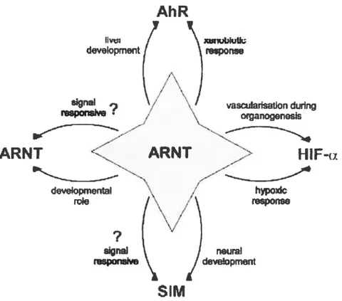
Figure 3. ARNT fonns both homodimers
and
heterodimers with MIR, HIF-Πand SIM which play roles both during mammalian developrnent and in response toAhR
jive, Uev&opment xm,ublulk rsponse sign& neural devolopment15
environmental stimuli in mammals. Symbol ‘?‘ indicates where these roles have
yet to be characterized (Kewley et al., 2004).
1.2.5. Crosstalk between bHLH-PAS protein-rnediated signalling pathways
The fact that bHLH-PAS proteins could be involved in more than one cellular pathway raises the possibility that signalling through one pathway could influence the responsiveness to another (Schmidt & Bradfield, 1996). Sucli a situation could arise when parallef pathways within the same ceil share a limiting common partner, such as ARNT. In support of this idea, it has been shown that AHR and HIF-la compete for binding to ARNT in vitro decreasing these signalling pathways (Chan et al.. 1999; Gassrnann et al., 1997). AÏthough the sirnplest explanation of these data is that ARNI is a limiting factor, these experiments do flot formally exciude other interpretations. such as the importance of other shared and limiting factors are important. In this regard, the significance of limiting heterologous factors is suggested by the interference between the dioxin and the progesterone signalling pathway. and between dioxin and oestrogen receptor signalling pathway (Kuil et al., 1998; Ohtake et al., 2003). Adding to the potential complexity of the cross-talk concept is the observation that certain target genes may be regulated by two bHLH-PAS heterodimers competing for the same binding sequence. For example, SIM1-ARNT and HIf-ARNT dimers can compete for binding to the HRE in the
Epo
enhancer (Woods & Whitelaw, 2002).Recently, a novel bHLH-PAS factor, NXF has been shown to compete with SIM2 for binding to target genes (Ooe et al., 2004). It has been demonstrated that reciprocal inhibitory crosstalk between the hypoxia and dioxin signal transduction pathways can occurwithin mammalian celis. For instance, the promoter ofhuman
Epo
gene is influenced by both the classical HREs in its 3’ regions, as well as anumber of degenerated XREs imrnediately upstream of its promoter (Chan et al., 1999). In addition to these paratiel interactions, bHLH-PAS proteins also can interact in a hierarchical manner. For example,
DrosophiÏa
sim positively and16
directly regulates its own expression through interaction with TANGO, a hornoiog of ARNT (Michaud et al., 2001). Such compiex interpiay between bHLH-PAS proteins mav enable the ccli to adapt its response to multiple environrnentai and developrnentai signais. and phsio1ogicai processes couid influence each other through interactions between bHLH-PAS proteins.
2. SIM proteins
2.1. DrosopÏ2iÏct sim. a regulator ofmidline deveiopment
sim functions as a master regulator ofthe dcv elopment ofthe midiine ofthe CNS in DrosophiÏa, acting upstream of ail known deveiopmental processes in this region of the ernbryos (Crews et ai., 1988; Nambu et ai.. 1993). sim protein is expressed in the midline cells throughout neurogenesis as well as in differentiated midiine neurons and glia. Nuil mutants of sim show a compiete absence of midiine ccli development: precursor celis fail to divide, and do flot undergo differentiation into neurons and glia. In contrast, when ectopically expressed, sim
can convert the laterai CNS to acquire the identity ofthe CNS midiine (Thomas et al., 1988; Nambu et ai., 1991). sim contains a C-terminal domain, acting as a transcriptional activator upon dimerization with Tango (Sonnenfeld et aÏ., 1997). Tango is localized to the cytopiasm in the absence of a dirnerizing bHLH-PAS protein (Ward et al., 1998). When sim is present, it forms a dirner with Tango and enters the nucieus (Shue & Kohtz, 1994). The sim-Tango complex binds a midiine enhancer eiement (CME, ACGTG core sequence) that resides on target genes. four sim target genes, breathtess (btl), sim, sut, and Toli (Tt), have been characterized. Each of these genes is reguiated differently: TÏ is expressed in midiine precursor ceÏls: si,iz is an autoregulatory target: sut is expressed in differentiated midiine glia celis; and btÏ is expressed in both midiine and tracheai celis (Nambu et aI., 1990; Crews, 1998).
17
2.2. Seguence similaritv ofthe mammalian SIM proteins
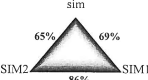
Two murine homologues of sim, Simi and Sim2, were isolated from a mouse genomic library at low stringency using a probe corresponding to the sim bHLH domain (Fan et al., 1996). The bHLH-PAS dornains ofthe DrosophiÏa sim gene product are highly conserved in the mouse (fig. 4). Overail, SIM1 and SIM2 share 69% and 65% amino acid identity with that of their Drosophila counterpart. Most remarkably, the arnino acid sequences of SIM1 and SIM2 bHLH-PAS domain show a striking identity (86% identify). However, no significant identity is seen at the C-terminus ofthe three proteins (fan et al.. 1996; Ema et al., 1996).
s im
65% 69%
SIM2
SIMÏ
86%
Figure 4. Arnino acid identity ofthree SIM proteins.
2.3. Transcriptional propeies ofmarnrnalian 51M proteins
Murine Sim hornologs share a number of functional similarities with DrosophiÏa sim. SIM1 and SIM2 are nuclear proteins, which bind the molecular chaperone hsp9O and undergo cytoplasrnic/nuclear shuttling before forming active heterodimers with ARNT or ARNT2 (Erna et al., 1996). In ceil transfection assays, the heterodirner 51M 1 -ARNT/2 transactivates reporter constructs containing the CME via the ARNT/2 C-terminus (Moffett & Pelletier, 2000). By itself, SIM1 has neither activation nor repression activity. Drosophula sim functions as a transactivator in the same assay. whereas SIM2 represses transcription by quenching ARNT/2 transactivation (Moffett et al., 1997; Moffett and Pelletier, 2000). The difference of activity between the 51M proteins is correlated with the observation that the C-termini of the three proteins have no
18
significant arnino acid homology. In addition to binding to classic CME, SIM proteins are capable of binding mammalian HRE sequences in combination with ARNT (Woods & Whitelaw, 2002). SIM1-ARNT heterodirner has been shown to activate transcription form erythropoietin hypoxic enhancer via the transcription activation domain (TAD) of ARNT. In contrast. SIM2-ARNT heterodimers do flot transactivate this construct, due to a repressive activity present within the C-terminus of SIM2. Moreover, it remains unclear whether the 51M proteins are ligand-responsive or exhibit their effects through their spatiatty and temporally restricted expression patterns. While no direct target genes have as yet been identified for $iml or S1m2. expression and knockout studies have given us insight into their possible roles.
2.4. Expression patterns ofmamrnalian$1m enes in the brain
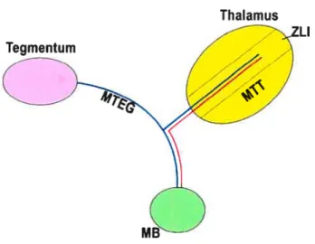
In situ hybridization of mouse ernbryos revealed that $1m] and $im2 expression in the brain is restricted. $imÏ is expressed in regions immediately adjacent to the ventral midiine of the spinal cord, mesencephalon and hypothalamus. Moreover,
$1m] is expressed in the zona lirnitans of the thalamus and in the nucleus of the
lateral olfactory tract (NLOT) of the arnygdala (Holder et al., 2004). $im2 is also expressed in the ventral mesencephalon and hypothalamus as well as in the zona limitans (Erna et al., 1996, Fan et al., 1996; Yamaki et al., 1996). In the hypothalamus, $im] is expressed at the highest levels in the paraventricular nucleus (P\TN), in the supraoptic nucleus (SON), in scattered cells located ventrornedially as well as in the mammillarv body (MB), whereas Sim2 is expressed in the PVN and in the MB, but not in the SON (Wang & Lufldn, 2000; Goshu et al., 2002) (fig. 5). $iml is continuously expressed in these hypothalamus and amygdala domains after birth, whereas Sim2 expression in the brain dramatically decreases afier E16.5. In the periphery, expression of$im2 is found in facial and trunk cartilage, as well as trunk muscles (Fan et al., 1996). $irn] and
19
Sim2 are expressed in the kidney during development as well as in adulthood (Erna et al.. 1996).
Figure 5. Expression pattern ofSirn] and Sim2 in the developing brain. Lefi side represents the of Sirni domains of expression, and the right side represents the Sim2 domains oJexpression. NLOT: nucleus ofthe lateral olfactory tract.
Simi
Si,;,2
ventromedia o oO o o o o 00 o o NLO20 3. The paraventricular nucleus ofthe hypothalamus
The hypothalamus is located in the most medial aspect of the brain, along the walls and floor ofthe 3rd ventricle (Alam et al., 1995; Markakis, 2002; Sherin, et al., 1996). The hypothalamus is an evolutionary ancient integrator of horneostasis that regulates basic processes such as food and water intake, energy expenditure, the response to stress, blood pressure and reproduction. Structurally, the hypothalamus is cornposed of a dozen srnall nuclei interspaced between less defined regions. A great body of work involving physiological and genetic studies has assigned specific functions to each of these nuclei and regions (See fig. 6, Caqueret et al., 2005.). One nucleus. the PVN, is of special interest since it has been shown to participate in several physiological processes including the control
ofenergy balance, stress responsiveness, thermogenesis and ofblood pressure.
3.1. Structure of paraventricular nue leus
The PVN ofthe anterior hypothalamus contains several subregions which harbour distinct neuronal ceil types. These neurons can be classified in at least three groups based on their axonal projections (Swanson & Sawchenko, 1983; Sawchenko et al., 1992):
1.) Magnocellular neurons project their axons to the posterior pituitary where they secrete two hormones, oxytocin (01) and vasopressin (VP), directly into the general circulation. OT prornotes lactation and labor whereas VP maintains blood pressure. The SON. which is derived from the PVN, is rnainly composed of magnocellular neurons that produce OT and VP neurons. The expression of 01 and VP in magnocellular neurons is mutually exclusive, defining two distinct ccl] types (fig. 6).
2.) Hypophvsiotropic parvocellular neurons project their axons to the medial erninence where they release several hormones, including thyrotropin-releasing hormone (TRH) and the corticotropin-releasing hormone (CRH). The ventrally contiguous anterior periventricular nucleus (aPV) contains somatostatin (SS)
21
expressing neurons that also project to the media! eminence. These hormones are transported by the portai vasculature to the anterior lobe of the pituitary where they modulate the secretion of severai pituitary hormones. The production of TRH, CRH and SS defines three distinct ceil types (fig. 6).
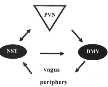
3.) A group ofparvocellular neurons project axons to the dorsal motor nucleus of the vagus nerve (DMV), located in the brainstem, which itselfprojects to virtuaily ail parasympathetic neurons inneiwating the proximai gastrointestinai (GI) tract, liver and the pancreas through the vagus nerve (Penicauci et ai., 2000). These vagaiefferents induce gastric secretion and motiiity, contributing to the feeling
bf
hunger. finaliy, the PVN aiso projects to the nucieus ofthe soiitary tract (NST), located adjacent to the DMV, which receives vagai afferences originating from the GI tract (Saper et ai., 1976; Sawchenko &
Swanson.
1982: Swanson & Kuypers, 1980; Rogers & Nelson, 1984; Lawrence & Pittrnan. 1985; Hornby & Piekut, 1928; Hardy, 2001). The NST integrates signais provided by these afferences to modulate the activity of the DMV directly, through sorne axonai projections (Sawchenko, 1983). The DMV. GI tract and the NST thus forrn a ioop (fig. 7). In vivo electrophysiological studies have shown that the PVN can modulate the activity of gastric distention-responsive NST and DMV neurons (Zhang et al., 1999). The criticai importance ofthe PVN in the controi offeeding behaviour is substantiated by the ciassicai observation that its selective destruction ieads to hyperphagia (Eimquist et ai., 1999).22
SON
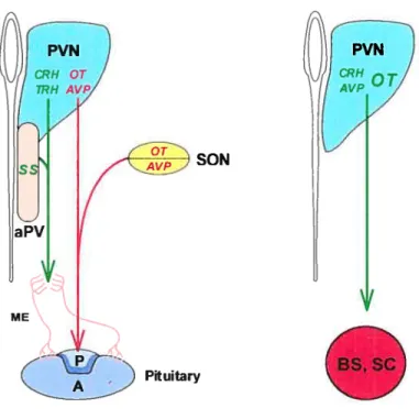
Figure 6. Structural and functional reÏationship between hypothalamic nucleï. MagnoceÏÏular neurons (in red) in the PVN and SON project to the posterior pituitary, whereas parvoceÏÏuÏar neurons (in green) of the PVN and aPV producing TRH, CRH and SS project to the medial eminence (ME). In addition, sarne parvoceÏÏuÏar neurons of the PVN can project to preganglion neurons of autonornic nervous system Ïocated in the brainstem (BS) and spinal cord (SC). OT expressing neurons represent about 10-20% ofthese fleurons. P: posterior; A: anterior.
23
/
__
vagus
17
pe ri plie ry
Figure 7. Interaction between the hypothalamus, the brainstem and the periphery. The dorsal medial nucleus of the vagus (DMV) influence gastric rnotility and secretion through the vagus nerve. Sensitive afferent fibres of the vagus originating from the gut project to the nucleus of the soiitary tract (NST). The DMV, the vagus nerve and the NST fonri a reguiatory loop. The PVN appears to regulate this loop through projection to the DMV and NST. Moreover, the PVN can indirectly perceive signais originating from the periphery through projection from the NST.
3.2. Regulation of energy banianc.e by the paraventricular nucleus
Adiposity signais provide the hypothalamus with information concerning the long-terrn status of the adipose mass. Leptin is the prototype of this type of signal (Friedman & Haiaas, 1998). The current paradigm is that an increase in the adipose mass resuils in an increased production of leptin which then feeds back in the hypothalamus to decrease food intake and increase energy expenditure. Mutations in leptin or in its receptor cause obesity in hurnan and in mice (Colernan, 1978; Clernent et al., 1998 Montague et aÏ.. 1997). Leptin has heen
24 shown to act in the arcuate nucleus (ARC) of the hypothalamus (Elias et al., 1999). Leptin decreases the expression of Npy and Agrp transcripts in mediai ceils and increases the expression of cocaine- and arnphetamine-regulated transcript (Cart,) and rneianocyte-stirnulating hormone (Msh) transcripts in lateral celis of the ARC (Elrnquist, et ai.. 1999). Genetic and physiological studies indicate that both neuropeptide Y (NPY) and agouti-related protein (AgRP) increase food intake and decrease energy expenditure (Billington et al., 1991; Graham et al., 1997; Ollrnann et al., 1997) whereas CARI and MSH, which is derived from pro-opiornelanocortin (POMC), have the opposite effects (fan et al., 1997; Kristensen et al., 1998). These changes of expression are expected to generate a similar physiological response as that of leptin, decrease of food intake and increase of energy expenditure.
ARC neurons expressing N’p/Agip and Cart/Msh project to the PVN (Elrnquist et ai., 1999 Elias et al.. 1999) (fig. 8). Electrophysiological studies have shown that individual neurons within the PVN are capable of detection and integration of the NPY and MSH signais (Cowley et ai., 1999). AgRP and MSH are in fact antagonist and agonist, respectively, of Mc4R, which is expressed in the PVN (Ollrnann et al., 1997; fan et al., 1997). The importance of the melanocortin pathway is illustrated by the identification of mutations in Fomc (Krude et al., 1998) and Mc4R (Vaisse et al., 1998; Yeo et al., 1998) in patients with extrerne obesity. Moreover, injections of NPY and MSH into the PVN have antagonistic effects on feeding and energy balance (Cowley et al., 1999).
A second class of peripheral signais provides information to the hypothalamus about the short-tetïri nutritionai status of the organisrn. These signais, generaliy produced by the GI tract in response to feeding, include peptides sucli as cholecystokinin (CCX) (fan et ai., 2004; Wynne et ai., 2005) and nutrirnents such as iinoÏeic acid (Randich et al., 2004). Several of these signais have been shown to induce satiety by acting directly on the ARC or by modulating the activity of the NST through vagal afferents (Fan et al., 2004). Ihe PVN can sense some of
25
these signais through projections from the NST. Ail together, these observations indicate that the PVN has the potential of integrating peripheral short and long terrn signais providing information about the nutritional status of the organisrn and of rnoduiating energy balance through its projections to the NST and preganglionic neurons of the autonomie nervous system.
Preganglionic • ••• sympathic and LHA parasympathic 4 fleurons • NPY/AgRP CART/MSH ARC
f igure 2. Interaction between the arcuate nucleus (ARC) and the PVN. The ARC is the main hypothaiamic site of ieptin action. In the ARC, leptin activate CART/MSH celis and inhibit NPY/AgRP ceils which project to the PVN and laterai hypothaiamic area (LHÀ). (Caqueret et ai., 2005)
Some observations suggest that regulation of food intake and energy expenditure by the PVN involves divergent pathways. For instance, NPY injection into the PVN induces feeding but it also decreases energy expenditure by decreasing the sympathetic outflow to brown adipose tissue. Both the feeding-stirnulatory and brown fat-inhibitory effects of NPY in the PVN are blocked by high doses of opioid antagonist adrninistered into the rostrai NST (rNST). However, at lower
ME
Pftuitary
26
doses of opioid antagonist. the feeding. but not the brown fat-inhibitory, effects of NPY in the PVN are biocked. These findings suggest that feeding signais originating on stimulation ofNPY receptors in the PVN ai-e routed through opioid patinvays in the rNST. whereas signais conveying energy expenditure information may be directed through opioid receptors present in neural centers nearby or within other regions of the NST (Kotz et aL. 1998). More recenly, a loxP rnodified, nuli Mc4r allele (ioxTB Mc4r) was generated. Mc4r gene codes a melanocortin receptor. Mice homozygous for the loxTB Mc4i- allele do not express MC4Rs and are rnarkedly obese. Restoration of MC4R expression in the PVN and a subpopulation of arnygdala fleurons, using SimÏ-Cre transgenic mice, prevented 60% of the obesity. The increased food intake of Mc4r nuli mice was cornpletely rescued while energy expenditure was unaffected. These findings demonstrate that the melanocortin pathway in the PVN and/or the arnvgdala controls food intake but that a melanocortin pathw’ay located elsewhere controls energy expenditure (Baithasar et al.. 2005).
4. Function of Sinil and Siïn2 in the paraventricular nucleus and in other parts of the brain
4.1. Roles ofSim] in the control ofneuron differentiation in PVN/SON/aPV Mutant analyses have demonstrated that Sim] functions to control developrnent of specific ceil types within the CNS like its Drosoph lia counterpart. PVN, aPV and SON cells expressing the neuropeptides TRH, SS, CRH, VP, and OT, define at least five major neuroendocrine ccli types (fig. 9, Michaud et al., 1998). Simi is required for the development of these hypothalamic neurons acting at the finai stages of their differentiation. Since 81m] is expressed in virtually ail PVN celis, it appears likely that its developrnent is aboiished in Simi mice. Simi functions upstrearn to maintain Brn2 expression. BRN2, a POU transcription factor, in turn directs the terminal differentiation ofa subset ofcells identified by the production of CRH, OT and VP. SIMI also acts in parallel with the transcription factor
27 orthopedia protein (OTP) for the development of the sarne lineages as those specified by SIM1 (Acampora et aI., 1999; Wang & Luflin, 2000). Yeast two hybrid studies do not provide evidence that SIMY and OTP interact directiv (Yang & Michaud, unpublished resuits). Several observations indicate that ARNT2 is the in vivo dimerization partner for 51M 1: 1.) Simi and Arnt2 are co expressed in the developing PVN/SON; 2.) SIM1 and ARNT2 can physically interact in yeast two-hybrid and co-immunoprecipitation assays; 3.) Siini and Arnt2 mutant mice show the same phenotype (Keith et al.. 2001; Hosoya et al., 2001); and 4.) Simi and Arnt2 function at the sarne stage of developrnent (Michaud et al., 2000; Ema et al., 1996; Goshu et al., 2004).
4.2. Role ofSim] in energy balance
Simi hornozygous mice (Simij die perinatally, presumably from the PVN developmental defect. In contract. Sinz] heterozvgous mice (Sim]) survive and
develop early-onset obesity with increased linear growth, hyperinsulinemia and hyperleptinernia. These mice are hyperphagic, even before they show increased weight gain, but their energy expenditure is not signiflcantly decreased (Michaud et al., 2001; Holder et al.. 2004). A balanced transiocation interrupting SIM] was found in a child with a sirnilar phenotype (Holder et al., 2000). Like Sim] mice, this child had early-onset obesity, increased linear growth, and a voracious appetite suggestive of hyperphagia. Mouse models suggest that SIMY haploinfflciency is responsible for the obesity in this child. The description of rnorbid obesity in chiidren with chrornosornal deletions in the 6q 16 region, which contains SIMJ, further strengthens this conclusion (Villa et al., 1995; Turleau et al., 1988; Gilhuis et al., 2000). Interestingly, a genome-wide search for childhood obesity traits bas shown signifïcant linkage on chromosome 6q22-q23, near the SIMJ locus (Meyre et al., 2004).
The mechanisms underlying the hyperphagia associated with 31m]
2$
the aduit liver, pancreas, muscle, white adipose tissue (WAT) and brown adipose tissue (BAT). Also, 81m] is flot expressed in the brainstem and in the intermediate/dorsal spinal cord where autonornic preganglionic neurons are found. Ail these information do not support the possibility that $iml functions in the peripheral tissues to regulate energy balance. One possibility is that dysfunction of the PVN causes this hyperphagia. This dysfunction could be of deveiopmental origin. Indeed, the PVN of Sim] mice is hypocellular, containing 24 % fewer ceils (Michaud et al., 2001). However, since Simi and its dirnerizing partner Arnt2 are strongly expressed in the PVN not only during its development but also throughout adult life, the possibility that Simi controls food intake physiologically is not excludcd. Finally, it is also possible that Sim] regulates food intake by acting in other regions of the brain such as scattered cells located in the anterior portion ofthe lateral hypothalamus or in the amygdale.
__
o
*1(Brn2)
f igure 9. Transcriptional regulation of anterior hypothalamic development. SIMY-ARNT2 and OTP function in parallel to control the developrnent of virtually ail neurons of the PVN/SON, of TRH-producing neurons of the preoptic region and of SS-producing neurons ofthe anterior periventricular nucleus, which is located ventrally contiguous to the PVN. These two complexes are required to
29
maintain Brn2 expression, which, in turn. directs the development of CRH-, OL and VP-producing neurons ofthe PVN and SON. SIMÏ-ARNT2 and OTP also act upstream of SIM2 for the deveÏoprnent of subsets of TRI-1 celis located in the PVN and preoptic region and of SS ceils found in the anterior periventricular nue leus.
4.3. Sirn2. a paralog of8tml
SimZ mice die within 3 days of birth due to respiratory failure, exhibiting reduced efficacy of lung inflation and several abnorrnalities involving the thorax (Goshu et al., 2002). $im2 regulates the growth or integrity of the ribs and vertebrae, although its precise function remains unclear. $im2 is also expressed in the anterior hypothalamus (Wang & Lu&in, 2000; Goshu et aÏ., 2004). Along the rostro-caudal axis, Sirn2 expression is detected in the anterior and mid-PVN, including the aPV. Toward the posterior PVN, Sim2 expression dirninishes. In these anterior areas, $im2 expression domain correlates with that of TRH and $8 expressing celis and is distinct from that of Brn2, which is necessary for the differentiation of OT, VP and C’RH neurons located more posteriorly. In the absence of Sirni function, there is no $i,ii2, TRTL and $8 expression, reflecting a general and upstrearn role of Sirni (Goshu et al., 2004). In the absence of $im2 function, TRH and $8 celis are reduced even with normal $im] gene dosage, reflecting a semidominant and downstream role of $im2. furthermore, in the absence of $im2 function, $im] dosage has an influence on TRH and $8 ceil numbers, dernonstrating that $iml lias some capacity to compensate for $irn2

![Figure 5. Expression pattern ofSirn] and Sim2 in the developing brain. Lefi side represents the of Sirni domains of expression, and the right side represents the Sim2 domains oJexpression](https://thumb-eu.123doks.com/thumbv2/123doknet/2056330.5754/37.918.163.811.250.808/expression-pattern-developing-represents-domains-expression-represents-ojexpression.webp)
![Figure 1. Targeting of the Simi locus. The targeting vector (tv), the $im] wild-type locus (wt) and the targeted locus (mt) are represented](https://thumb-eu.123doks.com/thumbv2/123doknet/2056330.5754/115.918.208.754.103.353/figure-targeting-simi-locus-targeting-vector-targeted-represented.webp)
