FEBS Letters 342 (1994) 23-28
ELSEVIER
FEBS 13820
LETTERS
Binding site-shaped repeated sequences of bacterial wall peptidoglycan
hydrolases
Jean-Marie Ghuysena**, Josette Lamotte-Brasseur”,
Bernard Jorisa, Gerald D. Shockmanb
“Centre d’lngknierie des Protkines, Universite de Lisge, Institut de Chimie. B6, B-4000 Sart Tilman (Lit?ge I), Belgium
bDepartment of Microbiology and Immunology, Temple University School of Medicine, 3400 North Broad Street, Philadelphia, PA 19140, USA
Received 7 February 1994
Abstract
The non-catalytic C-terminal regions of the N-acetylmuramidase (lysozyme) of Clostridium acetobutylicum and N-acetylmuramoyl(D-lactyl)-L- alanine amidases CwlA of Bacillus subtilis, ORFW and CwlL of Bacillus licheniformis were previously reported to have similarities with the amino acid sequence of the non-catalytic N-terminal module of the Streptomyces albus G Zn DD-peptidase. This peptidase is a bipartite protein of known three-dimensional structure. Its non-catalytic N-terminal module possesses, exposed at the surface, an elongated crevice which is defined by a loop-helix-loop-helix motif that consists of two repeats, each 16 amino acid residues long, connected by a heptapeptide and whose design is compatible with its possible functioning as a substrate recognition and binding site. Amino acid alignments suggest that cavities nearly identical in shape to that present in the non-catalytic module of the S. albus peptidase, are borne by the C-terminal regions of the CwL4 amidase (in one copy), the lysozyme and the ORFW and CwlL amidases (in two copies). Since a common feature of the five enzymes is their substrate, the bacterial cell wall peptidoglycan, we interpret the striking similarity of their non-catalytic N- or C-terminal modules to suggest that these modules are involved in the binding of these exocellular enzymes to their insoluble wall substrate.
Key MJO&: Protein modular design; Peptidoglycan hydrolase; Repeated sequence; 3-D Structure
1. Introduction
A number of bacterial wall lytic enzymes [l] and cell- surface-associated proteins of Gram-positive bacteria [2] appear to be constructed in a modular fashion. Whereas one domain contains the catalytic site, another domain, which is frequently at or near the N- or C-terminal end of the amino acid sequence, contains two or more tan- demly repeated sequences, 20 or more amino acids long. There appears to be several families of repeated se- quences and in more than one instance the repeats have been associated with ligand binding domains.
The most direct experimental evidence that such do- mains are involved in ligand binding was obtained by genetic engineering of pneumococcal bacteriolytic en- zymes [3,4]. For example, the portion of the gene encod- ing the catalytic N-terminal domain of the pneumococcal choline-dependent LytA amidase [5] was fused to the portion of the gene encoding the non-catalytic C-termi- nal domain of the choline-independent lysozyme, LC7, of bacteriophage CP7 [6]. The resultant chimeric enzyme expressed in Escherichia coli possessed an amidase activ- ity that was independent of the presence of choline. The reciprocal chimeric enzyme containing the N-terminal
*Corresponding author. Fax: (32) (41) 56 33 64.
domain of the bacteriophage choline-independent ly- sozyme fused to the C-terminal domain of the pneumo- coccal choline-dependent amidase hydrolysed the ly- sozyme-sensitive bonds only in choline-containing cell
walls. In spite of these advances, proof, in terms of three- dimensional structure, that the non-catalytic domains possess substrate recognition and binding sites is still lacking.
The goal of this report is to show that a potential binding site is present in the non-catalytic N-terminal module of the Streptomyces albus G metallo (Zn) DD- peptidase [7] and, by analogy, in the C-terminal regions of the Clostridium acetobutylicum lysozyme [8], Bacillus subtilis CwlA amidase [9,10], Bacillus licheniformis FD0120 CwlL amidase [l l] and B. lichenzjkmis MC14 0RFL3 gene product [12]. The function of the 0RFL3 gene product has not been determined but its derived amino acid sequence is similar to that of the CwlL ami- dase [ 111, strongly suggesting that the ORFL3 gene prod- uct is also an amidase. Although frequently referred to as a muramoyl pentapeptide carboxypeptidase, the S. albus peptidase hydrolyses C-terminal D-alanyl-(D)-di- amino acid linkages which serve as interpeptide bridges in the wall peptidoglycan of various bacterial species [I 3,141. Hence, the S. albus peptidase, the C. acetobutyl- icum lysozyme and the three Bacillus amidases are pepti- doglycan hydrolases of varying enzymatic specificities. They hydrolyse linkages within the peptide moiety, the
0014-5793/94/%7.00 0 1994 Federation of European Biochemical Societies. All rights reserved.
24 J-hf. Ehuysen et al. I FEBS Letters 342 (1994) 23-28
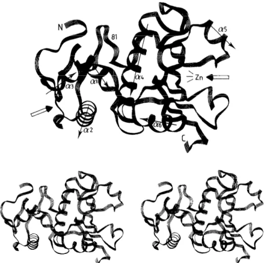
Fig. 1. Mono and stereo views of the folding of the u-carbon ~l~ptide chain of the Str~~to~yc~s albus G Zn DD-peptidase. The drawings and those of Figs. 3 and 4 were made by using the PLUTO programme on a Silicon Graphics workstation and the Insight programme of Biosym.
glycan chains and at the junction between the g&can and peptide moieties, respectively.
2. Results
The S. albus peptidase is a 213 amino acid residue bipartite protein. Its three-~mensional structure has been resolved to 0.18 nm [l&16]. Fig. 1 shows the folding of the a-carbon polypeptide. The catalytic, 132 amino acid residue C-terminal module possesses three tl-helices (c14a6) and five-stranded &sheet. The open arrow on the right points toward the ~in~~ont~nin~ site. Fused to the catalytic module, the non-~tal~i~, 81 amino acid residue N-terminal module possesses three a-helixes @I- &3). The open arrow on the left points toward a cavity which is defined by the sequence L36-477. This se- quence comprises the C-terminal portion of the loop that connects helix al to helix 012, helix 012, the loop that connects helix a2 to helix ~3, and helix 613.
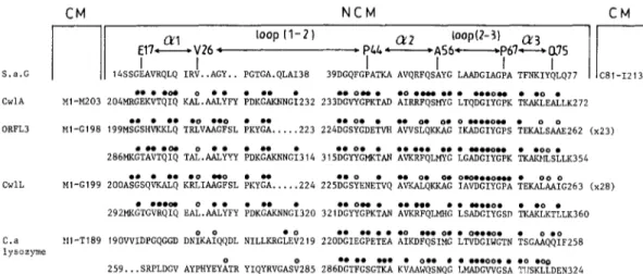
The amino acid sequence alignment of Fig. 2 high- lights the repeated design of the sequence L36-Q77. Re- peat 1 extends from L36 to Q54 and repeat 2 extends
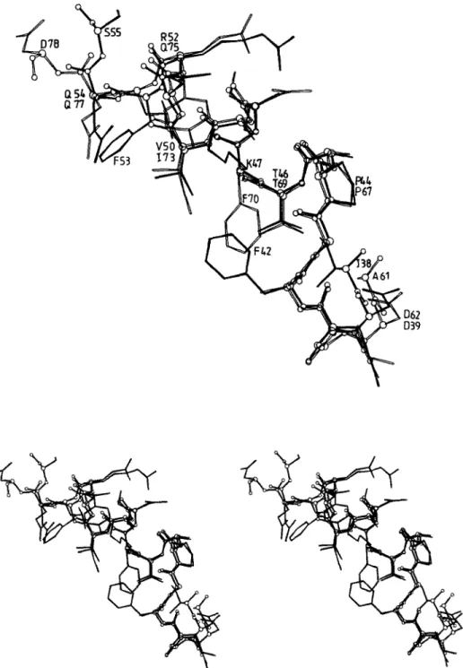
from L59 to 477. As shown in Fig. 3, repeat 1, from D39 to Q54, and repeat 2, from D62 to Q77, have almost identical polypeptide foldings. ~u~~mposition of the N-CE-C = 0 groupings occurs within a range of 0.02 to 0.05 nm. The side chains of the pair D39 and D62, the pair P44 and P67, the pair A45 and A68, the pair T46 and T69, the pair VSO and 173 (up to the Cy atoms) and
i i00p *17-( CXl 4 -16 I DGCYTWSGTLSEGSSGEAVRPLQIRV 57+ loopw3t *67&75 1 59 62 65 / 69 73 \ 76 17 YGLAADGIAGPATFNKIYQLQD fl.0.. o.... 0 0.
Fig, 2. Amino acid sequence of the N-terminal module of the Strepto- myces albus G Zn DD-peptidase. The positions of the secondary struc- tures are indicated. The alignment highlights the repeats LX-Q54 and LSQ75. Filled circles identify strict identities. Open circles identify hydrophobic residues occuring at equivalent places.
J-M. Ghuysen et al. IFEBS Letters 342 (1994) 23-28 25
Fig. 3. Mono and stereo views of the superimposed polypeptides D39-S55 (filled lines) and D62-D78 (open lines) of the N-terminal module of the Streptomyces albm G Zn DD-peptidase.
the pair R52 and 475 (up to the CS atoms) superimpose remarkably well. It thus follows that the cavity is defined by two superimposable repeats, D39-Q54 and D62-Q77 each 16 amino acid residues long, connected by the hep- tapeptide S55-A61.
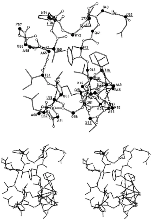
Fig. 4 presents a view of the cavity. It also shows the spatial disposition of the side chains of D39, F42, T46, V50, F53 and Q54 of repeat I, the side chains of D62, 164, T69, F70 and I73 of repeat 2 and the side chains of S55 and L59 of the intervening heptapeptide S55-A61. The side chains of D62 and 164 are at the left entrance
of the cavity, the side chains of D39 and 173 are at the right entrance, the side chains of T46, T69 and Q.54 and those of the hydrophobic amino acid residues F42, V50, F53 and L59 occupy central positions. The spanning distances between the 06 atoms of D39 and D62 and between the Oy atoms of T46 and T69 are 1.86 nm and 1.35 nm, respectively. The tetrapeptide Y74Q77 at the C-terminal end of repeat 2 and the side chains of S55 and F70 are outside the cavity.
On the basis of these structural data, we have used the non-catalytic, N-terminal module of S. albus peptidase
26 J-M. Ghuysen et al. IFEBS Letters 342 (1994) 23-28
Fig. 4. Mono and stereo views of the crevice created by the polypeptide D39-173 at the surface of the N-terminal module of the Streptomyces albus G Zn DD-peptidase. The a-carbon are filled circles. The N-CO atoms are open circles. The lateral chains of selected amino acid residues (underlined) are shown.
as a model in an attempt to shed light on the structural and therefore possible functional features of the non- catalytic C-terminal regions of the C. acetobutylicum ly- sozyme and Bacillus CwlA, CwlL and ORFL3 amidases. The amino acid alignments shown in Fig. 5 were made by using the best fit algorithm of the GCG package [17]. The following observations can be made.
(i) The C-terminal region of the CwlA amidase, from M204 to K272, has 50% identities with the N-terminal module of the S. albus peptidase, from S14 to Q77. (ii) The C-terminal regions of the 0RFL3 and CwlL amidases are of more complex design. Each is comprised
of three polypeptide segments. Two, M199-E262 and M286-K354 in ORFL3, and A200-G263 and M292- K360 in CwlL, are, respectively, 64 and 69 amino acid residues long and have 40% to 50% identities with the N-terminal module of the S. albus peptidase, from S14 to Q77. Each has an intervening peptide, 23 amino acid residues long in ORFL3 and 28 amino acid residues long in CwlL, connecting the two sequences described above. The connecting peptides also share about 50% identities with each other (not shown).
(iii) The C-terminal region of the C. acetobutylicum ly- sozyme, from V190 to N324, exhibits, still, another type
J-M. Ghuysen et al. IFEBS Letters 342 (1994) 23-28 21
CM NCM CM
ioop 11-2) a2 ioop(2-3) Q3 l PLL-AS6-P67-MS
I I I
S.a.G 14SSGEAVRQLQ IRV..AGY.. PGTGA.QLAI38 39DGQFGPATKA AVQRFQSAYG LAADGIAGPA TFNKIYQLQ,, .*..o. 0 . . . .a
CwlA Xl-M203 204MRGEKVTQIQ KAL.AALYN PDKGAKNNGf232 233t:;:KhD ::RiF;,;: ;TQ;:;$K &Ai%AtLK2,2 . . . . . .O ..O . . . . . 0. . . . 0. 0. 0 "..O" .
0RFL3 Ml-Cl98 199MSGSHVKXLQ TRLVAAGFSL PKYGA...223 224DGSYGDETVX AWSLQKXAG IKADGIYGPS T&S&262 (x23)
.".oa 0.. .u . . . "..0" . .00.
286MKGTAVTQIQ TAL.AALYYY PDKGAKNNG;3,4 3,5%%&N %g;LMYG LGADGIYGPK mnSLLK354 . . 0
CWlL Ml-G199 200A;&Qk& K&t&L P@KYz
0. Ome..O... .
. . ...224 225DGSYENE:VQ ?&QKXAG IAVDGIYGPA TEKA%;G263 (x28) . . ..a9 0 . . . . .
292MKGTGVRqIq EAL.AALYFY PDKGAKi'!NGbZO 32,%'%K:AN %&,H: :S~~;;&,, k$$TtLK360
. . . 0. . 0b.w.
C.a HI-T189 190VVIDPGqGGD D&.A&DL NILLKRG&t219 22O&&'h~ &DFqSIMG LTVDGIWGTN &G~Q~FF~~S 1ySOZpe
0.. l . . ..00.. .O ..a@ 259...SRPLDCV AYPHYEY&R YIQYRVGAS& 286::T::SGg K;AAtdQSNQG LMADGWGSA T!,SKLLDEN324
Fig. 5. Amino acid alignments of the non-catalytic, N-terminal module of the Streptomyces albus G Zn DD-peptidase (S.a.g.) and the non-catalytic, C-terminal modules of the Bacillus subtilis CwlA amidase, Bacillus licheniformis 0RFL3 and CwlL amidases and Clostridium acetobutylicum (Ca.) lysozyme. The secondary structures of the S. albus peptidase are indicated. Filled circles identify strict identities by reference to the S. albus peptidase. Open circles identified hydrophobic residues occurring at equivalent places. CM = catalytic module. NCM = non-catalytic module.
of design. It consists of two contiguous polypeptide seg- ments V190-F258 and S259-N324. The amino terminal portions, V190-V219 and S259-V285, of each of these segments are variable but the carboxy terminal portions, D220-F258 and D286-N324, each have 50% identities with the carboxy terminal portion, D39-Q77, of the N- terminal module of the S. albus peptidase.
(iv) With Pho = hydrophobic amino acid residue and using the amino acid numbering of the S. albus pepti- dase, the general consensus of the eight aligned se- quences of Fig. 5 is:
39 42 46 50 53 54 59
D G X Pho G X2 T X3 PhoX2 Pho Q X3GPhoX2
(E) *(El *
1
62 64 65 69 13 16 I?
D GPho Pho G X2 T X3 Pho X2 Pho X (E) *
(E)*: atposition of repeat1intheCwlLamidase;
at positions 223 of repeat land323 of repeat2 in the C. acetobutylicumlysozyme.
Remarkably, D39, D62, T46, T69, 454 and the hydro- phobic amino acid residues at positions 50, 53, 59, 64, 65 and 73 are conserved. As shown in Fig. 4, all these amino acid residues occupy topologically important po- sitions in the cavity of the non-catalytic module of the S. albus peptidase.
(v) As a corollary of the alignments shown in Fig. 5, the catalytic modules of the CwlA, 0RFL3 and CwlL ami- dases are about 200 amino acid residues long. They have similarity in their primary structures (not shown) and they lack the GSNRY (or analogue) motif thought to be a general marker of the amidases [18]. The catalytic and non-catalytic modules of the peptidoglycan amidases of known primary structure show variations in hydropho- bic cluster patterns, suggesting more than one family of amidases (unpublished data).
3. Discussion
Acquisition of a substrate binding module is an evolu- tionary advantage for exocellular enzymes that interact with and hydrolyse bonds in a polymeric insoluble sub- strate. In this respect, the data presented above allow the following conclusions to be drawn. As established by X-ray crystallography studies (and with CM = catalytic module and NCM = non-catalytic module), the modular design of the S. albus peptidase is NH,-NCM-CM- COOH. Its non-catalytic module possesses, exposed at the surface, an elongated cavity that bears a typical amino acid sequence signature in the form of two 01- helical repeats, 16 amino acid residues long, connected by a heptapeptide loop. As derived from amino acid alignments, the modular design of the B. subtilis CwlA amidase is NH,-CM-NCM-COOH, that of the C. ace- tobutylicum lysozyme is NH,-CM-NCM-NCM-COOH and (with IP = an intervening peptide about 25 amino acid residues long) that of the B. licheniformis 0RFL3
and CwlL amidases are NH,-CM-NCM-IP-NCM-
COOH. Each of these non-catalytic modules possesses a cavity whose shape and structure is comparable to that which is present in the non-catalytic module of the S. albus peptidase.
The likely function of these cavities as substrate recog- nition and binding sites would depend mainly on the glutamine residue, the two aspartic acid residues and the two threonine residues which are strictly conserved and the hydrophobic residues which occur at equivalent places along the cavity-defining motif. The peptidase, lysozyme and amidases under study hydrolyse different bonds in the bacterial cell wall peptidoglycan. Variations in the sequences of the binding motif due to the occur- rence of non-conserved amino acid residues could be responsible for substrate specificities and topologically directed activities. Full characterization of the non-cata-
28 J-M. Ghuysen et al. IFEBS Letters 342 (1994) 23-28
lytic modules as binding sites, however, requires identifi- cation of the recognized polymer and study of the ligand-binding site interactions by genetic engineering, molecular modelling and site-directed mutagenesis.
Acknowledgements: This work was supported in part by the Belgian
programme on Interuniversity Poles of attraction initiated by the Bel- gian State, Prime Minister’s Office, Science Policy Programming (PA1 No. 19) and the Fonds de la Recherche Scientifique Medicale (Contract No. 3.4531.92).
References
[l] Shockman, G.D. and Holtje, J.V. (1994) in: Bacterial Cell Wall (Ghuysen, J.M. and Hakenbeck, R. Eds.) New Comprehen- sive Biochemistry 27, pp. 131-136, Elsevier, Amsterdam, New York.
[2] Kehoe, M.A. (1994) in: Bacterial Cell Wall (Ghuysen, J.M. and Hakenbeck, R. Eds.) New Comprehensive Biochemistry 27, pp. 2 17-261, Elsevier, Amsterdam, New York.
[3] Lopez, R., Garcia, J.L., Garcia, E., Ronda, C. and Garcia, P. (1992) FEMS Microbial. Lett. 100, 439448.
[4] Diaz, E., Lopez, R. and Garcia, J.L. (1991) J. Biol. Chem. 266, 54645471.
[5] Garcia, P., Garcia, J.L., Garcia, E. and Lopez, R. (1986) Gene 43, 265-272.
[6] Garcia, P., Garcia, J.L., Garcia, E., Sanchez-Puelles, M and Lopez, R. (1990) Gene 86, 81-88.
[7] Duez, C., Lakaye, B., Houba, S., Dusart, J. and Ghuysen, J.M. (1990) FEMS Microbial. Lett. 71, 215-220.
[8] Croux, C. and Garcia, J.L. (1991) Gene 104, 25531.
[9] Kuroda, A. and Sekiguchi, J.J. (1990) J. Gen. Microbial. 136, 220992216.
[lo] Foster, J.J. (1991) J. Gen. Microbial. 137, 1987-1998.
[l l] Oda, Y., Nakayama, R., Kuroda, A. and Sekiguchi, J. (1993) Mol. Gen. Genet. 241, 38&388.
[12] Lee, J.W.K., Edwards, C.W. and Hulett, EM. (1991) J. Gen. Microbial. 137, 667-677.
[13] Ghuysen, J.M., Dierickx, L., Coyette, J., Leyh-Bouille, M., Guinand, M. and Campbell, J.N. (1969) Biochemistry 8,213-222. [14] Ghuysen, J.M., Leyh-Bouille, M., Bonaly, R., Nieto, M., Perkins, H.R., Schleifer, K.H. and Kandler, 0. (1970) Biochemistry 9, 2955-2961.
[I 51 Dideberg, O., Charlier, P., Dive, G., Joris, B., F&e, J.M. and Ghuysen, J.M. (1982) Nature 299, 469470.
[16] Wet-y, J.P. (1987) Ph.D. Thesis, University of Liege, Belgium. [17] Devereux, J., Haeberli, P. and Smithies, 0. (1984) Nucleic Acids
Res. 12, 387-395.
[18] Lazarevic, V., Margot, P., Soldo, B. and Karamata, D. (1992) J. Gen. Microbial. 138, 1949-1961.