1
Phthalates modulate steroid 5-reductase transcripts in the Western clawed frog embryo
1
Sonja Bissegger1, Marco A. Pineda Castro2, Viviane Yargeau2, Valerie S. Langlois1,3* 2
3
1Chemistry and Chemical Engineering Department, Royal Military College of Canada, Kingston, 4
ON, Canada 5
2Chemical Engineering Department, McGill University, Montreal, QC, Canada 6
3Institut de la recherche scientifique – Centre Eau Terre Environnement (INRS-ETE), Québec, 7 QC, Canada 8 9 10 11 12 13 14 15 16 17 18 19
*Author for correspondence and reprint requests: 20 21 Dr. Valerie S. Langlois 22 Associate Professor 23
Institut de la recherche scientifique – Centre Eau Terre Environnement (INRS-ETE) 24 490 rue de la Couronne 25 Québec, QC, G1K 9A9 26 Canada 27 Tel: + 1 (418) 654-2547 28 Fax: + 1 (418) 654-2600 29 Email: [email protected] 30
2
Abstract
31
Phthalates are used worldwide in the manufacturing of plastics, added to cosmetic products, 32
personal care products, pharmaceuticals, medical devices, and paints; and are widely detected in 33
soil, surface water, and organism tissues. Phthalate esters have been previously shown to 34
interfere with the endocrine system in vertebrates. However, few studies have investigated the 35
effects of phthalates on testosterone-converting enzymes that affect hormone levels and 36
reproduction. In the present study, we exposed the Western clawed frog (Silurana tropicalis) to 37
0.1, 1, and 10 µM diethylhexyl phthalate (DEHP), dibutyl phthalate (DBP), and diethyl phthalate 38
(DEP) during early amphibian embryonic development. Additional DBP exposures were 39
conducted ex vivo using mature frog testes. Malformations and mRNA levels of genes associated 40
to reproduction and oxidative stress were evaluated. 0.1 µM DEHP, DBP, and DEP induced an 41
array of malformations, including incomplete gut coiling, edemas, and eye malformations. 42
Moreover, all three phthalates increased the expression of androgen-related genes, such as 43
steroid-5α-reductase 1, 2, 3, steroid-5β-reductase, and androgen receptor at concentrations 44
ranging from 0.1 to 10 µM depending on the phthalate and gene. Data suggest that the phthalate 45
esters tested are teratogens to the amphibian embryo and that these phthalates exhibit 46
an androgenic activity in amphibians. 47
48 49
Keywords: Androgen disruption; DHEP; DBP; DEP; Srd5α1; Srd5α2; Srd5α3; Srd5β.
50 51
3
1. Introduction
52
Phthalates are used worldwide in the manufacturing of plastics (Daniels, 2009). Leaching 53
due to the non-covalent bonding of phthalates to polymers leads to the introduction of phthalates 54
into our ecosystems. Phthalates are also used as additives in various cosmetic products, medical 55
devices, personal care products, pharmaceuticals, and paints (reviewed in Magdouli et al., 2013). 56
Due to their wide use, phthalates are commonly detected in soil, surface water, and organism 57
tissues (Bauer and Herrmann, 1997; Blair et al., 2009). Diethylhexyl phthalate (DEHP) is one of 58
the most used plasticizers, and was detected in various environmental compartments (reviewed 59
in Magdouli et al., 2013). For example, DEHP was detected at concentrations ranging between 60
0.01 and 25 µg/L in rivers in Japan (Suzuki et al., 2001; Yuwatini et al., 2006) and reported in 61
the influent of a wastewater treatment plant in France at concentrations up to 44 µg/L (Dargnat et 62
al., 2009). Dibutyl phthalate (DBP) and diethyl phthalate (DEP) are two other plasticizers that 63
have been widely detected in waters. These phthalate esters were detected in the Tama River in 64
Japan at concentrations ranging from 0.088 to 0.54 µg/L DBP and 0.004 to 0.31 µg/L DEP 65
(Suzuki et al., 2001). The False Creek in Vancouver, BC, Canada also showed concentrations in 66
seawater of ~ 0.1 µg/L DEP (Blair et al., 2009). 67
Phthalate esters have been shown to interfere with vertebrate development on different 68
levels. The main mechanism of action behind phthalate induced transcriptional changes has been 69
reported to be the peroxisome proliferation-activated receptors (PPARs, Gazouli et al., 2002). In 70
addition, heat shock proteins have been shown to be modulated after phthalate exposure in 71
different species and are attributed to early warning signs of cellular stress (reviewed in Gupta et 72
al., 2010). Cellular oxidative stress is caused by the presence of reactive oxygen species, which 73
can lead to DNA damage in cells and result in abnormalities. For example, proteins such as 74
4
glutathione transferase, glutathione peroxidase, and heat shock protein 70 have been previously 75
reported to be altered when exposed to the phthalate DEHP (reviewed in Mathieu-Denoncourt et 76
al., 2015a). 77
In addition, research has shown that phthalates can have androgenic and/or anti-androgenic 78
properties and can adversely affect development and reproduction of male vertebrates (Latini et 79
al., 2006; Kay et al., 2014). For example, feminization of gonads by exposure to DBP was found 80
in juvenile Murray rainbowfish (Bhatia et al., 2015). In addition, disrupted spermatogenesis was 81
observed in the African clawed frog after DBP exposure (Lee and Veeramachaneni, 2005). The 82
mechanism of action by which phthalates mediate their action is still not completely understood 83
(Mathieu-Denoncourt et al., 2015a). Previous research suggested that phthalates interfere with 84
hormone synthesis by modulating the expression of sex steroid-related genes (Wong and Gill, 85
2002; Lehmann et al., 2004; Thompson et al., 2004). For example, decreased mRNA and protein 86
levels of StAR have been observed in rat testis after DEHP exposure (Borch et al., 2006). StAR 87
is responsible to transport cholesterol to the inner mitochondria in order to synthesize steroids, 88
including androgens. Decreased expression levels of star have also been correlated with reduced 89
levels of the androgen testosterone (T) (Borch et al., 2006), suggesting that T metabolism could 90
be directly affected by phthalate exposure. 91
Testosterone is converted to the potent androgen 5α-dihydrotestosterone (5α-DHT) by 92
steroid-5α-reductase (Srd5α) and to 5β-dihydrotestosterone (5β-DHT) by steroid-5β-reductase 93
(Srd5β). Few mammalian studies have investigated if phthalates are capable of modulating Srd5α 94
and Srd5β. Exposure to mono-ethylhexyl phthalate (MEHP) decreased Srd5α protein levels in a 95
primary cell culture of immature rat Leydig cells (Svechnikov et al., 2008). Similarly, exposure 96
to DBP in rats significantly decreased Srd5α2 protein in the proximal penis (Kim et al., 2010). In 97
5
contrast, a concentration dependent increase of Srd5α activity was detected in testis after DEHP 98
exposure of pubertal rat (Kim et al., 2003). These studies show that phthalates can modulate 99
Srd5α. However, no studies have addressed the effects of phthalates on Srd5β expression. 100
Srd5 are involved in vital biological functions (reviewed in Langlois et al., 2010a) and their 101
dysregulation leads to a variety of diseases in humans, in particular in the male reproductive 102
system and liver (reviewed in Azzouni et al., 2012). Thus, there is a need to determine how 103
phthalates with androgenic and/or anti-androgenic properties affect Srd5α and Srd5β in lower 104
vertebrates, such as amphibians. 105
The overall objective of the present study was to understand the effect of the three phthalates 106
DEHP, DBP, and DEP in the Western clawed frog (Silurana tropicalis). Specifically, we 107
exposed the Western clawed frog to DEHP, DBP, and DEP during early embryonic development 108
and investigated malformations and mRNA levels of genes involved in oxidative stress and 109
reproduction. As the frog embryos responded to DBP, we further chemically-challenged mature 110
frog testes ex vivo in order to analyze if DBP could interfere with normal testis regulation in 111
males. Thus, this study presents novel insights in regards to interactions between phthalates and 112
srd5 during two critical periods of S. tropicalis. 113
114
115
116 117
6
2. Materials and Methods
118
2.1 Experimental design
119
Maintenance of male and female S. tropicalis occurred in dechlorinated and aerated water at 120
the Queen’s University Animal Care facilities (Kingston, ON, Canada) in accordance with 121
guidelines of the Institution’s animal care protocols and the Canadian Council on Animal Care. 122
Animals were kept in a 12:12 h light:dark cycle with a water temperature of 26 ± 1 °C. 123
In vivo exposure was executed by exposing eggs of S. tropicalis to phthalates. Breeding 124
procedure was performed as described in Langlois et al. (2010b). Briefly, eggs were collected 125
and kept in Frog Embryo Teratogenesis Assay-Xenopus (FETAX) solution consisting of 625 mg 126
NaCl, 96 mg NaHCO3, 75 mg MgSO4, 60 mg CaSO4·2H2O, 30 mg KCl, 15 mg CaCl2/L, and 127
0.04 ppm gentamycin sulphate. The fertilized eggs were dejellied using 2% (w/v) L-cysteine. 128
200 embryos (divided in 5 jars) were exposed to 0.1, 1, and 10 µM DEHP (Sigma, Oakville, ON, 129
Canada), DBP (Sigma, Oakville, ON, Canada), or DEP (Sigma, Oakville, ON, Canada) once the 130
eggs reached Nieuwkoop and Faber (NF) stage 11 (Nieuwkoop, 1994). NF 11 embryos were also 131
exposed to two negative controls: a water only control and a solvent control (0.05% Ethanol) and 132
to a positive control to test alteration of srd5 mRNA levels; finasteride (100 µM; a known Srd5 133
inhibitor; Langlois et al., 2010c). The FETAX solution was kept at 26 °C and changed every 24 134
h. Daily water change also ensured a steady source of phthalate exposure, as phthalates are 135
known to degrade over time. Dead embryos were discarded once a day and recorded. Embryos 136
were sampled in pools (n = 10) at stage NF 46 (after an exposure time of 72 h) and flash frozen 137
on dry ice until RNA isolation. 138
7
The ex vivo assay used has been previously optimized for frogs and described in 139
Bissegger et al. (2014). Briefly, six male adult frogs were anesthetized in 0.1% MS-222 (ethyl 3-140
aminobenzoate methanesulfonate, Sigma, Oakville, ON, Canada). Testes were carefully 141
dissected from each animal. Each testis (n = 6 per treatment group, one testis per frog was used 142
as a control sample and the other testis was exposed to DBP) was weighed (to correct for steroid 143
production) and placed in a separate 1.5 mL centrifuge tube filled with 500 µL ice cold Lebovitz 144
(L-15 media, Sigma, Oakville, ON, Canada) containing 10 mM HEPES, 50 µg/mL gentamicin 145
(Fisher Scientific, Ottawa, ON, Canada) and 2% synthetic serum replacement (Sigma, Oakville, 146
ON, Canada) at pH 7.4. Once all animals were dissected, the testes were transferred into 147
designated wells in a 24-well plate containing 500 µL ice cold L-15 media. Prior to the start of 148
the incubation, the media in each well of the 24-well plate was replaced by 500 µL of L-15 149
media containing 0.05% ethanol (control samples) or L-15 media containing 10 µM of DBP. The 150
24-well plates were incubated for 6 h at 26 °C using an orbital shaker at 100 rpm. After 6 h, the 151
organs were snap-frozen on dry ice and stored at -80 °C for subsequent RNA isolation. The 152
media of each sample was also collected and stored at -80 °C for steroid analysis. 153
2.2 Malformation analysis
154
After the embryonic exposure, a subset (n = 46 – 103) of randomly collected animals at NF 155
46 was fixed in 10% formalin for each treatment in order to conduct morphological analysis. 156
Malformation analysis was performed based on the Atlas of Abnormalities (Bantle et al., 1998). 157
A Nikon SMZ18 microscope (Nikon, Mississauga, ON, Canada) was used to observe 158
malformations in eyes (reduction in size, asymmetric formation, incomplete separation from the 159
brain and cyclops), tails (shortening and flexure), hearts (failure to coil in an ‘S’ shape), guts 160
8
(failure to coil), gills (shredded appearance), and head and face (reduction in size and unusual 161
shape). 162
2.3 Analysis of phthalate concentration
163
Phthalate concentrations present in the water during the embryo exposure were measured 164
using liquid chromatography coupled to high resolution mass spectrometry (LC-HRMS) 165
optimized for DEHP, DBP, and DEP. Chromatographic separation was performed using an 166
Accela 600 LC system (Thermo Scientific, Waltham, MA, USA) with a Zorbax HDHR Eclipse 167
plus C18 column combined with C18 Eclipse plus (12.5 X 2.1 mm ID., 1.8 µm) guard column 168
(Agilent Technologies, Santa Clara, CA, USA) and using a gradient of two mobile phases. Initial 169
mobile phase conditions consisted of 2 mM ammonium formate in 0.1% formic acid buffer and 170
0.1% formic acid in methanol ran at a ratio of 60:40, respectively. From 1 to 6 min, the gradient 171
was changed gradually to 1:99 followed by a hold at 1:99 for 4.75 min. The initial buffer 172
composition was then held for 4.25 min until the next sample was started. The flow rate was 0.3 173
mL/min. Ten µL of the sample or its dilution were injected using an autosampler kept at 4 °C. As 174
an internal standard in each sample, dimethyl-d6 phthalate (CDN Isotopes, Pointe-Claire, QC, 175
Canada) was used at a concentration of 1 mg/L. MS detection was performed using a high 176
resolution Orbitrap detector with a pneumatic assisted heated electrospray ion (HESI) source set 177
to 3500V. Capillary and vaporization temperatures were 250 °C and 350 °C, respectively. 178
Helium was used as a collision gas at a pressure of 2.5 mTor. Data was analyzed using the 179
Thermo Xcalibur software (Thermo Scientific, Waltham, MA, USA). The limits of detection 180
were 0.95 µg/L for DEHP, 1.93 µg/L for DBP, and 1.12 µg/L for DEP. 181
9
2.4 RNA isolation and cDNA synthesis
182
Total RNA from embryo (NF 46) samples was isolated with the E.Z.N.A Total RNA kit II 183
(VWR, Mississauga, ON, Canada). Sample homogenization and disruption was carried out by a 184
Mixer Mill MM400 (Retsch, Newtown, PA, USA) at 20 MHz for 1 min. In contrast, total RNA 185
from testes was isolated using the Trizol reagent (Life Technologies Inc., Burlington, ON, 186
Canada) because of higher tissue weight. Sample homogenization and disruption was done using 187
a sonicator (Fisher Scientific, Toronto, ON, Canada). RNA was resuspended or eluted in 20 µL 188
nuclease free water for both embryo and testes samples and the concentration and quality was 189
assessed using the NanoDrop-2000 spectrophotometer (Fisher Scientific, Toronto, ON, Canada). 190
Residual genomic DNA was eliminated by the TURBO DNA-free kit (Life Technologies Inc., 191
Burlington, ON, Canada). Total cDNA from embryo samples was obtained from 1 µg RNA and 192
0.5 µg random primers utilizing the GoScript reverse transcriptase (Fisher Scientific, Toronto, 193
ON, Canada). In contrast, cDNA from testes samples was obtained from 0.5 µg RNA using the 194
QuantiTect Reverse Transcription Kit (Qiagen, Toronto, ON, Canada).
195
2.5 Real-time RT-PCR
196
The expression levels of genes associated with reproduction (srd5α1, srd5α2, srd5α3, srd5β, 197
star, aromatase (cyp19), estrogen receptor (erα), androgen receptor (ar)); oxidative stress 198
(glutathione transferase (gst), glutathione peroxidase (gpx), heat shock protein 70 (hsp70)); and a 199
nuclear receptor protein regulating DNA transcription (peroxisome proliferator activated 200
receptor γ (pparγ)) were determined relative to the reference gene ornithine decarboxylase (odc) 201
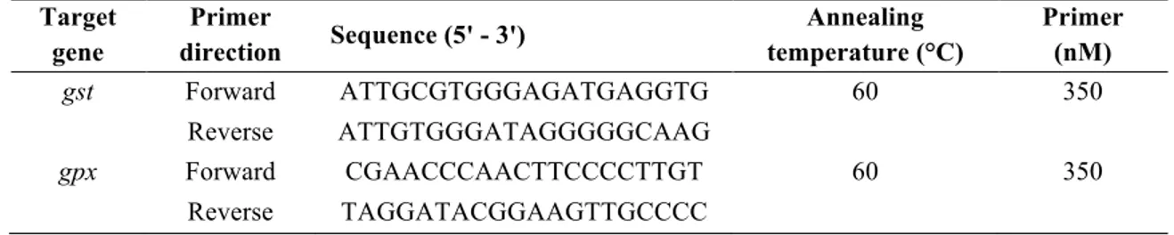
by real-time RT-PCR using the SYBR Green detection system. All primers except gst and gpx 202
were previously designed and optimized (Langlois et al. 2010c; Soriano et al., 2013; Mathieu-203
10
Denoncourt et al., 2015b). Primers for gst and gpx were designed and optimized in the present 204
study and gene products have been confirmed by sequencing. Assay conditions are presented in 205
Table 1. Each sample was diluted 1:80 (embryo samples) or 1:40 (testes samples) and analyzed 206
in duplicate using the GoTaq qPCR Master Mix (Fisher Scientific, Toronto, ON, Canada) with 207
the optimized concentration of forward and reverse primer (0.1 to 0.65 µM) on a CFX96 208
Touch™ real-time RT-PCR machine (Bio Rad, Mississauga, ON, Canada). The program used to 209
run all samples included an enzyme activation step at 95 °C for 2 min followed by 40 cycles with 210
95 °C for 15 sec and 58 or 62 °C (depending on target gene) for 1 min. After the amplification 211
phase, a dissociation curve was established in order to ensure the presence of a single amplicon. 212
Reaction efficiencies were 100 ± 10 % with an R2 > 0.990 and calculated by the CFX Manager 213
Software (Bio Rad, Mississauga, ON, Canada). 214
In each assay, a standard curve (0.048 to 50 ng), a no template control, and a no reverse 215
transcription control (to ensure the absence of genomic DNA in the samples) were run with the 216
samples. The standard curve was generated by pooling equal amounts of the treated and control 217
samples and was then serially diluted 1:4 to obtain concentrations from 50 to 0.048 ng. The 218
standard curve was then used to interpolate and calculate the mRNA level of target and reference 219
gene in each sample. The mRNA level of each target gene was calculated relative to the 220
reference gene odc. 221
2.6 Sex steroid measurement
222
For the ex vivo exposure, concentrations of T and 5α-DHT excreted from the testes into 223
the media were measured using commercially available enzyme-linked immunosorbent assays 224
(T: Cedarlane, Burlington, ON, Canada; 5α-DHT: Diagnostics Biochem Canada, Dorchester, 225
11
ON, Canada). Media samples were thawed on ice and diluted in the immunoassay buffer. The 226
immunoassay protocol was followed as described by the manufacturer. All samples were 227
measured in duplicate. The absorbance of samples after the designated incubation time of 1 h for 228
5α-DHT and 2 h for T were measured using a TECAN Infinit M1000 PRO microplate 229
absorbance reader (TECAN, Männedorf, Switzerland) at 415 nm for T and at 450 nm for 5α-230
DHT. The limit of detection according to the manufacturer was 6 pg/mL for T and 5α-DHT. 231
Hormone concentrations were normalized to tissue weight. 232
2.7 Statistical analysis
233
All statistical analysis except malformation analysis was performed using the software 234
GraphPad Prism 5.0 (GraphPad Software, La Jolla, CA, USA). Differences of phthalate 235
concentrations between 0 and 24 h, and differences between phthalate treatment concentrations 236
were analyzed using an unpaired t-test. Gene expression results of the embryo samples were 237
analyzed after removing outliers using one-way analysis of variance (ANOVA) with a 238
subsequent post-hoc test (Dunnett’s) to determine significant differences between treatments. 239
Gene expression results of the testes exposed to DBP ex vivo were analyzed using an unpaired t-240
test. The effect of DBP exposure on hormone concentration in ex vivo exposed testis was also 241
analyzed using an unpaired t-test. Analysis of malformation data was performed using the 242
software XLSTAT (2014.4.06, AddinsoftTM). Differences in malformation frequencies were 243
determined using chi-square tests on contingency tables followed by a post-hoc test based on 244
adjusted residuals for each malformation type and phthalate compound. Significant differences 245
were reported when p < 0.05. 246
12 248
3. Results
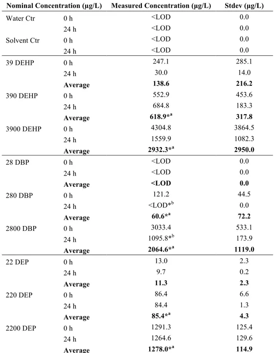
249 3.1 Water analysis 250LC-HRMS analysis showed that all water and solvent control samples were below the 251
detection limit for DEHP, DBP, and DEP. Average measured concentrations (before exposure) 252
were as follows: 138.6 µg/L, 618.9 µg/L, and 2932.3 µg/L for DEHP; <LOD, 60.6 µg/L, and 253
2064.6 µg/L for DBP; and 11.3 µg/L, 85.4 µg/L, and 1278.0 µg/L for DEP (Table 2). DBP 254
significantly degraded over the 24 h exposure time (85.4 µg/L: p = 0.042; 1278.0 µg/L: p = 255
0.017). The measured concentrations are lower than the nominal concentrations, which may be 256
due to losses associated with adsorption to glassware and material used during sampling and 257
sample preparation. 258
3.2 Mortality and malformation of embryos
259
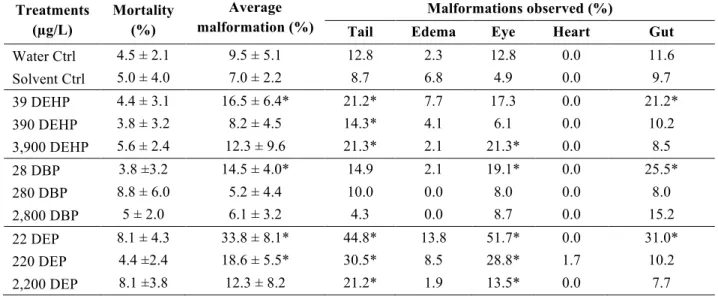
Survival rates of both, the water and solvent controls were above 90%, as recommended by 260
the American Society for Testing and Materials (2004; Table 3). Embryos exposed to DEHP, 261
DBP, and DEP did not result in a significantly higher mortality rate than control embryos. 262
However, phthalate exposure increased malformation rates. Exposure to DEHP, DBP, and DEP 263
increased both, incomplete gut coiling (21.2%, 25.5%, and 31%; respectively) and eye 264
malformations (17.3%, 19.1%, and 51.7%; respectively). Moreover, DEHP and DEP increased 265
tail abnormalities (21.2% and 44.8%; respectively), and DEP increased the occurrence of edemas 266
(13.8%). 267
13
3.3 Gene expression of larvae exposed to phthalates
268
Exposure to phthalate esters modulated androgen-related mRNA levels. Exposure to 685 269
µg/L DEHP increased the expression of srd5β and ar (ANOVA: srd5β: 1.4-fold increase, p = 270
0.0057; ar: 1.4-fold increase, p = 0.0228, Figures 1 and 2). Frogs exposed to DEHP at a 271
concentration 10X higher (i.e., 2,932 µg/L) responded with increased mRNA levels of all 272
four srd5 genes (ANOVA: srd5α1: 1.4-fold increase, p = 0.0001; srd5α2: 1.7-fold increase, p 273
= 0.0003; srd5α3: 1.5-fold increase, p = 0.0010; srd5β: 1.8-fold increase, p = 0.0001). 274
Interestingly, DBP resulted in significant changes at the lowest (below the detection limit of 275
2 µg/L) and highest (2,065 µg/L) concentrations only. Low DBP stimulated srd5β 276
transcription (ANOVA: 1.4-fold increase, p = 0.0302) and high DBP increased the mRNA 277
level of srd5α1, srd5α2, srd5α3, srd5β, and ar (ANOVA: srd5α1: 1.3-fold increase, p = 278
0.0046; srd5α2: 1.6-fold increase, p = 0.0029; srd5α3: 1.4-fold increase, p = 0.0178; srd5β: 279
1.6-fold increase, p = 0.0001; ar: 1.4-old increase, p = 0.0287). Exposure to DEP resulted in 280
a change of expression of srd5α1 (ANOVA: 1278 µg/L: 1.3-fold increase, p = 0.0094) and 281
srd5β (ANOVA: 85 µg/L: 1.5-fold increase, p = 0.0042). The three phthalates did not affect 282
the transcript levels for cyp19, erα, star, gst, gpx, or pparγ. As expected, finasteride exposure 283
significantly decreased the mRNA level of srd5α2 (ANOVA: 2-fold decrease, p = 0.01). In 284
addition to androgen related gene expression changes, hsp70 resulted in significantly 285
changed mRNA levels for <2 µg/L DBP (ANOVA: 1.5-fold decrease, p = 0.01) and 11 µg/L 286
DEP (ANOVA: 1.6-fold decrease, p = 0.014, Figure 3). 287
14
3.4 Frog testes exposed to DBP ex vivo
288
The total amount of T and 5α-DHT was measured in the media of control and DBP-289
exposed testes tissues. Interestingly, neither the secreted concentration of T or 5α-DHT into 290
the media was significantly altered after DBP exposure (p > 0.05; Figure 4). In addition, the 291
mRNA levels of srd5α1, srd5α2, srd5α3, and srd5β were also not significantly modified after 292 DBP exposure (Figure 5). 293
4. Discussion
294 295Both, phthalate esters and their metabolites have been linked to endocrine disruption in 296
wildlife (Mathieu-Denoncourt et al., 2015b). Amphibians, in particular, are at a high risk due to 297
their external egg development and permeable skin, which allows chemical penetration. One of 298
the phthalates, DBP, used in the present study showed significant degradation after 24 h, 299
suggesting that embryos have been also exposed to its major metabolite mono-n-butyl phthalate 300
(MBP; Silva et al., 2007). The contribution of DBP and its metabolites are thus confounded 301
when investigating the interference with the amphibian development and reproductive system in 302
the current study. As we were interested in sub-lethal effects, such as endocrine disruption, we 303
first established that the phthalate concentrations chosen did not affect the survival rate. 304
Similarly, previous studies have shown that exposure up to 1,000 µg/L DBP did not result in 305
decreased mortality of fathead minnow embryos (Mankidy et al., 2013), while exposure to 5,000, 306
10,000, and 15,000 µg/L DBP significantly decreased the survivability of Xenopus laevis larvae 307
(Lee et al., 2005). Likewise, higher DEP concentrations as the ones used in the present study 308
(e.g., 10,000 µg/L and above), decreased survival rate of fathead minnow and zebrafish embryos 309
15
(Kim et al., 2015; Mankidy et al., 2013). Similar to the findings in our study, 10 µg/L DEHP did 310
not result in significantly decreased mortality in Japanese medaka (Chikae et al., 2004). 311
In the present study, exposure to the lowest concentrations of DEHP, DBP, and DEP 312
augmented the occurrence of malformations in S. tropicalis, in particular the presence of 313
incomplete gut coiling, tail abnormalities, and eye malformations. Previous studies have shown 314
that endocrine disrupting chemicals don’t always follow a normal dose response curve where the 315
observable effects increase with increasing dose (reviewed in Vandenberg et al., 2012). Thus, the 316
present finding of a higher effect at low doses is not unexpected for phthalates. Other studies 317
have also observed that phthalate exposures to similar concentrations as used in this study 318
resulted in malformed animals. For example, exposure to DBP and DEP at concentrations 319
exceeding 500 µg/L significantly increased malformations, including abnormal gut coiling, 320
cardiac abnormalities, and malformed faces, eyes, and brains in X. laevis (Bantle et al., 1999; 321
Lee et al., 2005; Gardner et al., 2016). Furthermore, S. tropicalis exposed to dimethyl phthalate 322
(DMP) and dicyclohexyl phthalate (DCHP) also resulted in malformations, such as edemas, 323
improperly developed hearts, tail abnormalities, improperly coiled guts, and/or absent gills 324
(Mathieu-Denoncourt et al., 2016). 325
Malformations of the tail and gut are often attributed to cellular stress. Heat shock proteins 326
are known as early warning signs of cellular stress and have been previously shown to be 327
induced after phthalate exposure in different species (reviewed in Gupta et al., 2010). In the 328
present study, the lowest DBP and DEP concentrations significantly decreased mRNA levels of 329
hsp70. In contrary, 1000 µg/L butyl benzyl phthalate (BBP) and DEHP induced hsp70 mRNA 330
levels in Chironomus riparius larvae (Planelló et al., 2011) and exposure of S. tropicalis larvae 331
to DCHP also increased hsp70 mRNA levels (Mathieu-Denoncourt et al., 2016). Cellular 332
16
oxidative stress is caused by the presence of reactive oxygen species, which can lead to DNA 333
damage in cells. Cells can metabolize reactive oxygen species by producing antioxidant 334
enzymes, including glutathione transferase and glutathione peroxidase that transform reactive 335
oxygen species to less reactive compounds such as oxygen. In the present study, the expression 336
of gst and gpx did not change suggesting that the developmental abnormalities are not likely the 337
result of oxidative stress. However, in the current study, protein activity was not measured and it 338
is known that mRNA levels and protein activities do not always correlate (Koussounadis et al., 339
2015). Therefore, it is possible that proteins involved in oxidative stress changed in activity level 340
but no change in mRNA level was detected after phthalate exposure. 341
In addition to inducing malformations, phthalates are known to alter the endocrine system in 342
mammalian species. Previous studies have suggested that phthalates can interfere with 343
steroidogenesis and affect both the female and male reproductive systems. Multiple regulating 344
pathways involved in the maintenance of steroid homeostasis have been shown to be affected by 345
phthalates (reviewed in Mathieu-Denoncourt et al., 2015a). In order to examine the effects of the 346
studied phthalates in amphibians, a series of endocrine related targets were analyzed. First, the 347
expression of a gene involved in cholesterol transport (e.g., star) was analyzed as previous 348
reports that demonstrated that phthalates interfere with this critical step in steroid synthesis 349
(reviewed in Mathieu-Denoncourt et al., 2015a). In the present study, none of the phthalates of 350
interest modulated star transcription, reinforcing the point that each phthalate presents unique 351
molecular mechanisms of action. In addition, previous literature has also demonstrated that the 352
synthesis and signaling of the female sex steroids were affected by phthalate treatments. For 353
example, mRNA levels of cyp19, the enzyme responsible to aromatize T to estradiol, decreased 354
in rodent cell lines when treated with MEHP and DEHP (Lovekamp and Davis, 2001; Gupta et 355
17
al., 2010). In contrast, in amphibians, exposure to DCHP during embryogenesis in the Western 356
Clawed frog increased cyp19 mRNA level; however, DMP and its metabolite, MMP did not alter 357
cyp19 expression in the same species (Mathieu-Denoncourt et al., 2016). The later study is 358
similar to the data found in this study as none of DEHP, DBP, or DEP modulated the expression 359
of estrogen-related genes, such as cyp19, and erα. 360
Androgen synthesis is also known to be altered by phthalate exposure in mammalian species. 361
Generally, it is recognized that phthalates do not exert their action through ar (reviewed in 362
Rouiller-Fabre et al., 2015). As an example, juvenile and adult liver tissues exposed to MMP did 363
not alter transcript levels of ar (Mathieu-Denoncourt et al., 2015a). Similarly, ar expression was 364
not changed following exposure to 10,000 µg/L DEP and 1,000 µg/L DBP in fathead minnow 365
embryos (Mankidy et al., 2013). In contrast, our data revealed that exposure to 10 µM DEHP and 366
DBP increased ar transcription. Agonists of AR have been shown to induce transcriptional 367
changes (Li et al., 2017), suggesting that DEHP and DBP can act as agonists of AR. In addition 368
to the measured increase in ar mRNA level, srd5 expression was also augmented in the frog 369
larvae. All three phthalates increased srd5α1 transcripts. In addition, srd5α2 and srd5α3 370
expression levels were also increased by DEHP and DBP exposures. Similarly in mammalian 371
species, DEHP amplified the activity of SRD5α in the pubertal rat testes (Kim et al., 2003). An 372
increase of srd5α could lead to a higher than normal conversion of T to 5α-DHT. However, other 373
studies demonstrated that phthalate exposure decreases SRD5α activity. For example, a decrease 374
in Srd5α2 activity was demonstrated after DBP exposure in vitro in gonad microsomal 375
homogenates isolated from the common carp (Thibaut and Porte, 2004). Moreover, prenatal 376
exposure to DBP in rats significantly decreased Srd5α2 protein expression in the proximal penis 377
(Kim et al., 2010). In contrast, MMP, DMP, and DCHP did not alter srd5α2 mRNA level in S. 378
18
tropicalis (Mathieu-Denoncourt et al., 2016). These results suggest that the chemical nature of 379
the phthalates as well as the developmental stage or tissue are important as to how the specific 380
compound interferes with the level of certain genes/proteins. 381
Interestingly, not only srd5α isoforms, but also the srd5β transcript level increased after 382
DEHP, DBP, and DEP treatments. Srd5β is known to be involved in clearing excess steroids in 383
bird brains (Steimer and Hutchison, 1981). The observation that srd5β increased after phthalate 384
treatment may suggest a disturbance of the normal balance of sex steroids. However, no previous 385
studies have assessed the effects of phthalates on srd5β expression and limited studies have 386
examined this gene. Treatments with methyltrienolone and atrazine have been shown to alter 387
srd5β levels in human prostate cells and frog liver, respectively (Bolton et al., 2007; Langlois et 388
al., 2010a). Our results provide evidence that phthalates can interfere with srd5β and perhaps 389
result in adverse reproductive effects. Since srd5β is involved in many other biological functions, 390
including bile acid synthesis and erythropoiesis, other adverse effects may be observed. To test 391
this hypothesis, exposure of phthalates to animals throughout development and sexual 392
differentiation would be required. Taken together, these findings suggest that phthalates exert 393
their action through different mechanisms depending on species and tissues and affect androgen 394
synthesis in various ways. 395
As androgen-related genes were altered by DBP during frog early development, further 396
investigations were pursued in DBP-exposed testes of adult males due to a high androgen 397
synthesis in gonads. However, DBP ex vivo exposure did not alter the T or 5α-DHT steroid 398
levels nor did it alter srd5α expression in frog testes. Previous studies had demonstrated that 399
phthalates, including DEHP, DBP, DEP, MEP, monobutyl phthalate, dipentyl phthalate (DPeP), 400
monopentyl phthalate (MPeP), benzylbutyl phthalate (BzBP), mono-n-octyl phthalate (MnOP), 401
19
and MEHP decreased T levels in mammalian species (reviewed in Mathieu-Denoncourt et al., 402
2015a). As an example, DBP exposure of prenatal male rats resulted in significant decreased of 403
T levels, which was accompanied by a decreased expression of Srd5α2 in testicular tissues (Jiang 404
et al., 2016). Similarly, primary cultures of rat Leydig cells exposed to MEHP decreased Srd5α 405
activity in immature, but not in adult Leydig cells (Svechnikov et al., 2008). The lack of any 406
observed effect in our study may be due to the fact that the ex vivo exposure was stopped after 407
6h, which may have been too short of an exposure time to see any changes at the transcriptional 408
level. Nevertheless, our embryonic data suggest that phthalates can induce transcriptional 409
changes of genes associated with reproduction. 410
This study demonstrated that DEHP, DEP, and DBP interfere with normal frog development 411
by inducing an array of malformations to the developing animals. Exposure to these three 412
phthalates also increased the expression of androgen-related genes, in particular the four srd5 413
during amphibian embryogenesis, which suggests that DEHP, DEP, and DBP have an 414
androgenic activity in the amphibian embryo. Thus, this data shows evidence that certain 415
phthalates act via the srd5 signaling pathway. Furthermore, this finding also supports previous 416
studies suggesting that phthalate esters induce adverse effects in vertebrates by altering important 417
biological functions, including a hormonal imbalance. 418
Acknowledgements
419
This research project was funded by the support of the Natural Sciences and Engineering 420
Research Council of Canada Discovery Grant (NSERC-DG) and the Canada Research Chair 421
(CRC) to VSL. 422
20 424
References
425
American Society for Testing and Materials, 2004. Standard guide for conducting the frog 426
embryo teratogenesis assay––Xenopus (FETAX). 427
Bantle, J.A., Dumont, J., Finch, R.A., Linder, G., Fort, D.J., 1998. Atlas of abnormalities: a 428
guide for the performance of FETAX, second edition. ed. Printing services, Oklahoma 429
State University. 430
Bantle, J.A., Finch, R.A., Fort, D.J., Stover, E.L., Hull, M., Kumsher-King, M., Gaudet-Hull, 431
A.M., 1999. Phase III interlaboratory study of FETAX part 3. FETAX validation using 432
12 compounds with and without an exogenous metabolic activation system. J. Appl. 433
Toxicol. 19, 447–472. 434
Bauer, M.J., Herrmann, R., 1997. Estimation of the environmental contamination by phthalic 435
acid esters leaching from household wastes. Sci. Total Environ. 208, 49–57. 436
Bhatia, H., Kumar, A., Chapman, J.C., McLaughlin, M.J., 2015. Long-term exposures to di-n-437
butyl phthalate inhibit body growth and impair gonad development in juvenile Murray 438
rainbowfish (Melanotaenia fluviatilis). J. Appl. Toxicol. 35, 806–816. 439
Bissegger, S., Martyniuk, C.J., Langlois, V.S., 2014. Transcriptomic profiling in Silurana 440
tropicalis testes exposed to finasteride. Gen. Comp. Endocrinol. 203, 137–145. 441
doi:10.1016/j.ygcen.2014.01.018 442
Blair, J.D., Ikonomou, M.G., Kelly, B.C., Surridge, B., Gobas, F.A.P.C., 2009. Ultra-Trace 443
Determination of Phthalate Ester Metabolites in Seawater, Sediments, and Biota from an 444
Urbanized Marine Inlet by LC/ESI-MS/MS. Environ. Sci. Technol. 43, 6262–6268. 445
Bolton, E.C., So, A.Y., Chaivorapol, C., Haqq, C.M., Li, H., Yamamoto, K.R., 2007. Cell- and 446
gene-specific regulation of primary target genes by the androgen receptor. Genes Dev. 447
21, 2005–2017. 448
Borch, J., Metzdorff, S.B., Vinggaard, A.M., Brokken, L., Dalgaard, M., 2006a. Mechanisms 449
underlying the anti-androgenic effects of diethylhexyl phthalate in fetal rat testis. 450
Toxicology 223, 144–155. 451
Borch, J., Metzdorff, S.B., Vinggaard, A.M., Brokken, L., Dalgaard, M., 2006b. Mechanisms 452
underlying the anti-androgenic effects of diethylhexyl phthalate in fetal rat testis. 453
Toxicology 223, 144–155. 454
Chikae, M., Hatano, Y., Ikeda, R., Morita, Y., Hasan, Q., Tamiya, E., 2004. Effects of bis(2-455
ethylhexyl) phthalate and benzo[a]pyrene on the embryos of Japanese medaka (Oryzias 456
latipes). Environ. Toxicol. Pharmacol. 16, 141–145. 457
Daniels, P.H., 2009. A brief overview of theories of PVC plasticization and methods used to 458
evaluate PVC-plasticizer interaction. J. Vinyl Addit. Technol. 15, 219–223. 459
Dargnat, C., Teil, M.-J., Chevreuil, M., Blanchard, M., 2009. Phthalate removal throughout 460
wastewater treatment plant. Sci. Total Environ. 407, 1235–1244. 461
Gardner, S.T., Wood, A.T., Lester, R., Onkst, P.E., Burnham, N., Perygin, D.H., Rayburn, J., 462
2016. Assessing differences in toxicity and teratogenicity of three phthalates, Diethyl 463
phthalate, Di-n-propyl phthalate, and Di-n-butyl phthalate, using Xenopus laevis 464
embryos. J. Toxicol. Environ. Health A 79, 71–82. 465
Gazouli, M., Yao, Z.X., Boujrad, N., Corton, J.C., Culty, M., Papadopoulos, V., 2002. Effect of 466
21
peroxisome proliferators on leydig cell peripheral-type benzodiazepine receptor gene expression, 467
hormone-stimulated cholesterol transport, and steroidogenesis: role of the peroxisome 468
proliferator-activator receptor a. Endocrinol. 143, 2571–2583. 469
Gupta, R.K., Singh, J.M., Leslie, T.C., Meachum, S., Flaws, J.A., Yao, H.H.-C., 2010. Di-(2-470
ethylhexyl) phthalate and mono-(2-ethylhexyl) phthalate inhibit growth and reduce 471
estradiol levels of antral follicles in vitro. Toxicol. Appl. Pharmacol. 242, 224–230. 472
Gupta, S.C., Sharma, A., Mishra, M., Mishra, R.K., Chowdhuri, D.K., 2010. Heat shock proteins 473
in toxicology: How close and how far? Life Sci. 86, 377–384. 474
Jiang, J.-T., Zhong, C., Zhu, Y.-P., Xu, D.-L., Wood, K., Sun, W., Li, E.-H., Liu, Z.-H., Zhao, 475
W., Ruan, Y., Xia, S.-J., n.d. Prenatal exposure to di-n-butyl phthalate (DBP) 476
differentially alters androgen cascade in undeformed versus hypospadiac male rat 477
offspring. Reprod. Toxicol. 478
Kay, V.R., Bloom, M.S., Foster, W.G., 2014. Reproductive and developmental effects of 479
phthalate diesters in males. Crit. Rev. Toxicol. 44, 467–498. 480
Kay, V.R., Chambers, C., Foster, W.G., 2013. Reproductive and developmental effects of 481
phthalate diesters in females. Crit. Rev. Toxicol. 43, 200–219. 482
Kim, H.-S., Saito, K., Ishizuka, M., Kazusaka, A., Fujita, S., 2003. Short period exposure to di-483
(2-ethylhexyl) phthalate regulates testosterone metabolism in testis of prepubertal rats. 484
Arch. Toxicol. 77, 446–451. 485
Kim, S.-M., Yoo, J.-A., Baek, J.-M., Cho, K.-H., 2015. Diethyl phthalate exposure is associated 486
with embryonic toxicity, fatty liver changes, and hypolipidemia via impairment of 487
lipoprotein functions. Toxicol. In Vitro 30, 383–393. 488
Kim, T.S., Jung, K.K., Kim, S.S., Kang, I.H., Baek, J.H., Nam, H.-S., Hong, S.-K., Lee, B.M., 489
Hong, J.T., Oh, K.W., Kim, H.S., Han, S.Y., Kang, T.S., 2010. Effects of in utero 490
exposure to DI(n-Butyl) phthalate on development of male reproductive tracts in 491
Sprague-Dawley rats. J. Toxicol. Environ. Health A 73, 1544–1559. 492
Koussounadis, A., Langdon, S.P., Um, I.H., Harrison, D.J., Smith, V.A., 2015. Relationship 493
between differentially expressed mRNA and mRNA-protein correlations in a xenograft model 494
system, Sci Rep. 5, 10775. 495
Langlois, V.S., Zhang, D., Cooke, G.M., Trudeau, V.L., 2010a. Evolution of steroid-5α-496
reductases and comparison of their function with 5β-reductase. Gen. Comp. Endocrinol. 497
166, 489–497. 498
Langlois, V.S., Carew, A.C., Pauli, B.D., Wade, M.G., Cooke, G.M., Trudeau, V.L., 2010b. Low 499
Levels of the Herbicide Atrazine Alter Sex Ratios and Reduce Metamorphic Success in 500
Rana pipiens Tadpoles Raised in Outdoor Mesocosms. Environ. Health Perspect. 118, 501
552–557. 502
Langlois, V.S., Duarte-Guterman, P., Ing, S., Pauli, B.D., Cooke, G.M., Trudeau, V.L., 2010c. 503
Fadrozole and finasteride exposures modulate sex steroid- and thyroid hormone-related 504
gene expression in Silurana (Xenopus) tropicalis early larval development. Gen. Comp. 505
Endocrinol. 166, 417–427. 506
Latini, G., Del Vecchio, A., Massaro, M., Verrotti, A., De Felice, C., 2006. Phthalate exposure 507
and male infertility. Toxicology 226, 90–98. 508
22
Lee, S.K., Owens, G.A., Veeramachaneni, D.N.R., 2005. Exposure to Low Concentrations of Di-509
n-butyl Phthalate During Embryogenesis Reduces Survivability and Impairs 510
Development of Xenopus Laevis Frogs. J. Toxicol. Environ. Health A 68, 763–772. 511
Lee, S.K., Veeramachaneni, D.N.R., 2005. Subchronic Exposure to Low Concentrations of Di-n-512
Butyl Phthalate Disrupts Spermatogenesis in Xenopus laevis Frogs. Toxicol. Sci. 84, 513
394–407. 514
Lehmann, K.P., Phillips, S., Sar, M., Foster, P.M.D., Gaido, K.W., 2004. Dose-Dependent 515
Alterations in Gene Expression and Testosterone Synthesis in the Fetal Testes of Male 516
Rats Exposed to Di (n-butyl) phthalate. Toxicol. Sci. 81, 60–68. 517
Li, J., Chang, J., Li, W., Guo, B., Li, J., Wang, H., 2017. Disruption of sex-hormone levels and 518
steroidogenic-related gene expression on Mongolia Racerunner (Eremias argus) after exposure to 519
triadimefon and its enantiomers. Chemosphere 171, 554-563. 520
Lovekamp, T.N., Davis, B.J., 2001. Mono-(2-ethylhexyl) Phthalate Suppresses Aromatase 521
Transcript Levels and Estradiol Production in Cultured Rat Granulosa Cells. Toxicol. Appl. 522
Pharmacol. 172, 217–224. 523
Magdouli, S., Daghrir, R., Brar, S.K., Drogui, P., Tyagi, R.D., 2013. Di 2-ethylhexylphtalate in 524
the aquatic and terrestrial environment: A critical review. J. Environ. Manage. 127, 36– 525
49. 526
Mankidy, R., Wiseman, S., Ma, H., Giesy, J.P., 2013. Biological impact of phthalates. Toxicol. 527
Lett. 217, 50–58. 528
Mathieu-Denoncourt, J., Wallace, S.J., de Solla, S.R., Langlois, V.S., 2015a. Plasticizer 529
endocrine disruption: Highlighting developmental and reproductive effects in mammals 530
and non-mammalian aquatic species. Gen. Comp. Endocrinol. 219, 74–88. 531
Mathieu-Denoncourt, J., de Solla, S.R., Langlois, V.S., 2015b. Chronic exposures to 532
monomethyl phthalate in Western clawed frogs. Gen. Comp. Endocrinol., Disruption of 533
the thyroid and sex steroid hormone systems and their crosstalk in aquatic wildlife 219, 534
53–63. 535
Mathieu-Denoncourt, J., Martyniuk, C.J., Loughery, J.R., Yargeau, V., de Solla, S.R., Langlois, 536
V.S., 2016. Lethal and sublethal effects of phthalate diesters in Silurana tropicalis larvae. 537
Environ. Toxicol. Chem. 35, 2511-2522. 538
Nieuwkoop, P.D., 1994. Normal Table of Xenopus Laevis (Daudin): A Systematical and 539
Chronological Survey of the Development from the Fertilized Egg Till the End of 540
Metamorphosis. Garland Pub. 541
Planelló, R., Herrero, O., Martínez-Guitarte, J.L., Morcillo, G., 2011. Comparative effects of 542
butyl benzyl phthalate (BBP) and di(2-ethylhexyl) phthalate (DEHP) on the aquatic 543
larvae of Chironomus riparius based on gene expression assays related to the endocrine 544
system, the stress response and ribosomes. Aquat. Toxicol. 105, 62–70. 545
Rouiller-Fabre, V., Guerquin, M.J., N’Tumba-Byn, T., Muczynski, V., Moison, D., Tourpin, S., 546
Messiaen, S., Habert, R., Livera, G., 2015. Nuclear Receptors and Endocrine Disruptors 547
in Fetal and Neonatal Testes: A Gapped Landscape. Front. Endocrinol. 6. 548
Silva, M.J., Samandar, E., Reidy, J.A., Hauser, R., Needham, L.L., Calafat, A.M., 2007, 549
Metabolite profiles of di-n-butyl phthalate in humans and rats. Environ. Sci. Technol. 41, 550
7576-7580. 551
23
Soriano, J.J., Mathieu-Denoncourt, J., Norman, G., Solla, S.R. de, Langlois, V.S., 2013. Toxicity 552
of the azo dyes Acid Red 97 and Bismarck Brown Y to Western clawed frog (Silurana 553
tropicalis). Environ. Sci. Pollut. Res. 21, 3582–3591. 554
Steimer, T., Hutchison, J.B., 1981. Metabolic control of the behavioural action of androgens in 555
the dove brain: Testosterone inactivation by 5β-reduction. Brain Res. 209, 189–204. 556
Suzuki, T., Yaguchi, K., Suzuki, S., Suga, T., 2001. Monitoring of Phthalic Acid Monoesters in 557
River Water by Solid-Phase Extraction and GC-MS Determination. Environ. Sci. 558
Technol. 35, 3757–3763. 559
Svechnikov, K., Svechnikova, I., Söder, O., 2008. Inhibitory effects of mono-ethylhexyl 560
phthalate on steroidogenesis in immature and adult rat Leydig cells in vitro. Reprod. 561
Toxicol. 25, 485–490. 562
Thibaut, R., Porte, C., 2004. Effects of endocrine disrupters on sex steroid synthesis and 563
metabolism pathways in fish. J. Steroid Biochem. Mol. Biol. 92, 485–494. 564
Thompson, C.J., Ross, S.M., Gaido, K.W., 2004. Di(n-Butyl) Phthalate Impairs Cholesterol 565
Transport and Steroidogenesis in the Fetal Rat Testis through a Rapid and Reversible 566
Mechanism. Endocrinology 145, 1227–1237. 567
Vandenberg, L.N., Colborn, T., Hayes, T.B., Heindel, J.J., Jacobs Jr., D.R., Lee, D.-H., Shioda, 568
T., Soto, A.M, vom Saal, F.S., Welshons, W.V., Zoeller, R.S., Myers J.P., 2012. 569
Hormones and Endocrine-Disrupting Chemicals: Low-Dose Effects and Nonmonotonic 570
Dose Responses. Endocrine Reviews, 33, 378–455. 571
Wong, J.S., Gill, S.S., 2002. Gene Expression Changes Induced in Mouse Liver by Di(2-572
ethylhexyl) Phthalate. Toxicol. Appl. Pharmacol. 185, 180–196. 573
Yuwatini, E., Hata, N., Taguchi, S., 2006. Behavior of di(2-ethylhexyl) phthalate discharged 574
from domestic waste water into aquatic environment. J. Environ. Monit. 8, 191–196. 575
576 577
24 578
Table 1 qPCR primers and assay conditions of gpx and gst genes for S. tropicalis.
579 Target gene Primer direction Sequence (5' - 3') Annealing temperature (°C) Primer (nM) gst Forward ATTGCGTGGGAGATGAGGTG 60 350 Reverse ATTGTGGGATAGGGGGCAAG gpx Forward CGAACCCAACTTCCCCTTGT 60 350 Reverse TAGGATACGGAAGTTGCCCC 580 581
25 582
Table 2 Di(2-ethylhexyl) phthalate (DEHP), di-n-butyl phthalate (DBP), and diethyl phthalate (DEP) measured in
583
the water of exposed embryos at 0 h and after 24 h. Average concentrations are bold. Legend: Ctr, Control; LOD, 584
limit of detection; Stdev, standard deviation. 585
Nominal Concentration (µg/L) Measured Concentration (µg/L) Stdev (µg/L)
Water Ctr 0 h <LOD 0.0 24 h <LOD 0.0 Solvent Ctr 0 h <LOD 0.0 24 h <LOD 0.0 39 DEHP 0 h 247.1 285.1 24 h 30.0 14.0 Average 138.6 216.2 390 DEHP 0 h 552.9 453.6 24 h 684.8 183.3 Average 618.9*a 317.8 3900 DEHP 0 h 4304.8 3864.5 24 h 1559.9 1082.3 Average 2932.3*a 2950.0 28 DBP 0 h <LOD 0.0 24 h <LOD 0.0 Average <LOD 0.0 280 DBP 0 h 121.2 44.5 24 h <LOD*b 0.0 Average 60.6*a 72.2 2800 DBP 0 h 3033.4 533.1 24 h 1095.8*b 173.9 Average 2064.6*a 1119.0 22 DEP 0 h 13.0 2.3 24 h 9.7 0.2 Average 11.3 2.3 220 DEP 0 h 86.4 6.6 24 h 84.4 1.3 Average 85.4*a 4.3 2200 DEP 0 h 1291.3 125.4 24 h 1264.6 129.6 Average 1278.0*a 114.9
*a indicates a significant concentration difference to the previous lower concentration
586
*b indicates significant degradation between 0 h and 24 h of exposure
587 588
26
Table 3 Effects of di(2-ethylhexyl) phthalate (DEHP), di-n-butyl phthalate (DBP), and diethyl phthalate (DEP)
589
spiked water on mortality and malformation of S. tropicalis larvae at exposure completion (Nieuwkoop and Faber 590
stage 46). The mortality data are expressed as percent mean ± SD (%) and the malformation results are expressed as 591
a percentage (%) of malformed animals to the total number of animals analyzed. Asterisks indicate statistically 592
significant differences between treatments (DEHP, DBP, and DEP) and solvent (0.05% ethanol) control. 593 Treatments (µg/L) Mortality (%) Average malformation (%) Malformations observed (%)
Tail Edema Eye Heart Gut
Water Ctrl 4.5 ± 2.1 9.5 ± 5.1 12.8 2.3 12.8 0.0 11.6 Solvent Ctrl 5.0 ± 4.0 7.0 ± 2.2 8.7 6.8 4.9 0.0 9.7 39 DEHP 4.4 ± 3.1 16.5 ± 6.4* 21.2* 7.7 17.3 0.0 21.2* 390 DEHP 3.8 ± 3.2 8.2 ± 4.5 14.3* 4.1 6.1 0.0 10.2 3,900 DEHP 5.6 ± 2.4 12.3 ± 9.6 21.3* 2.1 21.3* 0.0 8.5 28 DBP 3.8 ±3.2 14.5 ± 4.0* 14.9 2.1 19.1* 0.0 25.5* 280 DBP 8.8 ± 6.0 5.2 ± 4.4 10.0 0.0 8.0 0.0 8.0 2,800 DBP 5 ± 2.0 6.1 ± 3.2 4.3 0.0 8.7 0.0 15.2 22 DEP 8.1 ± 4.3 33.8 ± 8.1* 44.8* 13.8 51.7* 0.0 31.0* 220 DEP 4.4 ±2.4 18.6 ± 5.5* 30.5* 8.5 28.8* 1.7 10.2 2,200 DEP 8.1 ±3.8 12.3 ± 8.2 21.2* 1.9 13.5* 0.0 7.7 594 595 596
27
Figure Captions
597
Figure 1 mRNA levels of A) srd5α1, B) srd5α2, C) srd5α3, and D) srd5β in frog embryos
598
following exposure to FIN, DEHP, DBP, and DEP. Data are expressed relative to the reference 599
gene odc. Bars represent the mean + SEM. Data were analyzed using one-way analysis of 600
variance (n = 4-8; p < 0.05). Legend: DBP, dibutyl phthalate; di-2-ethylhexyl phthalate DEHP, 601
diethyl hexyl phthalate; DEP, diethyl phthalate; FIN, finasteride; SC, Solvent Control; WC, 602
Water Control; *, significant different from control at p < 0.05. 603
Figure 2 mRNA levels of A) ar, B) erα, C) cyp19, and D) star in frog embryos following
604
exposure to FIN, DEHP, DBP, and DEP. Data are expressed relative to the reference gene odc. 605
Bars represent the mean + SEM. Data were analyzed using one-way analysis of variance (n = 4-606
8; p < 0.05). Legend: DBP, dibutyl phthalate; di-2-ethylhexyl phthalate DEHP, diethyl hexyl 607
phthalate; DEP, diethyl phthalate; FIN, finasteride; SC, Solvent Control; WC, Water Control; *, 608
significant different from control at p < 0.05. 609
Figure 3 mRNA levels of A) hsp70, B) gst, C) gpx, and D) pparγ in frog embryos following
610
exposure to FIN, DEHP, DBP, and DEP. Data are expressed relative to the reference gene odc. 611
Bars represent the mean + SEM. Data were analyzed using one-way analysis of variance (n = 4-612
8; p < 0.05). Legend: DBP, dibutyl phthalate; di-2-ethylhexyl phthalate DEHP, diethyl hexyl 613
phthalate; DEP, diethyl phthalate; FIN, finasteride;SC, Solvent Control; WC, Water Control; *, 614
significant different from control at p < 0.05. 615
Figure 4 Concentration of testosterone and 5α-dihydrotestosterone in the media after ex vivo
616
exposure of testes to dibutyl phthalate (DBP). Bars represent the mean + STD. Data were 617
analyzed using a two tailed t-test (n = 6). Legend: DBP, dibutyl phthalate; SC, Solvent Control. 618
28
Figure 5 mRNA levels of A) srd5α1, B) srd5α2, C) srd5α3, and D) srd5β in frog testes after ex
619
vivo exposure to 10 µM dibutyl phthalate (DBP). Data are expressed relative to the reference 620
gene odc. Bars represent the mean + SEM. Data were analyzed using a two tailed t-test (n = 4). 621
Legend: DBP, dibutyl phthalate; SC, Solvent Control. 622
29 624 625 626 0 .0 0 .5 1 .0 1 .5 2 .0 0 .0 0 .5 1 .0 1 .5 2 .0 0 .0 0 .5 1 .0 1 .5 2 .0 0 .0 0 .5 1 .0 1 .5 2 .0 srd5a1 srd5a2 srd5a3 srd5β A B C D R el ati ve mR N A e xp re ss ion / odc * * * * * * * * * * * * * Ctr FIN DEHP DBP DEP Phthalate concentration (µg/L) WC SC FIN 139 685 2932 < 2 61 2065 11 85 1278 WC SC FIN 139 685 2932 < 2 61 2065 11 85 1278 Fig 1
30 627 628 0 .0 0 .5 1 .0 1 .5 2 .0 0 .0 0 .5 1 .0 1 .5 2 .0 0 .0 0 .5 1 .0 1 .5 2 .0 0 .0 0 .5 1 .0 1 .5 2 .0 ar era cyp19 star R el ati ve mR N A e xp re ss ion / odc A B D C * * WC SC FIN 139 685 2932 < 2 61 2065 11 85 1278 Phthalate concentration (µg/L) Ctr FIN DEHP DBP DEP WC SC FIN 139 685 2932 < 2 61 2065 11 85 1278 Fig 2
31 629 630 0 .0 0 .5 1 .0 1 .5 2 .0 0 .0 0 .5 1 .0 1 .5 2 .0 0 .0 0 .5 1 .0 1 .5 2 .0 0 1 2 3 Ctr FIN DEHP DBP DEP hsp70 gst gpx pparγ A B D C R el ati ve mR N A e xp re ss ion / odc Phthalate concentration (µg/L) WC SC FIN 139 685 2932 <2 61 2065 11 85 1278 WC SC FIN 139 685 2932 <2 61 2065 11 85 1278 * * Fig 3
32 631 632 0 2 0 0 4 0 0 6 0 0 8 0 0 S te roi d p rod u ce d ( pg /mg) Testosterone 5α-dihydrotestosterone SC D BP SC D BP Fig 4
33 633 0 .0 0 .5 1 .0 1 .5 2 .0 s rd 5 a 1 s rd 5 a 2 s rd 5 a 3 s rd 5 b SC sr d5 α1 sr d5 α2 sr d5 α3 sr d5 β R el ati ve mR N A e xp re ss ion / odc Fig 5