353—
/
Université de Montréal
In situ-Forming Injectable Organogel Implant for
$ustained Release of Rivastigmine
par Anda Vintiloiu
Sciences pharmaceutiques Faculté de Pharmacie
Mémoire présenté à la Faculté des études supérieures en vue de l’obtention du grade de Maître en sciences (M.Sc.)
Sciences pharmaceutiques Option technologie pharmaceutique
Mai 2007
69V
h S
/ ‘TE. (7 71• fv’Ôo
n
Université
dl1
de Montréal
Direction des bibliothèques
AVIS
L’auteur a autorisé l’Université de Montréal à reproduire et dïffuser, en totalité ou en partie, par quelque moyen que ce soit et sur quelque support que ce soit, et exclusivement à des fins non lucratives d’enseignement et de recherche, des copies de ce mémoire ou de cette thèse.
L’auteur et les coauteurs le cas échéant conservent la propriété du droit d’auteur et des droits moraux qui protègent ce document. Ni la thèse ou le mémoire, ni des extraits substantiels de ce document, ne doivent être imprimés ou autrement reproduits sans l’autorisation de l’auteur.
Afin de se conformer à la Loi canadienne sur la protection des
renseignements personnels, quelques formulaires secondaires, coordonnées ou signatures intégrées au texte ont pu être enlevés de ce document. Bien que cela ait pu affecter la pagination, il n’y a aucun contenu manquant.
NOTICE
The author of this thesis or dissertation has granted a nonexclusive license allowing Université de Montréal to reproduce and publish the document, in part or in whole, and in any format, solely for noncommercial educational and research purposes.
The author and co-authors if applicable retain copyright ownership and moral
rights in this document. Neither the whole thesis or dissertation, nor
substantial extracts from it, may be printed or otherwise reproduced without the author’s permission.
In compliance with the Canadian Privacy Act some supporting forms, contact information or signatures may have been removed from the document. While this may affect the document page count, it does not represent any loss of content from the document.
Université de Montréal Faculté des études supérieures
Ce mémoire intitulé
Insitu-forming Injectable Organogel Implant for Sustained Release of Rivastigmine
présenté par Anda Vintiloiu
a été évalué par un jury composé des personnes suivantes: Dr. françoise Winnik, président-rapporteur
Dr. Jean-Chistophe Leroux, directeur de recherche Dr. Didier Hoarau, membre du jury
ci
iv
Résumé
Les organogels présentent un grand intérêt en pharmacie galénique en raison de leur facilité de préparation ainsi que leur capacité de libérer un principe actif de façon prolongée. Dans le cadre de ce projet, un organogel a été évalué comme implant injectable pour la libération soutenue de la rivastigmine, un inhibiteur de la cholinestérase utilisé pour le traitement de la maladie d’Alzheimer. Le gel a été préparé en dissolvant 5 — 15% (p/p) de N-lauroyle L-alanine methyl ester (SAM) dans l’huile de
carthame contenant la rivastigmine dissoute ou son sel d’hydrogène tartrate dispersé. Les mesures de températures de transition, obtenues par calorimétrie différentielle à balayage et par spectroscopie infrarouge, ont indiqué un affaiblissement de la structure de l’organogel suite à l’incorporation de drogue dissoute (4% p/p).
À
la même concentration, aucune modification en terme de température de transition n’a été détectée dans les gels contenant le principe actif sous forme sel, indiquant une interférence négligeable avec la matrice gélifiante. Ces mêmes gels ont démontré des profils de libération in vitro ayant le plus faible effet « burst », avec moins de 15% du principe actif libéré durant la première journée. Des études pharmacocinétiques réalisées avec des rats ayant reçu par la voie sous-cutanée des organogels 10% SAM ont mis en évidence une libération maintenant des concentrations plasmatiques de rivastigmine dans la fenêtre thérapeutique pendant 11 jours. De plus, cette formulation a permis une réduction de la concentration plasmatique maximale d’un facteur de 5 par rapport à la formulation contrôle constituée d’huile sans gélateur. En conclusion, ce projet a démontré la possibilité d’utiliser les organogels de SAM en tant que formulation à libération prolongée pour le traitement de la maladie d’Alzheimer.V
Mots clés: organogel, libération prolongée, implant autoformant in situ, rivastigmine, maladie d’Alzheimer.
vi
Abstract
Organogels are interesting systems holding great promise for drug delivery applications owing to their ease of preparation and potential for long-term release. This project investigated the use of such an organogel as an injectable implant for the sustained release of rivastigmine, a cholinesterase inhibitor used in the treatment of Alzheimer’s disease. Such a formulation is expected to provide a simplified dosing schedule and potentially reduce side effects associated to plasma peak concentrations. The gel was prepared by dissolving 5 - 15% (w/w) N-stearoyl L-alanine methyl ester (SAM) in
safflower oil containing either dissolved rivastigmine or its dispersed hydrogen tartrate sait. Differential scanning calorimetry and infrared spectroscopy indicated a weakening of the oleogel structure by the incorporation of dissolved drug (4% w/w), as proven by decreasing gel-sol transition temperatures. At the same concentration, no such changes were detected for gels containing the dispersed drug salt, indicating negligible interference of the latter with gel auto-assembly. In vitro release profiles of gels containing dispersed rivastigmine showed the lowest burst (<15% in the first day). A pharmacokinetic study, in which rats were injected subcutaneously with 10% SAM gels containing the dispersed drug, demonstrated a sustained rivastigmine release within therapeutic range for up to 11 days. Peak plasma levels were found to be well below the toxic threshold and up to five times lower than for the control oil formulation. Overall this project established SAM gels to be a promising option for a sustained-release formulation in the treatment ofAlzheimer’s disease.
Keywords: organogel, sustained release, in situ-forming implant, rivastigmine, Alzheimer’s disease.
vii Table of contents Résumé iv Abstract vi List of tables x List of figures xi
List of abbreviations xvi
Acknowledgements xviii
CHAPTER 1: ALZHEIMER’S DISEASE
1. Introduction 2 2. Pathophysiology 2 3. Treatment options 4 3.1. Cholinesterase inhibitors 4 3.2. Other therapies 6 References 8 CHAPTER 2:
SUSTAINED RELEASE FORMULATIONS 9
References 15
CHAPTER 3:
ORGANOGELS AND THEIR USE IN DRUG DELlVERY-A REVIEW
1. Abstract 18
2. Introduction 19
3. Organogel properties 22
3.1. Low molecular weight gelators 22
3.1.1. Solid-rnatrix organogels 23
3.1.].]. Generai gelation considerations 23
3. 1.1.2. Chiraiity efiècts 28
viii
3.1.2.1. Lecithin organogels 3]
3.1 2.2. Fatty-acid derived-sorbitan organogels 35
3.2. Polymeric gelators 36
4. Organogels in drug delïvery 38
4.1. Dermal and transdermal formulations 40
4.1.1. Lecithin organogels 41
4.1.2. Fatty-acid derived-sorbitan organogels 43 4.1.3. Organogels based on other low molecular weight gelators 43
4.1.4. Poly(ethylene) organogels 44
4.2. Parenterat depot formulations 44
4.3. Trans-mucosal and oral formulations 47
5. Summary and conclusions 50
6. Acknowledgements 51
Referen ces 52
CHAPTER 4: ARTICLE 57
1. Abstract 58
2. Introduction 69
3. Materials and methods 61
3.1. Materials 61 3.2. Preparation of implants 62 3.2.1. Dispersed-RHT gels 62 3.2.2. Dissolved-RB gels 62 3.3. Characterization 63 3.3.1. Rheology 63
3.3.2. Differential scanning calorirnetry (DSC) 63 3.3.3. Thermal Fourier-transform infrared spectroscopy (FuR) 64
3.3.4. Microscopy 65
3.4. In vitro release ofrivastigmine 65
3.4.1. Release study 65
ix
3.5. Pharmacokinetic study 67
3.6. Statistical analysis 6$
4. Results and discussion 69
4.1. Physicochemical characterization ofoleogel 69
4.1.1. Rheology 69 4.1.2. DSC 71 4.1.3. fuR 73 4.1.4. Microscopy 75 4.1.5. fn vitro release 77 4.1.6. Pharmacokinetic study 79 5. Conclusion $3 6. Acknowledgements 83 References $4
CHAPTER 5: RESULTS AND DISCUSSION $6
CHAPTER 6: CONCLUSION AND OUTLOOK 94
X
Lïst of tables
CHAPTER 1
Table I: Current drugs on the market or in clinical trials for the treatrnent ofAD.
5
CHAPTER 3
Table I: Organogel formulations used in drug delivery
39
CHAPTER 4
Table 1. Gel characterization: 10% (w/w) SAM oleogels containing various concentrations of rivastigmine incorporated by dissolution or dispersion were characterized by rheological analysis, DSC and IR (mean+ SD).
72
Table II: Pharmacokinetic parameters ofthe different formulations injected to rats (n=6) $0
xi
List of figures
CHAPTER 1
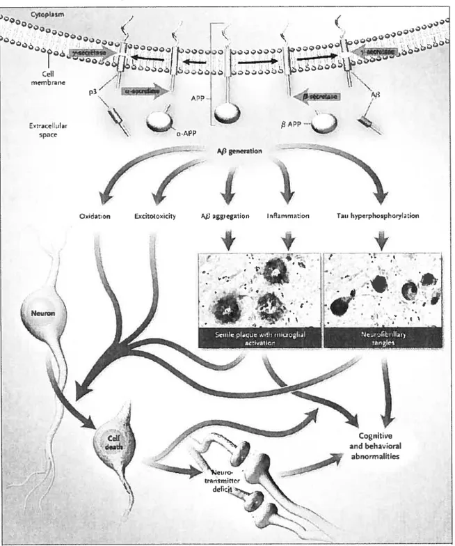
Figure 1: The hypothesis of the amyloid cascade proposes a progression from the generation of f3-amyloid precursor protein (APP), through multiple secondary steps, to ceil death.
3
CHAPTER 3
figure 1: Organogel classification.
20
Figure 2: Solid-matrix (strong) versus fluid-matrix (weak) organogels. A) Solid-matrix gels are more robust due to their permanent solid-like networks in which the junction points are relatively large (pseudo)crystalline microdomains (circled area). B) Fluid matrix gels have transient networks in which junctions points are most ofien simple chain entanglements. Additional kinetic features such as dynamic exchange of gelator molecules with the bulk liquid as well as chain breaking/recombination (arrows) may occur.
22
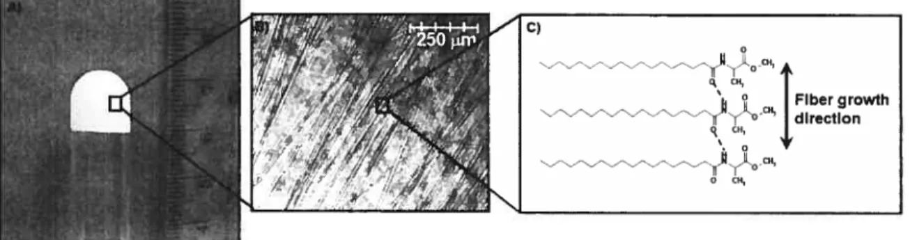
Figure 3: A) Photograph depicting the opaque N-stearoyl-L-alanine methyl ester organogel; B) optical micrograph showing the fibrous aggregates responsible for gelation; C) molecular packing within fibers.
25
figure 4: A) optical micrograph of a (4%) hexatriacontane (C36) organogel in octanol viewed tbrough crossed polars; B) a cartoon representation of the microplatelets
xii
observed in A); C) depiction of the lamellar orthorhombic molecular packing inside the platelets, showing the directions ofmicroplatelet growth.
25
Figure 5: A) Transmission electron micrograph of organogel of a crown ether
phthalocyanine in chloroform, showing a left-handed cou. B) Schematic representation of helical fibers in A). C) The helical aggregates are formed by the stacking of crown ether rings with a staggering angle, constant in magnitude and direction. D) Supercoiled structure is obtained from side-on aggregation of individual fibers.
29
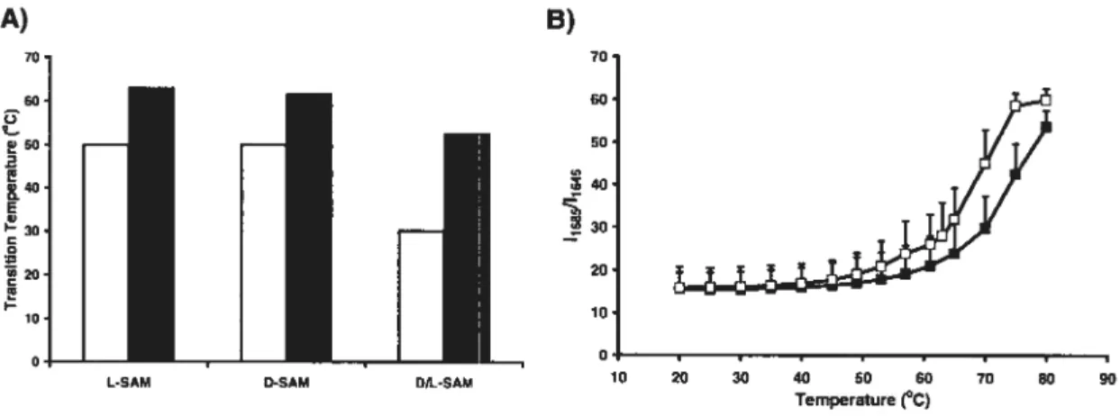
Figure 6: A) Gelator chirality effect showing a decrease in DSC-determined sol-gel
(white bars) and gel-sol (solid bars) transitions in racemic organogels (N-stearoyl D/L alanine methyl ester, D/L-SAM) with respect to enantiomerically-pure L- and D-SAM organogels, respectiveÏy (mean, n = 2). B) FuR analysis showing the proportion of free
gelator amide bonds in gel systems. as deterrnined from the band intensity ratio of amide I peaks at 1685 and 1648 cm1 (11685/1618), as a function of temperature.
Enantiomerically pure L-SAM (.) organogels showed higher gel-sol transition temperatures than D/L-SAM (o) organogels (mean± SD, n= 3).
30
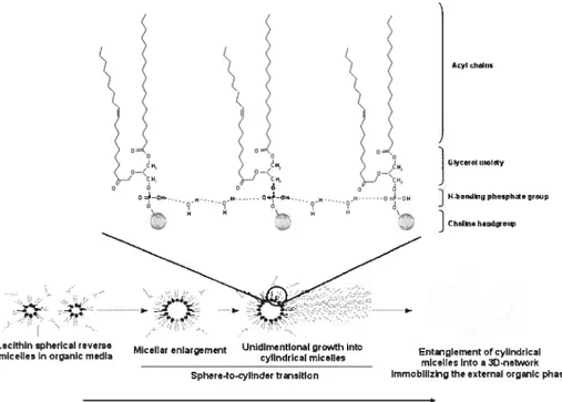
Figure 7: Formation of a three-dimensionat network of reverse cylindrical micelles in
lecithin organogel, involving hydrogen bonding between lecithin and polar solvent molecules.
32
Figure 8: Plot ofzero shear viscosityversus the water-to-lecithin molecular ratio (w0).
Dotted lines roughly indicate boundaries between various phase regions.
xiii
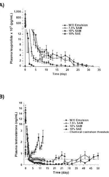
Figure 9: A) Plasma concentrations of leuprolide after the subcutaneous administration of a control w/o emulsion (squares) and various organogel formulations (circles and triangles; SAM and SAE: N-stearoyl methyl or ethyl ester. respectively). B) Plasma concentrations of testosterone after the administration of the formulations in a). The dotted une represents the chemical castration threshold (mean± SEM.
n=5-6).
4$
CHAPTER 4
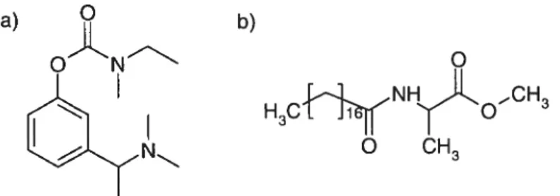
Figure 1: Molecular structures of a) rivastigmine base and b) N-stearoyl L-alanine methyl ester (SAM).
61
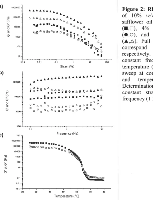
Figure 2: Rheology experiments of 10%
i’/w
SAM oleogel in safflower oil containing no drug (•,U), 4%w/w
dissolved RB (•.O), and 5% dispersed RHT (À,A). Full and empty symbols correspond to G’ and G”, respectively. a) Strain sweep at constant frequency (1 Hz) and temperature (25°C); b) Frequency sweep at constant strain (0.01%) and temperature (25°C): e) Determination of the TGS (I,D)at constant strain (0.005 %)and frequency (1 Hz).
69
Figure 3: DSC analyses showing gel-sol transitions for drug-free gels (n), for gels containing 5%
(w/w)
dispersed RHT(.),
and for gels containing RB dissolved at 4 (A), 20 (o),and 40%(w/w)
(K). Curves are offset on the y-axis for improved clarity. AH and TGS values were calculated from the area under the curve and the temperature corresponding to the peak transition, respectively.71
Figure 4: Thermal evolution, as probed by f TIR spectroscopy. The intensity ratio of the amide I peak components (11685/11648) was monitored as an indication of H-bond
xiv
disruption between SAM molecules during gel heating from 20 to 75 °C. Airows indicate changes from the gel (low temperatures) to the Iiquid (high temperatures) state.
73
Figure 5: fuR analysis showing the proportion of free amide bonds in drug-loaded gels. as deterrnined from the hand intensity ratio of amide I peaks at 1685 and 1642 cm
(11685/1164$), as a function of temperature for gels containing dissolved RB at different
concentrations: O (ri), 4 (A) and 40% (w/w) (o) (Mean± SD. n 3).
74
Figure 6: a) An inverted tube containing 10% (w/w) SAM gel is shown at room temperature to demonstrate the cohesiveness of the system. The inset shows an optical microscopy image ofthe oleogel network. ESEM images ofa 10% (w/w) SAM b) drug free oleogel and c) oleogel containing dissolved-RB (5% w/u’). Finally. an ESEM image is shown of d) a dried formulation, obtained after complete solvent evaporation from an organogel of 10% SAM in toluene.
76
Figure 7: In vitro release experiments for formulations varying in SAM and NMP content as well as in drug incorporation method (dissolution vs. dispersion). A control RHI solution in PBS (X) was tested. Release experiments from formulations containing dissolved RB are represented by solid symbols: ou formulation (.), 10% SAM gel with (.) and without NMP (Â); release experiments from gels containing dispersed RHT are represented by hollow symbols: ou formulation (o) and 10% SAM gel with (o) and without NMP (A) (Mean± SD. n = 3).
xv
Figure 8: Blood concentration of rivastigmine afler the s.c. administration ofa 10 mg/kg dose of saline RHT solution (À), and 1$ mg/kg doses of RHT dispersed in ou
(•),
in 5% (A), and in 10%(w/w)
SAM gel (n). (Mean + SEM, n 6) SEM rather than SDvalues were used for the error bars in order to reduce clutter and improve the clarity. $0
CHAPTER5
Figure 1: Average hardness of oleogels varying in SAM concentration, obtained by texture profile analysis (Mean+ SD, n= 3).
88
Figure 2: Gelator chirality effect showing a decrease in DSC-determined sol-gel (white bars) and gel-sol (solid bars) transitions in racemic organogels (D/L-SAM) with respect to enantiomerically-pure L- and D-SAM. (Mean, n = 2).
$9
Figure 3: FuR analysis showing the proportion of free amide bonds in drug-free gels, as determined from the band intensity ratio of amide I peaks at 1685 and 164$ cmt
(11685/1164s), as a function of temperature for gels prepared from 10%
w/w
enantiomerically pure L-SAM
(.)
and racemic D/L-SAM (o), respectively. (Mean± SD,n=3)
xvi
Lïst of abbreviations
AD Alzheirner’s disease
ANOVA analysis of variance
BSA bovine serum alburnin
ChEI cholinesterase inhibitor
D SC differential scanning calorimetry
FDA food and Drug Administration
FTIR fourier-transform infrared (spectroscopy)
HA haemagglutin
LMW low molecular weight
LO lecithin organogel
G’ storage modulus
G” loss modulus
NMDA N-methyl-D-aspartate
NMP nuclear magnetic resonance
NSAID non-steroidal anti-inflammatory drug
P(MAA-co-MAA) poly(methacrylic acid-co-methyl methacrylate)
PEO poly(ethyl oxide)
PLA poly(lactic acid)
PLGA poly(lactic acid-co-glycolic acid)
P0 poly(ethylene) organogel
RB rivastigmine base
RHT rivastigmine hydrogen tartrate
SAM N-stearoyl (L-)alanine methyl ester
SC stratum comeum
SMS sorbitan monostearate
‘GS gel-sol transition temperature
TSG sol-gel transition temperature
w/o water-in-oil
w0 polar solvent-to-lecithin ratio
xviii
Acknowledgements
I thank my research supervisor Dr. Jean-Christophe Leroux for the opportunity (and challenge!) of working in his lab as well as the continued guidance provided throughout the project (especially on the review!). Overali, the past two-something years have been rich in learning, both on professional and personal levels (my understanding of stamina was pushed to new levels). I thank my committee members, Dr. Françoise Winnik and Dr. Didier Hoarau, for their guidance in the bettering of this manuscript. I thank past and present members of the lab. particularly Aude Motulsky, David Ghattas, Anand Dhanikula, Roya Fattahie, Marie-Christine Jones, Marie-Hélène Dufrèsne, and Guillaume Bastiat, for their general help and the many constructive discussions shared. For backing me with their ftiendship and advice, my very heartfelt thanks go out to Steven, Irina, Luay, Marie-Eve, Nicolas, and François (my only human partner in the long hours spent with rats). I thank Jean-François for the warmth, love, and patience that make it all seem easy. And last (but n....) I would like to thank my family, Mihaela, Dan, and Razvan, for the unconditional love and support that give me courage to face anything (even grad studies).
Secrétariat du CDEA Division des animaleries Pavillon Roger-Gaudry
Renseignements complémentaires au renouvellement autorisé
Protocole 07-099 - Autorisé
Titre : Ri—Pharmacocinétique. biodistribution et efficcwité d’iminunoÏiposomes sensibles au pH
pottr la chimiothérapie de la leucémie myéloîde algue.
Directeur de recherche : Dr. Jean-Christophe Leroux
SECTION 9.1:
Seulement 2 souris ont étés utilisés l’année précédente, donc aucune justification sur la justesse des points limites ne peut être rapportée pour le moment.
SECTION 9.2:
A partir du premier jour d’inoculation des tumeurs, les animaux seront observés attentivement 2 fois par jour afin d’évaluer tout signe de douleur, de stress ou d’inconfort (i.e. réduction de l’activité, apparence voûtée, poil ébouriffé et yeux fermés, détresse respiratoire, etc). Les souris seront pesées au moins tous les 2 jours pour évaluer une perte de poids. Si la perte de poids est de plus de 15 à 20 %, les animaux seront sacrifiés. La personne responsable de ce protocole animal est Pierre Simard (étudiant au doctorat) et ce dernier s’assurera de remplir le registre d’utilisation des animaux.
CHAPTER 1
Alzheimer’s disease
2
1. Introduction
Alzheimer’s disease (AD) is the most common form of dementia amongst elderly and is estimated to afflict 1$ million people worldwide [1]. With an increasingly aging population, the prevalence of AD is on the rise. The disease takes an enormous toll on society, both at financial and social levels. Indeed, about $100 billion dollars in caring costs are spent annually in the United States alone. Meanwhile, with around 70% of patients living at home, great physical and emotional stress is placed on caregivers [1]. AD is a progressive neurotogical condition, evolving from short-term memory impairment to profound cognitive disturbances (such as severe memory loss, leaming difficulty, and language disorder), behavioural disturbances (such as aggression, depression, and wandering), as well as physical disability [2]. After diagnostic, patients usually survive 7 to 10 years, and typically die from bronchitis or pneumonia [3].
2. Pathophysiology
The disease is characterized, as determined from autopsies, by the extracellular deposition of f3-amyloid protein in senile plaques and the intracellular formation of neurofibrillary tangles. The process is thought to be initiated by the production and accumulation of the neurotoxic 13-amyloid peptide, the main component of 3-amyloid plaques. This leads to the formation of neurofibrillary tangles, oxidation, glutametergic excitotoxicity, inflammation, and finally an activation of neuronal apoptosis (Figure 1) [4].
3
t
7 •4 I Ere / ÀEip A gnrtoe1 OodtGn4
EXdtoto1Cit AaggetIon ffmmatn Ti hyperphc4sphory tion
Snile p1dquw4hmiwhal
Cugnitie and behavicral
Figure 1: The hypothesis of the amyloid cascade proposes a progression from the generation of 3-amy1oid precursor protein (APP), through multiple secondary steps, to ceil death. Reproduced from reference [4].
4
3. Treatment options
The celi dysfunction and death brought about in several neurotransmitter systems lead to deficits in acetylcholine, norepinephrine, and serotonin. This hypothesis underlies current pharmacological approaches to the disease (Table I). Amongst these, two classes of drugs are presently on the market, targeting the cholinergic and glutamate systems, respectively. Cholinesterase inhibitors (ChEI) increase acetylchoÏine levels at synapses by preventing its breakdown, thus prolonging neurotransmission in remaining cholinergic neurons [5j, while N-methyl-D-aspartate (NMDA) antagonists are believed to work by regulating levels of glutamate in the central nervous system.
3.1. Cholinesterase inhibitors
Since 1997, three second-generation ChEI have been approved by the f ood and Drug Administration (f DA) for treatment of mild to moderate AD: donepezil, rivastigmine, and galantamine (Table I). These agents are currently the first une of symptomatic treatment. Clinical trials have suggested that ChEI may help improve cognitive ability of AD patients. Nevertheless, the overati efficacy of these drugs is generally considered to be modest [4]. Similarly, their slowing of disease progression is controversial
[1].
ChEI drugs are generally well tolerated, with possible side-effects of gastrointestinal nature, such as nausea and anorexia. While the three active agents have similar efficacy and tolerability profiles, rivastigmine is the only marketed ChEI that has its effect targeted on the cortex and hippocampus, the areas most affected by the disease.5
Table I: Current drugs on the market or in clinical trials for the treatment of AD; adapted from reference
jlJ.
Drug Company Mechanism Status
donepezil approved for mild
-EisaiiPfizer ChEI
(Aricept) moderate AD
rivastigmine approved for mild
-Novartis ChEJ
(Exelon ) moderate AD
galantamine approved for mild
-® Johnson & Johnson ChEI
(Reminyl /Razadyne ) moderate AD
memantine approved for moderate
-Lundbeck/Forest NMDA receptor antagonist
(Namenda IEbixa ) severe AD
tramiprosate
(Alzhemed®) Neurochem amyloid plaque inhibition Phase 3
R-fiurbiprofen NSAID’ lowers j3-amyloid
Myriad Genetics Phase 3
(Flurizan ) levels
leuprolide acetate Voyager
Implantable leuprolide Phase 3
(Mernryte ) Pharmaceuticals
neramexane Merz & Co NMDA receptor antagonist Phase 3
xaliproden Sanofi-Aventis 5-HT la agonist Phase 3
lecozotan Wyeth 5-HT la agonist Phase 3
Targacept/ nicotinic acetylcholine
ispronicline Phase 2
AstraZeneca agonist
3-amyloid-speciflc
bapineuzumab Wyeth/Elan Phase 2
monoclonal antibody ‘NSAID: non-steroidal anti-inflammatory drugs
Also, as opposed to donepezil and galantamine. rivastigmine presents a tow risk for tolerance caused by enzyme induction and/or upregulation. Likewise, it shows a very low potential for drug-drug interactions and accumulation in the body, even with long term use [6]. Nevertheless, donepezil, the first second-generation Chu to enter the
6
market, has remained the leading product in its class. Antioxidants such as vitamin E are often recommended in conjunction with ChEI up into the late stages of the disease, but this practice is controversial due to the Yack of supporting clinical data [1].
3.2. Other therapies
While ChEI are only recommended up to moderate disease stages and have littie or no impact on disease progression, the NMDA antagonist memantine, approved by the FDA in 2003, has transformed the treatment of moderate to severe AD. This active agent is thought to interfere with glutamergic excitotoxicity and provide symptomatic improvement through effects on the function of hippocampal neurons [4]. Clinical trials have shown an improvement in cognitive function of memantine- over placebo-treated patients, although as for the other current therapies, improvements remain modest [4]. The potential for combination therapy has been explored, and whiÏe some studies have shown encouraging preliminary data, suggesting that treatment with memantine in addition to donepezil is superior to donepezil alone [7], others have concluded that no significant improvement was achieved [8].
While current therapies show modest symptomatic improvements in AD patients, the underlying degeneration of brain celis and consequent disease progression generally remain unaffected by these therapies. Consequently, ongoing research and clinical trials explore new treatment hypotheses (Table I). Several of these agents are aimed at countering amyloid plaque deposition, formation of neurofibrillary tangles, and neuroinflammation. It is argued that the most promising of these avenues is the targeting of the 3—amyloid peptide [1]. The most advanced drug in this class is tramiprosate,
7
which binds soluble 3—amyloid, preventing its aggregation into amyloid plaques. Clinical trials revealed the compound to be of moderate efflcacy in reducing f3—amyloid plaques [9,10]. Another interesting pharmacological approach is represented by bapineuzumab, a humanized monoclonal antibody against 3—amyloid, currently studied in Phase 2 trials. Immunotherapy leaves room for hope of a truly disease-modifying approach; however it also entails the greatest risks. Earlier drug candidates have caused cases of encephalitis [1]. It remains to be seen if any of the drugs currently under investigation will provide disease-modifying mechanisms and be safe enough for market release.
Given current pharmacological options for AD patients, a possible way of facilitating disease management could be the administration of sustained release formulations. One example such example bas been the commercialization of the oral once-daily formulation of galantamine (Razadyne®), an improvement over the previous twice-daily regimen. Simplified dosing schedules could possibly entail increased treatment adherence and compliance, while alleviating responsibility and workload for caregivers. Peak and trough plasma concentrations of the active agent are also potentially minimized, which can decrease side effects.
$
References
[1] C. Mount, C. Downton. Alzheimer disease: progress or profit?, Nat Mcd 12 (2006) 780-784.
[2] J.L. Cumrnings, G. Cole, Alzheimer’s Disease, JAMA 287 (2002) 2335-2338. [3] R. Brookmeyer, M.M. Corrada, F.C. Curriero, et al., Survival following a
diagnostic ofAlzheimer disease, Archives ofNeurology 59 (2002) 1764-1767. [4] J.L. Cummings, Alzheime?s disease, N Engi J Med 351 (2004) 56-67.
[5] A. Lleo, S.M. Greenberg, J.H. Growdon, Current pharmacotherapy for Alzheimer’s disease, Ann Rev Med 57 (2006) 5 13-533.
[6] f. Inglis, The tolerabiÏity and safety of cholinesterase inhibitors in the treatment ofdementia, Int J Clin Pract Suppi (2002) 45-63.
[7] P.N. Tariot, H.J. federoff, Current treatment for Alzheimer disease and future prospects, Alzheimer Dis Assoc Disord 17 Suppi 4 (2003) S105-1 13.
[8] A. Enz, C. Gentsch, Co-administration ofmemantine has no effect on the in vitro or ex vivo determined acetylcholinesterase inhibition of rivastigmine in the rat brain, Neuropharmacology 47 (2004) 408-413.
[9] S.M. Greenberg, J. Rosand, A.T. Schueider, et aï., A phase 2 study of tramiprosate for cerebral amyloid angiopathy, Alzheimer Dis Assoc Disord 20 (2006) 269-274.
[10] P.S. Aisen, D. Saumier, R. Briand, et al., A Phase II study targeting amyloid-beta with 3APS in mild-to-moderate Alzheimer disease, Neurology 67 (2006)
CHAPTER 2
rn
Parenteral sustained release systems act as reservoirs for the incorporated active agent, releasing the latter over time spans ranging between hours and months. These systems are generally administered via the peritoneal, subcutaneous, or intramuscular routes, and can be used for systemic or local treatment.
Sustained release systems gained popularity since the early eighties when the first polymeric implants were commercialized for the delivery of goserelin (Zoladex®) and levonorgestrel (Norplant®), for prostate cancer treatment and contraception, respectively [1]. Microspheres have been developed shortly afier for sustained release applications. These systems are constituted of polymeric particles, most commonly polyesters such as poly(lactic acid) (PLA) and poly(lactic acid-co-glycolic acid) (PLGA), which are injected subcutaneously or intramuscularly. Microsphere depot formulations containing leuprolide (Lupron®) were extensively studied t21 and finally marketed for the long-term release of the active agent with a consequent suppression of testosterone for up to 6 months. A number of studies have investigated the encapsulation of ChEI into biodegradable microspheres of PLA and PLGA [3-6]. These studies showed the potential of drug delivery over prolonged periods. Unfortunately, neither of the model drugs used (huperzine A and tacrine) is commonly used in AD management due to lack of FDA approval and hepatic toxicity, respectively.
Overall, microsphere systems are interesting since many are recognized as being biodegradable and biocompatible [7]. However, important shortcomings of these systems limit their use in drug delivery, notably the complexity of preparation for stable and
‘I
sterile formulations and the potential degradation of the active agent during this process. The possibility of microsphere migration away from the injection site can be of nuisance when local treatment is needed. More importantly yet, the formulation cannot be removed in the case of toxic effects following administration. Accordingly, in recent years, such systems have yielded the spotlight to in situ-forming systems allowing sustained drug release.
In situ-forming implants are liquid systems which solidify upon injection, as a resuit of changes in temperature and/or pH, or solvent diffusion [1]. Based on the specific solidification mechanisms, various categories of implants exist, notably thermoplastic pastes, precipitated polymers, hydrogels, and organogels. Each system will be briefly discussed in this section, while organogels will be presented in detail in Chapter 3.
Thermoplastic pastes are composed of polymers which are injected in the molten state and solidify upon injection as a result of temperature drop. These polymers have a melting point between 25 and 65°C and a glass transition temperature below the physiological temperature [8,9]. The latter characteristic is important since the system’s viscosity has to present a compromise; while being high enough to provide an adequate rate of release, it must be low enough to permit easy injection through conventional needles [9]. Typical polymers used are composed of lactic and glycolic acids, E
caprolactone, and orthoesters. Poly(ethylene oxide) (PEO) and albumin can be incorporated to provide accelerated release of the active agent. The inherent disadvantage of these systems is the relatively high temperature at which they must be injected. This
12
can trigger pain and possible necrosis at the administration site. The consequent formation of fibrous tissue around the implant may lead to erratic or insufficient release.
In situ-forming systems can also be produced by chemical or physical crosslinking of polymer chains, yielding solid three-dimensional matrices capable of offering a delaying effect on drug release. The main concem with such systems is the high burst release upon injection, produced by the delay in implant solidification [10]. This is greatly limiting for long-term sustained release applications. Also, polymer crosslinking subsequent to drug incorporation to the formulation involves the risk of chemical modification of the latter and ensuing loss oftherapeutic efficacy. Certain formulations have been developed which circumvent this problem, as is the case of a thiol-derivatized poly(ethylene oxide) system which circumvents the need for radical polymerization by allowing crosslinking at the thiol group via a linker [111. However, this formulation is not biodegradable. finally, another important shortcoming of crosslinked polymer systems is the inherent toxicity of initiators.
As a consequence to the problems associated with chemical crosslinking, polymeric systems relying on solidification triggered by physical interactions seem to be a more reasonable choice for pharmaceutical applications. These systems rely on hydrophobic and/or electrostatic interactions to form the solid systems. The constituting block copolymers contain hydrophilic and hydrophobic parts, which upon a play on temperature and/or pH induce a solidification of the system as a resuit of altered differential solubilities of the polymer moieties. for example, various such systems are
13
soluble in cofd aqueous media but form a gel upon an increase in temperature. This phenomenon is the resuit of dehydration of the hydrophobic chain. a consequent loss of solubility in water, and a formation of colloidal systems such as micelles. If colloid density exceeds a given threshold, typically encountered at polymer concentrations of 15-23%, phase mixing occurs between the hydrophilic micelle coronas and the hydrophobic cores, leading to chain entanglement and consequent gelation. [8]. Such polyrners most commonly include poloxamers and ABA-type polymers with PEO as the hydrophilic (A) and poly(propylene oxide) as the hydrophobic (B) moiety [12-14]. The major drawback of these systems is the fast release of small hydrophilic molecules through the large aqueous channels formed in the porous implant. furthermore, poloxamer gels are flot biodegradable and have been found to cause metabolic changes in rats t15]. Other
thermosensitive di- and tri-bloc copolymers composed of PEO and PLA, PLGA. and/or poly(E-caprolactone) have also been developed in efforts to transcend the problems associated with poloxamer gels [16,17]. These systems were shown to provide adequate release for model drugs, ranging from days to several weeks, for increasingly hydrophobic molecules. A change in pH can also trigger the physicat crosslinking of polymers [18,19]. Such systems are mostly used for oral formulations, given the high variations inpi-Ialong the gastrointestinal tract.
Finally. the precipitation of compounds as a result of temperature changes or in response to solvent diffusion is also a mechanism exploited for in situ-forming systems. An example is Atrigel, composed of PLA, PLGA, or poly(lactic acid-co-E-caprolactone) dissolved in N-methylpyrrolidone (NMP). Encouraging animal studies using this system have lead to the commercialization of Eligard’, a formulation providing therapeutic
14
plasma concentrations of leuprolide in humans for 3 to 6 months [20]. This constituted the first in situ-forming implant approved for use in humans. SABER® is another system ensuring sustained drug release following in silu-precipitation. This formulation is composed of sucrose acetate iso-butyrate, a water-insoluble compound, dissolved in ethanol at concentrations of 80-90% [21]. Injection of the low-viscosity system is followed by solvent diffusion into sunounding tissue and formation of a solid implant capable of providing sustained release of active agents, including proteins, for up to several weeks [21,22].
Organogels, composed ofa liquid organic medium immobilized by gelling molecules, are particularly interesting as a drug delivery platform, owing to the simplicity of their preparation. These systems will be discussed in detail in chapter 3, while chapter 4 will focus on the use of an organogel system for the sustained delivery of rivastigmine.
15
References
[1] A.J. Tipton, R.L. Dunn, In situ gelling systems, in: J. Senior, M. Radomsky (Eds.), Sustained-release injectable products, Interpharm Press, Englewood, 2000, pp. 241-27$.
[2] B.H. Woo, K.H. Na, B.A. Dani, et al., In vitro characterization and in vivo testosterone suppression of 6-month release poly(D,L-lactide) leuprolide microspheres, Pharm Res 19 (2002) 546-550.
[3] P. Gao, P. Ding, H. Xu, et al., In vitro and in vivo characterization of huperzine A loaded microspheres made from end-group uncapped poly(d,l-lactide acid) and poly(d,l-Iactide-co-glycolide acid), Chem Pharrn BuIl 54 (2006) 89-93.
[4] X. fu,
Q.
Ping, Y. Gao, Effects of formulation factors on encapsulation efficiency and release behaviour in vitro of huperzine A-PLGA microspheres, J Microencapsul 22 (2005) 705-7 14.[5] W.H. Liu, J.L. Song, K. Liu, et aï., Preparation and in vitro and in vivo release studies of huperzine A loaded microspheres for the treatment of Alzheimer’s disease, J Control Release 107 (2005) 4 17-427.
[6]
Q.
Yang, D. Williams, G. Owusu-Ababio, et aï., Controlled release tacrine delivery system for the treatment ofAlzheimer’s disease, Drug Dcliv 8 (2001) 93-98.[7] J.M. Anderson, M.S. Shive, Biodegradation and biocompatibility of PLA and PLGA microspheres, Adv Drug Deliv Rev 2$ (1997) 5-24.
[8] C.B. Packhaeuser, J. Scimieders, C.G. Oster, et al., Insitu forming parenteral drug delivery systems: an overview, Eur J Pharm Biopharm 5$ (2004) 445-45 5.
[9] A. Hatefi, B. Amsden, Biodegradable injectable in sittà’ forming drug delivery systems, J Control Release $0 (2002) 9-28.
[10] A. Motulsky, Caractérisation d’un organogel à base d’un dérivé amphiphile de la L-alanine, faculty ofpharmacy, University ofMontreal, 2005.
[11] Y. Qiu, K. Park, Environment-sensitive hydrogels for drug delivery, Adv Drug Dcliv Rev 53 (2001) 32 1-339.
[12] M. Katakam, W.R. Ravis, A.K. Banga, Controlled release of human growth hormone in rats following parenteral administration of poloxamer gels., J. Control. Release 49 (1997) 2 1-26.
[13] M.L. Veyries, G. Couarraze, S. Geiger, et al., Controlled release of vancomycin from poloxamer 407 gels, Int J Pharm 192 (1999) 183-193.
[14] J.G. Wenzel, K.S. Balaji, K. Koushik, et aï., Pluronic f127 gel formulations of deslorelin and GnRH reduce drug degradation and sustain drug release and effect in cattie, J Control Release 85 (2002) 5 1-59.
[15] K.M. Wasan, R. Subramanian, M. Kwong, et al., Poloxamer 407-mediated alterations in the activities of enzymes regulating lipid metabolism in rats, J Pharm Sci 6 (2003) 189-197.
[16] B. Jeong, Y.H. Bae, S.W. Kim, Drug release from biodegradable injectable thermosensitive hydrogel of PEG-PLGA-PEG triblock copolymers, J Control Release 63 (2000) 155-163.
[17] T. Kissel, Y. Li, f. Unger, ABA-triblock copolymers from biodegradable polyester A-blocks and hydrophilic poly(ethylene oxide) B-blocks as a candidate
16
for in situ forming hydrogel delivery systems for proteins, Adv Drug DeÏiv Rev 54 (2002) 99-134.
[18) 0. Sipahigil, A. Gursoy, f. Cakalagaoglu, et aï., Release behaviour and biocompatibility of drug-Ioaded pH sensitive particles, Int J Pharm 311(2006)
130-138.
[19] A. Kumar. S.S. Lahiri. H. Singh, Development of PEGDMA: MAA based hydrogel microparticles for oral insulin dcliv ery. Int J Pharm 323 (2006) 117-124. [20] 0. Sartor, Eligard: leuprolide acetate in a novel sustained-release delivery system,
Urology 61(2003)25-31.
[21] F.W. Okumu, N. Dao le, P.J. fielder, et al., Sustained delivery of human growth hormone from a nove! gel system: SABER, Biomaterials 23 (2002) 4353-4358. [22] S. Pechenov, B. Shenoy, M.X. Yang, et al., Injectable controlled release
CHAPTER 3
1$ 1. Abstract
Organogels are semi-solid systems, in which an organic Iiquid phase is immobitized by a three-dirnensional network composed of self-assembled, intertwined gelator fibers. Despite their majoritarily liquid composition, these systems demonstrate the appearance and rheological behaviour of solids. Investigative research pertaining to organogels has only picked up speed in the last few decades. Consequentty, rnany burning questions, such as the specific molecular requirernents guaranteeing gelation, stiil await definite answers. Nonetheless, the application of organogels to various areas of interest has been quick to follow their discoveries. Unfortunately, their use in drug delivery is stili quite limited by the scarce toxicology information available on organogelators, as well as by the few pharmaceutically-accepted solvents used in gel systems. This review aims at providing a global view of organogels, with special emphasis on the interplay between the gelators structural characteristics and the ensuing intermolecular interactions. A subsequent focus is placed on the application of organogels as drug delivery platforms for active agent administration vici diverse routes such as transderrnal, oral, and
19
2. Introduction
for the past few decades, gels have been presented. to the extent of a cliché, as being materials “easier to recognize than define”, a prophetic statement pioneered in the 192OEs by Lloyd [1]. Various definitions have followed. sometimes a same author providing descriptions ranging from the most elaborate, stating that a gel 1) bas a continuous structure of macroscopic dimensions that are permanent over the time-span of an experiment and 2) is solid-like in its rheological behaviour, to the more basic descriptions stating that if it looks like Jell-O”, it must be a gel [2]. It is now generally accepted that a gel is a semi-solid material composed of low concentrations (< 15%) of gelator molecules that, in presence of an appropriate solvent, self-assemble via physical or chemical interactions into an extensive mesh network preventing solvent ftow as a result of surface tension. Gels have been eloquently described as being the result of “crystallization gone awry [3]. Indeed, macroscopic phase separation into crystalline and liquid layers is avoided in these systems owing to the balance between gelator aggregating forces and solubilizing solvent-aggregate interactions. The result is a system ofcrystallized fibers that immobilize the liquid phase.
The specific process leading to the formation of the gelling matrix depends on the physicochemical properties of gel components and their resulting interactions. Figure I presents a fiow-chart compiling various accepted classifications of gels based on the nature of solvents, gelators, and intermolecular interactions.
20
Organogels, the focus of this review, can be distinguished from hydrogels by their predominanfly organic continuous phase and can then be further subdivided based on the nature of the gelling molecule: polymeric or low molecular weight (LMW) organogelators. Polymers immobilize the organic solvent by forming a network of either cross-linked or entangled chains for chemical and physical gels, respectively. The latter are possibly further stabilized by weak inter-chain interactions such as hydrogen bonding. van der Waals forces, and it-stacking. Likewise, the self-assembly of LMW organogelators depends on physical interactions for the formation of aggregates sufficiently long to overlap and induce soïvent gelation. Depending on the kinetic Figure 1: Organogel classification.
21 properties of aggregates, an important distinction amongst LMW organogels is made between those composed ofsolid (or strong) versus fluid (or weak) fiber networks.
Despite the numerous trends in gelling processes as well as the impressive variety of gelators identified [4], it remains difficuit to predict the molecular structure of a potentiat gelator, as well as one cannot readily foresee preferentially-gelled solvents. Today stiil, the discovery of gelators rernains serendipitous and is usually followed by investigative screening of different solvent systems potentiatly compatible with gelation. Prediction of gelation potential of a given molecule might seem possible by investigation of its propensity towards chemical or physical intermolecular interactions; however, no generalisations are so far possible. Many factors such as steric effects, rigidity, and polarity can counter the molecule’s aggregating tendency. Control over the gelation process as well as the systematic conception of new gelling molecules remain important challenges to face in the quest of new organogelators.
In the pharmaceutical field, organogels can be used for drug and vaccine delivery via
different administration routes, although relatively few such formulations have been investigated [5]. This review aims in its flrst part at providing a global view of the different existing organogelator categories while secondly providing a more focused discussion oftheir drug delivery applications.
22 3. Organogel properties
3.1. Low molecular weight organogelators
Amongst LMW organogels, a subtie but crucial distinction is made between those composed of entangled networks of solidversus fluid fibers (figure 2) [3].
A)
B)Figure 2: Solid-matrix (strong) versus fluid-matrix (weak) organogels. A) Solid-matrix gels are more robust due to their permanent solid-like networks in which the junction points are relatively large (pseudo)crystalline microdomains (circled area). B) f luid matrix gels have transient networks in which junctions points are most ofien simple chain entanglements. Additional kinetic features such as dynamic exchange of gelator molecules with the bulk liquid as well as chain breaking/recombination (arrows) may occur. Adapted with permission from reference [3].
The solid fibers, out of which most organogels are composed, are generally produced following a drop in temperature below the gelator’s solubility limit [61. Consequently, a fast partial precipitation of gelator molecules in the organic medium resuits in the formation of aggregates via cooperative intermolecular interactions (f igure 2A) [7]. On the other hand, fluid matrices are formed upon the incorporation of polar solvents to organic solutions of surfactants, which resuits in the reorganisation of surfactant molecules into mono- or bilayer cylindrical aggregates that immobilize the solvent (figure 23)
[71.
The key distinction between the two systems is the kinetic stability of23 the networks constituting the gel state. Strong gels are formed of permanent, most oflen crystalline, networks in which junction points are relatively large (pseudo)crystalline microdomains [3]. Conversely, weak gels are fonried of transient networks, characterized by the continuous breaking and recombination of the constituent rods, as in the case of reverse cylindrical micelles [8,9]. Similarly, aggregates undergo dynamic exchange of individual gelator molecules with the bulk liquid. Junction points in these fluid networks are simple chain entanglements, equally transient in nature.
The distinction between solid and fluid fibers is not much emphasized in the literature, although it is of great importance from a physicochemical stand point. Solid-matrix gels are more robust, as demonstrated by rheology studies [3]. This may be at least partially due to the fact that, while fluid fibers do not aggregate into higher-order structures, solid fibers are generally aligned in bundles as a result of their rigidity [7], likely conveying added robustness to the gel. Similarly, while molecular and supramolecular chirality plays a great role in the formation and stability of solid fibers, its effect is rare in fluid networks [6,7].
3.1.1. Solid matrix organogels
The vast rnajority of LMW organogelators discovered so far self assemble into solid networks when added to appropriate organic solvents.
3.1.1.1. Generat gelling considerations
In merely a century of organogel research, hundreds, if not thousands of LMW molecules with gelling properties have been discovered, most ofien by chance rather
24 than design. Several extensive reviews have been published on the topic in general [3,4], as well as on more pointed discussions: fiber formation mechanisms [10] and gelator families derived from various parent molecules such as fatty and amino acids [3], organometallic compounds [3], steroids [3,11], amide- or urea compounds [12], nucleotides [13], and dendrimers [14].
Solid-matrix gels are prepared by dissolving the gelator in the heated solvent, at concentrations typicalÏy inferior to 15%. Very low concentrations of less than 0.1% have been reported in the case of sugar-derived “supergelators” [15]. Upon cooling, the afflnity between organogelator and solvent molecules decreases and the former self assemble into solid aggregates held together by intermolecular physical interactions. The remaining solvent-aggregate affinity stabilizes the system by preventing complete phase separation. Aggregates are most often formed by the unidimentional growth into libers with high aspect (length-to-width) ratios, generally measuring a few tens of nanometers in width and up to several micrometers in length. One such example are L-alanine fatty acid derivatives which form opaque gels in pharmaceutical oils as a result of hydrogen bonding and van der Waals interactions [16,17] (Figure 3).
25
Although less common, examples exist of two-dimensional growth pattems, as in the case of hexatriacontane, a 36-carbon n-alkane (C36), which forms microplatelet arrangements (f igure 4).
C)
C /
B)
Figure 4: A) optical micrograph of a (4%) hexatriacontane (C36) organogel in octanot viewed through crossed polars; B) a cartoon representation of the microplatelets observed in A); C) depiction of the lamellar orthorhombic molecular packing inside the platelets, showing the directions of microplatetet growth. Adapted with permission from reference [19].
Figure 3: A) Photograph depicting the opaque N-stearoyl-L-alanine methyl ester organogel; B) optical micrograph showing the fibrous aggregates responsible for gelation; C) molecular packing within fibers. Adapted with permission from [1$].
Piatelet growth directions
26 Both one- and two-dirnentional aggregates are frequently crystalline in nature. The crystalline arrangement can be the same in the gel and the neat solid, as in the case of C36 molecules, of which the gel microplatelet arrangements are free of liquid molecules in the interlamellar spaces [19]. However. more ofien than flot, the crystalline packing differs between the gel and the neat solid [3]. Macroscopically, organogels range in appearance ftom white opaque to translucent, likely depending on aggregate size and the consequent gel’s abilïty to scatter incoming light. Sometimes, a sarne gel system can change in its degree of transparency upon small variations in composition [201.
While hydrophobic attractions are a major driving force for gelator aggregation in water, these interactions are of secondary importance in the case of organogels. In non-aqueous liquids, the attractive forces are mainly hydrogen-bonding, van der Waals interactions, rc-stacking. and metal-coordination bonds. Because of the strength and high directionality of their hydrogen-bonds, numerous emerging organogelators are derivatized peptides [4,12], sugars [15,21,22], and bis-urea-based compounds [12]. These are particularly eflicient organogelators because of their hydrogen-bonding core that provides a gelling scaffold which can be functionalized for extended versatility. Organogelators consisting of long n-alkanes (chain length varying from 24 to 36 carbon atoms) have proven to be of particular interest in dernonstrating the mechanisms of gelation [20]. They are not only rare examples in which hydrogen-bonding does not play a role in gel formation, but are even more unusual in that van der Waals forces alone lead to gelation. As a consequence of gelling solely through these weak physical interactions, such gels are not stable over long periods, eventually phase-separating due
27 to transitions towards thermodynamically-favoured packing arrangements. Not surprisingly, it was noted that gel sheif-life increased with gelator chain length as a resuit of extended van der Waals interactions, going from under a day to several months for C24 and C36, respectively [20].
A recent study involving a family of 3.5-diarninobenzoate derivatives dernonstrated, although flot for the first time, the implication and importance of aromatic stacking in the process of gelation [23]. Indeed, increasingly stronger gels were formed upon incorporation of additional aromatic substituents to gelator molecules. Another interesting class of gelators involving Tt—7t interactions are cholesterol-derivatized
molecutes. These can be very suitable for the design of functionalized organogelators because of their rernarkably high synthetic tunability [3.11]. The cholesterol group induces uni-directional self-association though van der Waals interactions, while functional groups added onto the cholesterol backbone stabilize the fiber via hydrogen bonding and/or 7t-lt interactions. The hydroxyl group at the C3 position of the
cholesterol molecule is crucial to gelation, likely due to its participation in hydrogen bonding [3]. Alternatively, steroids (S) derivatized at the C3 position with anthraquinone (A) via a linker (L) of varying length (ALS compounds) are known to forrn stable organogels. The fused aromatic rings of the anthraquinone group stabilize gel fibers by 7t-stacking.
2$ 3.1.1.2. Chirality Effi?cts
Chirality is neither necessary nor sufficient for gelation; however, despite flot being a gelling-force in itself, chirality seems to be intimately related to the growth and stability of the self-assembled fibrillar networks of LMW physical gels [3]. While this section strives at providing the reader with a general view of underlying principles of chirality and their impact on gelation, more extensive information can be found in a recent and excellent review by Brizard et al. [6].
Although the exact explanation remains yet to be formulated, a general empirical rule is that a molecule has a better chance of being a good gelator if it is chiral. Indeed, a large majority of existing organogelators possess at Ieast one stereogenic center, while non chiral gelators are generally cited as being exceptions to the rule [6]. Furthermore, it can be specified that chirality is only determinant in the case of solid fibers due to their marked rigidity, while rarely being of effect in fluid fibers which are highly dynamic in nature [7].
b further understand the stabilizing effect of chirality, it must first be said that it is known to play important roles both at the scale of individual molecules as well as that of resulting fibrillar aggregates. Indeed, molecular chirality is most ofien transferred to the morphology of self-assembled fibers, as shown by numerous studied systems [24-2$]. One such example are crown ether phthalocyanine organogels, composed of supercoiled helical fibers (Figure 5).
29
Figure 5: A) Transmission electron rnicrograph of organogel of a crown ether phthalocyanine in chloroform, showing a left-handed cou. B) Schematic representation of helical fibers in A). C) The helical aggregates are fornied by the stacking of crown ether rings with a staggering angle, constant in magnitude and direction. D) Supercoiled structure is obtained from side-on aggregation of individual fibers. Reproduced with permission from reference [27].
Initial molecular packing. driven by it-stacking between substituent aromatic rings, transfers molecular chirality to individual fibers, which further twist around each other into helical superstructures to maximize van der Waals interactions [27]. As opposed to flat aggregates, the contact area between such twisted structures is reduced due to their curvature. which makes them less prone to uncontrolled aggregation and to the resulting precipitation. This increases the chance of gelation by such chiral molecules. The opposite is generally true of racemic mixtures. These most often form flat aggregates that are more prone to uncontrolled crystallization [24,29].
30 N-stearoyl alanine methyl ester is an example of a compound showing superior gelling abilities when enantiomerically pure. Such gels exhibited higher transition temperatures than their racemic counterparts (Figure 6) [30].
A) B) 70 60 50 40 30 20 10
figure 6: A) Gelator chirality effect showing a decrease in DSC-determined sol-gel
(white bars) and gel-sol (solid bars) transitions in racemic organogels (N-stearoyl D/L alanine methyl ester, D/L-SAM) with respect to enantiomerically-pure L- and D-SAM organogels, respectively (mean, n = 2). B) FTIR analysis showing the proportion offree
gelator amide bonds in gel systems, as determined from the band intensity ratio of amide I peaks at 1685 and 164$ cm’ (116x5/11648), as a function of temperature.
Enantiomerically pure L-SAM (.) organogels showed higher gel-sol transition temperatures than D/L-SAM(E) organogels (mean+ SD. n =3) [30].
Meanwhile. racemic mixtures are often reported to form weaker gels, to crystallize [24], or to precipitate as flakes or pellets [6]. Nevertheless, a few examples have been reported in which racemates actually form stronger and/or more stable gels than their enantiomerically pure analogues, demonstrating that although racemates are often poorer
L-SAM D-SAM O/L-SAM 10 20 30 40 50 60 70 80 90 Temperature (°C)
31 3.1.2. f luid-matrix organogets
fluid fibers gel organic solvents in much the same way as solid fibers: aggregate size increases and the eventual entanglement of these structures immobilizes the solvent as a resuit of surface tension. Just as strong gels, fluid matrix systems are thermoreversible and can be transparent or opaque. The critical difference arises in the kinetic behaviour of the two types of matrices. Whule solid matrices have a robust and permanent morphology over the gel’s lifespan, fiuid matrices are transient structures in constant dynamic remodelling (f igure 2) [3]. Owing to the aggregate fiuidity and the transience ofjunction points, these structures are atso referred to as “worm-like” or “polymer-like” networks. This section presents two such systems, lecithin and sorbitan monostearate (SMS) (and/or sorbitan monopalmitate) organogels. which are both of very high interest in pharmaceutical sciences.
3.1.2.1. Leciihin organogels
From a drug delivery standpoint, lecithin organogels (LO) are very interesting systems, owing to their biocornpatibility, their amphiphilic nature, facilitating dissolution ofdrugs of various classes, as well as their permeation enhancement properties. Lecithin, or phosphatidylcholine (Table I), is the most abundant phospholipid in biological systems and is typically extracted from soy beans and egg yolk. Due to its amphiphilic structure, lecithin can assume many different forms such as mono- and bi-layer films. vesicles. liquid crystals, emulsions, and organogels [9]. When mixed to organic solvents, lecithin yields isotropic reverse-micellar solutions. Upon the addition of small amounts of polar solvents, cytindrical reverse micelles start to grow until they entangle into a gelling network (f igure 7).
32
Acyl cI,A*,
]
GeroI]
Ubo dbyj phoophA]
CI,oIiUa h.Lecithin sphericai reverse
Micellar enlargement Unidimentional growth into Entanglemerrtofcyiindrical micelles In organic media cylindrical micelles
micelles into a D-netwrk Sphereto-cylinder transition immobilizing the external organtc phase
Addition 0f polar solvent
Figure 7: formation of a three-dimensional network of reverse cylindrical micelles in
lecithin organogel, involving hydrogen bonding between lecithin and polar solvent molecules. Adapted with permission from reference [31].
Despite having been termed “weak” organogels, LO present very high viscosities, in several cases higher than that of gelatin [32]. Scartazzini et aÏ. [32] were the first to report a systematic investigation of LO, their resuits subsequently conflrmed by several groups. Indeed, evidence was presented suggesting that the rise in the systems’ viscosity was due to the growth and overlap of reverse tubular micelles [33-35] and flot to any form of liquid crystalline order [33]. as in the case ofbinary water-lecithin systems [9].
The hypothesis of entangled reverse micelles was proven by infrared spectroscopy studies showing a low-frequency shift of the PO vibration band for lecithin molecules upon gel formation, indicating the involvement of the phosphate group in H-bonding with the added polar solvent [34,36]. No indications were found of interactions at the
33 carbonyl groups and the glycerol residue of lecithin. Based on this evidence. a structural model was proposed, in which the lecithin phosphate group and polar solvent molecules are connected by H-bonds, thus forming a linear structure of alternating solvent and lecithin molecules, which ultimately self-assembles into overlapping worm-like reverse-micelles (Figure 7). Corroborating NMR studies [37-401 showed a une broadening for phosphorous and hydrogen resonances of the polar head-group upon an increasing molecular ratio of solvent-to-lecithin (w0). This suggested a progressive molecular stiffening of this part of the molecule upon polar solvent addition, consistent with the hypothesis ofinverted cylindrical micelle formation and entanglement.
Several polar solvents, many suitable for in vivo use, were found appropriate for induction of gelation. Glycerol provided maximal viscosity of the temary system at the lowest concentration (w0 = 1.7 - 1.9), followed by water (w0 = 3.6 - 3.8). formamide (w0
= 3.6 - 4.8), and ethylene glycol (w0> 5)
[341.
Other solvents such as ethyl alcohol anddiethylene glycol did flot induce organoget formation. In fact, it was suggested that the difference between gel-forming and non-gel-forming solvents was their orientation and localization between lecithin molecules, which in tum depended on their polarity [36]. Similariy, ail geiling-soivents were found to have a strong tendency towards hydrogen bonding, with hydrogen-bond donating potential seeming to be more important than hydrogen-bond acceptance. In terms of the hydrophobie organic solvents compatible with gel formation, Scartazzini et al. [32] concluded that the more apolar solvents such as alkanes, followed by cycloatkanes, allow a higher state of structural organization of the lecithin molecules. thus forming more stable gels.
34 Phase diagrams were obtained by severai groups, describing the variation of the lecithin system viscosity with the addition of increasing amounts of polar solvent. They rernained relatively constant for ail hydrocarbon solvents used [33,34,36] (figure g).
Organogel Two-phase Reverse- f ‘i. system: micellar
/
solution+solutIon / gel Two-phase
• / system: / solution+ I precîpitate
o.
I / > I--1. /I w - / —3 0 1 2 3 4 5 6 w0Figure 8: PLot ofzero shear viscosityversus the water-to-lecithin molecular ratio (w). Dotted unes roughly indicate boundaries between various phase regions. Adapted with permission from reference [36].
Siight variations occurred for certain other organic solvents [41]. but the evoiution ofthe lecithin system structure upon addition of polar solvent is a constant. The initial lecithin reverse-micelle solution aiways presents a sharp increase in viscosity upon the addition of criticai amounts of water, coinciding with organogel formation. f urther addition of water leads to a sharp decrease in viscosity, owing to the separation of a homogenous gel from the remaining low-viscosity fluid. Finaliy, the solidification ofthe separated gel into a non-transparent solid precipitate occctrs at even higher concentrations of polar soivent. More extensive physicochemical characterization can be found in the review by Shchipunov [42].
35 3.1.2.2. fatty acid derived organogels
Other biocompatible organogels extensively investigated in drug delivery are SMS (Span 60) and sorbitan monopalmitate (Span 40) organogels (Table I). Murdan et al. [43-461 were the flrst to report organic solvent gelation by these two compounds, both in the presence and absence of an aqueous phase. Anhydrous gels were obtained by dissolving low concentrations (i-10%) of the organogelator in aikanes (C>5), isopropyl myristate, and various vegetable oils at 60°C. Subsequent cooling of the system yielded white thermoreversible gels at room temperature. Alternatively, the drop-wise addition of an aqueous phase, in the form of either water or a suspension of niosomes (surfactant bilayer vesicles) to the hot organic surfactant solution yielded upon cooling a water-in ou (w/o) or a vesicle-in-water-in-oil (v/w/o) organogel system, respectively.
As with alt gelling mechanisms, the gelation point corresponds to decreased solvent gelator affinities resulting in a structural transition, in this case from the isotropic phase composed of reverse micelles to a system of entangled rod-shaped tubules that immobilizes the solvent [46,47]. Further organization of the amphiphiles inside the tubules was suggested to consist of concentric inverted bilayers, which as in the case of LO were found to be stabilized by hydrogen bonds between water and the amphiphiles’ polar heads [44,46,48].
The classification of these non-ionic surfactant organogels as either solid- or fluid matrix systems is less clear-cut than in the preceding cases. Although the bilayer arrangement within tubules intuitively suggests a certain fluidity and possible exchange of surfactant molecules with the surrounding bulk liquid, certain studies tend to point
36 away from the fluidity hypothesis. Indeed, when viewed under polarized light, the tubular aggregates exhibited crystallinity [48]. Nevertheless, X-ray diffraction measurements have shown the inverted bilayers to increase in width upon the addition of water, suggesting the accommodation of the aqueous phase between opposing polar groups of the amphiphilic bilayer [44] pointing towards the plasticity of the systems. A saturation point was reached, afier which excess water accumulated in separate droplets bound by surfactant film at the interface, followed by the eventual breakdown of the gel as aggregate integrity is substantially lost. The continuous modulation of the system to accommodate the polar solvent suggests a dominating fluid character for the constituting matrix.
3.2. Polymeric gelators
Polymeric gelators behave similarly to their LMW counterparts, solidifying organic solvents based on physical intermolecular interactions. Polymeric gels can vary from linear to star-shaped polymers. Three such polymeric systems with common or potential uses in drug delivery will be presented in this section.
Poly(ethylene) organogels (P0) are commonly used as ointrnent bases and are composed of 5% low molecular weight poly(ethylene) in mineral oil (Plastibase®) (Table I) [49-51]. The polymer is dissolved in the oil at about 130°C and ‘shock-cooled”. This leads to the partial precipitation of the polyrner chains and the formation of a colorless organogel [49-5 1]. Also of common application in pharmaceutics are copolymers of methacrylic acid (MAA) and methyl methacrylate (MMA) in 1:1 (Eudragit L®) and 1:2 (Eudragit S®) molar ratios (Table I). These can be used in the preparation of organogels
37
that have been evaluated as rectal sustained release preparations [52,53]. In the available studies, such gels typically consisted of a mode! drug dissolved in propylene glycol containing high concentrations of the gelling polymer (30 and 40 % for 1:1 and 1:2 P(MAA-co-MMA), respective!y). Basic drugs were found to weaken the gel structure more than acidic drugs, a phenomenon attributed to an increased disturbance of the hydrogen-bond interactions between polymer and propy!ene glycol molecules by the former.
Recent!y, Jones et al. [54] presented the preparation of star-shaped a!kylated poly(glycero! methacrylate) amphiphiles, capable of forming polymeric micelles in pharmaceutica!!y-acceptab!e apolar solvents such as ethyl oleate. It was found that organoge! formation occurred at high polymer concentrations (> 10 %) when the latter were derivatized with medium-length C12 and Ci4 alky! chains. On the other hand, gelation occurred at much lower concentrations
(
1 %) in the case of C18-derivatized polymers, showing the importance of intermolecular van der Waa!s interactions in the gelation mechanism. Hydrogcn bonding via the hydroxyl groups of the core polymers was suggested to be a driving force for gelation. The systems were shown to increase the so!ubility of hydrophilic compounds in oils, making them potentia!!y useful for the preparation of anhydrous peptide formulations. Potential drug delivery from these organogels remains an interesting option to be exp!ored.j
4. Organogels in drug delivery
Despite the large abundance and variety of organogel systems, relatively few have current applications in drug delivery, owing mostly to the lack of information on the biocompatibility and toxicity of organogelator molecules and their degradation products. This section focuses on organogel systems that have been geared towards pharmaceutical applications and are at various stages of devetopment, from pretiminary
in vitro experiments to clinical studies. Table I provides a summary of the key drug