Prognostic Effect of Long-Axis Left Ventricular Dysfunction and
B-Type Natriuretic Peptide Levels in Asymptomatic Aortic Stenosis
Patrizio Lancellotti, MD, PhD
a,*, Marie Moonen, MD
a, Julien Magne, PhD
a, Kim O’Connor, MD
a,
Bernard Cosyns, MD
b, Emilio Attena, MD
a, Erwan Donal, MD
c, and Luc Pierard, MD, PhD
aIn aortic stenosis (AS), the increased afterload results in progressive structural and func-tional changes that precede the development of symptoms. We hypothesized that the detection of abnormalities in left ventricular long-axis function could identify patients with asymptomatic AS at increased risk of events. We prospectively examined the outcome of 126 patients with asymptomatic AS who underwent a comprehensive echocardiographic examination, including tissue Doppler imaging. B-type natriuretic peptide (BNP) was measured in all patients. During a median follow-up period of 20.3 ⴞ 17.8 months, 6 patients died, 8 developed symptoms but did not undergo surgery, and 48 underwent aortic valve replacement. On multivariate Cox regression analysis, the parameters associated with the predefined outcome were gender (pⴝ 0.048), left atrial area index (p ⴝ 0.011), systolic annular velocity (pⴝ 0.016), E/Ea ratio (p ⴝ 0.024), late diastolic annular velocity (p ⴝ 0.023), and BNP (pⴝ 0.012). Using receiver operating characteristics curve analysis, a left atrial area index of >12.4 cm2/m2, systolic annular velocity of <4.5 cm/s, E/Ea ratio >13.8, late diastolic annular velocity of <9 cm/s, and BNP of >61 pg/ml were identified as the best cutoff values to predict events. In conclusion, in asymptomatic AS, tissue Doppler imaging and BNP measurements provide prognostic information beyond that from clinical and conventional echocardiographic parameters. © 2010 Elsevier Inc. All rights reserved. (Am J Cardiol 2010;105:383–388)
Aortic valve stenosis (AS) is the most common valvular disease and has become the most common cardiovascular disease, after coronary artery disease and hypertension, in developed countries.1AS is characterized by a long asymp-tomatic phase, lasting several decades, during which out-flow obstruction progressively develops.2 Aortic valve re-placement is the sole effective therapy for symptomatic patients. In contrast, the management of asymptomatic AS remains controversial.3,4 In these patients, the chronically increased afterload results in progressive left ventricular (LV) myocardial hypertrophy and interstitial fibrosis, dia-stolic dysfunction, elevated left atrial pressures, dilation of the left atrium, and, eventually, intrinsic myocardial dys-function.5These structural and functional changes precede symptom development, predict changes in clinical status, and trigger B-type natriuretic peptide (BNP) release.6,7 In AS, the BNP level correlates with the valve area, diastolic function, functional status, and symptomatic deterioration and might improve risk stratification.8 –11 Tissue Doppler measurement of mitral annular velocities is a sensitive method for the detection of early abnormalities in LV long-axis function and improves the assessment of LV diastolic function.12In asymptomatic AS, the incremental prognostic value of tissue Doppler imaging and BNP measurement
compared with validated parameters has never been inves-tigated. The present study was undertaken to prospectively assess the comparative usefulness in predicting the clinical outcomes of long-axis function and BNP level in a series of patients with asymptomatic severe AS.
Methods
Asymptomatic patients with severe AS were prospec-tively screened from our echocardiographic laboratory for inclusion in the present study. All the patients met the following criteria: (1) moderate to severe AS, as defined by an aortic valve area ofⱕ1.2 cm2; (2) no symptoms
accord-ing to a careful history taken by the referraccord-ing physician; (3) normal LV ejection fraction (ⱖ55%), as calculated by 2-di-mensional echocardiography; (4) no more than mild asso-ciated cardiac valve lesions; (5) sinus rhythm; and (6) serum creatinine ⬍16 mg/L. A total of 126 patients met these criteria. The relevant institutional review boards approved the protocol, and all patients gave written informed consent. A comprehensive Doppler echocardiographic study, in-cluding M-mode, 2-dimensional echocardiography, color Doppler, and pulsed-wave and continuous-wave Doppler measurements was performed, in all patients using a VIVID 7 ultrasound machine (General Electric Healthcare, Little Chalfont, United Kingdom). The images were stored on a dedicated workstation for off-line analysis. For each mea-surement, ⱖ2 cardiac cycles were averaged. Continuous-wave Doppler was used to measure the aortic transvalvular maximal velocities; the peak and mean gradients were cal-culated using the simplified Bernoulli equation. The aortic valve area was calculated from the continuity equation.13
aDepartment of Cardiology, University Hospital Sart Tilman, Liège,
Belgium;bCHIREC, Braine l’Alleud, Belgium; andcCHU de Ponchaillou,
Rennes, France. Manuscript received July 2, 2009; revised manuscript received and accepted September 8, 2009.
*Corresponding author: Tel: (⫹32) 7194; fax: (⫹32) 4-366-7195.
E-mail address:plancellotti@chu.ulg.ac.be(P. Lancellotti).
0002-9149/10/$ – see front matter © 2010 Elsevier Inc. All rights reserved. www.AJConline.org
The LV end-diastolic and end-systolic volumes and ejection fraction were measured using the bi-apical Simpson disk method.14The left atrial area was obtained by planimetry in 2 end-systolic frames of the apical 4-chamber view. Color
tissue Doppler imaging was performed in the apical views (2 and 4 chamber) to assess longitudinal myocardial func-tion (frame rateⱖ115/s).14Off-line peak systolic velocities obtained at the level of the septal, lateral, inferior, and anterior mitral annulus were measured separately and then averaged. An effort was made to align each of the LV walls as near to 0° as possible to the long-axis motion. For diastolic function, the peak velocities of early (E) and late (A) diastolic filling, E/A ratio, deceleration time, and iso-volumic relaxation time were derived from Doppler record-ings of LV inflow. Using pulsed-wave tissue Doppler, the peak velocities during early (Ea) and late (Aa) diastole obtained at the level of the septal and lateral mitral annulus
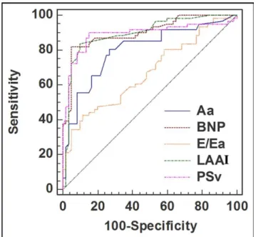
Figure 1. Receiver operating characteristics curves of predictors of out-come. Aa⫽ late diastolic annular velocity; LAAI ⫽ left atrial area index; PSv⫽ peak systolic velocity.
Table 1 Patient characteristics Variable No Events (n⫽ 64, 51%) Events (n⫽ 62, 49%) Univariate Multivariate
p Value p Value Hazard Ratio (95% CI)
Age (years) 68⫾ 10 67⫾ 12 0.51 — —
Women 16 (25%) 35 (56%) 0.00024 0.048* 0.53 (0.28–0.99)*
Hypertension 29 (43%) 32 (52%) 0.51 — —
Diabetes mellitus 12 (19%) 12 (19%) 0.97 — —
Hypercholesterolemia 28 (44%) 26 (42%) 0.34 — —
Systolic arterial pressure (mm Hg) 144⫾ 18 141⫾ 19 0.55 — —
B-type natriuretic peptide (pg/ml) 39.9⫾ 27.3 166⫾ 237 ⬍0.0001 0.012* 1.001 (1–1.003)*
Serum creatinine (mg/l) 8.2⫾ 1.8 8.7⫾ 1.7 0.14 — —
Abnormal response to exercise 6 (9) 26 (42) 0.007 0.88 0.95 (0.49–1.8)
Aortic valve area (cm2) 0.86⫾ 0.13 0.79⫾ 0.16 0.05 0.80 1.49 (0.06–34)
Peak aortic velocity (m/s) 4.03⫾ 0.55 4.35⫾ 0.58 0.0013 0.58 1.31 (0.5–3.4)
Aortic peak pressure gradient (mm Hg) 69.7⫾ 19.7 77⫾ 21 0.01 — —
Aortic mean pressure gradient (mm Hg) 42.4⫾ 13.4 46.7⫾ 13. 0.046 0.32 1.02 (0.9–1.06)
Left ventricular mass (g) 174⫾ 86 169⫾ 67 0.85 — —
Left ventricular end-diastolic volume (ml) 92.8⫾ 26.5 96.9⫾ 30.3 0.94 — —
Left ventricular end-systolic volume (ml) 32.2⫾ 14.2 32.5⫾ 13 0.91 — —
Left ventricular ejection fraction (%) 65.8⫾ 7.4 67.4⫾ 7.5 0.52 — —
Left atrial area index (cm2/m2) 10.3⫾ 2.1 14.8⫾ 3.4 ⬍0.0001 0.011* 1.06 (1.01–1.11)*
Mitral early diastolic filling wave (cm/s) 77⫾ 24 83⫾ 26 0.12 — —
Mitral late diastolic filling wave (cm/s) 87⫾ 29 89⫾ 28 0.29 — —
Mitral early/late diastolic filling ratio 0.92⫾ 0.32 0.99⫾ 0.48 0.53 — —
Mitral early diastolic filling wave deceleration time (ms) 219⫾ 78 237⫾ 97 0.34 — —
Peak systolic velocity (cm/s) 5.2⫾ 0.9 3.6⫾ 1.6 ⬍0.0001 0.016* 0.73 (0.57–0.94)*
Peak early diastolic annular velocity (cm/s) 9.95⫾ 1.7 7.9⫾ 1.8 0.025 0.91 1.01 (0.84–1.2)
Peak late diastolic annular velocity (cm/s) 8.7⫾ 2 7.6⫾ 2.1 ⬍0.0001 0.023* 0.81 (0.67–0.97)*
Early diastolic filling/annular velocity (average annuli) 10⫾ 3.3 13.5⫾ 6.6 0.009 0.024* 0.94 (0.88–0.99)* * Parameters selected on the multivariate Cox regression analysis.
Table 2
Area under curve, sensitivity, specificity, and optimal cutoff values of significant variables for predicting outcome
Data at Inclusion Cutoff Value
Area Under Curve
Sensitivity Specificity
Left atrial area index
(cm2/m2) ⱖ12.4
0.90 83.9% 90.6%
Peak systolic velocity (cm/s)
ⱕ4.5 0.87 88.7% 82.8%
Peak Aa velocity (cm/s) ⱕ9 0.81 80.6% 75%
Early diastolic filling/ annular velocity
⬎13.8 0.67 42% 88%
B-type natriuretic peptide (pg/ml)
were measured separately and then averaged. The E/Ea ratio was then calculated.
Venous blood samples for BNP measurement were drawn before echocardiography, after 10 minutes of supine rest. Chilled ethylenediaminetetraacetic acid tubes were centrifuged immediately at 4,000 rpm (4°C) for 15 minutes. Separated plasma samples were processed by immunofluo-rescence assay (Biosite, Beckman Coulter, San Diego, Cal-ifornia). The inter- and intra-assay variation was 5% and 4%, respectively. The assay detection limit was 1 pg/ml.
Symptom-limited graded bicycle exercise tests were per-formed at inclusion for all patients. After an initial workload of 25 W maintained for 2 minutes, the load was increased by steps of 25 W every 2 minutes. The exercise test was interrupted when the age-related maximum heart rate was reached or if symptoms, hypotension, or significant ventric-ular arrhythmias developed. The test was considered abnor-mal if the patient presented withⱖ1 of the following cri-teria: (1) angina, (2) evidence of dyspnea, (3) dizziness, (4) syncope or near-syncope, (5) and increase in systolic blood pressure during exercise of ⬍20 mm Hg or a decrease in blood pressure, and (6) ventricular tachycardia or ⬎4
pre-mature ventricular complexes in a row. Because the present study was conducted before the official recommendations of 2007, the results of the exercise test, even when abnormal, did not affect patient treatment.
Follow-up information was obtained from interviews with the patients, their relatives, or their physicians every 6 to 12 months, according to the guidelines. Particular care was taken to obtain information regarding the development of symptoms, eventual aortic valve replacement, and death. The clinical management was determined independently by the patient’s personal physician using all information avail-able. The combined end point included the onset of symp-toms (angina, dyspnea, syncope, heart failure), cardiac-re-lated death, and the need for aortic valve replacement.
Continuous variables are expressed as the mean⫾ SD, unless otherwise specified. Group comparisons for categor-ical variables were obtained using the chi-square test and for continuous variables with 1-way analysis of variance. Anal-ysis was performed by censoring follow-up data at cardiac surgery, if eventually performed. To detect independent predictors of events, a multivariate Cox proportional haz-ards regression procedure was used to compare the patients
Figure 2. The four figures represent the Kaplan-Meier event-free survival curves according the following categorical variables which were selected in the multivariate model to predict the outcome in asymptomatic patients with AS. (A) BNP: B-Type Natriuretic Peptide; (B) LA: left atrial area; (C) PSv: peak systolic velocity; (D) Aa: late diastolic annular velocity.
who remained asymptomatic during follow-up and those who experienced an event (STATISTICA, version 7, Stat-soft, France). All clinically relevant variables with p⬍0.10 were included in the multivariate model. p Values ⬍0.05 were considered significant. Receiver operating character-istic curve analysis was performed to determine the cutoff values that best distinguished the issue (area under the curve). Survival curves were established using the Kaplan-Meier method, and statistical significance was determined using the log-rank test.
Results
The mean patient age was 67 ⫾ 10 years (range 41 to 84). From the patient history and echocardiographic analy-sis findings, the suspected origin of AS was calcification of a trileaflet (n⫽ 104), bicuspid (n ⫽ 15), rheumatic disease (commissural fusion and calcification most prominent along the edges of the cusps on echocardiography; n ⫽ 5), and undetermined (n⫽ 2). The aortic valve area range was 0.38 to 1.2 cm2(mean 0.82⫾ 0.15), and the peak aortic pressure gradient was 77 ⫾ 21 mm Hg. The mean aortic pressure gradient was 26 to 86 mm Hg (mean 45⫾ 14). The mean LV ejection fraction was 67⫾ 7% (range 55% to 84%), and the mean peak annular systolic velocity was 4.5⫾ 1.5 cm/s (range 0.84 to 9). The mean BNP level was 102 ⫾ 178 pg/ml (range 5 to 1,500). At inclusion, the exercise test findings were abnormal (dyspnea in 20, decrease or increase in systolic blood pressure during exercise of⬍20 mm Hg in 6, and combined parameters in 6) in 32 patients (25%). During a median follow-up period of 20.3 ⫾ 18.7 months (interquartile range 9 to 22), the predefined end point oc-curred in 62 patients. Of the 62 patients, 6 patients died from cardiovascular causes: 3 suddenly and 3 from
progres-sive heart failure. Aortic valve replacement was required for the development of symptoms in 34 patients, new-onset atrial fibrillation in 1, newly positive exercise test findings in 7, and equivocal symptoms in 6. Finally, 8 patients developed symptoms but refused to undergo surgery.
The clinical and echocardiographic characteristics of the patients who remained asymptomatic or experienced an event are listed inTable 1. No clinical data, except female gender, allowed the distinction between the 2 groups. Pa-tients who experienced an end point had a smaller aortic valve area, lower tissue Doppler annular systolic and dia-stolic velocities, and a greater E/Ea ratio, left atrial area, and BNP level. The response to exercise was more often abnor-mal in these patients. On multivariate Cox regression anal-ysis, the parameters independently associated with the pre-defined composite outcome were gender, left atrial area index, systolic annular velocity, E/Ea, late diastolic annular velocity (Aa), and BNP level (Figure 1,Table 2).Figure 2
shows the survival curves for categorical variables, and
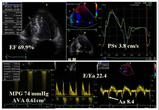
Figure 3 shows an example of a patient with impaired long-axis function. In the subgroup of patients with a normal response to exercise, except for the E/Ea, the other parameters remained independently associated with the outcome.
Discussion
The decision to perform surgery on asymptomatic pa-tients with AS remains controversial.3,4The immediate risks of surgery are often weighed against the later risk of events without intervention.15Therefore, reliable risk stratification is clinically important.16 –18In the present prospective study, we report, for the first time, the added prognostic value of tissue Doppler imaging and BNP measurement compared to
Figure 3. Example of patient with severe AS and impaired long-axis function. Aa⫽ late diastolic annular velocity; AVA ⫽ aortic valve area; EF ⫽ ejection fraction; MPG⫽ mean transaortic pressure gradient; PSv ⫽ peak systolic velocity.
the classical clinical and echocardiographic parameters in a prospective series of patients with asymptomatic moderate to severe AS. Patients, particularly females, with impaired long-axis function and increased BNP levels were at in-creased risk of untoward events.
In AS, the chronically increased afterload results in pro-gressive LV remodeling and myocardial hypertrophy.19 Al-though the increase in LV wall thickness is a compensatory mechanism that reduces systolic wall stress, it can result in impaired LV relaxation, reduced LV compliance, and in-creased metabolic demands. The ability of the LV to ade-quately fill under normal pressures is thus altered, and the LV diastolic pressure increases. As a result, LV filling becomes more dependent on the atrial contribution. In the advanced stage of the disease, the chronic increase in LV filling pressure is accompanied by progressive left atrial enlargement and dysfunction.20Elevated LV diastolic pres-sure also limits the coronary perfusion prespres-sure with a reduction of the subendocardial coronary flow reserve, which leads to myocardial ischemia even in the absence of significant coronary artery disease.21 Despite appropriate hypertrophy and accumulation of collagen in the subendo-cardial region, the LV ejection fraction can be classically maintained within the normal range for years.22This reflects the preserved contribution of radial thickening to LV func-tion. In contrast, the LV longitudinal function will be im-paired because of subendocardial dysfunction. Initially, sub-endocardial dysfunction might normalize after aortic valve surgery.23
In the present study, except for female gender, neither the clinical findings (i.e., age, risk factors, exercise test) nor standard echocardiographic parameters (i.e., severity of ste-nosis, LV mass, LV volume, LV ejection fraction, extent of valve calcification) emerged as independent predictors of outcome. However, the prognostic effect of these factors was probably weakened by the inclusion of almost specif-ically elderly patients (in which the positive predictive value of exercise testing is limited) and the analysis of more powerful predictors of outcome such as BNP level and tissue Doppler parameters.24,25 In these patients, the in-creased release of BNP from the ventricles could reflect the substantial gender differences in the LV adaptive responses to AS.26 BNP has been shown to be associated with the prognosis in several cardiovascular diseases such as AS.7–11 In heart failure, the predictive value of BNP seems to be greater in women than in men.27Our results have confirmed the strong prognostic value of BNP in a cohort of patients with asymptomatic AS.
The current guidelines consider surgery as reasonable in patients with asymptomatic severe AS and reduced LV ejection fraction (⬍50%).3,4However, the LV ejection frac-tion is often normal in these patients. In contrast to this crude estimate of the LV systolic function, tissue Doppler imaging is superior for detecting subtle changes in myocar-dial function.12Measurements of myocardial systolic annu-lar velocities reflect longitudinal motion owing to the lon-gitudinally directed fibers, which are mainly located in the subendocardium and subepicardium. When the velocities are averaged from the 4 regions of the annulus, they reflect the global long-axis function. In patients with asymptomatic AS, impaired subendocardial function has been shown to be
associated with impaired exercise tolerance and changes in symptomatic status during short-term follow-up.6,22 Our results have extended these preliminary data by showing that the decrease in long-axis function can identify a subset of patients with moderate to severe asymptomatic AS at greater risk of developing cardiac events.
In the present study, we found that the left atrial area, late diastolic annular velocity (Aa), and E/Ea ratio were strong independent predictors of outcome. The left atrial size re-flects the chronicity of diastolic burden.28A value ofⱖ12.4 cm2/m2 provided high predictive accuracy for cardiac events. The late diastolic annular velocity reliably quantifies the left atrial contribution to LV filling. In these patients, the left atrial booster pump function has a crucial role in coun-teracting the increased LV end-diastolic pressure and the delayed LV untwisting to maintain optimal cardiac output.29 A reduction in left atrial function might thus favor clinical deterioration. In our population, a late diastolic annular velocity ofⱕ9 cm/s was associated with an excess risk of death, symptoms, or surgery. An increased E/Ea ratio, an estimate of LV pre-A pressure, has been shown to be an important marker of adverse events in patients with pre-served LV function.12In the present study, we found that an E/Ea ratio⬎13.8 identified a subset of patients with AS at greater risk of future events.
1. Rahimtoola SH. Valvular heart disease: a perspective on the asymp-tomatic patient with severe valvular aortic stenosis. Eur Heart J 2008;29:1783–1790.
2. Otto CM. Valvular aortic stenosis: disease severity and timing of intervention. J Am Coll Cardiol 2006;47:2141–2151.
3. Bonow RO, Carabello BA, Kanu C, de Leon AC Jr, Faxon DP, Freed MD, Gaasch WH, Lytle BW, Nishimura RA, O’gara PT, O’Rourke RA, Otto CM, Shah PM, Shanewise JS, Smith SC Jr, Jacobs AK, Adams CD, Anderson JL, Antman EM, Faxon DP, Fuster V, Halperin JL, Hiratzka LF, Hunt SA, Lytle BW, Nishimura R, Page RL, Riegel B. ACC/AHA 2006 guidelines for the management of patients with valvular heart disease: a report of the American College of Cardiology/ American Heart Association Task Force on Practice Guidelines.
Cir-culation 2006;114:e84 – e231.
4. Vahanian A, Baumgartner H, Bax J, Butchart E, Dion R, Filippatos G, Flachskampf F, Hall R, Iung B, Kasprzak J, Nataf P, Tornos P, Torracca L, Wenink A. Guidelines on the management of valvular heart disease: the Task Force on the Management of Valvular Heart Disease of the European Society of Cardiology. Eur Heart J 2007;28: 230 –168.
5. Hachicha Z, Dumesnil JG, Bogaty P, Pibarot P. Paradoxical low-flow, low-gradient severe aortic stenosis despite preserved ejection fraction is associated with higher afterload and reduced survival. Circulation 2007;115:2856 –2864.
6. Tongue AG, Dumesnil JG, Laforest I, Theriault C, Durand LG, Pibarot P. Left ventricular longitudinal shortening in patients with aortic ste-nosis: relationship with symptomatic status. J Heart Valve Dis 2003; 12:142–149.
7. Weber M, Arnold R, Rau M, Elsaesser A, Brandt R, Mitrovic V, Hamm C. Relation of N-terminal pro B-type natriuretic peptide to progression of aortic valve disease. Eur Heart J 2005;26:1023–1030. 8. Lim P, Monin JL, Monchi M, Garot J, Pasquet A, Hittinger L, Vanoverschelde JL, Carayon A, Gueret P. Predictors of outcome in patients with severe aortic stenosis and normal left ventricular function: role of B-type natriuretic peptide. Eur Heart J 2004;25: 2048 –2053.
9. Gerber IL, Stewart RA, Legget ME, West TM, French RL, Sutton TM, Yandle TG, French JK, Richards AM, White HD. Increased plasma natriuretic peptide levels reflect symptom onset in aortic stenosis.
Circulation 2003;107:1884 –1890.
10. Gerber IL, Legget ME, West TM, Richards AM, Stewart RA. Useful-ness of serial measurement of N-terminal pro-brain natriuretic peptide
plasma levels in asymptomatic patients with aortic stenosis to predict symptomatic deterioration. Am J Cardiol 2005;95:898 –901. 11. Bergler-Klein J, Klaar U, Heger M, Rosenhek R, Mundigler G, Gabriel
H, Binder T, Pacher R, Maurer G, Baumgartner H. Natriuretic peptides predict symptom-free survival and postoperative outcome in severe aortic stenosis. Circulation 2004;109:2302–2308.
12. Yu CM, Sanderson J. Tissue Doppler imaging: a new prognosticator for cardiovascular diseases. J Am Coll Cardiol 2007;49:1903–1914. 13. Skjaerpe T, Hegrenaes L, Hatle L. Noninvasive estimation of valve
area in patients with aortic stenosis by Doppler ultrasound and two-dimensional echocardiography. Circulation 1985;72:810 – 818. 14. Lancellotti P, Cosyns B, Piérard LA. Dynamic left ventricular
dyssyn-chrony contributes to B-type natriuretic peptide release during exercise in patients with systolic heart failure. Europace 2008;10:496 –501. 15. Rosenhek R, Binder T, Porenta G, Lang I, Christ G, Schemper M,
Maurer G, Baumgartner H. Predictors of outcome in severe, asymp-tomatic aortic stenosis. N Engl J Med 2000;343:611– 617.
16. Pellikka PA, Sarano ME, Nishimura RA, Malouf JF, Bailey KR, Scott CG, Barnes ME, Tajik AJ. Outcome of 622 adults with asymptomatic, hemodynamically significant aortic stenosis during prolonged follow-up. Circulation 2005;111:3290 –3295.
17. Otto CM, Burwash IG, Legget ME, Munt BI, Fujioka M, Healy NL, Kraft CD, Miyake-Hull CY, Schwaegler RG. Prospective study of asymptomatic valvular aortic stenosis: clinical, echocardiographic, and exercise predictors of outcome. Circulation 1997;95:2262–2270. 18. Lancellotti P, Lebois F, Simon M, Tombeux C, Chauvel C, Pierard
LA. Prognostic importance of quantitative exercise Doppler echocar-diography in asymptomatic valvular aortic stenosis. Circulation 2005; 112:I377–I382.
19. Lund O, Erlandsen M, Dorup I, Emmertsen K, Flo C, Jensen FT. Predictable changes in left ventricular mass and function during ten years after valve replacement for aortic stenosis. J Heart Valve Dis 2004;13:357–368.
20. Dalsgaard M, Egstrup K, Wachtell K, Gerdts E, Cramariuc D, Kjaer-gaard J, Hassager C. Left atrial volume in patients with asymptomatic
aortic valve stenosis (the Simvastatin and Ezetimibe in Aortic Stenosis study). Am J Cardiol 2008;101:1030 –1034.
21. Hein S, Arnon E, Kostin S, Schönburg M, Elsässer A, Polyakova V, Bauer EP, Klövekorn WP, Schaper J. Progression from compensated hypertrophy to failure in the pressure-overloaded human heart: struc-tural deterioration and compensatory mechanisms. Circulation 2003; 107:984 –991.
22. Lafitte S, Perlant M, Reant P, Serri K, Douard H, DeMaria A, Roudaut R. Impact of impaired myocardial deformations on exercise tolerance and prognosis in patients with asymptomatic aortic stenosis. Eur J Echocardiogr 2009;10:414 – 419.
23. Poulsen SH, Sogaard P, Nielsen-Kudsk JE, Egeblad H. Recovery of left ventricular systolic longitudinal strain after valve replacement in aortic stenosis and relation to natriuretic peptides. J Am Soc Echocar-diogr 2007;20:877– 884.
24. Das P, Rimington H, Chambers J. Exercise testing to stratify risk in aortic stenosis. Eur Heart J 2005;26:1309 –1313.
25. Monin JL, Lancellotti P, Monchi M, Lim P, Weiss E, Piérard L, Guéret P. Risk score for predicting outcome in patients with asymptomatic aortic stenosis. Circulation 2009;120:69 –75.
26. Legget ME, Kuusisto J, Healy NL, Fujioka M, Schwaegler RG, Otto CM. Gender differences in left ventricular function at rest and with exercise in asymptomatic aortic stenosis. Am Heart J 1996; 131:94 –100.
27. Christ M, Laule-Kilian K, Hochholzer W, Klima T, Breidthardt T, Perruchoud AP, Mueller C. Gender-specific risk stratification with B-type natriuretic peptide levels in patients with acute dyspnea: in-sights from the B-type natriuretic peptide for acute shortness of breath evaluation study. J Am Coll Cardiol 2006;48:1808 –1812.
28. Rossi A, Tomaino M, Golia G, Anselmi M, Fucá G, Zardini P. Echocardiographic prediction of clinical outcome in medically treated patients with aortic stenosis. Am Heart J 2000;140:766 –771. 29. Poh KK, Chan MY, Yang H, Yong QW, Chan YH, Ling LH.
Prog-nostication of valvular aortic stenosis using tissue Doppler echocardi-ography: underappreciated importance of late diastolic mitral annular velocity. J Am Soc Echocardiogr 2008;21:475– 481.