Université de Montréal
Function of the immunoregulatory CD4-CD8- T cells
in the context of autoimmune diabetes
par Erin Hillhouse
Département de Microbiologie et Immunologie Faculté de Médecine
Thèse présentée à la Faculté des Études Supérieures en vue de l’obtention du grade de Ph.D.
en Microbiologie et Immunologie
Février, 2013 © Erin Hillhouse, 2013
Université de Montréal
Faculté des études supérieures et postdoctorales
Cette thèse intitulée:
Function of the immunoregulatory CD4-CD8- T cells in the context of autoimmune diabetes
Présentée par : Erin Hillhouse
a été évaluée par un jury composé des personnes suivantes :
Dr Jacques Thibodeau, président-rapporteur Dre Sylvie Lesage, directeur de recherche
Dre Nathalie Arbour, membre du jury Dre Li Zhang, examinateur externe
RÉSUMÉ
La tolérance immunitaire dépend de la distinction entre le soi et le non soi par le système immunitaire. Un bris dans la tolérance immunitaire mène à
l'auto-immunité, qui peut provoquer la destruction des organes, des glandes, des articulations ou du système nerveux central. Le diabète auto-immun, également connu sous le nom diabète juvénile et diabète de type 1, résulte d'une attaque auto-immune sur les cellules β pancréatiques sécrétrices d’insuline, localisées au niveau des îlots de Langerhans du pancréas. Bien que le diabète auto-immun soit traitable par une combinaison d’injections quotidiennes d’insuline d’origine exogène, de régime et d'exercices, beaucoup de complications chroniques peuvent se manifester chez les patients, y compris, mais non limitées à, la cécité, les maladies
cardiovasculaires, l’insuffisance rénale et l'amputation. En raison des nombreuses complications liées au diabète auto-immun à long terme, la recherche continue afin de mieux comprendre tous les facteurs impliqués dans la progression de la maladie dans le but de développer de nouvelles thérapies qui empêcheront, renverseront et/ou traiteront cette maladie.
Un rôle primordial dans la génération et l'entretien de la tolérance immunitaire a été attribué au nombre et à la fonction des sous-populations de cellules régulatrices. Une de ces populations est constituée de cellules T CD4-CD8- (double négatives, DN), qui ont été étudiées chez la souris et l'humain pour leur contribution à la
tolérance périphérique, à la prévention des maladies et pour leur potentiel associé à la thérapie cellulaire. En effet, les cellules de T DN sont d'intérêt thérapeutique parce qu'elles montrent un potentiel immunorégulateur antigène-spécifique dans divers cadres expérimentaux, y compris la prévention du diabète auto-immun. D’ailleurs, en utilisant un système transgénique, nous avons démontré que les souris prédisposées au diabète auto-immun présentent peu de cellules T DN, et que ce phénotype
contribue à la susceptibilité au diabète auto-immun. En outre, un transfert des cellules T DN est suffisant pour empêcher la progression vers le diabète chez les souris prédisposées au diabète auto-immun. Ces résultats suggèrent que les cellules T DN
puissent présenter un intérêt thérapeutique pour les patients diabétiques. Cependant, nous devons d'abord valider ces résultats en utilisant un modèle non-transgénique, qui est plus physiologiquement comparable à l'humain.
L'objectif principal de cette thèse est de définir la fonction immunorégulatrice des cellules T DN, ainsi que le potentiel thérapeutique de celles-ci dans la prévention du diabète auto-immun chez un modèle non-transgénique. Dans cette thèse, on démontre que les souris résistantes au diabète auto-immun présentent une proportion et nombre absolu plus élevés de cellules T DN non-transgéniques, lorsque comparées aux souris susceptibles. Cela confirme une association entre le faible nombre de cellules T DN et la susceptibilité à la maladie. On observe que les cellules T DN éliminent les cellules B activées in vitro par une voie dépendante de la voie perforine et granzyme, où la fonction des cellules T DN est équivalente entre les souris
résistantes et prédisposées au diabète auto-immun. Ces résultats confirment que l'association au diabète auto-immun est due à une insuffisance en terme du nombre de cellules T DN, plutôt qu’à une déficience fonctionnelle. On démontre que les cellules T DN non-transgéniques éliminent des cellules B chargées avec des antigènes d'îlots, mais pas des cellules B chargées avec un antigène non reconnu, in vitro. Par ailleurs, on établit que le transfert des cellules T DN activées peut empêcher le développement du diabète auto-immun dans un modèle de souris non-transgénique. De plus, nous observons que les cellules T DN migrent aux îlots pancréatiques, et subissent une activation et une prolifération préférentielles au niveau des ganglions pancréatiques. D'ailleurs, le transfert des cellules T DN entraîne une diminution d'auto-anticorps spécifiques de l'insuline et de cellules B de centres germinatifs directement dans les îlots, ce qui corrèle avec les résultats décrits ci-dessus. Les résultats présentés dans cette thèse permettent de démontrer la fonction des cellules T DN in vitro et in vivo, ainsi que leur potentiel lié à la thérapie cellulaire pour le diabète auto-immun.
Mots-clés : Cellules T double négatives, diabète auto-immun, immunorégulation, souris transgénique, souris non-transgénique, antigène-spécifique.
ABSTRACT
Immune tolerance is dependent on the immune system discriminating between self and non-self. A break in immune tolerance results in autoimmunity, which can lead to the destruction of healthy organs, glands, joints or the central nervous system. Any disease that results from such an aberrant immune response is termed an
autoimmune disease. Autoimmune diabetes, which is also referred to as juvenile diabetes and type 1 diabetes, results from an autoimmune attack on the insulin-producing β cells located within the islets of Langerhans of the pancreas. Although autoimmune diabetes is treatable through a combination of insulin therapy, diet and exercise, many chronic complications may arise in patients, including, but not limited to, blindness, cardiovascular disease, kidney failure and amputation. Due to the many complications associated with long-term autoimmune diabetes, research continues to better understand all the factors implicated in disease progression in order to develop new therapies that will prevent, reverse and/or cure this disease.
A prominent role in the generation and maintenance of immune tolerance has been attributed to the number and function of regulatory cell subsets. One of these regulatory cell populations, namely CD4-CD8- (double negative, DN) T cells, have been studied in both mice and humans for their contribution to peripheral tolerance, disease prevention and their potential for use in cellular therapy. DN T cells are of particular therapeutic interest because they exhibit an antigen-specific immunoregulatory potential in various experimental settings, including the prevention of autoimmune diabetes. Indeed, using a transgenic system, we have shown that autoimmune diabetes-prone mice carry fewer DN T cells and that this phenotype contributes to autoimmune diabetes susceptibility, where a single transfer of DN T cells is sufficient to prevent diabetes progression in otherwise autoimmune diabetes-prone mice. These results suggest that DN T cells may be of therapeutic interest for diabetic patients. However, we must first validate these results using a non-transgenic setting, which is more physiologically relevant to humans.
The main objective of this thesis is to determine the immunoregulatory function of the DN T cells as well as the therapeutic potential of these cells in the prevention of autoimmune diabetes in the non-transgenic setting. Here, we show that diabetes-resistant mice present with a higher proportion and cell number of DN T cells than diabetes-susceptible mice in the non-transgenic setting, which associates a deficiency in DN T cell number with disease susceptibility. We determine that DN T cells eliminate activated B cells in vitro via a perforin/granzyme-dependent pathway, where the function of DN T cells is equal between the diabetesresistant and -susceptible mice, demonstrating that the association to autoimmune diabetes is due to a deficiency in DN T cell number rather than function. Interestingly, we show that non-transgenic DN T cells eliminate B cells loaded with islet antigen, but not B cells loaded with an irrelevant antigen, in vitro. Importantly, we establish that the transfer of activated DN T cells could prevent autoimmune diabetes development in the non-transgenic setting. Interestingly, we reveal that DN T cells migrate to the pancreatic islets and undergo preferential activation and proliferation within the pancreatic lymph nodes. Moreover, the transfer of DN T cells results in a decrease in both germinal center B cells directly within the pancreatic islets as well serum insulin autoantibody levels, which correlates with the aforementioned findings. Altogether, the results presented in this thesis have allowed us to enhance our understanding of the function of DN T cells both in vitro and in vivo as well as demonstrate the therapeutic potential for DN T cells as a novel cellular therapeutic for autoimmune diabetes.
Keywords : Double negative T cells, autoimmune diabetes, immunoregulation, transgenic mouse, non-transgenic mouse, antigen-specific.
TABLE OF CONTENTS
RÉSUMÉ ... i ABSTRACT ... iii TABLE OF CONTENTS ... v LIST OF FIGURES ... ix LISTE OF ABBREVIATIONS ... x ACKNOWLEDGEMENTS ... xiii CHAPTER 1 Introduction ... 1 1.1 Autoimmunity ... 1 1.2 Autoimmune diabetes ... 2 1.2.1 The pancreas ... 2 1.2.2 Insulin ... 31.2.3 Clinical signs and diagnosis ... 4
1.2.4 Prevalence ... 5 1.2.5 Risk factors ... 6 1.2.5.1 Genetics ... 7 1.2.5.2 Environment ... 8 1.2.5.2.1 Dietary influence ... 8 1.2.5.2.2 Hygiene hypothesis ... 8 1.2.5.2.3 Viral infections ... 9 1.2.6 Pathogenesis ... 10 1.2.6.1 T cells ... 12
1.2.6.2 B cells ... 13
1.2.6.3 Macrophages ... 15
1.2.6.4 Natural killer cells ... 16
1.2.6.5 Dendritic cells ... 17
1.3 The non-obese diabetic mouse model ... 19
1.4 Autoimmune diabetes therapy ... 22
1.4.1 Transplantation ... 22
1.4.1.1 Pancreas transplantation ... 22
1.4.1.2 Islet transplantation ... 23
1.4.2 Immunotherapy ... 24
1.4.2.1 Cyclosporine A ... 24
1.4.2.2 Anti-CD3 monoclonal antibodies ... 25
1.4.2.3 Anti-CD20 monoclonal antibody ... 25
1.5 Immunoregulatory CD4-CD8- (DN) T cells ... 29
1.5.1 Challenge associated with the specific identification of immunoregulatory DN T cells ... 30
1.5.1.1 Distinguishing DN T cells from NK T cells ... 31
1.5.2 DN T cell phenotype... 32
1.5.2.1 Phenotype of TCR transgenic DN T cells ... 33
1.5.2.2 Phenotype of non-transgenic DN T cells ... 33
1.5.2.3 Phenotype of human DN T cells ... 35
1.5.3 Targets and mechanisms of action of DN T cells ... 36
1.5.3.1 The immunoregulatory function of TCR transgenic DN T cells ... 36
1.5.3.2 The immunoregulatory function of non-transgenic DN T cells ... 38
1.5.3.3 The immunoregulatory function of human DN T cells ... 40
1.5.3.4 The in vivo function of immunoregulatory DN T cells ... 42
1.5.4 DN T cells in transplantation and disease models ... 43
1.5.4.1 Graft tolerance ... 43
1.5.4.2 Graft-vs-host disease (GVHD) ... 46
1.5.4.3 Cancer ... 48
1.5.4.5 Autoimmune lymphoproliferative syndrome (ALPS) ... 50
1.5.4.6 Autoimmune diabetes ... 51
1.5.4.6.1 Comparing DN T cells and NK T cells and their role in autoimmune diabetes pathology ... 54
1.6 Future directions ... 56
1.7 Rationale, hypothesis and objectives ... 57
CHAPTER 2 Article 1 ... 58
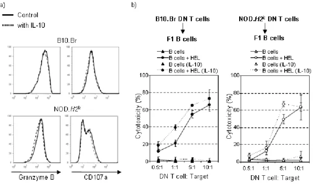
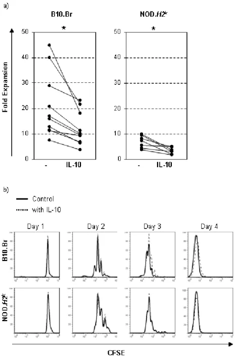
2.1 IL-10 limits the expansion of immunoregulatory CD4-CD8- T cells in autoimmune-prone NOD mice ... 58
2.2 Abstract ... 60 2.3 Introduction ... 61 2.4 Results ... 64 2.5 Discussion ... 69 2.6 Methods ... 74 2.7 Acknowledgements ... 77 2.8 References ... 78 2.9 Figures ... 84 CHAPTER 3 Article 2 ... 91
3.1 Immunoregulatory CD4-CD8- T cells, a novel cellular therapeutic preventing autoimmune diabetes ... 91 3.2 Abstract ... 93 3.3 Introduction ... 94 3.4 Results ... 97 3.5 Discussion ... 102 3.6 Acknowledgements ... 104 3.7 Methods ... 105 3.8 Figures ... 110
3.9 References ... 121
CHAPTER 4 Discussion ... 125
4.1 Non-transgenic DN T cells: Phenotype and proportion ... 125
4.2 The function of DN T cells ... 128
4.3 The in vivo role of non-transgenic DN T cells in autoimmune diabetes ... 134
4.4 The in vivo mechanism of action of non-transgenic DN T cells ... 138
4.5 Conclusion and future perspectives ... 144
BIBLIOGRAPHY ... xv APPENDIX 1 ... xlvii APPENDIX 2 ... lxxxi APPENDIX 3 ... l OTHER CONTRIBUTIONS ... li
LIST OF FIGURES AND TABLES
Figure 1: Schematic representation of islet and pancreas cell types.Figure 2: Model for the development of autoimmune diabetes.
Figure 3: Mechanisms of action of anti-CD20 antibodies inducing B cell depletion.
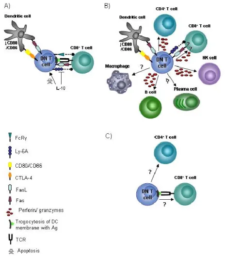
Figure 4: The targets and mechanism of action of DN T cells.
Figure 5: Illustration of the potential adoptive DN T cell therapy protocol for the treatment of autoimmune diabetes in humans.
Figure 6: Representation of the roles of autoreactive B cells in autoimmune disease, such as autoimmune diabetes.
Figure 7: Representation of the B cell developmental pathways in the germinal center.
Figure 8: Working hypothesis of possible cellular targets of DN T cells leading to a decrease of antigen-specific antibody levels in vivo.
LIST OF ABBREVIATIONS
51Cr: chromium-51
αGalCer: alpha-galactosylceramide
AHCT: allogeneic hematopoietic cell transplantation ALPS: autoimmune lymphoproliferative syndrome AML: acute myeloid leukemia
APC: antigen-presenting cell B10: C57BL/10SnJ
B6: C57BL/6
CBV: coxsackie B virus
CFSE: carboxyfluorescein diacetate succinimidyl ester C-peptide: connecting peptide
CTLA-4: cytotoxic T-lymphocyte-associated protein 4 CTV: cell trace violet
CXCR5: chemokine (C-X-C motif) receptor 5 DC: dendritic cell
DLI: donor leukocyte infusion DN: double negative, CD4-CD8-
DP: double positive, CD4+CD8+
EGTA: ethylene glycol tetraacetic acid ELISA: enzyme-linked immunosorbent assay EMCV: encephalomyocarditis virus
Fas: apoptosis stimulating fragment
FasL: apoptosis stimulating fragment ligand FcRγ: Fc gamma receptor
FLT-3L: Fms-like tyrosine kinase 3 ligand FOXP3: forkhead box P3
GAD65: 65-kDa isoform of glutamic acid decarboxylase G-CSF: granulocyte colony-stimulating factor
gp: glycoprotein
GVHD: Graft-vs-host disease HbA1c: haemoglobin A1c
HEL: hen egg lysozyme
HLA: human leukocyte antigen IA-2: insulinoma-associated-2 IAA: insulin autoantibody Idd: insulin-dependent diabetes IFNγ: interferon gamma Ig: immunoglobulin
iGb3: isoglobotrihexosylceramide IL: interleukin
IL-10: interleukin-10
IL-10R: interleukin 10 receptor IL2RA: interleukin 2 receptor, alpha iNKT: invariant NKT
INS: insulin
IPEX: immunodysregulation, polyendocrinopathy, enteropathy, X-linked LCMV: lymphocytic choriomeningitis virus
lpr: lymphoproliferation LPS: lipopolysaccharide
MHC: major histocompatibility complex MLR: mixed-lymphocyte reaction
NK: natural killer NKT: natural killer-like NOD: non-obese diabetic OVA: ovalbumin
PLN: pancreatic lymph node
PP: pancreatic polypeptide PPI: preproinsulin
PTPN22: protein tyrosine phosphatase, non-receptor type 22 Rag: recombination activating gene
RIP: rat insulin promoter
SCID: severe combined immunodeficient SD: standard of deviation
SIRP: signal regulatory protein SLE: systemic lupus erythematosus TCR: T cell receptor
TGFβ: transforming growth factor beta Th2: T helper 2
TNF-α: tumor necrosis factor alpha Treg: regulatory T cells
ZnT8: zinc transporter 8
β -GalCer : β-galactosylceramide β-GlcCer: β-D-glucopyranosylceramide
ACKNOWLEDGEMENTS
First and foremost, I would like to thank Dr. Sylvie Lesage, my PhD
supervisor. 5 1/2 years ago you accepted me into your lab as a Master's student and, at the time, I had no idea of the amazing journey that lay ahead for me. I consider myself extremely fortunate to have come across a supervisor that not only offered me a project that I would become faithfully passionate about, but also cared enough about my education and career path to guide and encourage me every step of the way. Being under your direction not only helped me to accomplish this goal of writing a PhD thesis, but I believe that with the completion of this degree that I will be taking with me knowledge and experience above and beyond what might be normally expected at the end of one's PhD. I will forever be grateful to you and proud that I was privileged enough to study under such a remarkable researcher who I also consider to be an exceptional teacher.
I cannot deny that a huge reason for my positive experience during my PhD training can be explained by the people I worked with on a daily basis in the lab. In particular, I would like to thank Véro for guiding me during my training, a gesture that would be invaluable to my project, Fanny for being a friend and someone I could talk, and complain, about diabetes to, Geneviève for always being there for me when times were challenging during this project and keeping my spirits up, and Adam for just being silly to the point that I could never be in a bad mood while at work. Not only do I consider you all, both past and present members, incomparable colleagues that assisted me during my training, both technically and intellectually, but amazing people that made coming to work every day an honest pleasure. Eating lunch together every afternoon, after work gatherings, singing and maybe sometimes even dancing
(Adam) in the lab, and our famous Friday talks; it is undeniable that the bar has been set impossibly high for the work environments that I may come to cross in the future.
I would also like to thank Marie-Josée Guyon, Fany de Wilde, and Martine Dupuis, without your expertise and assistance I would not have been able to accomplish even half of what I did during my PhD.
I am forever indebted to my family and friends who have never ceased to encourage me every step of the way. Their pride in my accomplishments pushed me past the difficult times and made me believe in myself and my ability to achieve my goals. To my parents and brother who guided and encouraged me to believe that I could do anything I set my head to; without you I wouldn't be where I am today. To my husband, Kevin, for supporting me throughout the years and never complaining when I locked myself in a room for days or weeks at a time. I know you couldn't have married me for my salary as a student, so thank you for loving me for me and making me feel like the richest girl in the world. Lastly, I would like to thank my unborn child who hasn't made the process of writing my thesis easy at times during the last couple of months, but who was thoughtful enough to give me a break near the end when I needed it the most. I hope you are proud of your mom.
I could not complete this section without acknowledging my doctors who cared for me since the age of 9 when I was diagnosed with type 1 diabetes. I was lucky to have had Dr. Celia Rodd to oversee me at the Montreal Children's Hospital during the years when glucose control would prove to be the most difficult, yet of most crucial importance. I would like to thank Dr. Natasha Garfield who has looked after me for the last decade and consistently and genuinely enquires about the progress of my research. Last, but definitely not least, I dedicate this thesis to my paediatrician, the late Dr. Werner Woelber, who saved my life 20 years ago this month of February. I am forever indebted to you and I will continue to live each day in a way that makes you proud.
CHAPTER 1: INTRODUCTION
1.1 Autoimmunity
A low level of autoreactivity is physiologic and crucial to normal immune function, where autoantigens helps to form the repertoire of mature lymphocytes and promote the survival of naive T cells and B cells in the periphery1. Contrary to this
physiological low level of autoreactivity, autoimmunity results from the failure of the immune system to discriminate self from non-self, leading to an immune response against cells and tissues. Any disease that results from such an aberrant immune response is termed an autoimmune disease. Autoimmune diseases are typically defined as either systemic, in which the autoimmune reaction occurs against various organs, tissues and cells of the body, such is the case for systemic lupus
erythematosus, or organ-specific, in which the autoimmune reaction targets a single organ as the name implies, as in the case of autoimmune diabetes.
1.2 Autoimmune diabetes
Autoimmune diabetes, which is also referred to as juvenile diabetes and type 1 diabetes, results from the destruction of the insulin-producing β cells located within the islets of Langerhans of the pancreas.
1.2.1 The pancreas
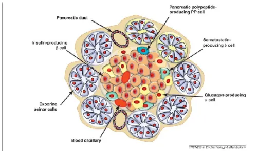
The pancreas is made up of exocrine and endocrine tissues (Figure 1). The exocrine is made up of cellular clusters called acinus, which consist of acinar cells that secrete digestive enzymes into the intestine, including proteases, amylases, lipases and nucleases2. The endocrine cells, which secrete hormones, are contained
within the islets of Langerhans, which are compact spheroidal clusters embedded in the exocrine tissue2. There are four major cell types of endocrine cells, namely α, β, δ
and PP cells, where the β cells make up approximately 70% of the islet mass. Together the and cells regulate blood glucose levels, where the cells secrete glucagon, which promotes the conversion of stored glycogen in the liver into glucose that can be released into the bloodstream, and the cells secrete insulin, which is necessary for glucose uptake from the blood into cells. The cells secrete
somatostatin, and the PP cells secrete pancreatic polypeptide (PP), both of which are implicated in modulating the secretory properties of the other cell types2.
Figure 1. Schematic representation of islet and pancreas cell types. Adapted from 3.
1.2.2 Insulin
During insulin synthesis, preproinsulin is first secreted from the β cells of the pancreas with an A-chain, a connecting peptide (C-peptide), a B-chain, and a signal sequence, which, once cleaved, gives rise to proinsulin. Subsequently, the C-peptide is removed, leaving the A-chain and B-chain that constitute the mature insulin
molecule. The mature insulin is stored in the β cells of the islets of Langerhans and its release is controlled by the level of glucose within the blood. Following insulin release, insulin binds to its receptor on the cell surface2. The signal transduction
subsequently induces various physiological effects, including but not limited to the influx of glucose into cells, glycogen synthesis and lipid synthesis. As a result, insulin is a vital requirement for life2.
Autoimmune diabetes was considered a fatal disease until the early 1920s when doctors Banting and Best, who were positioned in Toronto, demonstrated that the injection of concentrated pancreatic extracts, namely insulin, from dogs into human diabetic patients could maintain blood glucose homeostasis4. In 1923, this
discovery would result in Dr. Banting being awarded the Nobel Prize at the age of 325. Today, diabetic patients use human biosynthetic insulin to treat their disease.
1.2.3 Clinical signs and diagnosis
The classical symptoms associated with autoimmune diabetes include polyuria (excessive production of urine), polydipsia (excessive thirst), glucosuria (excretion of sugar in the urine) and tiredness, which are all connected to
hyperglycemia, or high blood glucose/sugar levels. The average plasma glucose concentration over a 3-4 month period can be established by measuring haemoglobin A1c (HbA1c) levels, where HbA1c is the glycated haemoglobin which forms due to the
haemoglobin's exposure to plasma glucose.
At the time of the diagnosis, the mass of β cells is estimated to represent 10 to 33% of its initial value based on a glucose tolerance test that measures your blood glucose levels following glucose stimulation, which is insufficient to provide the insulin contribution necessary following glucose ingestion6. The insulin response
continues to decline following diagnosis, where two years after diagnosis, the insulin response now represents 28 ± 8.4% of the response at diagnosis7. Indeed, there is a
significant inverse relationship between the insulin secretory response and glucose control, which is reflected by the HbA1c levels7. Therefore, increased HbA1c levels
are indicative of a decrease in the insulin secretory response.
The insulin secretory response following a glucose tolerance test assesses β cell function by measuring either insulin or C-peptide levels found in the plasma6,
where lower insulin levels would indicate a decrease in β cell function, thus the progression to diabetes. However, insulin is known to have a short half-life and an unpredictable hepatic extraction as well as peripheral clearance, making it difficult to assess insulin secretion based on insulin plasma levels following glucose
stimulation8. On the contrary, C-peptide plasma levels are a more accurate
measurement of insulin secretion as C-peptide, which is co-secreted with insulin from β cells at an equimolar concentration, does not undergo significant hepatic extraction, maintains a constant metabolic clearance rate and exhibits a longer half-life6, 8.
used to measure β cell function, where a decrease in β cell function is an indicator of autoimmune diabetes progression.
Another factor used to identify autoimmune diabetes development is the presence of autoantibodies to insulin (IAA) and other islet antigens, including autoantibodies targeting the 65-kDa isoform of glutamic acid decarboxylase, (GAD65) and autoantibodies targeting the phosphatase-related
insulinoma-associated-2 (IA-2) molecule9. Moreover, the presence of these autoantibodies can
also be used to predict the onset of disease10, 11, 12, 13. Indeed, IAA, GAD65 and IA-2
autoantibodies may arise before any signs of hyperglycemia, where the time interval between the emergence of autoantibodies and disease onset can range from a few months to several years14. Notably, not everyone who presents with autoantibodies
progresses to autoimmune diabetes. However, as the number of autoantibody types increases, the risk of developing the disease is also amplified, where an individual who presents with three to four autoantibody types has a 60–100% risk of progressing to autoimmune diabetes14. Autoantibodies to the zinc transporter ZnT8, which is a
member of the large cation efflux family, were recently discovered to be present in pre-diabetic and new-onset diabetic patients, where ZnT8 antibodies generally emerged later than IAA and GAD65 antibodies in pre-diabetic patients11. Indeed, the
inclusion of ZnT8 autoantibody with IAA, GAD65 and IA2 autoantibody detection can increase the prognostic value to 98% rather than the previous 90%11. With the
combination of these 4 autoantibodies, we are closer to detecting pre-diabetes in a general pediatric population with a family history of disease11.
Altogether, the symptoms of autoimmune diabetes development include a decrease in insulin and C-peptide plasma levels that correlates with an increase in blood glucose levels and its associated symptoms as well as the potential presence of insulin and islet antigen autoantibodies.
Currently, there are 347 million people worldwide living with diabetesi. Of
these diabetic patients, an estimated 5–10% constitute people living with autoimmune diabetes15, which amounts to approximately 17-35 million people worldwide. In
Canada alone, more than 300,000 people are living with this diseaseii. Importantly, it
is believed that the worldwide rate of diabetes incidence among children under the age of 14 will increase by three per cent annually16. Therefore, until a cure is
discovered, this disease will continue to burden an increasing number of children and adults all over the world.
1.2.5 Risk factors
Importantly, both genetic and environmental factors have been implicated in disease susceptibility. Studies involving twins have helped to reveal the relative importance of environmental and genetic factors with regards to autoimmune diabetes. Indeed, in comparison to dizygous (non-identical) twins, who express a disease concordance of 6%–10%, monozygous twins (genetically identical) express an increased disease concordance of approximately 30-50%17, 18 and a concordance of
at least 66% for the presence of persistent islet cell autoantibodies and/or disease17.
These results clearly demonstrate that genetic factors contribute to autoimmune diabetes development. However, as there is a significant fraction of monozygous twins that do not show disease concordance, it is also clear that environmental factors play an important role in disease development, as well. For instance, a study has revealed a higher prevalence of autoimmune diabetes ( ≥ 20/ 100,000 per year) for many developed countries, such as the United Kingdom, Sweden, and Canada, whereas a very low prevalence (<1/100,000 per year) can be detected for various underdeveloped countries, such as Peru, Paraguay and Venezuela19. An additional
indication of an environmental factor is the north–south gradient of disease frequency, where the incidence of disease decreases from north to south in the Northern Hemisphere20. Altogether, these results demonstrate that both genetic and
i WHO website
environmental factors are implicated in autoimmune disease susceptibility. Although the genetic factors involved in disease development have been very compelling, the precise environmental causes have remained rather controversial.
1.2.5.1 Genetics
The major histocompatibility complex (MHC), also called human leukocyte antigen (HLA) in humans, remains the greatest genetic contributor to autoimmune diabetes susceptibility. Indeed, the genes located within the MHC region account for approximately 45% of the genetic component implicated in autoimmune diabetes21.
The function of these genes is well known such that they are involved in the
presentation of antigenic peptides to T lymphocytes. More specifically, MHC class I is expressed on all nucleated cells and presents peptides derived from cytosolic proteins, or extracellular proteins that have undergone cross-presentation, to CD8+ T
cells. On the other hand, the expression of MHC class II is limited to
antigen-presenting cells (APCs) and presents peptides from the extracellular medium to CD4+
T cells. The MHC class II region actually encodes the strongest genetic contribution to autoimmune diabetes21. More specifically, the HLA-DQ β chain in humans is
directly associated with autoimmune diabetes susceptibility. Accordingly, the lack of the aspartic amino acid residue at position 57 of the β chain22, which is responsible
for the HLA-DQ conformational stability23, affects antigen presentation. However,
there exist HLA class II genes in humans that also protect from autoimmune diabetes, such as the DQB1*0602 haplotype, which confers dominant protection even in the presence of islet autoantibodies24. Overall, certain HLA haplotypes provide
protection (ex. DQA1*0102, DQB1*0602, DRB1*1501) while others form susceptibility (ex. DQA1*0301, DQB1*0302, DRB1*0401) for disease development25.
Multiple non-MHC genes also clearly contribute to disease susceptibility26, 27, 28. These include the insulin gene (INS) on chromosome 11p1529, the cytotoxic
T-lymphocyte-associated protein 4 (CTLA4) gene on chromosome 2q3330, the protein
and the interleukin 2 receptor, alpha (IL2RA, also known as CD25) gene on chromosome 10p1528. Moreover, a meta-analysis of 3 distinct genome-wide
association studies uncovered over forty non-MHC candidate genes associated with autoimmune diabetes, which, in addition to those mentioned above, include CD69 and interleukin-10 (IL-10)26, 28, 32. Altogether, although the MHC, or HLA in humans,
represents the greatest genetic contributor to autoimmune diabetes susceptibility, a multitude of non-MHC factors also play an important role in disease development. 1.2.5.2 Environment
Several environmental risk determinants, in addition to those mentioned above have been investigated for their potential role in autoimmune diabetes development, including early infant diet, vitamin D, social mixing and viral infections.
1.2.5.2.1 Dietary influence
As mentioned above, countries located further from the equator, such as the United Kingdom and Canada, express a higher incidence of autoimmune diabetes20.
Due to the association between sun exposure and vitamin D synthesis, a lack of the daily requirement of vitamin D has also been implicated as a risk factor for disease development. Indeed, two studies have demonstrated that vitamin D supplementation during infancy was associated with a decreased risk of autoimmune diabetes33, 34. In
addition to the role of vitamin D during infancy, various Finnish reports have suggested that breastfeeding during infancy and early exposure to cow's milk have a respective beneficial and negative outcome on autoimmune diabetes development in children35, 36, 37. In contrast, both the DAISY study performed in the United States of
America38 and the BABYDIAB study performed in Germany39 have not documented
any significant outcomes of the aforementioned factors. Therefore, a dietary influence on autoimmune diabetes development remains controversial.
In 1989, David P. Strachan published an article demonstrating that both hay fever and eczema were less common in children from larger families, which would suggest a greater likelihood of exposure to more infectious agents through increased interactions with siblings40. This article was the first to corroborate the 'hygiene
hypothesis', which suggests that a lack of early childhood exposure to infectious agents increases susceptibility to disease development by suppressing natural development of the immune system. The hygiene hypothesis can also be applied to autoimmune diabetes as studies have demonstrated that daycare attendance and social mixing during the first year of life, which facilitates the exposure to multiple
infections, is associated with a significantly reduced risk of autoimmune diabetes41, 42.
Moreover, both the number of children in the daycare setting and the number of sessions attended were significantly associated with protection from disease41.
Altogether, these studies suggest that an increase in exposure to infections at an early age correlates with a reduced risk of developing autoimmune diabetes. Interestingly, an inverse association between the incidence of prototypical infectious diseases and the incidence of immune disorders has been observed between 1950 to 2000 in the United States20. Accordingly, the hygiene hypothesis may help to clarify why over
the years, while standards of sanitation have continued to improve, disease incidence has continued to increase as well.
1.2.5.2.3 Viral infections
As explained by the hygiene hypothesis, exposure to viral infections may help to prevent the development of autoimmune diabetes. This hypothesis has been
supported by studies using mouse models. Indeed, infecting young,
diabetes-susceptible mice with either Mycobacteria avium 43 or lymphocytic choriomeningitis
virus44 results in autoimmune diabetes inhibition. The coxsackie B virus (CBV) is
another example of this theory as the exposure to this virus can help to prevent autoimmune diabetes development in young diabetes-susceptible mice which have yet to undergo pancreatic islet infiltration45. However, in the presence of islet
infiltration, the CBV virus will not inhibit, but rather accelerate the onset of disease45.
The association of CBV with autoimmune diabetes has been further suggested from studies involving diabetic patients. Indeed, CBV proteins could be found within the islets of patients46, 47, while CBV RNA has been detected in the blood48, of newly
diagnosed autoimmune diabetes patients. In addition to CBV, a diabetogenic variant of the picornavirus encephalomyocarditis virus (EMCV) induces autoimmune
diabetes in otherwise diabetes-resistant mice49. These studies demonstrate that viruses
can either promote or inhibit autoimmune diabetes as well as suggest that the timing of viral exposure can have opposing consequences on disease development.
Although it remains controversial which environmental aspects play a clear role in autoimmune diabetes development, it is incontrovertible that both genetic and environmental factors contribute to disease susceptibility.
1.2.6 Pathogenesis
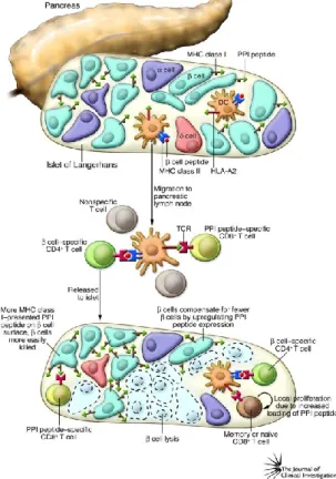
Autoimmune diabetes results from the destruction of the pancreatic β cells. As illustrated in Figure 2, β cell-derived antigens, such as preproinsulin (PPI), are taken up by APCs in the islets resulting in their maturation and subsequent migration to the draining pancreatic lymph nodes (PLNs). Within the PLN, the APCs will present the β cell-derived antigens to naive β cell-reactive T cells, thus activating them.
Following their activation, the autoreactive T cells acquire the ability to migrate to the islets where they will encounter cognate antigen expressed by the β cells, leading to the destruction of the insulin-producing β cells50.
The source of islet antigen is believed to be the wave of physiological pancreatic β cell death that occurs during pancreatic tissue remodelling. Indeed, in mice 51, 52, pigs53 and humans54, this wave of β cell death is evident early in life. In
mice, this neonatal wave of β cell death peaks at about 2 weeks of age51, 52, which
correlates with the initiation of insulitis, which describes the infiltration and inflammation of the pancreatic islets, in diabetes-susceptible mice50. In humans, a
decrease in β cell proliferation is observed starting at 17-32 weeks of gestation up until 6 months of age, while the frequency of β cell apoptosis increases up to 6
months of age and subsequently decreases54. Evidence that this wave in β cell death is
implicated in disease development is demonstrated through the removal of the PLNs from NOD mice prior to 3 weeks of age, when insulitis onset occurs, which results in protection from T1D55. Moreover, it has recently been shown that β cell death in
young, female NOD mice induces the recruitment and activation of various innate immune cells to the pancreas56. This was not observed, however, when 1 week-old
NOD mice were treated with a pan-caspase inhibitor to block β cell death, a treatment that also prevented T1D development up to 30 weeks of age56. Altogether, these
results suggest that the onset of insulitis and subsequent progression of T1D is related to the wave of β cell death that occurs in the islets. However, as β cell death does not appear to be unique to the diabetes-susceptible NOD strain and is also apparent in diabetes-resistant strains 52, 57, 58, it cannot be said that this wave of β cell death is the
trigger that leads to T1D onset despite its importance in the process.
During the first stage of autoimmune diabetes, the pancreatic islets are invaded by a mix of leukocytes, including dendritic cells, macrophages, NK cells, B cells, and T cells.
1.2.6.1 T cells
The utilization of mouse models has greatly enhanced our understanding of autoimmune diabetes pathogenesis. Accordingly, the non-obese diabetic (NOD) mouse model, which develops spontaneous autoimmune diabetes comparable to disease progression in humans (refer to Section 1.3), has become an important tool for researchers. Using this mouse model, it has been shown that T cell-enriched splenocytes from diabetic mice, contrary to T cell-depleted splenocytes, are able to transfer disease to young NOD mice60, demonstrating that T cells are involved in
disease development. Moreover, T cell-modulating therapies, such as anti-CD3 antibody61 and cyclosporin62, inhibit disease development. Altogether, these results
demonstrate that autoimmune diabetes is a T cell-mediated disease.
The role of the diverse T cell subsets in disease development has been investigated. Accordingly, it was shown that a CD4+ T cell clone required the
co-transfer of CD8+-enriched T cells in order to induce autoimmune diabetes in
immunodeficient NOD.SCID (severe combined immunodeficient) mice, which lack both T cells and B cells63. Because of these results, some believed that CD8+ T cells,
and not CD4+ T cells, were essential for autoimmune diabetes development. However, it is now clear that both CD4+ and CD8+ T cells play a role in the
development of disease. Indeed, autoimmune diabetes is impeded in the absence of CD4+ T cells, as shown by the treatment of NOD mice with a monoclonal antibody
targeting CD4+ T cells64, 65 as well as in mice that lack CD4+ T cells66, demonstrating
that CD4+ T cells are involved in disease progression. In addition, autoimmune
diabetes is inhibited in mice that are deficient in CD8+ T cells, either by anti-CD8
antibody injection into young mice67 or of β2 microglobulin-deficient mice, in which
few CD8+ T cells develop68. Altogether, these studies demonstrate that autoimmune
On the contrary, another T cell population, the CD4+CD25+Foxp3+ regulatory
T cells (Tregs), have been shown to play a role in autoimmune diabetes prevention rather than induction. Indeed, patients with IPEX (immunodysregulation,
polyendocrinopathy, enteropathy, X-linked) syndrome, which is caused by mutations in the transcription factor FOXP3 (forkhead box P3), which is important for the development of Tregs, can also develop autoimmune diabetes69. Moreover,
CD28-deficient NOD mice, which exhibit a drastic decrease in Tregs, develop accelerated disease70. In addition, the transfer of splenocytes from diabetic CD28-deficient mice,
thus lacking Tregs, to NOD.SCID mice induced autoimmune diabetes, while the co-transfer of the Treg-deficient splenocytes and Tregs resulted in the prevention of disease transfer70. Therefore, these results demonstrate that Tregs play a role in
autoimmune diabetes prevention.
Altogether, these results demonstrate that while CD4+ and CD8+ T cells are
involved in the promotion of autoimmune diabetes, Tregs are involved in the prevention of disease.
1.2.6.2 B cells
While T cells are implicated in the destruction of pancreatic islets, B cells are also implicated in disease development. Indeed, diabetes incidence is much reduced in NOD mice lacking B cells 71, 72, 73, 74, 75, and similarly, the elimination of most B
cells using anti-CD20 treatment reverses or delays diabetes progression in both mice and humans76, 77. Interestingly, diabetes susceptibility was restored in irradiated B
cell-deficient NOD mice that were reconstituted with syngeneic bone marrow mixed with NOD B lymphocytes, but not in mice reconstituted with syngeneic bone marrow only78. Therefore, B cells contribute to disease progression.
Importantly, the antigen-presenting function of B cells is believed to play a role in autoimmune diabetes development. Accordingly, NOD mice for which the class II MHC molecule I-Ag7 expression was specifically deleted in B cells are
resistant to the development of disease, despite the presence of peri-insulitis, which describes infiltration surrounding the islets, of the pancreas79. Moreover, T cells from
GAD65-primed B cell-deficient NOD mice failed to respond upon antigenic
restimulation in vitro in the presence of either B cell-sufficient or -deficient APCs78.
However, the reconstitution of irradiated B cell-deficient NOD mice with syngeneic bone marrow mixed with NOD B lymphocytes, but not with syngeneic marrow alone, restored T cell responses to GAD65 autoantigen78. These results suggest that
antigen-presenting B cells are required for the initial in vivo priming of GAD65-reactive T cell responses in NOD mice. B cells are also activated in the presence of another autoantigen, the insulin antigen, where the costimulatory molecule, CD86, which is important for T cell crosstalk, is upregulated on autoreactive B cells following insulin autoantigen exposure80. When insulin B cells are specifically eliminated by
anti-insulin monoclonal antibody therapy, diabetes incidence is inhibited in NOD mice80.
Altogether, B cells can present β cell autoantigens to autoreactive T cells, which overcomes a checkpoint in T cell tolerance to islet β cells and promotes autoimmune diabetes development.
H2-O (HLA-DO in humans) expression can influence the participation of B cells in the germinal centers, which are the site of B cell maturation 81. More specifically, H2-O is a MHC class II-like protein that inhibits
peptide loading onto MHC II by inhibiting the function of the MHC class II-like protein H2-M (HLA-DM in humans) 82. Recent publications have demonstrated that
germinal center B cells exhibit a decreased H2-O expression 83. Moreover, it has been
shown that H2-O-deficient antigen-specific B cells preferentially populate germinal centers in comparison to H2-O-sufficient B cells due to their enhanced ability to obtain antigen-specific T cell help, where T cell help is dependent upon antigen presentation by B cells 84. Interestingly, H2-O expression is cell
maturation-dependent as both mature B cells and DCs express H2-M and H2-O whereas developing immature B cells and DCs express very little H2-O 85, 86. These results
suggest that, as H2-O limits peptide exchange on MHC class II, loss of function of H2-O might lead to autoimmunity as presentation of self-peptides by
antigen-presenting cells such as B cells would not be attenuated. Although it has been shown that H2-O-deficient mice do produce higher titers of autoantibodies 87, H2-O
expression on B cells and its direct association with autoimmune diabetes has not been evaluated.
B cells also contribute to autoimmune diabetes through the production of autoantibodies. Indeed, it has been demonstrated that maternally transmitted
autoantibodies contribute to the disease onset in NOD offspring88, 89. Accordingly, the
NOD progeny from B cell–deficient NOD mothers that were mated with a NOD males, as well as the NOD progeny of diabetes-resistant mothers that were implanted with the NOD embryos, displayed a reduction in autoimmune diabetes incidence88.
Thus, the maternal transmission of antibodies is implicated in disease development.
Altogether, it is clear B cells play a role in autoimmune diabetes development.
1.2.6.3 Macrophages
Macrophages are present in the pancreatic islet infiltrates of NOD mice prior to T cell infiltration64, 90, suggesting that macrophages are involved in autoimmune
diabetes progression. Macrophages are believed to participate in disease pathogenesis through the production of various pro-inflammatory cytokines, such as tumor
necrosis factor alpha (TNF-α) and interleukin-1 (IL-1), which contribute to β cell dysfunction90, 91, and interleukin-12 (IL-12), which promotes the differentiation of
diabetogenic CD8+ cytotoxic T cells in the diabetes-prone setting92. Interestingly, the
injection of a monoclonal blocking antibody that is specific for the myelomonocytic adhesion-promoting type-3 complement receptor (CD11b/CD18), which targets macrophages but not T cells, prevented islet infiltration by both macrophages and T cells and inhibited disease onset93. Interestingly, TNF-α and IL-1β-producing
macrophages have also been observed in pancreatic islet infiltrates from patients with recent-onset autoimmune diabetes in humans 94. Therefore, these studies demonstrate
1.2.6.4 Natural killer cells
Natural killer (NK) cells are composed of effector lymphocytes, known to play a crucial role in the control of viral infection and the prevention of cancer95, as
well as regulatory cells95. Indeed, while NK cells can kill immature DCs in humans
and mice thereby influencing DC homeostasis 96, 97, the interaction between NK cells
and immature monocyte-derived DCs in humans can also promote cytokine
production by DCs 96. Similarly, it has been shown that NK cells can both promote
the priming of CD4+ T cells through the secretion of IFN-γ98 or kill activated T cells
that express insufficient amounts of classical or non-classical MHC class I molecules
99. Moreover, IL-10-secreting NK cells inhibit antigen-specific T cell proliferation
while maintaining their natural cytotoxic activity 100. Therefore, NK cells are not a
homogenous subset and are composed of both effector and regulatory subsets.
NK cells are another cell population implicated in autoimmune diabetes. Indeed, similar to macrophages, NK cells also infiltrate the pancreas before T cells in NOD mice101 and can be found directly within the islets of diabetic patients47.
Moreover, the depletion of NK cells using an anti-asialo-Gm1 antibody prevented autoimmune diabetes in NOD mice 102. Interestingly, NK cells from
diabetes-susceptible NOD mice have impaired immune functions compared with NK cells from diabetes-resistant mouse strains103, 104. Accordingly, NK cells isolated from the
pancreas of NOD mice are hyporesponsive such that they produce significantly less interferon gamma (IFNγ) and express less CD107a, which is a marker of
degranulation101. Interestingly, NOD mice congenic for the NK cell surface marker
NK1.1, which is expressed in the diabetes-resistant C57BL/6 strain but not in the NOD strain, exhibit a reduced disease incidence105. This protection from diabetes
development also correlates with improved NK cell performance, as compared with wild-type NOD mice105. Thus, it is possible that the functional deficiency of NK cells
from diabetes-susceptible mice, rather than an efficient NK cell function, contributes to autoimmune diabetes progression. Altogether, these results demonstrate that NK
cells infiltrate the pancreas and play a role in autoimmune diabetes. However, whether NK cells promote or protect from autoimmune diabetes remains controversial.
1.2.6.5 Dendritic cells
Antigen-presenting dendritic cells (DCs) play an important role in autoimmune diabetes pathogenesis. Accordingly, DCs are able to capture
autoantigens released after β cell death, which occurs physiologically in NOD mice at 2 weeks of age during tissue remodelling52, as described earlier in section 1.2.6.
Subsequently, the autoantigen-specific DCs migrate to the PLN where they can present the autoantigen to islet antigen-specific T cells, which initiates the
diabetogenic response52. Indeed, a large frequency of DCs presenting β cell antigens
can be found within the PLNs of NOD mice52. Interestingly, in diabetes-susceptible
NOD mice that constitutively overexpress human HLA-DO in DCs, where, as previously explained, the association of HLA-DO with HLA-DM modulates the peptide loading function of HLA-DM thus hampering the presentation of self-antigens and the inappropriate activation of T cells 82, 106, disease development is
completely blocked107. This inhibition of autoimmune diabetes occurred despite a
comparable diabetogenic T cell repertoire and level of peri-insulitis to control NOD mice 107, suggesting that HLA-DO expression dampens the presentation of
islet-derived autoantigens by DCs to T cells and can consequently promote T cell tolerance. Altogether, it is clear that the antigen-presenting function of DCs plays a key role in disease initiation.
DC involvement in autoimmune diabetes has been demonstrated via the in vivo treatment using Fms-like tyrosine kinase 3 ligand (FLT-3L), which expands DC populations. Indeed, FLT-3L treatment can accelerate autoimmune diabetes
development in NOD mice108. Nevertheless, the contribution of DCs to autoimmune
diabetes development seems to depend on the timing of FLT-3L treatment and the presence of autoreactive T cells such that FLT-3L treatment can prevent autoimmune
diabetes in NOD mice at early stages of autoimmune diabetes progression109, whereas
it can also accelerate disease development at later stages when islet antigen-specific CD8+ T cells are already detectable in the blood108. Altogether, the contribution of
DCs to autoimmune diabetes development seems to depend on both the stage of disease development and the presence of autoreactive T cells.
1.3 The non-obese diabetic mouse model
Murine models are a useful tool for research as they allow for the characterization of the cellular mechanisms involved in the progression of autoimmune disease. Autoimmune diabetes can be experimentally induced with various chemicals, such as streptozotocin110 and cyclophosphamide111, or by genetic
manipulation, which is observed for transgenic mice. However, spontaneous murine models of autoimmune diabetes also exist. In particular, the non-obese diabetic (NOD) mouse strain is a key tool for investigating the aetiology of human autoimmune diabetes112. Indeed, the NOD mouse strain spontaneously develops
autoimmune diabetes, which shares many similarities to disease development in humans, such as the presence of autoreactive CD4+ and CD8+ T cells as well as
pancreas-specific autoantibodies. Moreover, the broadly immunosuppressive anti-CD3 therapy, which has been successful in the treatment of new-onset type 1 diabetic patients during clinical trials, is a protocol initially developed in the NOD mouse 113, 114, further demonstrating a parallel in disease development between humans and
mice.
Investigation of the genetic control of autoimmune diabetes in the NOD mouse has revealed an interesting parallel with genetic susceptibility to disease in humans28, 32, 115, 116. First, the MHC haplotype H-2g7 expressed by the NOD mouse
does not express an MHC class II I-E molecule because of a defective Eα locus. In addition, similar to the human autoimmune diabetes-associated HLA-DQ β chain (HLA-DQB1*0302) 22, 117, 118, NOD mice have a unique MHC class II I-A molecule
containing a non-aspartic acid substitution at position 57 of the β chain119, which
substantially alters the repertoire of MHC binding peptides presented by this allele120.
Furthermore, autoimmune diabetes is multigenic in both mice121, 122 and humans123
with the MHC being the major locus contributing to disease development123.
Accordingly, NOD.B10-H-2b mice, for which the H-2g7 is replaced by the H-2 region
from the diabetes-resistant C57BL/10SnJ (B10) strain, do not develop autoimmune diabetes124. However, in B10.NOD-H-2g7 mice, for which the H-2 region from the
diabetes-resistant B10 strain is replaced by the NOD H-2g7, diabetes development
does not ensue105, 108, demonstrating that non-MHC genes are also implicated in
disease susceptibility122, 125. Indeed, in the NOD mouse, over 20 insulin-dependent
diabetes (Idd) loci have been identified with the MHC composing the Idd1 locus126.
Accordingly, most of the human and mouse alleles conferring risk for autoimmune diabetes reveal common candidate genes that are concordant between humans and the NOD mouse, including the aforementioned INS ( within the Idd2 locus) and CTLA-4 (within the Idd12 locus) genes115, 116, 127. Hence, the NOD mouse represents a useful
tool for the study of the genetic factors influencing autoimmune diabetes susceptibility.
In terms of autoimmune diabetes pathogenesis in the NOD mouse, infiltration begins around 3-4 weeks of age such that few immune cell infiltrates surround the islet (peri-insulitis). By 10 weeks of age, the cell infiltrates progress and invade the islets (insulitis). Overt diabetes is observed beginning at 12-14 weeks of age in females and slightly later in males. By 30 weeks of age, the incidence of disease observed in NOD mice is 80% in females and less than 20% in males128.
Moreover, the pathological examination of the pancreas in NOD mice revealed a high frequency of lymphocyte infiltration around and/or into the pancreatic islets as well as a reduction in the number and size of pancreatic islets in the overt diabetic mice128.
Finally, the NOD mouse also displays key diabetic symptoms that also arise in diabetic patients, such as polyuria, polydipsia, hyperglycemia, and glucosuria128.
Because of these many similarities, the NOD mouse is a key tool for autoimmune diabetes research.
Nevertheless, it must be noted that there also exists various differences between NOD mice and humans. Firstly, there exists various differences between the innate and adaptive immunity of mice and humans, including variances in the balance of leucocyte subsets, γ T cells, cytokines and cytokine receptor expression, to name a few 129. Moreover, additional differences have been noted between NOD mice and
humans with respect to autoimmune diabetes manifestation. For instance, the islet infiltration in NOD mice is characterized by peri-insulitis followed by insultis. In
contrast to what is observed in NOD mice, few leukocytes are detectable within the inflamed islets of human patients 130. Furthermore, while CD4+ T cells are the
predominant T cell subset found in the NOD mouse islet infiltrate131, CD8+ T cells are
the most abundant cell population detected during insulitis in human patients 132. In
addition, the presence of maternal islet autoantibodies in the offspring of diabetic mice has been identified as a diabetogenic factor in mice 88, as described in Section
1.2.6.2. However, children of mothers with autoimmune diabetes who were
autoantibody positive at birth had significantly lower risks for developing the disease than children of mothers with autoimmune diabetes who were islet autoantibody negative at birth 133, suggesting that fetal exposure to islet autoantibodies may be
protective against disease development in humans. Lastly, the gender bias observed for autoimmune diabetes development in NOD mice, which is described above, is not observed in humans134. Therefore, despite many similarities, the NOD mouse does
not fully mimic all aspects of autoimmune diabetes observed in humans.
Altogether, because of its many similarities to autoimmune diabetes in human patients, the NOD mouse has been used extensively in autoimmune diabetes research and as a pre-clinical tool for the development of new therapeutic strategies for the treatment of this disease.
1.4 Autoimmune diabetes therapy
Due to the many complications associated with long-term autoimmune
diabetes, the life expectancy of people living with the disease may be shortened by as much as 15 yearsiii. As a result, research continues to better understand all the factors
implicated in autoimmune diabetes progression in order to develop new therapies that will prevent, reverse and/or cure the disease. The main therapeutic avenues that are discussed in this section include the transplantation of pancreatic islets or of whole pancreas as well as immunotherapy.
1.4.1 Transplantation
Some patients who have experienced severe complications associated with autoimmune diabetes have seeked an alternative therapy, namely either pancreatic or islet transplantation, in an attempt to cure their disease and achieve glucose tolerance and an alleviation of their complications.
1.4.1.1 Pancreas transplantation
A pancreas transplant involves implanting a healthy pancreas from a recently deceased individual, or a partial pancreas from a living donor, into the diabetic patient. As mentioned above, the majority of pancreas transplant recipients have complicated autoimmune diabetes. Since 75% of diabetic patients with kidney failure do not survive longer than 5 years while receiving dialysis135, approximately 90% of
pancreas transplant recipients are either uremic (in kidney failure) or post-uremic. Importantly, mortality rate was decreased by approximately 50% when patients with autoimmune diabetes and end-stage renal failure received a simultaneous pancreas and kidney transplant135.
In patients who receive a pancreas transplant alone, the actual patient and pancreas survivals at 5 years is 98.6% and 73.2%, respectively136. Long-term
follow-up of transplant recipients has demonstrated that pancreas transplants are successful in returning normal glycemia to diabetic patients for periods as long as 1-2
decades137. Indeed, the half-life of the pancreas graft currently averages at 16.7
years138. Additional benefits of successful pancreatic transplant are improved
retinopathy, blood pressure, as well as neuropathic and cardiac function139. The
inability to maintain insulin-independence following transplantation is mostly explained by technical failure in the postoperative phase and rejection in the long-term period138, where graft rejection remains the leading cause of pancreas loss140.
Autoimmunity is also increasingly recognized as a cause of graft loss141.
Nevertheless, improvements in graft preservation, surgical techniques,
immunosuppression, and prophylactic treatments are expected to further improve the results of pancreas transplantation138.
Altogether, pancreas transplantation consistently induces
insulin-independence in diabetic patients, however this is at the cost of major surgery and its associated complications as well as life-long immunosuppression138, hence the reason
why only patients who have severe complications associated with the disease, and thus have no other options, consider this means of treatment.
1.4.1.2 Islet transplantation
Islet transplantation is similar to pancreas transplantation in that it entails the transplantation of isolated islets from a donor pancreas into a diabetic patient. However, this transplantation is considered to be a more attractive alternative to pancreas transplantation since the islets can simply be infused into the liver of the patient through a catheter. Once transplanted, the islets will begin to produce insulin leading to glucose regulation.
In 1988, Ricordi and colleagues first reported on the semi-automated method for human pancreatic islet isolation making it feasible to consistently reproduce the
difficult process of separating insulin-producing β cells from the rest of the pancreas142. In 2000, a group in Edmonton developed a steroid-free
immunosuppressive regime termed the Edmonton protocol, which promoted long-term islet survival and function in patients143. With the development of the Edmonton
protocol, 100% of patients, in comparison to the previous 8%, did not require insulin therapy 1 year after transplantation143. The initial success of the “Edmonton Protocol”
quickly created an international passion and many centers adopted this technique. Unfortunately, 5 years after transplantation, more than 90% of the patients were again dependent on exogenic insulin144. Although much work is left to be done with regards
to islet transplantation, researchers are still confident that this form of therapy will prove to be a success.
Indeed, both pancreas and islet transplantations result in the restoration of proper insulin production and, consequently, blood glucose regulation. Therefore, both methods have the potential to cure autoimmune diabetes, at least for a given amount of time. Nevertheless, both surgeries are accompanied by a lifelong regimen of immunosuppressive drugs to prevent graft rejection, where both the surgery itself and permanent immunosuppression is considered to be far more dangerous than continued insulin replacement therapy.
1.4.2 Immunotherapy
Several immunosuppressive agents have been tested for their ability to prevent/delay/reverse autoimmune diabetes development due to their ability to target either T cells or B cells, both of which are involved in disease progression.
1.4.2.1 Cyclosporine A
Cyclosporine A is an immunosuppressive agent that is widely used to prevent graft rejection due to its ability to interfere with T cell activity145. As T cells are
directly involved in the destruction of pancreatic β cells, cyclosporine A was studied as a preventative therapy for autoimmune diabetes in mice and humans. NOD mice between the ages of 4 and 9 weeks that were treated with cyclosporine every 2 days
until 23 weeks of age demonstrated a significant reduction in disease incidence in comparison to control mice62. These results also correlated with a decrease in blood
glucose levels, islet infiltration and islet destruction62. However, when cyclosporine
treatment was started after development of glucose intolerance, the treatment appeared to have little therapeutic effect on disease development62. In humans,
approximately 50% of patients who were treated with cyclosporine within 6 weeks of diagnosis and for a duration of 2 to 12 months became insulin-independent and showed C-peptide plasma concentrations in the normal range as well as decreasing titres of islet cell antibodies146. Nevertheless, this agent demonstrated multiple side
effects, including nephrotoxicity, making it unsuitable for the treatment of disease147.
1.4.2.2 Anti-CD3 monoclonal antibodies
Anti-CD3 antibodies, including teplizumab and otelixizumab, target T cells by binding to the CD3 molecule located on their cell surface. The ability of a CD3-specific antibody to reverse autoimmune diabetes onset in 64-80% of female NOD mice61 paved the way for clinical trials in diabetic patients. Recent-onset diabetic
patients who received the anti-CD3 antibody otelixizumab showed a sustained remission of the disease for up to four years148. Success of the treatment correlated
with the initial residual β cell function where patients presenting with a higher functional β cell mass before starting the treatment responded better. Indeed, 18 months post-treatment, nearly 75% of patients in this subgroup were clinically
insulin-independent148. Disease remission following anti-CD3 treatment is believed to
be a result of the simultaneous action of the antibody on effector T cells, a fraction of which was deleted by apoptosis, and on Tregs, whose function was preserved and even increased149, 150. Therefore, anti-CD3 antibodies can be used to reverse and delay
autoimmune diabetes progression. 1.4.2.3 Anti-CD20 monoclonal antibody
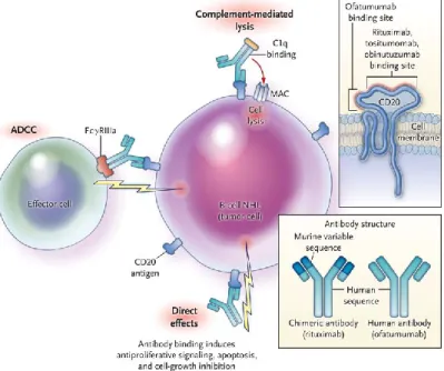
The anti-CD20 monoclonal antibody, rituximab, is a chimeric monoclonal antibody, incorporating human immunoglobulin heavy-chain sequences and murine immunoglobulin variable regions151, that eliminates B cells by binding to the
extracellular portion of CD20 molecules, which are expressed on the cell surface of all B cells except for plasma cells. More specifically, rituximab depletes B cells via three mechanisms of action152, 153 (Figure 3). The first mechanism is via the
modulation of the apoptotic signal transduction pathway resulting in direct cytotoxicity. The second mechanism is via antibody-dependent cell-mediated cytotoxicity, in which NK cells and macrophages bind to the Fc portion of the anti-CD20 antibody by means of their Fcγ receptor and subsequently release effector molecules such as perforin leading to cell lysis. The third mechanism is via
complement-dependent cytotoxicity, for which complement binds to the Fc portion of the anti-CD20 antibody thereby activating the complement cascade and formation of the membrane attack complex154. Although the mechanism by which B cell depletion
ameliorates disease is not completely understood, the effect on antibody production and antigen presentation to T cells are thought to be important.
Figure 3. Mechanisms of action of anti-CD20 antibodies inducing B cell depletion. Adapted from 151.