Cyclophosphamide inhibits the generation and function of CD8
⫹regulatory T cells
Ilaria Traverso
a, Daniela Fenoglio
a,b, Simone Negrini
a,b, Alessia Parodi
a, Florinda Battaglia
a,
Francesca Kalli
a, Giuseppina Conteduca
a, Samuele Tardito
a, Paolo Traverso
a,c, Francesco Indiveri
a,b,
Gilberto Filaci
a,b,*
aCentre of Excellence for Biomedical Research, University of Genoa, Genoa 16132, Italy bDepartment of Internal Medicine, University of Genoa, Genoa 16132, Italy cDepartment of Surgical Sciences, University of Genoa, Genoa 16132, Italy
A R T I C L E I N F O Article history:
Received 7 October 2011 Accepted 21 December 2011 Available online 14 January 2012 Keywords:
Cyclophosphamide CD8⫹
Treg Cancer Autoimmunity
A B S T R A C T
CD8⫹regulatory T cells (Treg) and CD4⫹CD25⫹Treg infiltrate human cancers, thus favoring tumor immune escape. Therefore, in the setting of antitumor therapeutic protocols, it is important to associate antitumor treatment with agents that are able to inhibit Treg function. Cyclophosphamide (CY) has been demonstrated to be effective in counteracting CD4⫹CD25⫹Treg activity. Hence, we tested its inhibitory efficacy on human CD8⫹Treg. Because CY is a prodrug, 4-hydroperoxycyclophosphamide (4-HC), a derivative of CY that in aqueous solution is converted to 4-hydroxycyclophosphamide, an active metabolite of CY, was used. 4-HC significantly inhibited CD8⫹Treg generation and function but only at the higher tested concentration (0.5 g/mL), that is, in the therapeutic range of the drug. The lower 4-HC concentration tested (0.1 g/mL) was almost ineffective. 4-HC inhibitory effects were related to apoptosis/necrosis induction. When CD8⫹CD28⫹ non-Treg were analyzed for comparative purposes, significantly lower cytotoxic rates among these cells were observed than among CD8⫹Treg, which were differentiated because they did not express the CD28 molecule. These data demonstrate that CD8⫹Treg are inhibited through cytotoxic phenomena by CY, thus supporting the use of this drug at adequate concentrations and schedules of administration as a Treg inhibitor in combinatorial chemo- or immunotherapeutic anticancer protocols.
䉷 2012 American Society for Histocompatibility and Immunogenetics. Published by Elsevier Inc. All rights reserved.
1. Introduction
Regulatory T cells (Treg) play a pivotal role in the maintenance of immunologic self-tolerance, downregulating the activation/pro-liferation of self-reactive T cells and thus preventing the develop-ment of various autoimmune diseases[1,2]. However, Treg have been detected among tumor-infiltrating lymphocytes[3,4]. Treg infiltrating cancer lesions are thought to be responsible for the inhibition of antitumor immune responses, thus favoring tumor immune escape. Indeed, administration of ipilimumab[5], an agent blocking the CTLA4-CD80/CD86 interaction— one of the molecular circuits adopted by Treg for suppressing immune responses[6]— has therapeutic effects against cancer[7]. The importance of coun-teracting tumor-infiltrating Treg function to set antitumor proto-cols of chemo- and/or immunotherapy that are more efficient than those currently in use is therefore clear. However, Treg belong to different T cell lineages[8,9]and each Treg subtype uses different mechanisms to induce immune suppression[10,11]. Hence, the search for additional agents that can block tumor-infiltrating Treg
is mandatory. The chemotherapeutic agent cyclophosphamide (CY) has been reported to be effective on Treg. CY is a nitrogen mustard alkylating agent that exhibits great cytotoxicity against cells ac-tively replicating their DNA[12,13]. CY is an inactive prodrug that requires activation by the hepatic cytochrome P-450 enzyme sys-tem to form the active metabolite 4-hydroxycyclophosphamide, which is in equilibrium with its tautomer aldophosphamide. These 2 intermediate metabolites rapidly diffuse out of hepatic cells into the circulation and are subsequently taken up by other cells, includ-ing cancer cells. Within the cells, aldophosphamide decomposes to form the cytotoxic phosphoramide mustard, which produces the interstrand DNA cross-links responsible for the cytotoxic proper-ties of the drug. The selective toxicity on tumor cells occurs because the concentration of the enzymes converting aldophosphamide into the cytotoxic metabolite is higher in tumor cells than in normal cells[14]. CY is one of the most successful and widely used drugs for the treatment of hematologic and solid malignancies[15,16], as well as for the treatment of different autoimmune disorders [17], and therefore is commonly considered an immunosuppressive drug. However, evidence exists that CY may have immunostimulatory ef-fects (i.e., enhancing the therapeutic activity of adoptive T cell immu-* Corresponding author.
E-mail address:gfilaci@unige.it(G. Filaci).
Contents lists available atSciVerse ScienceDirect
0198-8859/$36.00䉷 2012 American Society for Histocompatibility and Immunogenetics. Published by Elsevier Inc. All rights reserved. doi:10.1016/j.humimm.2011.12.020
notherapy)[18]. Recent studies have linked the immunostimulating effect of CY to the selective inhibition/depletion of CD4⫹CD25⫹Treg in both experimental[19 –21]and human[22,23]tumors. However, the Treg repertoire also includes CD8⫹ Treg; no information on the sensitivity of CD8⫹Treg to CY is currently available. This article reports data on the effects of 4-hydroperoxycyclophosphamide (4-HC), a derivative of CY that in aqueous solution is converted to 4-hydroxycyclophosphamide (the active metabolite of CY)[24], on CD8⫹Treg function and viability.
2. Subjects and methods
2.1. Generation of CD8+Treg from peripheral blood
CD8⫹Treg were generated as described previously[25]. Briefly, peripheral blood mononuclear cells (PBMC) were isolated from buffy coats by centrifugation on a Ficoll–Hypaque gradient (Bio-chrom AG, Berlin, Germany) for 30 minutes at 1800 rpm. PBMC were incubated in RPMI 1640 culture medium (Gibco by Life Tech-nologies Ltd., Paisley, UK) with 10% fetal calf serum (Invitrogen) in culture flasks (Corning Life Sciences, Amsterdam, The Netherlands) at 37⬚C overnight. CD8⫹T lymphocytes were purified from
nonad-herent cells by magnetic bead separation using microbeads conju-gated with monoclonal antibody (mAb) specific for the CD8 antigen (Dynal CD8 positive isolation kit, Invitrogen by Life Technologies Ltd., Paisley, UK). Purified CD8⫹T lymphocytes (2⫻ 105cells/well)
resuspended in culture medium consisting of RPMI 1640 (Gibco by Life Technologies Ltd., Paisley, UK) with 10% fetal calf serum (Invit-rogen by Life Technologies Ltd., Paisley, UK) were incubated with 20 U/mL of interleukin (IL)-2 (Proleukin, Eurocetus, Amsterdam, The Netherlands) and 10 ng/mL of IL-10 (PeproTech) in 96-well flat-bottom plates (Corning Life Sciences, Amsterdam, The Nether-lands) at 37⬚C for 7 days.
At the end of the incubation, the cells were collected, washed, counted, and used as suppressors in a proliferation suppression assay.
2.2. Proliferation suppression assay
The suppressive activity of Treg was evaluated by monitoring the inhibition of dye dilution in PBMC stained with carboxyfluores-cein succinimidyl ester (5M; Molecular Probes, Invitrogen by Life Technologies Ltd., Paisley, UK) before the test. Thereafter, the cells were pulsed with anti-CD3 UCHT1 mAb (5g/mL, BD Bioscience, Franklin Lakes, NJ) and cultured for 5 days in a 96-well round-bottom plate (1⫻ 105
cells/well) in the presence (or absence) of in
vitro– generated CD8⫹Treg (1⫻ 105
cells/well). At the end of the incubation the samples were washed in phosphate-buffered saline and analyzed using a FACSCanto flow cytometer (BD Bioscience, Franklin Lakes, NJ) using FACSDiva software (BD Biosciences, Franklin Lakes, NJ).
2.3. 4-HC treatment
4-HC was purchased from Niomech–IIT GmbH (University of Bielefeld, Bielefeld, Germany). The drug was opportunely diluted to be used at a final concentration of 0.5 or 0.1g/mL, concentrations within the therapeutic range of 4-HC[26]. To evaluate the effects on CD8⫹Treg generation, the drug was added to cultures during the 7-day incubation with IL-2 and IL-10. To analyze 4-HC effects on already generated CD8⫹Treg, the drug was added to cultures on the 6th day of generation for 24 hours.
2.4. Immunofluorescence analyses
Cell expression of membrane antigens was analyzed by immu-nofluorescence by incubating the cells (1⫻ 105
cells in 100L of
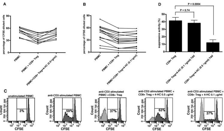
Fig. 1. Analysis of 4-hydroperoxycyclophosphamide (4-HC) effects on CD8⫹regulatory T cell (Treg) generation. (A and B) Anti-CD3 monoclonal antibody (mAb)-induced peripheral blood mononuclear cell (PBMC) proliferation (expressed as a percentage of carboxyfluorescein succinimidyl ester [CFSE]-diluted cells) in the presence or absence of CD8⫹Treg generated with or without 4-HC used at 0.5 (A) or 0.1 (B)g/mL. (C) Representative results of proliferation analysis of anti-CD3 mAb-stimulated PBMC from donor 16 cultured in the presence or absence of CD8⫹Treg generated in the presence or absence of either 0.5 or 0.1g/mL of 4-HC. Percentages of proliferating cells are indicated. (D) Comparison between the mean suppressive activity of untreated CD8⫹Treg and that of CD8⫹Treg generated in the presence of either 0.1 or 0.5g/mL 4-HC.
phosphate-buffered saline) with specific mAbs at 4⬚C for 30 min-utes in the dark. The following mAbs were used: fluorescein iso-thiocyanate (FITC)-conjugated anti-CD45RA, phycoerythrin (PE)-conjugated anti-CD127, allophycocyanin-(PE)-conjugated anti-CD39, PE– cyanin 7– conjugated CD8, and PerCP-conjugated anti-CD28. After staining procedures, the cells were acquired and ana-lyzed by a FACSCanto flow cytometer using FACSDIVA software.
2.5. Analysis of apoptotic and necrotic cells
CD8⫹purified T cells, preexposed or not to 4-HC, were incubated
with Pe– cyanin 7– conjugated anti-CD8 and allophycocyanin-conjugated anti-CD28 for 15 minutes at room temperature in the dark, following the manufacturer’s instructions. The percentage of apoptotic cells was assessed by flow cytometry after cell incubation with FITC-conjugated annexin V (BD Pharmingen, San Diego, CA). To quantify necrotic cells, 7-aminoactinomycin D (BD Biosciences, Franklin Lakes, NJ) was added to the cells immediately before flow cytometric analysis. Cells stained only by FITC-conjugated annexin V were considered apoptotic, whereas cells stained by both FITC-conjugated annexin V and 7-aminoactinomycin D were enumer-ated as necrotic cells.
2.6. Statistical analysis
All results are expressed as means⫾ standard deviation. Statis-tically significant differences between mean percentage data (re-lated to apoptosis, necrosis, and suppressive activity of different T cell subsets treated or not with 4-HC) were analyzed using the Mann–Whitney test for nonparametric values. Correlations be-tween variables were analyzed by Spearman’s correlation test. Dif-ferences were considered significant when p⬍ 0.05. The statistical
analyses were performed using GraphPad Prism 4.0 Software (GraphPad Software Inc., San Diego, CA).
3. Results
3.1. Analysis of the effects of 4-HC on CD8+Treg generation
and function
To evaluate the effects of 4-HC on in vitro generation of CD8⫹Treg, ex vivo purified CD8⫹T cells were incubated for 7 days with the drug used at 0.1 or 0.5g/mL concentrations. The suppres-sive activities of CD8⫹Treg generated in the presence or absence of the drug were comparatively analyzed. Control CD8⫹Treg exerted a suppressive activityⱖ25% in all analyzed samples. CD8⫹Treg
pre-incubated with 0.5 g/mL of 4-HC exhibited significantly lower suppressive activity than control CD8⫹Treg (12% vs 41%, p ⫽ 0.0004; Fig. 1). To the contrary, the suppressive activity of CD8⫹Treg pretreated with 4-HC at 0.1g/mL concentration was comparable to that of control CD8⫹Treg (38% vs 41%, p⫽ 0.74; Fig. 1).
To also assess the effects of 4-HC on the function of already generated CD8⫹Treg, in vitro– generated CD8⫹Treg were incubated for 24 hours with the drug used at the same concentrations as above. Again, the suppressive activities of treated or untreated CD8⫹Treg treated were comparatively analyzed. CD8⫹Treg incu-bated with 0.5g/mL of 4-HC exhibited significantly reduced sup-pressive activity compared with control CD8⫹Treg (20% vs 41% p⫽
0.0004;Fig. 2). Instead, the suppressive activity of CD8⫹Treg incu-bated overnight with 4-HC at 0.1g/mL concentration was compa-rable to that of control CD8⫹Treg (38% vs 41%, p⫽ 0.45;Fig. 2).
Fig. 2. Analysis of 4-hydroperoxycyclophosphamide (4-HC) effects on CD8⫹regulatory T cell (Treg) suppressive activity. (A and B) Anti-CD3 monoclonal antibody (mAb)-induced peripheral blood mononuclear cell (PBMC) proliferation (expressed as a percentage of carboxyfluorescein succinimidyl ester [CFSE]-diluted cells) in the presence or absence of in vitro– generated CD8⫹Treg incubated for 24 hours with or without 4-HC used at 0.5 (A) or 0.1 (B)g/L (C) Representative results of proliferation analysis of anti-CD3 mAb-stimulated PBMC from donor 16 cultured in the presence or absence of CD8⫹Treg incubated or not for 24 hours in the presence of either 0.5 or 0.1 g/mL of 4-HC. Percentages of proliferating cells are indicated. (D) Comparison between the mean suppressive activity of untreated CD8⫹Treg and that of in vitro– generated
3.2. 4-HC does not alter the phenotype of CD8+
Treg
CD127, the IL-7 receptor, is downmodulated in Treg subpopula-tions [27,28]so it is commonly monitored for assessing T cell differentiation toward regulatory function. CD39 is a nucleosidase often expressed by Treg cells[29,30]and CD45RA is expressed on non-antigen-specific CD8⫹Treg[9,31]. To assess whether the inhi-bition of CD8⫹Treg suppressive activity was dependent on pheno-typic changes eventually induced by 4-HC, the expression of CD127 (IL-7 R), CD39, and CD45RA antigens was analyzed.Table 1 demon-strates that 4-HC did not cause significant variations of surface antigen expression when added to CD8⫹ T cell cultures during CD8⫹Treg generation. Comparable findings were achieved when in
vitro– generated CD8⫹Treg were incubated for 24 hours with 4-HC (Table 2).
3.3. 4-HC induces CD8+
Treg apoptosis and necrosis
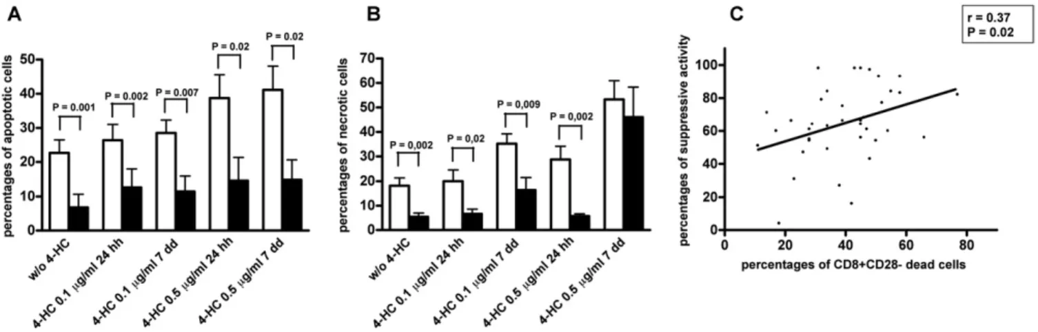
4-HC is an antiblastic drug; hence, its pharmacologic activity could be related to cell death induction. Therefore, the rates of apoptotic and necrotic cells in CD8⫹Treg exposed to 4-HC were analyzed. Because peripheral blood– derived CD8⫹Treg do not ex-press the CD28 antigen [25], the percentages of apoptotic and necrotic cells were calculated by gating CD8⫹CD28⫺ and CD8⫹ CD28⫹T cells separately. Interestingly, the percentage of apoptotic cells among CD8⫹CD28⫺ Treg generated in the presence of 0.5 g/mL of 4-HC was significantly higher than that observed among untreated CD8⫹CD28⫺ Treg (Fig. 3A). Similarly, a significantly higher percentage of apoptotic cells was detected among in vitro– generated CD8⫹CD28⫺Treg incubated for 24 hours with 0.5g/mL of 4-HC compared with untreated CD8⫹CD28⫺Treg (Fig. 3A). To the contrary, comparable rates of apoptotic cells between treated and untreated cells were observed when CD8⫹CD28⫺Treg were incu-bated with 0.1g/mL of 4-HC either during or after Treg generation (Fig. 3A).
The analysis of necrosis in CD8⫹CD28⫺Treg demonstrated a significantly increased percentage of necrotic cells among CD8⫹ CD28⫺Treg generated in the presence of 4-HC, independent of drug concentration, compared with untreated CD8⫹CD28⫺ Treg (Fig. 3B). To the contrary, no significant differences in necrotic cell per-centage were detected between CD8⫹CD28⫺Treg incubated or not for 24 hours with 4-HC (Fig. 3B).
To achieve comparative information on sensitivity to 4-HC by Treg (CD8⫹CD28⫺) and non-Treg (CD8⫹CD28⫹) T cell populations, the percentages of apoptotic or necrotic cells were also analyzed within the CD8⫹CD28⫹T cells. CD8⫹CD28⫹T cells incubated for 7
days with 0.5g/mL of 4-HC exhibited higher apoptosis percent-ages compared with untreated cells (Fig. 3C). Similarly a signifi-cantly higher percentage of apoptotic cells was detected among CD8⫹CD28⫹ T cells after their incubation for 24 hours with 0.5 g/mL of 4-HC compared with untreated cells. To the contrary, comparable rates of apoptotic cells between treated and untreated cells were detected when CD8⫹CD28⫹T cells were incubated for 24 hours or 7 days with 0.1g/mL of 4-HC (Fig. 3C). Significantly increased percentages of necrotic T cells were only observed among CD8⫹CD28⫹ T cells incubated for 7 days with 0.1 or 0.5 g/mL of 4-HC, but not among CD8⫹CD28⫹T cells incubated for 24
hours with 4-HC, compared with untreated cells (Fig. 3D). Interest-ingly, the comparison of apoptotic cell percentages between CD8⫹CD28⫺and CD8⫹CD28⫹T cells subpopulations indicated higher
apoptotic rates in the former than in the latter T cell subpopulation under all culture conditions (Fig. 4A). Similarly, comparison of ne-crotic cell percentages between CD8⫹CD28⫺ and CD8⫹CD28⫹ T cells indicated a higher level in the former than in the latter T cell subpopulation under all culture conditions, with the exception of cells incubated for 7 days with 0.5g/mL of 4-HC (Fig. 4B).
To verify whether 4-HC-mediated inhibition of suppressive ac-tivity could be strictly related to its cytotoxic effects on CD8⫹Treg, the suppressive activity of CD8⫹Treg variably exposed to 4-HC was correlated with drug-induced mortality (considering the sum of percentages of apoptotic and necrotic cells):Fig. 4C illustrates a clear correlation between the 2 variables, thus supporting the con-cept that the 4-HC impact on CD8⫹Treg activity is mainly mediated by cytotoxic phenomena.
4. Discussion
The results of this study indicate the following: (1) 4-HC, at a dose of 0.5g/mL, inhibits both CD8⫹Treg generation and
func-tion; and (2) 4-HC effects on CD8⫹Treg correlate with apoptosis/ necrosis induction.
Several previous studies have established that Treg cells are crucial for maintaining peripheral tolerance by suppressing im-mune responses against self-antigens and that numeric or func-tional defects of these cells have been linked to the development of autoimmunity[2,32,33]. By contrast, high concentrations of Treg cells were observed in the peripheral blood and in the tumor envi-ronment of patients affected with a wide range of human cancers, where they suppress, rather than enhance, immune responses, thus leading to immune tolerance toward cancer cells and promoting tumor growth[4,34,35]. Therefore, the depletion of Treg or inter-Table 1
Comparative analysis of antigen expression on basal CD8⫹T cells as well as on CD8⫹regulatory T cells (Treg) generated in the presence or absence of different concentrations of 4-hydroperoxycyclophosphamide (4-HC) Antigen Basal CD8⫹T cells A CD8⫹Treg B CD8⫹Treg⫹0.1g/mL 4-HC C CD8⫹Treg⫹0.5g/mL 4-HC p values CD127 76a⫾ 16 46⫾ 19 39⫾ 11 39⫾ 22 A vs B: p⫽ 0.5; A vs C: p ⫽ 0.5 CD39 2⫾ 3 3⫾ 2 4⫾ 4 5⫾ 4 A vs B: p⫽ 0.8; A vs C: p ⫽ 0.4 CD45RA 75⫾ 11 72⫾ 20 64⫾ 27 62⫾ 27 A vs B: p⫽ 0.7; A vs C: p ⫽ 0.7
aData are expressed as percentages of positive cells.
Table 2
Comparative analysis of antigen expression on basal CD8⫹T cells as well as on CD8⫹regulatory T cells (Treg) incubated or not with different concentrations of 4-hydroperoxycyclophosphamide (4-HC) for 24 hours
Antigen Basal CD8⫹T cells A CD8⫹Treg B CD8⫹Treg⫹0.1g/mL 4-HC C CD8⫹Treg⫹0.5g/mL 4-HC p values CD127 76a⫾ 16 46⫾ 19 44⫾ 13 40⫾ 16 A vs B: p⫽ 0.9; A vs C: p ⫽ 0.5 CD39 2⫾ 3 3⫾ 2 3⫾ 2 4⫾ 5 A vs B: p⫽ 1; A vs C: p ⫽ 0.8 CD45RA 75⫾ 11 72⫾ 20 70⫾ 23 70⫾ 25 A vs B: p⫽ 0.9; A vs C: p ⫽ 0.9
ference with their activity could represent an important strategy to prevent tumor immune escape. In the past, several approaches have been tried to target regulatory T cells[36]; among them, the use of cyclophosphamide was reported to be effective in inhibiting CD4⫹CD25⫹Treg activity[19]. However, to be considered a valid Treg inhibitor, an agent must target not only CD4⫹CD25⫹but also CD8⫹Treg, which have been observed to be highly represented within tumor-infiltrating lymphocytes[4]and able to strongly in-hibit antitumor immune responses[37,31]. Here we demonstrate that 4-HC counteracts both CD8⫹Treg generation and function. This
finding is important because it supports the use of CY as an inhibi-tor of the regulainhibi-tory immune response in combinainhibi-torial protocols in which this drug is associated with other cytotoxic drugs, as well as with antitumor immunotherapies. However, the effects on CD8⫹ Treg were mainly evident when the drug was used at a concentra-tion of 0.5g/mL, which corresponds to the serum concentration observed after intravenous administration of 10 to 20 mg/kg of CY [26]. To the contrary, low or null effects on CD8⫹Treg were ob-served at the lower drug concentration (0.1g/mL), which corre-sponds to a CY dose of about 2 mg/kg. Although relevant pharma-Fig. 3. Analysis of apoptosis and necrosis induction by 4-hydroperoxycyclophosphamide (4-HC) during or after CD8⫹Treg generation. Purified CD8⫹T cells were cultured with interleukin (IL)-2 and IL-10 for 7 days to generate CD8⫹Treg. 4-HC was added at the beginning of incubation (black columns) or on the 6th day of culture for 24 hours (white columns). Cultures performed without addition of 4-HC served as controls (gray columns). Immunofluorescence analyses for the evaluation of apoptotic (A and C) or necrotic (B and D) cells were performed separately at the end of incubation gating for CD8⫹ CD28⫺(A and B) or CD8⫹CD28⫹(C and D) T cell subpopulations.
Fig. 4. Comparative analysis of 4-hydroperoxycyclophosphamide (4-HC) cytotoxic effects between CD8⫹CD28⫺regulatory T cells (Treg; white columns) and CD8⫹CD28⫹T cells (black columns) and correlation of CD8⫹CD28⫺Treg mortality with their suppressive activity.
cokinetic changes may occur between different species, it is noteworthy that CY was still reported to be effective in inhibiting CD4⫹CD25⫹Treg function when administered in mice at very low doses (2 mg/mouse corresponding to⬃100 mg/kg)[20]. Moreover, a reduction in circulating CD4⫹ CD25⫹Treg numbers was achieved
in humans with an administered dose of CY as low as⬃1.5 mg/kg/ day[22]. These observations likely suggest that the dosage at which 4-HC is effective on CD8⫹ Treg (0.5 g/mL) is higher than that targeting CD4⫹CD25⫹Treg. Moreover, data relative to the
admin-istration of an intravenous CY dosage determining a drug serum concentration close to 0.5g/mL indicated only a moderate reduc-tion in CD4⫹non-Treg percentage[38]. This finding agrees with our unpublished observation that 4-HC added at 0.5g/mL concentra-tion to unselected PBMC or purified CD4⫹T cells does not signifi-cantly modify the percentage of CD4⫹CD25⫺T cells (not shown). Subsequently, new schedules of CY treatment, tailored using tim-ing and dosages of administration appropriate for each of the 2 different Treg subsets, should be set to efficiently target the whole Treg compartment.
Another consideration pertains to the field of treatment of au-toimmune diseases. CY has been largely adopted as an immunosup-pressive drug[17]administered at doses providing serum concen-trations comparable to those demonstrated here to be able to inhibit CD8⫹Treg. Indeed, the prolonged use of CY at such doses may lead to the progressive eradication of the whole Treg compart-ment, thus exposing patients to the risk of becoming resistant to the drug and/or being affected with severe inflammatory rebounds. Experimental evidence of these events has been reported[39,40].
The mechanism by which 4-HC exerts its effects on CD8⫹Treg is cytotoxity. Indeed, 4-HC inhibition of CD8⫹Treg function was
re-ported to be directly correlated with the amount of apoptotis/ necrosis– dependent mortality of the target cells. Because periph-eral blood CD8⫹ Treg do not express the CD28 molecule, it was possible to differentiate a CD8⫹CD28⫺Treg subset from a CD8⫹CD28⫹ non-Treg subpopulation. Interestingly, when the rates of apoptosis/ necrosis induction was comparatively analyzed in CD8⫹CD28⫺Treg and CD8⫹CD28⫹non-Treg, a significantly higher sensitivity to 4-HC was observed in the former of the 2 T cell subpopulations, reminiscent of what was already demonstrated when sensitivity to CY of CD4⫹CD25⫹and CD4⫹CD25⫺T cell subsets was comparatively ana-lyzed[20]. Because increased sensitivity to CY seems to be a common feature of both CD4⫹CD25⫹and CD8⫹Treg, it is possible to envisage the setting of newly articulated protocols for CY administration spe-cifically aimed at deleting Treg sparing non-Treg subpopulations (i.e., using shortened or pulsed timing of administration). Clinical trials specifically designed to investigate this issue could clarify whether CY can be really considered a valid inhibitor of Treg function to be used in anticancer treatment protocols.
Acknowledgments
This work was supported in part by a grant from Compagnia di San Paolo entitled “Immunoterapia anti-tumorale: analisi d’efficacia dei principali protocolli tradizionali d’immunizzazione e validazione dell’efficacia terapeutica dell’inibizione dell’interleuchina 10 nel trat-tamento del melanoma” and in part by a PRIN grant from MIUR entitled “Immunoterapia anti-tumorale operata attraverso l’inibizione dei circuiti regolatori citochino-dipendenti.”
References
[1] Sakaguchi S. Regulatory T cells: key controllers of immunologic self-tolerance. Cell 2000;101:455– 8.
[2] Suzuki M, Konya C, Goronzy JJ, Weyand CM. Inhibitory CD8⫹T cells in autoim-mune disease. Hum Immunol 2008;69:781–9.
[3] Beyer M, Schultze JL. Regulatory T cells in cancer. Blood 2006;108:804 –11. [4] Filaci G, Fenoglio D, Fravega M, Ansaldo G, Borgonovo G, Traverso P, et al. CD8⫹
CD28⫺T regulatory lymphocytes inhibiting T cell proliferative and cytotoxic functions infiltrate human cancers. J Immunol 2007;179:4323–34.
[5] Keler T, Halk E, Vitale L, O’Neill T, Blanset D, Lee S, et al. Activity and safety of CTLA-4 blockade combined with vaccines in cynomolgus macaques. J Immunol 2003;171:6251–9.
[6] Sansom DM, Walker LS. The role of CD28 and cytotoxic T-lymphocyte anti-gen-4 (CTLA-4) in regulatory T-cell biology. Immunol Rev 2006;212:131– 48. [7] Graziani G, Tentori L, Navarra P. Ipilimumab: a novel immunostimulatory
monoclonal antibody for the treatment of cancer. Pharmacol Res 2011. Epub ahead of print.
[8] Shevach EM. From vanilla to 28 flavors: multiple varieties of T regulatory cells. Immunity 2006;25:195–201.
[9] Filaci G, Fenoglio D, Indiveri F. CD8(⫹) T regulatory/suppressor cells and their relationships with autoreactivity and autoimmunity. Autoimmunity 2011;44: 51–7.
[10] Joosten SA, Ottenhoff TH. Human CD4 and CD8 regulatory T cells in infectious diseases and vaccination. Hum Immunol 2008;69:760 –70.
[11] Sakaguchi S, Wing K, Onishi Y, Prieto-Martin P, Yamaguchi T. Regulatory T cells: how do they suppress immune responses? Int Immunol 2009;21:1105–11. [12] Arnold H, Bourseaux F, Brock N. Chemotherapeutic action of a cyclic nitrogen
mustard phosphamide ester (B 518-ASTA) in experimental tumours of the rat. Nature 1958;181:931.
[13] Brock N. The history of the oxazaphosphorine cytostatics. Cancer 1996;78: 542–7.
[14] Gomori G. Histochemical demonstration of sites of phosphamidase activity. Proc Soc Exp Biol Med 1948;69:407–9.
[15] Emadi A, Jones RJ, Brodsky RA. Cyclophosphamide and cancer: golden anniver-sary. Nat Rev Clin Oncol 2009;6:638 – 47.
[16] Bass KK, Mastrangelo MJ. Immunopotentiation with low-dose cyclophosph-amide in the active specific immunotherapy of cancer. Cancer Immunol Immu-nother 1998;47:1–12.
[17] Brodsky RA. High-dose cyclophosphamide for autoimmunity and alloimmu-nity. Immunol Res 2010;47:179 – 84.
[18] North RJ. Cyclophosphamide-facilitated adoptive immunotherapy of an estab-lished tumor depends on elimination of tumor-induced suppressor T cells. J Exp Med 1982;155:1063–74.
[19] Ghiringhelli F, Larmonier N, Schmitt E, Parcellier A, Cathelin D, Garrido C, et al. CD4⫹CD25⫹regulatory T cells suppress tumor immunity but are sensitive to
cyclophosphamide which allows immunotherapy of established tumors to be curative. Eur J Immunol 2004;34:336 – 44.
[20] Lutsiak ME, Semnani RT, De Pascalis R, Kashmiri SV, Schlom J, Sabzevari H. Inhibition of CD4(⫹)25⫹ T regulatory cell function implicated in enhanced immune response by low-dose cyclophosphamide. Blood 2005;105:2862– 8. [21] Motoyoshi Y, Kaminoda K, Saitoh O, Hamasaki K, Nakao K, Ishii N, et al.
Different mechanisms for anti-tumor effects of low- and high-dose cyclophos-phamide. Oncol Rep 2006;16:141– 6.
[22] Ghiringhelli F, Menard C, Puig PE, Ladoire S, Roux S, Martin F, et al. Metronomic cyclophosphamide regimen selectively depletes CD4⫹CD25⫹regulatory T cells and restores T and NK effector functions in end stage cancer patients. Cancer Immunol Immunother 2007;56:641– 8.
[23] Audia S, Nicolas A, Cathelin D, Larmonier N, Ferrand C, Foucher P, et al. Increase of CD4⫹CD25⫹regulatory T cells in the peripheral blood of patients with metastatic carcinoma: a phase I clinical trial using cyclophosphamide and immunotherapy to eliminate CD4⫹CD25⫹T lymphocytes. Clin Exp Immunol 2007;150:523–30.
[24] Diamantstein T, Willinger E, Reiman J. T-suppressor cells sensitive to cyclo-phosphamide and to its in vitro active derivative 4-hydroperoxycyclophosph-amide control the mitogenic response of murine splenic B cells to dextran sulfate. A direct proof for different sensitivities of lymphocyte subsets to cyclophosphamide. J Exp Med 1979;150:1571– 6.
[25] Balashov KE, Khoury SJ, Hafler DA, Weiner HL. Inhibition of T cell responses by activated human CD8⫹T cells is mediated by interferon-gamma and is defec-tive in chronic progressive multiple sclerosis. J Clin Invest 1995;95:2711–9. [26] Wagner T, Heydrich D, Voelcker G, Hohorst HJ. [Blood level and urinary
excre-tion of activated cyclophosphamide and its deactivaexcre-tion products in man (author’s transl)]. J Cancer Res Clin Oncol 1980;96:79 –92.
[27] Fenoglio D, Ferrera F, Fravega M, Balestra PC, Battaglia f, Proietti M, et al. Advancements on phenotypic and functional characterization of non-antigen-specific CD8⫹CD28- regulatory T cells. Hum Immun 2008;69:745–50. [28] Liu W, Putnam AL, Zhou X, Szot GL, Lee MR, Zhu S, et al. CD127 expression
inversely correlates with FoxP3 and suppressive function of human CD4⫹T reg cells. J Exp Med 2006;7:1701–11.
[29] Borsellino G, Kleinewietfeld M, Di Mitri D, Sternjak A, Diamantini A, Giometto R, et al. Expression of ectonucleotidase CD39 by Foxp3⫹ Treg cells: hydrolysis of extracellular ATP and immune suppression. Blood 2007;110:1225–32. [30] Bernuzzi F, Fenoglio D, Battaglia F, Fravega M, Gershwin ME, Indiveri F, et al.
Phenotypical and functional alterations of CD8 regulatory T cells in primary biliary cirrhosis. J Autoimm 2010;35:176 – 80.
[31] Filaci G, Fravega M, Negrini S, Procopio F, Fenoglio D, Rizzi M, et al. Nonantigen specific CD8⫹T suppressor lymphocytes originate from CD8⫹CD28⫺T cells and inhibit both T-cell proliferation and CTL function. Hum Immunol 2004;65: 142–56.
[32] Dejaco C, Duftner C, Grubeck-Loebenstein B, Schirmer M. Imbalance of regu-latory T cells in human autoimmune diseases. Immunology 2006;117:289 – 300.
[33] Dinesh RK, Skaggs BJ, La Cava A, Hahn BH, Singh RP. CD8⫹ Tregs in lupus, autoimmunity, and beyond. Autoimmun Rev 2010;9:560 – 8.
[34] Wolf AM, Wolf D, Steurer M, Gastl G, Gunsilius E, Grubeck-Loebenstein B. Increase of regulatory T cells in the peripheral blood of cancer patients. Clin Cancer Res 2003;9:606 –12.
[35] Wang HY, Wang RF. Regulatory T cells and cancer. Curr Opin Immunol 2007; 19:217–23.
[36] Zou W. Regulatory T cells, tumour immunity and immunotherapy. Nat Rev Immunol 2006;6:295–307.
[37] Filaci G, Fravega M, Fenoglio D, Rizzi M, Negrini S, Viggiani R, et al. Non-antigen specific CD8⫹ T suppressor lymphocytes. Clin Exp Med 2004;4:86–92.
[38] Lacki JK, Mackiewicz SH, Wiktorowicz KE. Contrasting effect of oral and intravenous cyclophosphamide treatment on phenotypes of human peripheral blood lymphocytes. Arch Immunol Ther Exp 1994;42: 291– 4.
[39] Brode S, Raine T, Zaccone P, Cooke A. Cyclophosphamide-induced type-1 diabetes in the NOD mouse is associated with a reduction of CD4⫹CD25⫹Foxp3⫹ regula-tory T cells. J Immunol 2006;177:6603–12.
[40] Brode S, Cooke A. Immune-potentiating effects of the chemotherapeutic drug cyclophosphamide. Crit Rev Immunol 2008;28:109 –26.