HAL Id: tel-01402074
https://tel.archives-ouvertes.fr/tel-01402074
Submitted on 24 Nov 2016HAL is a multi-disciplinary open access archive for the deposit and dissemination of sci-entific research documents, whether they are pub-lished or not. The documents may come from teaching and research institutions in France or abroad, or from public or private research centers.
L’archive ouverte pluridisciplinaire HAL, est destinée au dépôt et à la diffusion de documents scientifiques de niveau recherche, publiés ou non, émanant des établissements d’enseignement et de recherche français ou étrangers, des laboratoires publics ou privés.
Improvement of pancreatic islets viability in the
bioartificial pancreas
Aida Rodriguez-Brotons
To cite this version:
Aida Rodriguez-Brotons. Improvement of pancreatic islets viability in the bioartificial pancreas. En-docrinology and metabolism. Université de Strasbourg, 2016. English. �NNT : 2016STRAJ013�. �tel-01402074�
ECOLE DOCTORALE DES SCIENCES DE LA VIE ET DE LA SANTE
Centre européen d’étude du Diabète
THESE
Présentée pour l’obtention du grade de
DOCTEUR DE L’UNIVERSITE DE STRASBOURG
Discipline : Science de la Vie et de la Santé
Spécialité : Aspect Cellulaire et Moléculaire de la Biologie
Amélioration de la viabilité des îlots pancréatiques
dans le pancréas bioartificiel
Par Aida RODRIGUEZ-BROTONS
THÈSE dirigée par :
Mme Séverine Sigrist Directeur de recherche, Université de Strasbourg
RAPPORTEURS :
M. Pierre-Yves Benhamou Directeur du Departement Endocrinologie et Diabetologie, CHU Grenoble
M. Bertrand Duvillié Chercheur, INSERM Paris
AUTRES MEMBRES DU JURY :
M. Joffrey Zoll Chercheur, Université de Strasbourg
M. Pierre Gianello Directeur de recherche, Université Catholique du Louvain
INVITÉS :
3
ACKNOWLEDGMENTS
First, I would like to thank Prof Michel Pinget for welcoming into his laboratory, the Centre européen d’étude du Diabète.
I’m truly grateful to Dr Séverine Sigrist to accept lead my thesis during those three years. Thank you for giving me the opportunity to learn and gain experience and knowledge in the field of diabetes.
I’m indebted to Dr Elisa Pedracini, her continuous supervision and helpful advices were a major push forward that brought me to this achievement. During all this time she always trusted me and made me see the positive way of the research, especially at the beginning when the qPCR did not work. She always saw a light when for me was everything black. I sincerely thank the jury members for the honor to have agreed to judge this work: Pr Pierre Gianello, Research director in the Université Catholique du Louvain (Cellular therapy, encapsulation and immunology), Prof Pierre-Yves Benhamou, Head of the Department of Endocrinology and Diabetology at the CHU Grenoble, Dr Joffrey Zoll, Researcher in the Physiology Institute Strasbourg (EA3072: "Mitochondria, oxidative stress and muscular protection”), Dr Bertrand Duvillié, Researcher in INSERM Paris (Molecular Biology).
I’m grateful to the whole CEED team, starting with the technical team for their help during all my experiments, William, Cynthia, Carole and Elodie.
I would like especially thank to Claude, my personal bike mechanic, my thesis will not be the same without you. Always happy, trying to help the others. You have been the person who makes me smile in the sad days and the most important is that even if your English was not the best and my French either we always had a special communication talking “Frenchglish”. Also thanks to Harzo for all the advices and her support, for let me be part of the Obi’s life when we went to the kinder garden to pick up him. I have to confess that I was a little bit afraid each time that she drive, thank you so much to keep me alive and for giving me a lot of chocolate. I will wait you in San Francisco darling.
The rest of the CEED team, Allan for being always happy and making dirty jokes all the time, Steph always ready for party in the Christmas dinner, Roman, my left desk neighbor, for all the intellectual conversations about six packs and always happy to answer all my questions and doubts and the new CEED signing Karim, thank you for pushing me to go to the gym to disconnect from my manuscript.
Thank to Carine and Nathalie for all the administrative part. Mr Seyller, Estelle, ASDIA, Redom, Redom Jeunes and ETP to share the good moments during all this time in Strasbourg. To finish with the lab team, just to say that it was a pleasure to share moments with the rest of the PhD students and trainings. Mamadou, with his nice personality and always smiling and Sahla for showing me another culture, Sarah for all the good moments laughing during 6 months next to me giving me advice and Charlotte who made me the corrections easier thanks to the breaks every 10 minutes Anais, for all the travels and congress that we went together even if we had to sleep in the same bed, all the technics that she trained me at the beginning
4 and all the funniest moments and parties together. Elodie, that even if we couldn’t share a lot of moments as a PhD student, we did a lot of things together during her master internship. Thank you for the nights in Barco, Salamandra, Café…..And the last and not the less important ‘mon cacahuet’, from the first day until the last you were here. Travels, good and bad moments, fights, laughter, supermegafactories, shitty personality, but always there. I hope to keep you in my life for a long time.
Thanks to the Defymed team, Fred, Jordan, Aladin, Charles-Thibault and especially Richard, it was a really grateful to work with them. Thank you Richard to introduce me a lot of people and invite me to all the parties from the first weekend that I arrived to Strasbourg.
My greatest thanks to all my friends Andres, Isa, Sandra and Esther who believe in me from the beginning and always have been there giving me support, both in the university and in this new adventure. To my shopping queen Margot for all the good moments in Strasbourg, without you my closed will be empty and my bank account full.
At last, to my pivots, my family, that have been there always no matter what happened in my life or in which part of the world I am. Especially to my parents who fight to gave me the best education that they could and without them I will not be here. To my grandparents, the more important persons in my life and to my brother Alberto and sisters, Maria and Carla.
5
SUMMARY
1 INTRODUCTION ... 13 1.1 Pancreas ... 13 1.1.1 Anatomy ... 13 1.1.2 Function ... 14 1.1.2.1 Exocrine tissue ... 14 1.1.2.2 Endocrine tissue ... 15 1.1.2.3 Islet vascularization ... 16 1.2 Insulin ... 18 1.2.1 Structure ... 18 1.2.2 Insulin biosynthesis ... 191.2.3 Control of insulin secretion ... 20
1.2.4 Insulin receptor: structure and action ... 22
1.3 Diabetes ... 25
1.3.1 Definition criteria and classification of DM ... 25
1.3.2 The natural history of DM ... 26
1.3.3 Diabetes diagnostic ... 27
1.3.4 Description of the principals type of diabetes ... 28
1.3.4.1 Type 1 diabetes ... 28
1.3.4.2 Type 2 diabetes ... 30
1.3.4.3 Other types of diabetes ... 30
1.3.5 Diabetes complications ... 31
1.4 Diabetes treatments ... 32
1.4.1 Oral antidiabetic drugs (OAD) for type 2 ... 32
1.4.1.1 Metformin ... 33
1.4.1.2 Insulin secretagogues ... 33
1.4.1.2.1 Sulfonylureas ... 33
1.4.1.2.2 Glinides ... 34
1.4.1.2.3 Dipeptidyl Peptidase-IV inhibitors: gliptins ... 34
1.4.1.2.4 GLP-1 analogs or GLP-1 receptor agonist ... 34
1.4.1.3 SGLT2 inhibitors ... 35
1.4.1.4 α-glucosidase inhibitors ... 35
1.4.2 Insulin therapy ... 35
1.4.2.1 Fast-acting human insulin ... 36
6
1.4.2.3 Insulin analogs ... 36
1.4.2.3.1 Long-acting injected insulin analogs ... 37
1.4.2.3.2 Rapid-acting injected insulin analogs ... 38
1.4.2.4 Insulin administration devices ... 38
1.4.2.4.1 Insulin pens ... 38 1.4.2.4.2 Insulin pumps ... 39 1.4.2.4.3 Artificial pancreas ... 39 1.4.3 Transplantation ... 39 1.4.3.1 Pancreas transplantation ... 40 1.4.3.2 Islet transplantation ... 41 1.4.3.2.1 Story ... 42
1.4.3.2.2 Islet transplantation procedure ... 44
1.4.3.2.2.1 Donor selection ... 44 1.4.3.2.2.2 Recipient selection ... 45 1.4.3.2.2.3 Pancreas preservation ... 46 1.4.3.2.2.4 Digestion ... 48 1.4.3.2.2.5 Purification ... 48 1.4.3.2.2.6 Culture ... 49 1.4.3.2.2.7 Islet infusion ... 49
1.4.3.2.3 Islet transplantation and graft loss ... 49
1.4.3.2.3.1 Rejection ... 49
1.4.3.2.3.2 Vascularization ... 50
1.4.3.2.3.3 Immunosuppressive regimen ... 50
1.4.3.2.3.4 IBMIR ... 50
1.5 Islets encapsulation: Bioartificial pancreas ... 51
1.5.1 Concept ... 51 1.5.2 Intravascular device ... 52 1.5.3 Extravascular device ... 53 1.5.3.1 Microencapsulation ... 53 1.5.3.2 Macroencapsulation ... 54 1.5.4 Biocompatibility ... 54 1.5.5 Immunoprotection ... 55
1.5.6 Prototypes of bioartificial pancreas ... 55
1.5.6.1 TheraCyte® ... 55
1.5.6.2 Beta-air ... 56
1.5.6.3 MAILPAN® ... 56
7
1.5.7.1 Transplantation site ... 58
1.5.7.2 Confinement ... 59
1.5.7.3 Hypoxia ... 59
1.5.7.3.1 Hypoxia Inducible Factor (HIF-1α) ... 59
1.5.7.3.2 Angiogenesis ... 61
1.5.7.4 Strategies to improve hypoxia in islet macroencapsulation ... 62
1.5.7.4.1 Pro-angiogenic factors ... 62
1.5.7.4.2 Hypoxia resistant cells ... 63
1.5.7.4.3 Oxygen delivery ... 63
1.5.7.4.3.1 Perfluorocarbons (PFCs) ... 63
1.5.7.4.3.2 HEMOXcell® ... 64
1.5.7.5 Inflammation ... 65
1.5.7.5.1 The NF-κB signaling pathway ... 65
1.5.7.5.2 CREB pathway ... 67
1.5.7.5.3 Inflammatory mediators implicated in NF-κB and CREB pathway ... 68
1.5.7.5.3.1 Cyclooxygenase-2 (COX-2) ... 68
1.5.7.5.3.2 Interleukin-6 (IL-6) and MCP-1 ... 69
2 OBJECTIVE ... 71
3 MATERIALS AND METHODS ... 72
3.1 Pancreatic islets isolation ... 72
3.2 Pancreatic islet culture ... 73
3.2.1 Density seeding ... 73
3.2.2 Islet culture in presence of molecules ... 73
3.2.2.1 Preparation of the medium with the different molecules ... 74
3.3 Pancreatic islets viability and functionality tests ... 75
3.3.1 Islets viability ... 75
3.3.2 Islets functionality ... 76
3.3.2.1 ATP levels ... 76
3.3.2.2 Glucose stimulation insulin assay (GSIS) ... 76
3.4 Macrophages migration: in vitro test ... 76
3.5 Protein extraction ... 77
3.5.1 Protein extraction ... 77
3.5.2 Protein measurement ... 77
3.5.3 Enzyme Linked Immunosorbent Assay (ELISA) ... 78
3.5.4 Western Blot ... 78
3.6 Gene expression ... 80
8
3.6.2 ARN Reverse transcription ... 80
3.6.3 ARN quantification: Real time-PCR ... 80
3.7 Oxygen measurements ... 81
3.8 Histology ... 81
3.9 Statistical analysis ... 81
4 RESULTS ... 82
4.1 Islet confinement under hypoxia ... 82
4.2 Improvement of hypoxia in encapsulated islet ... 101
4.3 Improvement of inflammation in encapsulated islet ... 120
4.3.1 Introduction ... 120
4.3.2 Results ... 121
4.3.3 Discussion ... 125
5 DISCUSSION, CONCLUSION AND PERSPECTIVES ... 128
9
FIGURES LIST
Figure 1-1 Pancreas anatomy ... 14
Figure 1-2 Activation of Trypsin and Chymotrypsin ... 15
Figure 1-3 Human and rodent pancreatic islets of Langerhans ... 16
Figure 1-4 Islet vascularization in the pancreas ... 17
Figure 1-5 Microvascularization of small and large islet ... 18
Figure 1-6 Basic structural parameters of mature human insulin ... 19
Figure 1-7 Quaternary arrangement of insulin ... 19
Figure 1-8 Mechanisms of conversion of proinsulin to insulin ... 20
Figure 1-9 Intracellular steps of insulin secretion ... 20
Figure 1-10 Insulin secretion... 21
Figure 1-11 Insulin secretion curve ... 22
Figure 1-12 Insulin receptor: β and α subunits... 23
Figure 1-13 Different cellular effects of insulin ... 23
Figure 1-14 Insulin-mediated glucose uptake ... 24
Figure 1-15 Mechanisms of glycogenesis ... 25
Figure 1-16 Worldwide diabetes distribution prevalence in 2015 ... 26
Figure 1-17 Microvascular complications associated with A1C increase ... 28
Figure 1-18 Diabetes complications ... 32
Figure 1-19 Action mechanism of metformin ... 33
Figure 1-20 Action mechanism of sulfonylureas ... 34
Figure 1-21 Insulin characterization in function of the onset, peak and duration ... 36
Figure 1-22 Modification sites in the insulin sequence... 37
Figure 1-23 Insulin oligomerization ... 37
Figure 1-24 Insulin pen ... 38
Figure 1-25 Insulin pump ... 39
Figure 1-26 Graft survival among adult pancreas transplant recipients in the ... 41
Figure 1-27 Semi-automated human islet isolation procedure ... 42
Figure 1-28 Pancreatic islets transplantation ... 43
Figure 1-29 Islet allograft recipient ... 44
Figure 1-30 Rates of insulin independence after allogeneic islet infusion ... 44
Figure 1-31 Two layer method for pancreas preservation ... 47
Figure 1-32 Encapsulation principle ... 52
Figure 1-33 Intravascular devices ... 53
Figure 1-34 Extravascular devices ... 53
Figure 1-35 Microencapsulation ... 54
Figure 1-36 Bioartificial pancreas concept ... 54
Figure 1-37 TheraCyte® ... 55
Figure 1-38 β-air II ... 56
Figure 1-39 MAILPAN® ... 57
Figure 1-40 Regulation of HIF-1α protein ... 60
Figure 1-41 Factors involved in HIF-1 activation of hypoxia-response genes ... 61
Figure 1-42 VEGF activation induces by hypoxia ... 62
Figure 1-43 Chemical structure of cyclic perfluorocarbon, perfluorodecalin ... 64
Figure 1-44 Structure and conformation of Arenicola marina hemoglobin ... 64
Figure 1-45 NF-κB canonical signaling pathway ... 66
Figure 1-46 NF-κB alternative signaling pathway ... 66
10
Figure 1-48 Molecular regulation of PG synthesis ... 68
Figure 3-1 Rat pancreas isolation ... 72
Figure 3-2 Hypoxic chamber ... 73
Figure 4-1 Summary of the issues in encapsulated islets ... 83
Figure 4-2 Impact oxygen carrier molecules tested in islets ... 101
Figure 4-3 Islet viability. ... 121
Figure 4-4 Islet functionality ... 122
Figure 4-5 Pro-Inflammation. ... 123
Figure 4-6 Chemokines activation and macrophages recruitment. ... 124
Figure 4-7 Impact anti-inflammatory molecules tested in islets ... 127
11
TABLES LIST
Table 1-1 Diagnostic criteria for diabetes ... 27
Table 1-2 Criteria for recipient inclusion or exclusion in islets transplantation ... 46
Tableau 3-1 Listes des Kits ELISA utilisés ... 78
Table 3-2 Liste des anticorps utilisés avec le Western Blot ... 79
12
ABBREVIATIONS
ADE – adenosine
ADP – adenosine diphosphate ATP – adenosine triphosphate BSA – bovine serum albumin
CREB – c-AMP Response Element-binding COX – cyclooxygenase
DNA – deoxyribonucleic Acid ECM – extracellular matrix
ELISA – enzyme-linked immunosobent assay ESCs – embrionic stem cells
FDA – fluorescein diacetate GLUT – glucose transporter
GSIS – glucose stimulated insulin secretion HBSS – Hank’s buffered saline sotution HIF-1α – hypoxic inducible factor HRP – horseradish peroxidase IAK – islet after kidney
IBMIR – instant blood mediated inflammatory reaction IEQ – islet equivalents
IL – interleunkin
ITA – islet transplantation alone KRB – krebs ringer solution
MCP-1 – monocyte chimoattractant protein-1 mRNA – messenger ribonucleic acid
NSAIDs – non-steroideal anti-inflammatory drugs NF-κB – nuclear factor-kappa B
NO – nitric oxide
PAK – pancreas after kidney PBS – phosphate buffered saline
PCR – quantitative polymerase chain reaction PFC – perfluorocarbons
PGE2– prostaglandins
PI – propidium iodide PO – oxygen pressure
PTA – pancreas transplantation alone PVDF – polyvinylidene difluoride ROS – reactive oxygen species SPK – simultaneous pancreas kidney STZ – streptozotocine
TBS – tris buffered saline TLM – two layer method
UWS – university wisconsin solution VEGF – vascular endothelial growth factor
13
1
INTRODUCTION
1.1
Pancreas
1.1.1
Anatomy
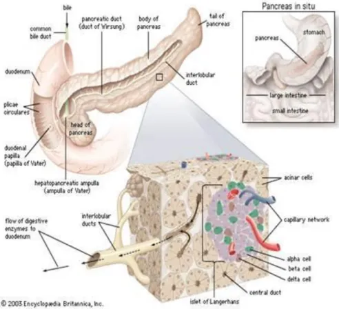
The pancreas is a retroperitoneal digestive organ connected to the duodenum through the pancreatic duct. It has 15cm length and around 70-80g of weight. It is located in the front of the aorta and behind the stomach.
Anatomically, the pancreas is divided in 4 parts: head, neck, body and tail (Fig. 1-1). The head is situated in the concavity of the duodenum, the body is placed just behind the stomach, and the tail is next to the spleen. The duodenum and the pancreas are irrigated from the celiac trunk and the superior mesenteric artery. The body and tail of the pancreas are supplied mostly by branches of the splenic artery, namely the great, superior, caudal, and dorsal pancreatic arteries.
The pancreas is a heterocrine gland with an exocrine and an endocrine tissue. The exocrine tissue represent between 98% and 99% of the pancreas by weight while endocrine tissue makes up the other 1 to 2%. The endocrine tissue is made of islets (pancreatic or Langerhans) which will be detailed thoroughly later in the manuscript. The exocrine tissue is arranged in acini, acinar cell clusters surrounding tiny ducts. Acinar cells produce and release digestive enzymes into the ducts. The ducts of many acini connect to form larger ducts which run into the main pancreatic ducts [1].
The pancreas has two main ducts: the major pancreatic duct (duct of Wirsung or pancreatic duct) and the accessory pancreatic duct. The function of these ducts is the delivery of pancreatic secretions to aid digestion into the duodenum. The main pancreatic joins the common bile duct right before entering the duodenum, so they both dump their contents via the greater (major) duodenal papilla (comprised of the ampulla of Vater, or hepatopancreatic ampulla, and the sphincter of Oddi). The main pancreatic duct crosses the whole pancreas collecting the pancreatic juice from all acinar cells. The accessory duct drains into the duodenum via the lesser (minor) duodenal papilla [1, 2].
14
Figure 1-1 Pancreas anatomy
Source: http://www.aboutcancer.com
1.1.2
Function
The pancreas because of the mixed tissue has two complementary functions. The exocrine tissue is involved in digestion and the endocrine tissue involved in glycemia regulation [2].
1.1.2.1 Exocrine tissue
Exocrine tissue is composed of acinar cells, ducts and centroacinar cells. The ducts and the centroacinar cells produce bicarbonate and water to insure the flush of the pancreatic juice in the ducts [3]. All enzymes secreted by the pancreas have the capacity to reduce digestible macromolecules into small molecules able of being absorbed [4].
For an efficient digestion three groups of enzymes are needed:
Proteases: Different proteases, like pepsin, are synthesized in the pancreas and secreted into the lumen of the small intestine, and then the digestion of protein is initiated in the stomach by the pepsin. Trypsin and chymotrypsin are the most important pancreatic proteases. These enzymes are synthetized as an inactive proenzymes trypsinogen and chymotrypsinogen which are packaged into secretory vesicles. The trypsinogen is activated by the enterokinase and is converted in the active form, trypsin. Immediately chymotrypsinogen is activated in chymotrypsin by trypsin. This activation occurs in the lumen of the small intestinal in order to do the protein digestion (Fig. 1-2). When the pancreatic secretions reach the small intestine, all the proteases are active and the digestion process starts [4]. The function of trypsin and
15 chymotrypsin is the digestion of proteins into peptides and peptides. There are other proteases also from the pancreas like carboxypeptidases which are able to digest the peptides and proteins into single amino acids. However, the digestion into single amino is mainly due by peptidases which are on the surface of small intestinal epithelial cells [4].
Figure 1-2 Activation of Trypsin and Chymotrypsin [4]
Pancreatic lipase: triglycerides are the major component of the diet and the intestinal mucosa is not able to absorb directly this molecule. Triglycerides are hydrolyzed by the pancreatic lipase which converts the triglycerides in monoglycerides and free fatty acids. Both lipases are needed to get a proper digestion of triglycerides producing free fatty acids and monoglyceride that can be absorbed.
Amylase: this enzyme is present in the pancreatic secretions and in the saliva. Its function is the starch hydrolysis into maltose, trisaccharide maltotriose and small fragments called limit dextrins [4].
1.1.2.2 Endocrine tissue
The endocrine portion of the pancreas takes the form of small clusters of cells called islets of Langerhans or pancreatic islets. Islets produce and release important hormones directly into the bloodstream. Two of the main pancreatic hormones are insulin, which acts to lower blood sugar, and glucagon, which acts to raise blood sugar. Maintaining proper blood sugar levels is crucial to the functioning of key organs including the brain, liver, and kidneys. Human pancreas contains around one million of islets [5].
Pancreatic islets are composed of five cell types; each type produces a different endocrine product and has a specific role [5]:
- α cells are responsible for the glucagon secretion (hyperglycemic hormone) and
represent around 15-20% of the total tissue. These cells have dense secretory vesicles. The release is regulated by the influence of different factors (nutritional factors, hormones and neurotransmitters).
- β cells produce insulin (hypoglycemic hormone) and are the most abundant of the
islet, around 60 to 80% of endocrine cells. Insulin is stocked in hexamers inside vesicles recognized by the crystalline core surrounded by a nimbus. Insulin secretion
16 is regulated by the synergic actions of nutritional factors, hormonal messengers and nervous.
- δ cells secrete somatostatin and represent around 5% of the endocrine cell population.
Somatostatin cells are located in many species in the β cells periphery because there is a close link between them. The role of the somatostatin is to inhibit the pancreas secretion with the inhibition of certain enzymes like insulin and glucagon.
- PP cells contain pancreatic polypeptides, are located in the periphery related to β cells
and represent the 1% of the endocrine cells. Pancreatic polypeptides play an important role in the regulation of insulin secretion, increasing basal insulin concentration in plasma but do not affect glucose stimulated secretion of insulin or glucagon.
- Ghrelin cells produce the peptide hormone ghrelin and represent 1% of endocrine cell
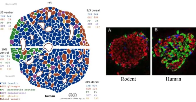
in adult pancreas. Ghrelin play a role in glucose-stimulated insulin secretion [6]. Depending upon the species, the distribution of the different cell types is different. In rodents, islets have a well-defined structure with a central core of β cells (representing 60–80% of islet cells) and a layer of other endocrine cells surrounding the core including α cells (15–20%), δ cells (<10%) and PP cells (<1%). In human islets, the α-, β- and δ- cells appear to be randomly distributed throughout the islet. The proportion of β cells is higher in rodent islets than in human cells, 77% vs. 55% [7] (Fig. 1-3).
Figure 1-3 Human and rodent pancreatic islets of Langerhans
Source: Suckale at al. 2008 (left), Diabetes Research Institute, Miami (right)
1.1.2.3 Islet vascularization
In the pancreas, islets are richly vascularized, which permits a fine tuning in glucose detection in blood and the subsequent insulin release. Although islets represent only 1-2% of the mass of the pancreas, they receive about 10 to 15% of the pancreatic blood flow. Capillaries surrounding islets show a remarkable number of small pores called fenestrates. Via these structures, nutrient exchanges occur, glucose permeate through the capillaries is detected by β cells, which exert glycemic control in response. Additionally, they are
17 innervated by parasympathetic and sympathetic neurons, and nervous signals clearly modulate secretion of insulin and glucagon [5] (Fig. 1-4).
Figure 1-4 Islet vascularization in the pancreas
Source: Encyclopedia Britanica, 2003
The organization of islet vascularization depends on islet size. Afferent arterioles are connected with acinar and ductular microvessels forming insulin-acinar portal system. Small islets (60-160 µm diameter) are not closely associated with duct or vessels. Intermediate islets (160-260 µm diameter) are found along secondary vessels and large islets (260-800 µm diameter) are often clumped at a major branches of blood vessels and in close association with major ducts. Small islets have one afferent vessel, an arteriole and intermediate of large big ones have got 1 to 3 short arterioles entering the islet directly from the large artery/arteriole. From the interior of this structure emerge efferent capillaries that extend beyond the islet periphery (Fig. 1-5) [8].
18
Figure 1-5 Microvascularization of small and large islet [8]
Through the endocrine capillaries, islets are relatively over-perfused under basal conditions, and their blood supply regulation is independent of the exocrine pancreas. Pancreatic islets are highly metabolically active, depending on oxygen consumption and glucose oxidation for their formation of ATP and subsequent insulin secretion. The highly specialized glomerular-like vascular structure of islets greatly facilitates proper glucose sensing of ambient blood glucose concentration and the distribution of secreted hormones to target organs. The mechanisms that have evolved to regulate islet perfusion are complex and mediated by signals from the nervous, hormonal, and circulatory systems. Nutrients are known regulators of islet function. Glucose is the most important insulin secretagogue, and glucose administration almost doubles islet perfusion through multiple mechanisms. A suggested mechanism for this phenomenon is that immediate or anticipatory islet vasodilation is mediated by the nervous system, followed by maintenance and more precise regulation of islet perfusion by locally produced factors. Similarly, induction of hypoglycemia by insulin administration leads to an immediate increase in islet blood flow, presumably to promote glucagon influx into the circulation [9]. The capillary density combined with a high blood perfusion of the islets delivers high amounts of oxygen to the islets, keeping PO2 metabolically active islet tissue in
equilibrium to that of venous blood (40 mmHg), and permit the secretion of insulin [10-12].
1.2
Insulin
1.2.1
Structure
Circulating and biologically active insulin is monomeric and its molecular weight is 5800 Da. It is composed of two polypeptide chains: chain A has 21 amino acids and chain B has 30 amino acids (in humans). Two disulfide bridges covalently the chains and chain A is characterized by an internal disulfide bridge (Fig. 1-6). The positions of these three disulfide bonds are invariant in mammalian forms of insulin. At micromolar concentrations, insulin dimerizes, and in the presence of zinc, it further associates into hexamers [8].
19
Figure 1-6Basic structural parameters of mature human insulin
Source: http://dailymed.nlm.nih.gov
In the core of the protein there is a cluster of hydrophobic residues which helps the protein stability using the constraint of the polypeptide backbone by the disulfide bridges. Outside the core there is a first flat and aromatic nonpolar surface buried upon dimer formation contributing to an antiparallel sheet structure. The second nonpolar surface is more extensive and is buried upon hexamer formation (Fig. 1-7). The surface that insulin uses for binding to his receptor is the same surface used for self-assembled [8].
Figure 1-7 Quaternary arrangement of insulin
Source: http://www.cell.org
1.2.2
Insulin biosynthesis
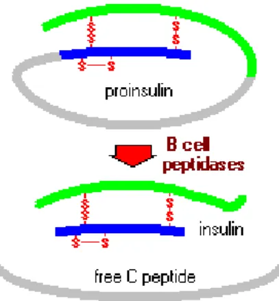
Insulin is a hormone produced exclusively by β cells in the pancreas. The insulin mRNA is translated as an inactive protein precursor called preproinsulin which contains N-terminal signal peptide. The N-N-terminal signal peptide interacts with the signal recognition particle (SRP), which facilitates the entry of the preproinsulin in the rough endoplasmic reticulum (RER) from the cytosolic compartment. The N-terminal signal sequence is cleaved to generate proinsulin. It is composed of three domains: an amino-terminal B chain, a carboxy-terminal A chain and a connecting peptide in the middle known as the C peptide. In the lumen of the ER, proinsulin undergoes protein folding. The folded proinsulin and C peptide are then delivered to the Golgi apparatus and packaged in secretory granules [8] (Fig.
20
Figure 1-8 Mechanisms of conversion of proinsulin to insulin
Source: Bowen, 1999
The conversion of proinsulin into insulin takes place in the secretory granules, where insulin is stored. Insulin is secreted from the β cells in the pancreas by exocytosis and circulates into islet capillary blood. The C peptide is also secreted into blood, but has no known biological activity [8] (Fig. 1-9).
Figure 1-9 Intracellular steps of insulin secretion
Source: http:// www.nbs.csudh.edu
1.2.3
Control of insulin secretion
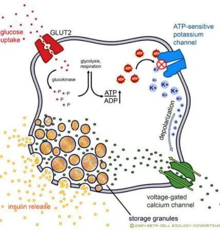
Glucose stimulation is detected thanks to sensing devices able to measure circulating glucose like the glucose transporter 2 (GLUT2), constitutively expressed in β cells. Glucose is internalized in β cells after mobilization of GLUT2 to the plasma membrane; this mechanism is insulin-independent ensuring high glucose influx. After entering, glucose is phosphorylated
21 by the glucokinase, which acts as a glucose sensor because of its ‘low’ affinity for glucose and becomes active only when glucose concentration increases. Glucokinase catalyzes the phosphorylation of the glucose in glucose-6-phosphate. This product is then transformed into pyruvate, which is then oxidized through the tricarboxylic acid cycle by mitochondria in β cells to produce ATP [13].
Production of ATP and increase in ATP/ADP ratio results in closure of ATP dependant potassium (KATP) channels. Accumulation of potassium ions increased the positive charges in
the vicinity of the membrane which triggers its depolarization. The change in membrane polarization leads to the activation in the voltage-gated calcium channels and the entry of calcium ions into the cell. The increase of the influx calcium concentrations produced the fusion of insulin granules with the plasma membrane and exocytosis of granule content. Once insulin is exported from the β cells, it is diffused in the blood vessels to control the blood glucose levels providing the first pic in insulin secretion [8] (Fig. 1-10).
Figure 1-10 Insulin secretion
Source: http://www.ciitn.missouri.edu
Then, the level of insulin go back to basal levels and after 10 minutes of glucose stimulation, the second phase of secretion starts (during 45 minutes). The second phase slower than the first phase explains the biphasic response in the insulin secretion curve (Fig. 1-11) [14].
22
Figure 1-11 Insulin secretion curve [14]
1.2.4
Insulin receptor: structure and action
Once release into the blood stream, insulin circulates in blood until meet its receptor. The main organ that predominantly clears insulin from circulation is the liver. In a non-diabetic patient the liver clears about 60% of endogenous insulin via the hepatic portal vein. Once glucose is transported inside the hepatocytes, insulin stimulates glycogen synthesis and storage and inhibits glucose production (glycogenolysis) [8].
The insulin receptor is a tyrosine kinase, an enzyme family whose members play critical regulatory roles in development, cell division, and metabolism. The insulin receptor is composed of two α-subunits and two β-subunits linked by disulfide bonds located in the plasma membrane. The α-subunit has a molecular mass of 130 kDa and contains the insulin binding domains located in both sites of a rich cysteine sequence which allows the formation of disulfide bonds. In the absence of insulin, the α-chains have an inhibitory function and the receptor is in an inactive configuration. The β-subunit has a molecular mass of 95 kDa and is composed by three compartmentalized regions: the extracellular, transmembrane and cytosolic domains. The cytosolic domain has ATP-binding and tyrosine kinase activity with a regular loop which covers the catalytic site and keeps the receptor inactive (Fig. 1-12). When insulin binds to the α-subunit, there is induction of the tyrosine autophosphorylation of the receptor β-subunit, which in turn is activated and phosphorylates its substrates, among which IRS-1 [8].
23
Figure 1-12 Insulin receptor: β and α subunits
Source: http://www.vivocolastate.edu
IRS-1 acts as a type of docking center for recruitment and activation of other enzymes implicated in insulin’s pleiotropic action. For instance, IRS-1 interacts with and recruits phosphatidylinositol (PI) 3-kinase. These events lead in particular to activation signaling pathways required for insulin as GLUT4 membrane translocation [15] (Fig. 1-13).
Figure 1-13 Different cellular effects of insulin
Source: http://themedicalbiochemistrypage.org/insulin.php
Insulin-stimulated glucose uptake is achieved by insulin-sensitive glucose transporters (GLUT4) present on the plasma membrane of muscle cells, adipocyte, hepatocytes and other targeted tissues. When glucose and circulating insulin decrease, the GLUT4 transporters shift back to the storage vesicles waiting for future insulin signaling (Fig. 1-14). In skeletal muscles, glucose uptake depends on the coordination of three steps: (1) increased delivery of glucose to the muscle fiber by increased blood flow, (2) increased glucose transport across the plasma membrane (GLUT4 and other facilitative transporters), and (3) phosphorylation of glucose by hexokinase [16].
24
Figure 1-14 Insulin-mediated glucose uptake
Source: cell biology consortium
Insulin signaling ends with the degradation of insulin and the dephosphorylation of the receptor. Insulin-receptor complexes are internalized together with other receptors through the formation of clathrin-coated vesicles, and then complexes are delivered to endosomes, where the acidic pH induces the dissociation of insulin molecules from insulin receptors. Subsequently, insulin molecules are targeted to late endosomes and lysosomes where they are degraded and receptors are recycled back to the cell surface in order to be reused [17].
Once the glucose is entered in the cells, it is transformed and stored into glycogen. Glycogen synthesis (Fig. 1-15) is stimulated by insulin which inhibits and activates specific enzymes involved in this pathway. It is a very large branched polymer of glucose residues that can be broken down to yield glucose molecules when energy is needed. It consists in a glucose branch linked by α-1,4-glycosidic bonds and is ramified in α-1,6-glycosidic bonds every 7 from 11 residues. It is a polymer of chemical formula (C6H10H5)n. Glucose stock is produced
by the glycogen synthetase. This enzyme avoids, after the digestion, the accumulation of glucose in the blood (hyperglycemia). When glucose is needed, degradation of glycogen in glucose (glycogenolysis) by the glycogen phosphorylase is triggered by the glucagon or adrenaline[8].
25
Figure 1-15 Mechanisms of glycogenesis
Source: Biochemistry, 2012
1.3
Diabetes
1.3.1
Definition criteria and classification of DM
Diabetes mellitus (DM) is a group of metabolic diseases characterized by hyperglycemia resulting from defects in insulin secretion, insulin action, or both. Several pathogenic processes are involved in the development of diabetes. These range from autoimmune destruction of the β cells of the pancreas with consequent insulin deficiency to abnormalities that result in resistance to insulin action [18]. The current World Health Organization ( WHO) diagnostic diabetes when the fasting plasma glucose is higher than 7 mM (1.26g/L) or 11 mM (2g/L) two hours after hyperglycemia produced by oral route (OGTT).
According to the International Diabetes Federation (IDF), diabetes prevalence in 2015 worldwide was estimated than half a million children aged 14 and under living with type 1 diabetes. It is also estimated that actually there are 415 million adults aged 20-79 with diabetes worldwide, including 193 million who are undiagnosed. A further 318 million adults are estimated to have impaired glucose tolerance, which puts them at high risk of developing the disease. If this rise is not halted, by 2040 there will be 642 million people living with the disease (Fig. 1-16). There are different types of diabetes such as type 1, type 2, monogenic, secondary and gestational between others.
26
Figure 1-16 Worldwide diabetes distribution prevalence in 2015 (IDF)
1.3.2
The natural history of DM
Glucose is the main nutrient of the body's cells. Therefore every individual consumes continuous glucose (2 mg/min/kg on average); mainly muscle during inter period absorptions. The digestive tract becomes the main consumer in postprandial periods, reducing the glucose levels used by the brain. If exogenous glucose from the meals is not enough, there are stocks of glucose in the liver (75%) and in the kidney (25%). Normoglycemia is maintained when the glucose intake perfect fit for consumption, however, if it is not the case the insulin-glucagon system will be implicated.
This system does not work properly in some cases due to insulin deficiency and insulin resistance. As a consequence there is a reduction in glucose consumption and an increase in hepatic glucose production (as a result of insulin deficiency and excessive glucagon) and renal (by increasing tubular reabsorption of glucose).
The imbalance between the reduced consumption and increased production of glucose, results in hyperglycemia. Early symptoms of hyperglycemia such as frequent urination, thirst, blurred vision, fatigue and headache can develop a pre-diabetic status.
27
1.3.3
Diabetes diagnostic
The effects of high glucose concentration in blood are multiple and can be used for the diagnosis (Table. 1-1):
Table 1-1 Diagnostic criteria for diabetes
Source: American Diabetes Association
Fasting plasma glucose (FPG) test is the easiest and the first to be performed after the patient has gone at least 8 hours without food or drink. It is performed analyzing the blood glucose levels through a blood test.
The random plasma glucose (RPG) test is a simple blood sugar test. There is no need to fast for the RPG. The test is simple and can be performed taking a blood test and having the levels of glucose. A normal blood glucose level reading, without fasting first, of under 140 mg/dl is considered normal. However, a level of over 200 mg/dl, especially with symptoms of frequent urination, excessive thirst, etc. will indicate a strong possibility of diabetes [19].
What makes the FPG different from the RPG test is that through fasting the body of a person without diabetes will produce and process insulin in response to increased glucose in the blood. For a person with diabetes, the body will not have that same response and their blood glucose levels will remain high. Receiving two readings of blood glucose levels of 126 mg/dl or over usually means the person has diabetes. A normal level for a person without diabetes should be in the 70-110 mg/dl range after fasting [19].
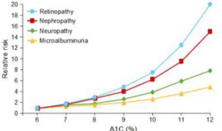
The A1C test is based on the percentage of the hemoglobin, the protein that carries oxygen, is coated with glucose (glycated). In the body, red blood cells are constantly forming and dying, but typically they live for about 3 months. Thus, the A1C test reflects the average of a person’s blood glucose levels over the past 3 months. The A1C test result is reported as a percentage. The higher the percentage, the higher a person’s blood glucose levels have been. A normal A1C level is below 5.7 %. The test plays a critical role in the management of the patient with diabetes, since it correlates well with both microvascular and, to a lesser extent,
28 macrovascular complications and is widely used as the standard biomarker for the adequacy of glycemic management (Fig. 1-17).
Figure 1-17 Microvascular complications associated with A1C increase
Source: Internal Medicine & Pediatric Associates
The last test given is called an oral glucose tolerance test. The patient needs to fast, usually overnight, for 10-16 hours before the test is taken. First blood sample is performed to give a basal reading. Then, the patient is given a cola drink with high content of sugar (75 grams of glucose) and blood test is taken 2 hours after the drink. The two blood tests after the drink with high glucose content will show the rise and fall of blood sugar levels over time, as a normal profile [19]. In the first measure, a normal level should be in the 70-110 mg/dl range for non-diabetic and >150 mg/dl for diabetic person. The second measure after 2 hours, a normal level should be >140 mg/dl range for non-diabetic and >200 mg/dl for diabetic person. There are two principal types of diabetes, type 1 diabetes (T1DM) and type 2 diabetes (T2DM), both caused by a combination of genetic predisposition and environmental risk factors:
1.3.4
Description of the principals type of diabetes
1.3.4.1 Type 1 diabetes
Type 1 diabetes mellitus (T1DM), or insulin dependent diabetes is an autoimmune disease that occurs when T cells specifically attack and destroy most of the β cells in the pancreas. It appears most often in childhood and involves 10% of patients. It is management requires daily administration of exogenous insulin.
Risk factors related to T1DM [20]:
- Geography: the incidence of T1DM tends to increase when the geographical latitude (distance from the equator) increases. People living in Finland and Sardinia have the highest incidence of T1DM.
- Genetic factors: familial aggregation of T1DM has been recognized for many years, and 10–13% of newly diagnosed children have a first-degree relative affected with type 1 diabetes. Risk of developing islet autoimmunity varies depending on which relative(s) have type 1 diabetes.
- Environmental factors: environmental agents that are suspected to trigger β cell autoimmunity in genetically susceptible individuals include dietary factors and
29 common viral infections. By today, however, no single factor has been identified that can induce the process of autoimmune β cell destruction.
Type 1 diabetes is associated with the appearance of humoral and cellular islet autoimmunity and a defective immunoregulation in the 90% of type 1 diabetic patients. The disease has different stages starting with a genetic susceptibility then, autoimmunity without clinical disease and subsequent clinical diabetes. First signs of islet autoimmunity appear in early childhood under 10 years of age, with a peak with 2 years of age. Not all children develop the autoantibodies before 2 years, but children with a later development have a slower progression to multiple antibodies. The first antibodies detected are the autoantibodies to insulin (IAAs). After the first response of IAA follows a development of autoantibody to GAD (GADAs) and autoantibody of tyrosine-phosphatase (IA-2 and IA-2β). Once islet autoantibodies appear, they usually persist. Association of anti-GAD and anti-IA2 is correlated with high affinity in the progression of type 1 diabetes. However, IAA is the least persistent because they may be transferred during pregnancy from the mother with type 1 diabetes. If antibodies are detected in a child early in life, it is important for the assignment of diabetes risk to distinguish whether these antibodies are indeed de novo-produced antibodies of the child or rather antibodies acquired from the mother.
In most children, first peak antibody levels decline and autoantibodies against other β cell antigens may arise sequentially over several years, suggesting regulation and spreading of islet autoimmunity in childhood. Different factors like genes, environment and age are implicated in the progress of the disease [20].
The other 10% of type 1 diabetic patients is due to idiopathic diabetes. The pathophysiologic mechanisms involved in its etiology is unknown, but could be due to glucose desensitization, lipotoxicity, environmental factors, and/or genetic defects in nuclear transcription factors involved in fuel metabolism. Despite the fact that most patients do well for a few years after the diagnosis of diabetes, when treated with diet/oral agents, it seems that insulin therapy maintains lower A1C over the long duration [21].
Β cell autoimmunity, marked by the development of islet reactive autoantibodies, portends the development of activated autoreactive T cells capable of destroying β cells, resulting in a progressive and predicable loss in insulin secretory function. Clinical T1D does not present until >80%–90% of the β cells have been destroyed, and there is a marked gap between the onset of autoimmunity and the onset of diabetes. In addition, the degree of β cell destruction required for symptomatic onset is also of growing question, with recent studies suggesting that 40%–50% β cell viability may be present at the onset of hyperglycemia, an aspect that may be related to subject age, among other factors (e.g., body mass index, physical activity, etc.). This may explain why, despite persistent autoimmunity, insulin secretory function can remain stable for long periods of time in persons with T1D. A loss of first-phase insulin response is usually followed by a period of glucose intolerance and a period of clinically “silent” diabetes. Finally, the “slope” reflective of β cell loss in the pre-diabetic period has also recently been subject to considerable debate, with some proposing that the disorder may see its symptomatic onset only following a period of relapsing/remitting like autoimmunity [22].
The symptoms of type 1 diabetes are polyuria (frequent and abundant urination), the polydipsia (thirst), constant hunger, weight loss, impaired vision and fatigue. These symptoms may occur suddenly although the damage to the β cells may begin much earlier and progress slowly and silently [20].
30
1.3.4.2 Type 2 diabetes
Type 2 diabetes mellitus (T2DM) is one of the most common types of DM, accounted for 90-95% of the diabetic cases worldwide. It is the result of genetic and environmental factors like family history, excessive fat and sugar intake, age and physical inactivity. This diabetes is link with a progressive desensitization of the receptor for the insulin [23, 24]. Glucose is not translocated from the blood stream into the cells because there is a defective regulation of GLUT4 protein due to the inhibition of tyrosine phosphorylation of insulin receptor substrate-1 (IRS-1) [25]. In response, pancreatic β cells produce more insulin in order to force the glucose uptake by cells. The increase in insulin production is accompanied by increased islet size and pancreatic proportion of β cells [26]. In presence of hyperinsulinism, the recycle/internalization cycles of GLUT4 can produces a decrease in the number of receptors in the membrane and this effect could be related with the insulin resistance. At this stage, decreased β cells mass is due to apoptosis of β cells mainly caused by glucotoxicity, lipotoxicity, and deposits of islets amyloid polypeptide (IAPP) [27-29]. Pre-diabetes and metabolic syndrome, which follows it, are parts of interrelated common clinical disorders that are accompanied by symptoms of obesity, insulin resistance, glucose intolerance, lipid abnormalities, moderate glycation (A1C 5,7-6,4%), impaired fasting glucose (110-125 mg/dl), and impaired glucose tolerance (140-200 mg/dl). Some studies have reported that approximately 5-10% of pre-diabetic population would suffer from diabetes and problems associated with it in around a year such as heart problems, imbalance in glucose and lipid metabolism, and vascular disorders. Timely interventions in this population can preserve pancreatic β cells, and improve their performance. Current medications to control blood glucose and lipid profile may have dangerous side effects over time such as increased risk of weight gain, liver toxicity, and cardiovascular diseases. Thus, we need to use stronger alternatives with fewer side effects. In this line, nutritional interventions, change in lifestyle, and behavioral therapy are on the rise. However, these interventions may not be effective alone to prevent the development of type 2 diabetes [30].
Although T2DM cannot be cured, it can be treated with a healthy lifestyle such as diet, exercise and weight control which can provide the foundation for managing of T2DM. However, anti-diabetic agents are required to regulate blood glucose levels. These drugs can cause side effects for instance, weight gain which consequently increases the risk of insulin resistance leading to a further enhance in drug dose [31].
1.3.4.3 Other types of diabetes
Monogenic diabetes: Some rare forms of diabetes result from mutations in a single gene and are called monogenic. Monogenic forms of diabetes account for about 1-5% of all cases of diabetes in young people. In most cases of monogenic diabetes, the gene mutation is inherited; in the remaining cases the gene mutation develops spontaneously. Most mutations in monogenic diabetes reduce the body's ability to produce insulin. Neonatal diabetes mellitus (NDM) and maturity-onset diabetes of the young (MODY) are the two main forms of monogenic diabetes. MODY is much more common than NDM. NDM first occurs in newborns and young infants; MODY usually first occurs in children or adolescents but may be mild and not detected until adulthood [32].
31 Secondary diabetes: It is developed from pancreatic disease, endocrine disease, or administration of certain drugs. When diabetes is secondary to pancreatic disorders, particularly when β cell mass is greatly reduced as in malignancy or pancreatectomy, or when diabetes is due to chemical agents toxic to the β cell, overt diabetes with or without ketoacidosis will often result depending on the extent of β cell loss. In contrast, when diabetes is secondary to endocrinopathies leading to counter regulatory hormone production, overt diabetes or ketoacidosis is unusual, mainly owing to the compensatory responsiveness of the normal β cell mass. The net metabolic outcome in patients with secondary diabetes thus depends on the direct or indirect impact of the underlying disorders on insulin secretion, insulin-sensitivity, and/or unmasking of genetic diabetes [33].
Gestational diabetes: It is developed during pregnancy. It occurs in about 4% of all pregnancies. It is usually diagnosed in the later stages of pregnancy and often occurs in women who have never had diabetes. It is thought to arise because the many changes that occur in the body during pregnancy lead some women to become resistant to insulin or the insulin production is inefficient. There are two reasons why the mother needs more insulin: because during pregnancy some hormones block the insulin action or because the baby increases the mother's need for insulin.
1.3.5
Diabetes complications
Glycemia fluctuations lead to acute or chronic complications. Acute complications include hypoglycemia diabetic ketoacidosis (DKA), and hyperosmolar hyperglycemic state (HHS):
Hypoglycemia is defined by low blood glucose (<4mM), this may result in a variety of symptoms including clumsiness, trouble talking, confusion, loss of consciousness, seizures, or death.
Ketoacidosis (DKA) is defined by the insulin deficiency with hyperglycemia (glucose levels usually >200 mg/dl) with increased lipolysis, increased ketone production, hyperketonemia and acidosis. Precipitation factor are infection, acute illnesses, lack of diabetes education, poor self-care, inadequate glucose monitoring and indeterminate causes. Morbidity is related to the severity of acid-base and electrolyte disturbances which may result in coma and death [34].
Hyperosmolar Hyperglycemic Nonketotic Syndrome (HHNS) is characterized by the presence of relative insulin deficiency and hyperglycemia, usually >600 mg/dl with associated elevated serum osmolality, dehydration, and stupor, progressing to coma correlated with renal failure. Causes of the HHNS are dehydration, medications such as steroids and thiazides, acute illness, cerebral vascular disease and advanced age. The consequences consist of coma and impaired neurologic function with a predisposition to vascular occlusive disease from dehydration or poor perfusion [34]. However, chronic complications can have deleterious impact on numerous tissues including eyes, heart, kidneys, nerves, brain and feet [35]. 60% of the diabetes complications have a cardiovascular disease origin and 30% are from organ degeneration (Fig. 1-18):
Cardiovascular disease (micro- and macroangiopathy) are characterized by lesions of the small and large blood vessels of the legs, heart and brain. The elevated blood glucose concentration can produce blood coagulation and increase the blood vessels obstruction near to the heart (heart attack), brain (AVC) or feeds (gangrene).
32 Nephropathy is refers to the presence of elevated urinary protein excretion in a person with diabetes as a consequence of the high glucose levels which produce an excessive filtration in the kidney. Other factors associated with the development of diabetic nephropathy include diabetes duration, hypertension, hyperglycemia, and smoking. Control of blood glucose and blood pressure reduce the rate of progression of renal disease in diabetes. The presence of albumin is associated with a nearly risk of death from cardiovascular disease.
Neuropathy is the result of damage of the peripheral or autonomic nerves, produced by prolonged exposure to high blood glucose. It can result in traumatic injuries, infections, metabolic problems, inherited causes, exposure to toxins and urinary tract infections.
Retinopathy is characterized by alterations in the small blood vessels in the retina. High blood pressure, early age at onset of diabetes and longer duration of diabetes are associated with increased risk of progression of retinopathy. Causes of decreased vision in patients with diabetes are cataract (clouding of the lens), glaucoma (damage to the optic nerve), and corneal disease.
Figure 1-18 Diabetes complications
Source: Sanofi
1.4
Diabetes treatments
1.4.1
Oral antidiabetic drugs (OAD) for type 2
Antidiabetic agents aim to achieve normoglycemia and relieve diabetes symptoms, such as thirst, polyuria, weight loss and ketoacidosis in type 2 diabetic patients. The long term goals are to prevent the development or slow the progression of long term complications of the disease.
33
1.4.1.1 Metformin
Metformin, a biguanide that acts directly against insulin resistance, is regarded as an insulin sensitizing drug. Available formulations include Glucophage®, Glucophage XR®, Riomet®, Fortamet®, Glumetza®, Obimet®, Dianben®, Diabex® and Diaformin®. Because of
its safety and efficacy, metformin is the cornerstone of monotherapy, and joint guidelines recommend that metformin be initiated as first line monotherapy unless a contraindication such as renal disease, hepatic disease, gastrointestinal intolerance or risk of lactic acidosis coexists [36].
Despite being the most widely used OAD in the world, metformin can reach a plateau of effectiveness due to progressive β cell failure. Metformin is only effective when there is sufficient endogenous or exogenous insulin and, because of this, patients are unable to maintain tight glycemic control as their disease progresses [37, 38] (Fig. 1-18).
Figure 1-19 Action mechanism of metformin [39]
1.4.1.2 Insulin secretagogues
These main classes include agents that stimulate insulin secretion in β cells through specific receptors (sulphonylureas and glinides) or agents that increase GLP-1, insulin secretagogue hormone from the pancreas. The GLP-1 increase could be related with a reduction of the endogenous GLP-1 destruction by DPP4 (DPP4 inhibitors and gliptins) or with a GLP-1 resistance to DPP4 (GLP-1 analogs and GLP-1 agonist receptors).
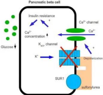
1.4.1.2.1 Sulfonylureas
Sulfonylureas (SUs) are the oldest and most widely used medications for the treatment of T2DM. Although SU therapy effectively lowers blood glucose concentrations (average decrease in FPG of 2-4 mmol/l, accompanied by a decrease in A1C of 1–2%) by stimulating insulin secretion from β cells, treatment with SUs is associated with a progressive linear decline in β cell function. Eventual inability to maintain glycemic control reflects an advanced stage of β cell failure. Hypoglycemia is the most common and most serious adverse event
34 associated with SU therapy, mainly because of insulin release being initiated even when glucose concentrations are below the normal threshold for normal physiologic glucose-stimulated insulin release. Weight gain, regarded as a class effect of SUs, is thought to result from an anabolic effect of increased insulin concentration. Owing to decreased effectiveness of SUs over time and an associated decline in the insulin secretory reserve, combination therapy has focused mainly on adding insulin-sensitizing medications, including metformin and thiazolidinediones [40-42] (Fig. 1-20).
Figure 1-20 Action mechanism of sulfonylureas
Source: http://typetwodiabetes.info
1.4.1.2.2 Glinides
Meglitinides such as repaglinide and nateglinide are prandial insulin releasers that stimulate rapid insulin secretion. Repaglinide (NovoNorm®, Prandin®, GlucoNorm®) is the
first clinically available insulin secretagogue that specifically enhances early-phase prandial insulin response by increasing the sensitivity of β cells to elevated glucose levels, producing a greater insulin release under hyperglycemic conditions. Rapid-acting insulin releasers can be suitable for lifestyles where meals are unpredictable or missed. Lower risk of hypoglycemia makes these agents an attractive option for some elderly patients, in particular when other agents may be contraindicated [43, 44].
1.4.1.2.3 Dipeptidyl Peptidase-IV inhibitors: gliptins
Dipeptidyl peptidase-IV (DPP-IV) inhibitors such as gliptins, suppress the degradation of a variety of bioactive peptides, including glucagon-like peptide-1 (GLP-1), leading to an enhancement of their action. GLP-1 inhibits glucagon release, which in turn increases insulin secretion and decreases blood glucose levels. DPP-IV inhibitors are orally administered drugs with a significant effect on glucose tolerance and lasting improvement of A1C. DPP-IV inhibitors are weight-neutral and well tolerated [45].
1.4.1.2.4 GLP-1 analogs or GLP-1 receptor agonist
GLP-1 analogs have therefore been developed to produce a longer terminal elimination half-life, and these have been shown to cause weight loss and improvement in
35 glycemia in the diabetic population. A meta-analysis of GLP-1 analogues showed that these were able to reduce A1C by 0.97%. It has been postulated that shorter acting GLP-1 analogues (exenatide and lixisenatide) reduce hyperglycemia primarily by slowing gastric emptying, whereas longer acting GLP-1 analogs (liraglutide, exenatide long-acting release, albiglutide and dulaglutide) predominantly lower postprandial glucose levels by insulinotropy and glucagon inhibition [46].
1.4.1.3 SGLT2 inhibitors
Sodium-glucose co-transporter 2 (SGLT2) inhibitors are a new class of diabetic medications. In conjunction with exercise and a healthy diet, they can improve glycemic control. They have been studied alone and with other medications including metformin, sulfonylureas, pioglitazone, and insulin. Inhibition of SGLT2 leads to the decrease in blood glucose due to the increase in renal glucose excretion. The mechanism of action of this new class of drugs also offers further glucose control by allowing increased insulin sensitivity and uptake of glucose in the muscle cells, decreased gluconeogenesis and improved first phase insulin release from the β cells.
1.4.1.4 α-glucosidase inhibitors
α-glucosidase inhibitors, including acarbose, are competitive inhibitors of membrane-bound intestinal α-glucosidases that hydrolyze oligosaccharides, trisaccharides and disaccharides to glucose and other monosaccharides in the small intestine and thereby delay postprandial glucose absorption. These agents are available as a first-line treatment in patients with slightly raised basal glucose concentrations and marked postprandial hyperglycemia (average decrease in A1C of 0.5–1%). The use of α-glucosidase inhibitors in combination with sulfonylureas, metformin or insulin can improve glycemic control. Despite their good [42, 47].
1.4.2
Insulin therapy
Insulin administration is performed in type 1 diabetic as first treatment and in type 2 diabetes patients for whom oral antidiabetic drug therapy become inefficient. Insulin is usually administered subcutaneously using a syringe, insulin pen or insulin pump. Which insulin regimen is best for patient depends on factors such as the type of diabetes, blood sugar fluctuation throughout the day and lifestyle.
It is important where insulin is injected in the body, because it affects the blood glucose level. Depends of injection site, insulin goes into the blood with different speeds. For this reason, it is preferable to inject insulin always in the same site. The fastest site is in the abdomen and from the upper arms is slower [45].
Each insulin type is characterized by the starting time (onset), maximal efficiency (peak) and the lifespan (duration) (Fig. 1-21):
36
Figure 1-21 Insulin characterization in function of the onset, peak and duration
Source: University of California, San Francisco, UCSF
1.4.2.1 Fast-acting human insulin
Synthetic human insulin administered by all routes (intravenous, intramuscular, subcutaneous and intraperitoneal). The onset of action depends of the administration route. The effect when is injected subcutaneously starts 15-20 minutes after injection, reach the peak between 2 and 4 hours and finish after 6 hours. It is presents in a clear solution that can be used at all times in all injection systems: syringes, pens and pumps. However, it is less stable than ultrafast analogs in external pumps. In common use, it is administered subcutaneously in single or multiple daily injections usually associated with other long-acting insulin injections.
1.4.2.2 Insulin NPH
NPH (Neutral Protamine Hagedorn) is an intermediate-acting biogenetic human insulin with a protamine sulfate added, for delaying and increasing its duration of action. This insulin is still widely used under the names Umuline® NPH (Eli Lilly) or Insulatard® (Novo
Nordisk) as intermediate-acting insulin. It is in the form of an injectable suspension in vial or pen. The duration of action of NPH insulin continues for 14 to 18 hours. This period is called intermediate because it is between the fast-acting insulin and the long-acting insulin.
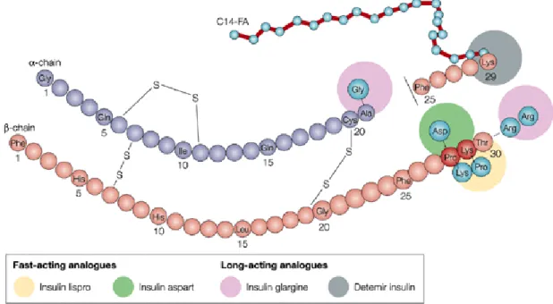
1.4.2.3 Insulin analogs
An insulin analogue is a modified form of human insulin but still functional on the patient to perform the same action than human insulin in terms of glycemic control. By genetic engineering, the amino acids of the insulin can be changed to modify its absorption characteristics, distribution, metabolism and excretion (Fig. 1-22). These modifications have been used to create two types of insulin analogues:
37 Those which are slowly released over a period between 8 and 24 hours. They are used
to maintain basal insulin throughout the day.
Those that are more readily absorbed from the injection site and thus act more quickly. They are intended for use with prandial bolus.
Figure 1-22 Modification sites in the insulin sequence
Source: Nature Reviews, Drug Discovery
1.4.2.3.1 Long-acting injected insulin analogs
Long-acting insulin analogues can be obtained by modifying the spatial conformation of insulin. Indeed, in the presence of zinc, insulin forms a hexamer: three dimers set around a threefold axis passing through 3 zinc atoms. So, their diffusion is slowed, which contributes to increase the duration of action of insulin (Fig. 1-23).
Figure 1-23 Insulin oligomerization
These long-acting analogues arise from modification of the structure of human insulin, but this modification is intended to increase their duration of action. Their release is performed in a constant way reducing the risk of hypoglycemia. There are two long-acting analogs:
38 Insulin glargine or Lantus® (Sanofi-Aventis) is soluble at pH 4 and is in the form of a clear solution. Once injected into the subcutaneous tissue, it precipitates in the microcrystals allowing its gradual release. Its action begins within 2 to 4 hours and extends regularly and consistently without peak until 25 hours. This kinetic allows the one daily injection at a fixed time.
Insulin detemir or Levemir® (NovoNordisk) is in the form of a clear neutral solution.
It binds in the subcutaneous tissue which gives it its delay effect. It begins to work 2 to 4 hours after injection. Its action continues without peak until 14 hours after injection.
1.4.2.3.2 Rapid-acting injected insulin analogs
They are injected subcutaneously and insulin action begins to act more quickly than soluble human insulin within 10 minutes after injection. They maintain lower blood glucose within 4 hours after the meal, with a maximum effect occurring 1-3 hours after injection. Their action duration is lower than soluble human insulin and there is a rise in blood glucose meals distances beyond 4 hours after injection. These analogs generally replace fast insulin in emergency treatment and in multiple injection regimes. They are administered immediately before the meal so that the human insulin should be 30 minutes before. They allow better reduce postprandial glucose elevations and avoid episodes of hypoglycemia between meals. Ultrafast analogs are perfectly stable in the external pumps but not in implantable pumps. There are three fast analogs (Fig. 1-22):
that resulting from the reversal of the two penultimate amino acids on the β chain, taking the lysine the β28 position and the proline the β29 position to give insulin lispro or Humalog sell by Eli Lilly,
that obtained by replacing proline in position β28 by an aspartate to give insulin aspart or Novorapid developed by the laboratory Novo Nordisk,
that replacing the aspartate in position β3 by the lysine and the lysine in position β29 with a glutamate to give insulin glulisine or Apidra (Sanofi-Aventis).
1.4.2.4 Insulin administration devices 1.4.2.4.1 Insulin pens
Insulin dose is dialed on the pen, and is injected through a needle, much like using a syringe. Cartridges and pre-filled insulin pens only contain one type of insulin. Two injections must be given with an insulin pen if using two types of insulin. Those pens are light, easy to transport and discreet. However, the repeated injections can be painful and it can produce lipoatrophy, lipohypertrophy or allergic reactions (Fig. 1-24) [35].
![Figure 1-2 Activation of Trypsin and Chymotrypsin [4]](https://thumb-eu.123doks.com/thumbv2/123doknet/14517206.721947/16.892.223.671.217.491/figure-activation-trypsin-chymotrypsin.webp)
![Figure 1-19 Action mechanism of metformin [39]](https://thumb-eu.123doks.com/thumbv2/123doknet/14517206.721947/34.892.209.674.412.755/figure-action-mechanism-of-metformin.webp)
![Figure 1-27 Semi-automated human islet isolation procedure [73]](https://thumb-eu.123doks.com/thumbv2/123doknet/14517206.721947/43.892.136.776.756.1066/figure-semi-automated-human-islet-isolation-procedure.webp)