Coma and Disorders of Consciousness: Scientific Advances and Practical
Considerations for Clinicians
Olivier Bodart, Steven Laureys, Olivia Gosseries
Department of Neurology, Coma Science Group, Cyclotron Research Center, University of Liège, University Hospital of Liège, Liège, Belgium
Abstract
Recently, neuroscientists and clinicians have seen the rapid evolution of diagnoses in disorders of consciousness. The unresponsive wakefulness syndrome-vegetative state, the minimally conscious state plus and minus, and the functional locked-in syndrome have been defined using new neuroimaging techniques. Diffusion tensor imaging, positron emission tomography, functional magnetic resonance imaging, electroencephalography, and transcranial magnetic stimulation techniques have all promoted important discoveries in the field of disorders of
consciousness. This has led to a better understanding of these patients' condition and to the development of new prognosis, therapeutic, and communication tools. However, low sensitivity and artifacts problems need to be solved to bring these new technologies to the single-patient level; they also need to be studied in larger scale and randomized control trials. In addition, new ethics questions have arisen and need to be investigated.
Keywords : Unresponsive wakefulness syndrome-vegetative state ; Minimally conscious state ; Consciousness ;
Functional magnetic resonance imaging (fMRI) ; Neuroimaging
Rapid evolution of neuroimaging technologies and paradigms has led to profound modifications in the nosology, prognosis, and treatment of patients with disorders of consciousness. We will first review the evolution of the revised diagnoses, then the neuroimaging techniques and paradigms that promoted these changes will be discussed. Finally, we investigate the potential impact this new knowledge may have on clinical practice.
Evolution of Nosology in Disorders of Consciousness
More than 10 years ago, Giacino et al1 defined the diagnosis criteria for a new state of altered consciousness, the minimally conscious state. Until then, patients with disorders of consciousness were considered either in coma, in a persistent or permanent vegetative state, or in a severely disabled state. Patients in a vegetative state recover arousal (i.e., eyes opening), but not awareness of themselves and their environment.
This condition has been given a widely accepted name in 1972,2 and diagnosis criteria, etiology, and prognosis were summarized in 1994.3,4 However, recently this state has been widely studied, leading to changes in terminology. First, a new name has been proposed, the unresponsive wakefulness syndrome (UWS),5 as it better describes the condition (the patients are indeed awake—showing eye opening, but unresponsive—in the absence of command following, and can have several etiologies to their clinical signs—which are thus included in a syndrome); it also removes the impression given to the public that these patients are "vegetable-like". Second, this population of unresponsive wakefulness syndrome patients has been shown to be in fact composed of different states of altered consciousness. With the application of the diagnosis criteria for the minimally conscious state,1 many patients have been rediagnosed. Patients in the minimally conscious state can show signs of consciousness such as command following (even if inconsistent), visual pursuit, localization to noxious stimulation, and appropriate responses to emotional stimuli without being able to functionally communicate. Recently, it has been proposed to distinguish in this patient population those who show higher-order signs of consciousness (e.g., command following, intelligible verbalization, and nonfunctional communication) from those who show only low-level signs of consciousness (e.g., visual pursuit of a salient stimulus, noxious stimulation localization, appropriate emotional response).6 Advances in the field of neuroimaging, as we will discuss below, have also recently led to the proposition of the diagnosis of functional locked-in syndrome.6 Classical locked-in patients usually have most of their cognitive functions preserved, but because of the brainstem injury their motor output is near null (except for eye movements).7 Functional locked-in syndrome
represents patients who are behaviorally in an unresponsive wakefulness syndrome or minimally conscious state, but on neuroimaging show better consciousness than expected, with command following and even sometimes functional communication.6 Figure 1 summarizes the different diagnostic entities that can be encountered after a severe brain injury.
So far, the gold standard for consciousness assessment remains the behavioral evaluation.8 With the development of dedicated and validated scales such as the Coma Recovery Scale-Revised (CRS-R),9 trained clinicians and neuroscientists can make diagnosis of coma, unresponsive wakefulness syndrome, and the minimally conscious state and emergence from this state. The CRS-R is the best scale to distinguish the minimally conscious state from unresponsive wakefulness syndrome10 as it is composed of six subscales (auditory, visual, motor, oromotor, communication, and arousal) in which signs of consciousness are sought and is now widely used, as assessed by the number of validated translation in various languages that exist.10 At the acute stage, the Glasgow Coma Scale (GCS)11 is the most commonly used validated scale to predict the probability of recovery. The more recent Full Outline of Unresponsiveness (FOUR) Scale12 has proved to share good predictive value of functional outcome with the GCS, with the advantages of being appropriate for intubated patients and for the detection of minor signs of consciousness such as visual pursuit.13
Even with the availability of validated scales, the rate of misdiagnosis remains high (~40%).14,15 There are many reasons for this. On the patient side, there might be limitations from motor dysfunction (paralysis, spasticity, hypotonic status), impaired cognitive processing (language processing, apraxia), a sensory deficit (such as deafness/blindness), fluctuation of vigilance (from the disorder of consciousness itself or from drugs), pain, etc. A lack of repeated evaluation, or evaluation over too short a time span can also lead to misdiagnosis.16 With the consequences that such errors can have on the management of these patients, neuroimaging and
electroencephalography techniques were sought.
Fig. 1 Proposed nosology of the diagnostic entities after a coma based on clinical behavioral assessment and on recent functional neuroimaging studies. UWS, unresponsive wakefulness syndrome; MCS, minimally conscious state; LIS, locked-in syndrome; fUS, functional locked-in syndrome. (Adapted from Bruno et al.96 Unresponsive wakefulness to minimally conscious PLUS and functional locked-in syndromes: recent advances in our
understanding of disorders of consciousness. J Neurol 2011;258(7):1373-1384)
Evolution in Neuroimaging Techniques
A simple way to study the patients' injured brain is through magnetic resonance imaging (MRI). Magnetic resonance imaging provides valuable information on the number, nature, severity, and localization of brain
lesions. Given the high variety of lesions, researchers tried to establish a correlation between the localization, the nature and the number of lesions, and the patient's state of consciousness. The number of lesions correlates with the Glasgow Outcome Scale17 (GOS).18 Lesions in the thalamus, brainstem, and basal ganglia were found to be of poor prognosis, especially when they are bilateral.19-24 However, patients with unresponsive wakefulness syndrome and minimally conscious state patients cannot be differentiated on MRI. Indeed, some patients share the same kind of structural lesions with different states of disorders of consciousness.
The development of diffusion-weighted imaging (DWI) and its derived diffusion-tensor imaging (DTI) brought new tools for the structural evaluation of these patients.25 These imaging sequences are based on the principle that water-molecule movement (diffusion) is dependent on its molecular environment, with axon membranes acting as barriers. DTI can give information on the structural integrity of axon tracts, resulting in the ability to evaluate the anatomic connectivity between different parts of the brain. This technique reflects the level of consciousness and distinguishes between the group of unresponsive wakefulness syndrome patients and the group of minimally conscious state patients.26 Figure 2 illustrates structural damage highlighted by DTI in one disorder of consciousness patient as compared with a healthy subject. Diffusion-tensor imaging can also be used as a prognostic tool: specific brain areas (i.e., the internal capsule, the corpus callosum, the cerebral peduncle, and other white matter tracts) correlate with outcome.27,28 Moreover, DTI may have demonstrated axonal regrowth paralleling clinical improvement in one patient in a chronic minimally conscious state.29 Still, DTI only gives information on structural connectivity and not on its functionality.
Fig. 2 Structural brain images from diffusion tensor imaging technique showing white matter tracts in a healthy control and in a patient with chronic disorders of consciousness.
The most basic state of the brain to study is the brain at rest. In this state, the patient receives no specific task or stimulus. This "resting state" has been studied using fluorodeoxyglucose positron emission tomography (FDG-PET), which has shown a global decrease in brain metabolism in unresponsive wakefulness syndrome patients. 30-32 However, recovery of consciousness in these patients is not always paralleled with an increase in global metabolism.33 Rather some areas of the brain are necessary for the emergence of consciousness. Indeed, studies have demonstrated that a widespread thalamocortical network (encompassing the thalamus; the precuneus; and the mesiofrontal, prefrontal, and posteroparietal cortices) shows impaired metabolism in unresponsive
wakefulness syndrome patients,34,35 and its functional connectivity is restored when the level of consciousness improves.36,37
This resting state was also studied with functional MRI (fMRI), with the identification of a somewhat similar widespread thalamocortical network named the default mode network. Functional connectivity in this network correlates with the level of consciousness,38 being null in brain-dead patients.39 Its preserved connectivity at the acute stage could hold good prognostic value.40
Electrophysiological studies of this brain state using electroencephalography (EEG) have also shown prognostic value, as occipital alpha band power could be a predictor of recovery in unresponsive wakefulness syndrome patients.41 Using entropy measurements of the EEG signal, it is also possible to distinguish in the early stages unconscious (coma and unresponsive wakefulness syndrome) from minimally conscious patients.42
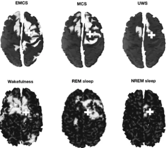
Instead of measuring structural or functional connectivity (each can be present without the other one), transcranial magnetic stimulation combined with high-density EEG (TMS/hd-EEG) has been used recently to determine effective connectivity by stimulating a brain area and recording the subsequent electrical activity. Effective connectivity can be defined as the capacity of one element of a network to causally influence another.43 Effective connectivity assessed by this technique correlates with the level of consciousness, being high in healthy awake subjects, in locked-in patients and in patients who emerged from the minimally conscious state, intermediate in rapid eye movement sleep44 and in the minimally conscious state,45 and low in deep slow-wave sleep,46 under general anesthesia,47 and in unresponsive wakefulness syndrome patients45 (Fig. 3). Besides being able to distinguish between unresponsive wakefulness syndrome and minimally conscious state patients, effective connectivity parallels recovery of consciousness,45 and is thus a potentially valuable prognostic tool. However, more studies need to be performed to assess the prognosis utility of this new technology.
Fig. 3 Effective connectivity assessed by transcranial magnetic stimulation combined with high-density electroencephalography in a healthy control during wakefulness, rapid eye movement (REM), and non-REM sleep (NREM), and in patients in emergence from the minimally conscious state (EMCS), a minimally conscious state (MCS), and an unresponsive wakefulness syndrome (UWS). The crosses indicate the site of stimulation and effective connectivity is represented in white (i.e., brain activation following the initial stimulation, in the order of several hundred milliseconds). (Adapted from Massimini et al.97 Cortical reactivity and effective connectivity during REM sleep in humans. Cogn Neurosci 2010;1(3):176-183 and Rosanova et al. Recovery of cortical effective connectivity and recovery of consciousness in vegetative patients. Brain 2012;135(Pt 4):1308-1320)
In addition to having been considered at rest, brain activity has also been studied with different kinds of sensory stimulation using 15O-radiolabeled water-PET. In passive tasks using auditory, visual, and somatosensory stimuli, unresponsive wakefulness syndrome patients experienced respective activation of primary auditory,48,49 primary visual,50 and primary somatosensory cortices.51-53 In minimally conscious state patients, however, these stimuli activated a wider network of associative areas, similar to those observed in healthy controls.52 fMRI studies confirmed these findings.54-56 Event-related potentials (ERP; averages stimulus-induced EEG responses) in EEG studies showed that late components can be used to evaluate the conscious processing of information.57 A selective disruption of top-down processes from high levels of a cortical hierarchy (e.g., from frontal to temporal cortex in an auditory paradigm) can lead to loss of consciousness in brain-damaged patients, and can differentiate unresponsive wakefulness syndrome from minimally conscious state patients at the group level.58 The problem with these paradigms resides in their passive design. Even if brain activation similar to controls is found, it is hard to definitely affirm this is a sign of consciousness in total absence of a volitional component from the patient.
To better deal with this problem, active protocols were designed. In the most widely known fMRI paradigm, one unresponsive wakefulness syndrome patient was asked to imagine playing tennis and to imagine walking through a house,59 resulting in the activation of the supplementary motor area and the parahippocampal cortex, respectively, as observed in healthy controls.60 This command-following protocol has been derived in several other studies using motor,61-63 language,64 and visual65 tasks, which showed similar results. In an attempt to duplicate these findings at the bedside, EEG-active protocols were designed. In the case of a total locked-in syndrome patient (i.e., complete immobility, including all eye movement), consciousness was detected only through the use of this method.66 Other EEG-based studies were conducted in patients with altered
consciousness.67-70 The original paradigm was later used to try to establish communication with non communicative patients with disorders of consciousness.63 Interestingly, one patient with unresponsive
wakefulness syndrome (diagnosis on admission; subsequent CRS-R testing showed fluctuating behavioral signs of awareness, albeit no capacity to communicate) was able to answer a yes or no questionnaire via brain activity modulation. This was repeated in the Bardin et al study.62 Some practitioners are beginning to use this kind of protocol to create real-time communication interfaces for these patients with disorders of consciousness using fMRI71 or ERPs.72
Caution should, however, be taken with negative results.73 The absence of command using brain-activity modulation does not mean the patient is unconscious. Indeed, there are many causes for a false-negative. Technically, the number of potential sources of artifact is great.74 Fluctuation of arousal and cognitive dysfunction in language, auditory, visual, or memory processing can all lead to inadequate or absence of a patient response. Nevertheless, willful modulation of brain activity following command is an indisputable sign of consciousness. This leads to the question: What is the real state of consciousness for behaviorally diagnosed unresponsive wakefulness syndrome patients who can follow command using neuroimaging and
electroencephalography techniques? The same question is valuable for unresponsive wakefulness syndrome and minimally conscious state patients who can communicate only through these cutting-edge tools. That is why the diagnosis of functional locked-in syndrome was proposed to better describe the condition of these patients.6 This also suggests the need to include ancillary examinations in the evaluation of patients with altered consciousness. Before this can be done, several problems need to be solved, including low sensibility and high amount of artifacts, especially in fMRI.74 This will render single-subject-level evaluation possible, which is of absolute necessity to be part of a diagnostic process in the clinical routine.
Clinical Implications
The variety of diagnoses and their determination has drastically changed in the last decade, as discussed above. Making an accurate diagnosis is all important regarding a patient's prognosis, treatment management, and related ethical considerations, as we will see below. To accurately detect signs of consciousness, one should know the most subtle ones, and those that are not in fact signs of awareness. In the CRS-R, visual pursuit and visual fixation are labeled as signs of the minimally conscious state. However, recently our team has shown that visual fixation, at least in anoxic patients, is not a sign of consciousness.75 Visual pursuit is still regarded as a sign of consciousness, and it should be assessed by using salient stimuli (a mirror is the best tool to detect visual pursuit76). Finally, blinking to threat is probably reflexive, and is not a sign of awareness recovery.77 Patients' relatives are understandably highly concerned about prognosis and possible outcome. Etiology is determinant for prognosis. Indeed, unresponsive wakefulness syndrome patients with traumatic brain injury have better outcome at 12 months than nontraumatic ones.4 With the latter definition of a minimally conscious state, studies have demonstrated a better prognosis for this population than for the unresponsive one.78 The rate of
recovery at the acute stage is another predictor of better recovery and better outcome.79 Less data are available on the long-term prognosis of these patients. Recovery later than 3 months post-injury for non traumatic
unresponsive wakefulness syndrome is unlikely; it has been reported as associated with poor functional outcome.80 Late recovery of minimally conscious state patients is more frequent, with up to 30% improvement more than one year after the loss of consciousness, once again with a poor functional outcome.81
Along with the etiology, the time from injury, and the level of residual consciousness, many paraclinical markers have been tested as potential predictors of recovery or of poor recovery. Auditory ERPs are the most studied, and some components such as the N100 (negative component at 100 ms indication of the sensory cortex processing of the stimulus) hold prognostic value.82 In a meta-analysis, the P300 (positive component occurring 300 ms after the stimulus) and the mismatch negativity (MMN; obtained with a deviant tone in an otherwise repetitive auditory stimulus, reflecting sound discrimination) predict recovery of some degree of consciousness in patients recovering from coma.83 A tree-based classification uses the MMN, pupillary light reflexes, and somatosensory evoked potentials to predict awakening or non recovery with good accuracy.84 As discussed above, conventional MRI, DTI, and resting-state connectivity all carry prognostic value.
Accurate diagnosis is also important to evaluate probable residual brain process, and more importantly pain processing. Like somatosensory stimulations in the passive paradigms, pain processing is different in
unresponsive wakefulness syndrome from the minimally conscious state.51,52 This has led clinicians to think that unresponsive wakefulness syndrome patients were not "feeling" pain in the sense healthy individuals do, thus affecting decisions concerning pain management85 and even end-of-life decisions.86 Better management of pain could be obtained if its evaluation in non communicative patients was easier. Hence, a dedicated scale, the Nociception Coma Scale (NCS) was designed.87 This allows for the fine-tuning of pain treatment—avoiding inefficient treatment leaving the patient in pain while avoiding excessive painkiller doses, possibly leading to a sedative side effect.
A more accurate diagnosis is necessary to better evaluate the efficacy of therapeutic options for the promotion of arousal and awareness in patients with disorders of consciousness. Recently, the first large-scale randomized control trial studying the effect of amantadine (a N-methyl-D-aspartate and dopaminergic agonist) on traumatic unresponsive wakefulness syndrome and minimally conscious state patients88 has shown interesting positive results in both subpopulations. Zolpidem (a non benzodiazepine agonist of GABA receptors) is another medication being tested in unconscious patients. Although there is no large-scale randomized control trial available, preliminary results have shown impressive results,89,90 but only in a small proportion of the patients91 and only transiently (the effect lasts ~4 h, then the patient falls into his or her previous state). Large-scale randomized control trials are waited to better objective the potential effect of this therapeutic option. Other pharmacological treatments have been tested on smaller scales with poor results.92 As a non pharmacological intervention, deep brain stimulation (DBS) has shown the best effect. In one traumatic and chronic minimally conscious state patient, behavioral improvement was noticed after the activation of the electrodes implanted in the thalamus.93 The theory behind this invasive technique is related to the findings in the resting-state studies, showing functional disconnection in the thalamocortical network. Meanwhile, physical and cognitive rehabilitation, as well as sensory stimulation programs, are essential in the therapy planning for patients with disorders of consciousness.94
Finally, accurate diagnosis is necessary when one considers the seriousness of the ethics questions in relation to non ^communicative patients. Pain management is one issue. Given the high rate of misdiagnosis and the indirect measures of pain perception, painkillers should be given to all patients likely to be in pain as assessed with the NCS. A hard-to-make choice is an end-of-life decision, usually through artificial nutrition and hydration (ANH) withdrawal. According to a European study, ANH is acceptable if the patient is in an unresponsive wakefulness syndrome, less if in a minimally conscious state.86 U.S. laws also reflect this.95 It would be best to communicate with the concerned patient. With the recent development of ancillary tools62,63 and the continued development of brain-computer interfaces, this communication could become a reality. Clinicians would be able to address these issues with the patient, who would then make the final decision. But can we really rely on such indirect measures?
Conclusion
Recent scientific advances in the field of disorders of consciousness have changed clinicians and neuroscientists view of severely brain-injured patients. With a better understanding of their conditions, one can expect better management possibilities, such as therapeutics options and communication tools. Information given to patients' relatives should also become more precise, with prognosis and outcome more easily understood. But there is still
a lot of work to be done. There cannot be any therapeutic guideline without more large-scale randomized control trials. There cannot be correct information on prognosis without long-term studies of the newly defined
diagnosis. The neuroimaging and electrophysiological tools that permit much of the recent discoveries cannot be included in the clinical setting until they can be used at the single-subject level. In addition, all of these studies should be accompanied by ethical debate to frame further decisions.
Acknowledgments
This work was supported by the Belgian National Funds for Scientific Research (FNRS), Fonds Léon Frédéricq, Belgian interuniversity attraction pole, James S. McDonnell Foundation, Mind Science Foundation, European Commission (Mindbridge, DISCOS, DECODER & COST), Concerted Research Action (ARC 06/11-340), Public Utility Foundation "Université Européenne du Travail," "Fondazione Europea di Ricerca Biomedica," and the University of Liège. We also thank Erik Ziegler and Francisco Gomez for their help with the illustrations.
References
1 Giacino JT, Ashwal S, Childs N, et al. The minimally conscious state: definition and diagnostic criteria. Neurology 2002;58(3): 349-353 2 Jennett B, Plum F. Persistent vegetative state after brain damage. A syndrome in search of a name. Lancet 1972;1(7753):734-737 3 The Multi-Society Task Force on PVS. Medical aspects of the persistent vegetative state (1). N Engl J Med 1994;330(21): 1499-1508 4 The Multi-Society Task Force on PVS. Medical aspects of the persistent vegetative state (2). N Engl J Med 1994;330(22): 1572-1579 5 Laureys S, Celesia GG, Cohadon F, et al; European Task Force on Disorders of Consciousness. Unresponsive wakefulness syndrome: a new name for the vegetative state or apallic syndrome. BMC Med 2010;8:68
6 Bruno MA, Vanhaudenhuyse A, Thibaut A, Moonen G, Laureys S. From unresponsive wakefulness to minimally conscious PLUS and functional locked-in syndromes: recent advances in our understanding of disorders of consciousness. J Neurol 2011;258(7):1373-1384 7 Gosseries O, Bruno M-A, Vanhaudenhuyse A, et al. Consciousness in the locked-in syndrome. In: Laureys S, Tononi G, eds. The Neurology of Consciousness: Cognitive Neuroscience and Neuropathology. Oxford, United Kingdom: Elsevier; 2009:191-203
8 Seel RT, Sherer M, Whyte J, et al; American Congress of Rehabilitation Medicine, Brain Injury-Interdisciplinary Special Interest Group, Disorders of Consciousness Task Force. Assessment scales for disorders of consciousness: evidence-based recommendations for clinical practice and research. Arch Phys Med Rehabil 2010;91(12):1795-1813
9 Giacino JT, Kalmar K, Whyte J. The JFK Coma Recovery Scale-Revised: measurement characteristics and diagnostic utility. Arch Phys Med Rehabil 2004;85(12):2020-2029
10 Seel RT, Sherer M, Whyte J, et al; American Congress of Rehabilitation Medicine, Brain Injury-Interdisciplinary Special Interest Group, Disorders of Consciousness Task Force. Assessment scales for disorders of consciousness: evidence-based recommendations for clinical practice and research. Arch Phys Med Rehabil 2010;91(12):1795-1813
11 Teasdale G, Jennett B. Assessment of coma and impaired consciousness. A practical scale. Lancet 1974;2(7872):81-84
12 Wijdicks EF, Bamlet WR, Maramattom BV, Manno EM, McClelland RL. Validation of a new coma scale: the FOUR score. Ann Neurol 2005;58(4):585-593
13 Bruno MA, Ledoux D, Lambermont B, et al. Comparison of the Full Outline of UnResponsiveness and Glasgow Liege Scale/Glasgow Coma Scale in an intensive care unit population. Neurocrit Care 2011;15(3):447-453
14 Schnakers C, Vanhaudenhuyse A, Giacino J, et al. Diagnostic accuracy of the vegetative and minimally conscious state: clinical consensus versus standardized neurobehavioral assessment. BMC Neurol 2009;9:35
15 Gill-Thwaites H. Lotteries, loopholes and luck: misdiagnosis in the vegetative state patient. Brain Inj 2006;20(13-14):1321-1328 16 Godbolt AK, Stenson S, Winberg M, Tengvar C. Disorders of consciousness: preliminary data supports added value of extended behavioural assessment. Brain Inj 2012;26(2):188-193
17 Jennett B, Bond M. Assessment of outcome after severe brain damage. Lancet 1975;1(7905):480-484
18 Yanagawa Y, Tsushima Y, Tokumaru A, et al. A quantitative analysis of head injury using T2*-weighted gradient-echo imaging. J Trauma 2000;49(2):272-277
imaging. Lancet 1998;351(9118):1763-1767
20 Firsching R, Woischneck D, Diedrich M, et al. Early magnetic resonance imaging of brainstem lesions after severe head injury. J Neurosurg 1998;89(5):707-712
21 Hoelper BM, Soldner F, Choné L, Wallenfang T. Effect of intracerebral lesions detected in early MRI on outcome after acute brain injury. Acta Neurochir Suppl (Wien) 2000;76:265-267
22 Paterakis K, Karantanas AH, Komnos A, Volikas Z. Outcome of patients with diffuse axonal injury: the significance and prognostic value of MRI in the acute phase. J Trauma 2000;49(6):1071-1075
23 Karantanas A, Paterakis K. Magnetic resonance imaging and brainstem injury. J Neurosurg 2000;92(5):896-897
24 Weiss N, Galanaud D, Carpentier A, et al. A combined clinical and MRI approach for outcome assessment of traumatic head injured comatose patients. J Neurol 2008;255(2):217-223
25 Tshibanda L, Vanhaudenhuyse A, Galanaud D, Boly M, Laureys S, Puybasset L. Magnetic resonance spectroscopy and diffusion tensor imaging in coma survivors: promises and pitfalls. Prog Brain Res 2009;177:215-229
26 Fernández-Espejo D, Bekinschtein T, Monti MM, et al. Diffusion weighted imaging distinguishes the vegetative state from the minimally conscious state. Neuroimage 2011;54(1):103-112
27 Huisman TA, Schwamm LH, Schaefer PW, et al. Diffusion tensor imaging as potential biomarker of white matter injury in diffuse axonal injury. AJNR Am J Neuroradiol 2004;25(3):370-376
28 Perlbarg V, Puybasset L, Tollard E, Lehéricy S, Benali H, Galanaud D. Relation between brain lesion location and clinical outcome in patients with severe traumatic brain injury: a diffusion tensor imaging study using voxel-based approaches. Hum Brain Mapp
2009;30(12):3924-3933
29 Voss HU, Uluç AM, Dyke JP, et al. Possible axonal regrowth in late recovery from the minimally conscious state. J Clin Invest 2006;116(7):2005-2011
30 Tommasino C, Grana C, Lucignani G, Torri G, Fazio F. Regional cerebral metabolism of glucose in comatose and vegetative state patients. J Neurosurg Anesthesiol 1995;7(2):109-116
31 Beuthien-Baumann B, Handrick W, Schmidt T, et al. Persistent vegetative state: evaluation of brain metabolism and brain perfusion with PET and SPECT. Nucl Med Commun 2003; 24(6): 643-649
32 Rudolf J, Ghaemi M, Ghaemi M, Haupt WF, Szelies B, Heiss WD. Cerebral glucose metabolism in acute and persistent vegetative state. J Neurosurg Anesthesiol 1999;11(1):17-24
33 Laureys S, Owen AM, Schiff ND. Brain function in coma, vegetative state, and related disorders. Lancet Neurol 2004;3(9):537-546 34 Laureys S, Goldman S, Phillips C, et al. Impaired effective cortical connectivity in vegetative state: preliminary investigation using PET. Neuroimage 1999;9(4):377-382
35 Nakayama N, Okumura A, Shinoda J, Nakashima T, Iwama T. Relationship between regional cerebral metabolism and consciousness disturbance in traumatic diffuse brain injury without large focal lesions: an FDG-PET study with statistical parametric mapping analysis. J Neurol Neurosurg Psychiatry 2006;77(7):856-862
36 Laureys S, Faymonville ME, Luxen A, Lamy M, Franck G, Maquet P. Restoration of thalamocortical connectivity after recovery from persistent vegetative state. Lancet 2000;355(9217):1790-1791
37 Thibaut A, Bruno MA, Chatelle C, et al. Metabolic activity in external and internal awareness networks in severely braindamaged patients. J Rehabil Med 2012;44(6):487-494
38 Vanhaudenhuyse A, Noirhomme Q, Tshibanda LJ, et al. Default network connectivity reflects the level of consciousness in non-communicative brain-damaged patients. Brain 2010;133(Pt 1):161-171
39 Boly M, Tshibanda L, Vanhaudenhuyse A, et al. Functional connectivity in the default network during resting state is preserved in a vegetative but not in a brain dead patient. Hum Brain Mapp 2009;30(8):2393-2400
40 Norton L, Hutchison RM, Young GB, Lee DH, Sharpe MD, Mirsattari SM. Disruptions of functional connectivity in the default mode network of comatose patients. Neurology 2012;78(3):175-181
41 Babiloni C, Sarà M, Vecchio F, et al. Cortical sources of resting-state alpha rhythms are abnormal in persistent vegetative state patients. Clin Neurophysiol 2009;120(4):719-729
syndrome and minimally conscious state. Funct Neurol 2011;26(1):25-30
43 Friston KJ. Functional and effective connectivity: a review. Brain Connect 2011;1(1):13-36
44 Massimini M, Ferrarelli F, Murphy M, et al. Cortical reactivity and effective connectivity during REM sleep in humans. Cogn Neurosci 2010;1(3):176-183
45 Rosanova M, Gosseries O, Casarotto S, et al. Recovery of cortical effective connectivity and recovery of consciousness in vegetative patients. Brain 2012;135(Pt 4):1308-1320
46 Massimini M, Ferrarelli F, Huber R, Esser SK, Singh H, Tononi G Breakdown of cortical effective connectivity during sleep. Science 2005;309(5744):2228-2232
47 Ferrarelli F, Massimini M, Sarasso S, et al. Breakdown in cortical effective connectivity during midazolam-induced loss of consciousness. Proc Natl Acad Sci U S A 2010;107(6):2681-2686
48 Boly M, Faymonville ME, Peigneux P, et al. Auditory processing in severely brain injured patients: differences between the minimally conscious state and the persistent vegetative state. Arch Neurol 2004;61(2):233-238
49 Schiff ND, Ribary U, Moreno DR, et al. Residual cerebral activity and behavioural fragments can remain in the persistently vegetative brain. Brain 2002;125(Pt 6):1210-1234
50 Giacino JT, Hirsch J, Schiff N, Laureys S. Functional neuroimaging applications for assessment and rehabilitation planning in patients with disorders of consciousness. Arch Phys Med Rehabil 2006;87 (12, Suppl 2):S67-S76
51 Laureys S, Faymonville ME, Peigneux P, et al. Cortical processing of noxious somatosensory stimuli in the persistent vegetative state. Neuroimage 2002;17(2):732-741
52 Boly M, Faymonville ME, Schnakers C, et al. Perception of pain in the minimally conscious state with PET activation: an observational study. Lancet Neurol 2008;7(11):1013-1020
53 Silva S, Alacoque X, Fourcade O, et al. Wakefulness and loss of awareness: brain and brainstem interaction in the vegetative state. Neurology 2010;74(4):313-320
54 Owen AM, Menon DK, Johnsrude IS, et al. Detecting residual cognitive function in persistent vegetative state. Neurocase 2002;8(5):394-403
55 Qin P, Di H, Liu Y, et al. Anterior cingulate activity and the self in disorders of consciousness. Hum Brain Mapp 2010;31(12): 1993-2002
56 Di HB, Yu SM, Weng XC, et al. Cerebral response to patient's own name in the vegetative and minimally conscious states. Neurology 2007;68(12):895-899
57 Vanhaudenhuyse A, Laureys S, Perrin F. Cognitive event-related potentials in comatose and post-comatose states. Neurocrit Care 2008;8(2):262-270
58 Boly M, Garrido MI, Gosseries O, et al. Preserved feedforward but impaired top-down processes in the vegetative state. Science 2011;332(6031):858-862
59 Owen AM, Coleman MR, Boly M, Davis MH, Laureys S, Pickard JD. Detecting awareness in the vegetative state. Science 2006;313(5792):1402
60 Boly M, Coleman MR, Davis MH, et al. When thoughts become action: an fMRI paradigm to study volitional brain activity in non-communicative brain injured patients. Neuroimage 2007;36(3):979-992
61 BekinschteinTA, Manes FF, Villarreal M, Owen AM, Della-Maggiore V. Functional imaging reveals movement preparatory activity in the vegetative state. Front Hum Neurosci 2011;5:5
62 Bardin JC, Schiff ND, Voss HU. Pattern classification of volitional functional magnetic resonance imaging responses in patients with severe brain injury. Arch Neurol 2012;69(2):176-181
63 Monti MM, Vanhaudenhuyse A, Coleman MR, et al. Willful modulation of brain activity in disorders of consciousness. N Engl J Med 2010;362(7):579-589
64 Rodriguez Moreno D, Schiff ND, Giacino J, Kalmar K, Hirsch J. A network approach to assessing cognition in disorders of consciousness. Neurology 2010;75(21):1871-1878
65 Monti MM, Pickard JD, Owen AM. Visual cognition in disorders of consciousness: From V1 to top-down attention. Hum Brain Mapp 2013;34(6):1245-1253
66 Schnakers C, Perrin F, Schabus M, et al. Voluntary brain processing in disorders of consciousness. Neurology 2008;71(20):1614-1620 67 Cruse D, Chennu S, Chatelle C, et al. Bedside detection of awareness in the vegetative state: a cohort study. Lancet
2011;378(9809):2088-2094
68 BekinschteinTA, Dehaene S, Rohaut B, Tadel F, Cohen L, Naccache L. Neural signature of the conscious processing of auditory regularities. Proc Natl Acad Sci U S A 2009;106(5):1672-1677
69 Cruse D, Owen AM. Consciousness revealed: new insights into the vegetative and minimally conscious states. Curr Opin Neurol 2010;23(6):656-660
70 Goldfine AM, Victor JD, Conte MM, Bardin JC, Schiff ND. Determination of awareness in patients with severe brain injury using EEG power spectral analysis. Clin Neurophysiol 2011;122(11):2157-2168
71 Sorger B, Reithler J, Dahmen B, Goebel R. A real-time fMRI-based spelling device immediately enabling robust motor-independent communication. Curr Biol 2012;22(14):1333-1338
72 Lulé D, Noirhomme Q, Kleih SC, et al. Probing command following in patients with disorders of consciousness using a brain-computer interface. Clin Neurophysiol 2013;124(1):101-106
73 Bardin JC, Fins JJ, Katz DI, et al. Dissociations between behavioural and functional magnetic resonance imaging-based evaluations of cognitive function after brain injury. Brain 2011;134(Pt 3):769-782
74 Soddu A, Vanhaudenhuyse A, Demertzi A, et al. Resting state activity in patients with disorders of consciousness. Funct Neurol 2011;26(1):37-43
75 Bruno MA, Vanhaudenhuyse A, Schnakers C, et al. Visual fixation in the vegetative state: an observational case series PET study. BMC Neurol 2010;10:35
76 Vanhaudenhuyse A, Schnakers C, Brédart S, Laureys S. Assessment of visual pursuit in post-comatose states: use a mirror. J Neurol Neurosurg Psychiatry 2008;79(2):223
77 Vanhaudenhuyse A, Giacino J, Schnakers C, et al. Blink to visual threat does not herald consciousness in the vegetative state. Neurology 2008;71(17):1374-1375
78 Noé E, Olaya J, Navarro MD, et al. Behavioral recovery in disorders of consciousness: a prospective study with the Spanish version of the Coma Recovery Scale-Revised. Arch Phys Med Rehabil 2012;93(3):428-433.e12
79 Whyte J, Gosseries O, Chervoneva I, et al. Predictors of short-term outcome in brain-injured patients with disorders of consciousness. Prog Brain Res 2009;177:63-72
80 Estraneo A, Moretta P, Loreto V, Lanzillo B, Santoro L, Trojano L. Late recovery after traumatic, anoxic, or hemorrhagic long-lasting vegetative state. Neurology 2010;75(3):239-245
81 Luauté J, Maucort-Boulch D, Tell L, et al. Long-term outcomes of chronic minimally conscious and vegetative states. Neurology 2010;75(3):246-252
82 Luauté J, Fischer C, Adeleine P, Morlet D, Tell L, Boisson D. Late auditory and event-related potentials can be useful to predict good functional outcome after coma. Arch Phys Med Rehabil 2005;86(5):917-923
83 Daltrozzo J, Wioland N, Mutschler V, Kotchoubey B. Predicting coma and other low responsive patients outcome using event-related brain potentials: a meta-analysis. Clin Neurophysiol 2007;118(3):606-614
84 Fischer C, Luauté J, Némoz C, Morlet D, Kirkorian G, Mauguière F. Improved prediction of awakening or nonawakening from severe anoxic coma using tree-based classification analysis. Crit Care Med 2006;34(5):1520-1524
85 Demertzi A, Schnakers C, Ledoux D, et al. Different beliefs about pain perception in the vegetative and minimally conscious states: a European survey of medical and paramedical professionals. Prog Brain Res 2009;177:329-338
86 Demertzi A, Ledoux D, Bruno MA, et al. Attitudes towards end-of-life issues in disorders of consciousness: a European survey. J Neurol 2011;258(6):1058-1065
87 Schnakers C, Chatelle C, Vanhaudenhuyse A, et al. The Nociception Coma Scale: a new tool to assess nociception in disorders of consciousness. Pain 2010;148(2):215-219
88 Giacino JT, Whyte J, Bagiella E, et al. Placebo-controlled trial of amantadine for severe traumatic brain injury. N Engl J Med 2012;366(9):819-826
2007;62(1):102-105
90 Clauss RP, Güldenpfennig WM, Nel HW, Sathekge MM, Venkanna-gari RR. Extraordinary arousal from semi-comatose state on Zolpidem. A case report. S Afr Med J 2000;90(1):68-72
91 Whyte J, Myers R. Incidence of clinically significant responses to Zolpidem among patients with disorders of consciousness: a preliminary placebo controlled trial. Am J Phys Med Rehabil 2009;88(5):410-418
92 Chew E, Zafonte RD. Pharmacological management of neurobe-havioral disorders following traumatic brain injury—a state-of-the-art review. J Rehabil Res Dev 2009;46(6):851-879
93 Schiff ND, Giacino JT, Kalmar K, et al. Behavioural improvements with thalamic stimulation after severe traumatic brain injury. Nature 2007;448(7153):600-603
94 Gosseries O, Thonnard M, Laureys S. Pharmacological treatment. In: Schnakers C, Laureys S, eds. Coma and disorders of consciousness. London, UK: Springer-Verlag; 2012:121-138
95 Larriviere D, Bonnie RJ. Terminating artificial nutrition and hydration in persistent vegetative state patients: current and proposed state laws. Neurology 2006;66(11):1624-1628
96 Bruno MA, Vanhaudenhuyse A, Thibaut A, Moonen G, Laureys S. From unresponsive wakefulness to minimally conscious PLUS and functional locked-in syndromes: recent advances in our understanding of disorders of consciousness. J Neurol 2011;258(7): 1373-1384 97 Massimini M, Ferrarelli F, Murphy M, et al. Cortical reactivity and effective connectivity during REM sleep in humans. Cogn Neurosci 2010;1(3):176-183