HAL Id: inserm-00461666
https://www.hal.inserm.fr/inserm-00461666
Submitted on 17 Jun 2011HAL is a multi-disciplinary open access archive for the deposit and dissemination of sci-entific research documents, whether they are pub-lished or not. The documents may come from teaching and research institutions in France or abroad, or from public or private research centers.
L’archive ouverte pluridisciplinaire HAL, est destinée au dépôt et à la diffusion de documents scientifiques de niveau recherche, publiés ou non, émanant des établissements d’enseignement et de recherche français ou étrangers, des laboratoires publics ou privés.
FMRI language mapping in children: a panel of
language tasks using visual and auditory stimulation
without reading or metalinguistic requirements.
Clément de Guibert, Camille Maumet, Jean-Christophe Ferré, Pierre Jannin,
Arnaud Biraben, Catherine Allaire, Christian Barillot, Elisabeth Le Rumeur
To cite this version:
Clément de Guibert, Camille Maumet, Jean-Christophe Ferré, Pierre Jannin, Arnaud Biraben, et al.. FMRI language mapping in children: a panel of language tasks using visual and auditory stimulation without reading or metalinguistic requirements.. NeuroImage, Elsevier, 2010, 51 (2), pp.897-909. �10.1016/j.neuroimage.2010.02.054�. �inserm-00461666�
Article reference :
de Guibert C., Maumet C., Ferré J.-C., Jannin P., Biraben A., Allaire C., Barillot C., Le Rumeur E. (2010). FMRI language mapping in children: a panel of language tasks using visual and auditory stimulation without reading or metalinguistic requirements, NeuroImage, 51, 897–909.
FMRI language mapping in children: a panel of language tasks using visual and
auditory stimulation without reading or metalinguistic requirements.
Clément de Guibert a,b,c,d,e*, Camille Maumet a,b,c, Jean-Christophe Ferré a,b,c,f, Pierre Jannin a,b,c, Arnaud Biraben g, Catherine Allaire e, Christian Barillot a,b,c and Elisabeth Le Rumeur f
a
INSERM, U746, Faculty of Medicine, CS 34317, F-35043 Rennes Cedex, France
b
INRIA, VisAGeS Unit/Project, F-35042 Rennes, France
c University of Rennes 1, CNRS, UMR 6074, IRISA, F-35042 Rennes, France d
European University of Britain-Rennes2, Language Sciences, LAS-EA 2241, CS 24307, F-35043, Rennes Cedex, France.
e
University Hospital, Regional center for language and learning disorders,
f
University Hospital, Department of Neuroradiology,
g
University Hospital, Department of Neurology, Pontchaillou, 2 rue H. Le Guilloux, F-35033 Rennes Cedex 9, France.
* Corresponding author. Clément de Guibert Sciences du langage Université Rennes 2 Place H. Le Moal CS 24307
F-35043 Rennes Cedex, France. Tel: 33/02 99 14 19 11.
Fax: 33/02 99 14 19 05.
Abstract
In the context of presurgical mapping or investigation of neurological and developmental disorders in children, language fMRI raises the issue of the design of a tasks panel achievable by young disordered children. Most language tasks shown to be efficient with healthy children require metalinguistic or reading abilities, therefore adding attentional, cognitive and academic constraints that may be problematic in this context. This study experimented a panel of four language tasks that did not require high attentional skills, reading, or metalinguistic abilities. Two reference tasks involving auditory stimulation (words generation from category, “category”; auditory responsive naming, “definition”) were compared with two new tasks involving visual stimulation. These later were designed to tap spontaneous phonological production, in which the names of pictures to be named involve a phonological difference (e.g. in French poule/boule/moule; “phon-diff”) or change of segmentation (e.g. in French car/car-te/car-t-on; “phon-seg”). Eighteen healthy children participated (mean age: 12.7 ± 3 years). Data processing involved normalizing the data via a matched pairs pediatric template, and inter-task and region of interest analyses with laterality assessment. The reference tasks predominantly activated the left frontal and temporal core language regions, respectively. The new tasks activated these two regions simultaneously, more strongly for the phon-seg task. The union and intersection of all tasks provided more sensitive or specific maps. The study demonstrates that both reference and new tasks highlight core language regions in children, and that the latter are useful for the mapping of spontaneous phonological processing. The use of several different tasks may improve the sensitivity and specificity of fMRI.
Keywords: Pediatric fMRI; language; semantic fluency; auditory responsive naming;
Introduction
Functional magnetic resonance imaging (fMRI) is a safe and non-invasive method for determining the brain functional localization and lateralization of language in children, which is an important issue both for pediatric clinical applications and research purposes (Gaillard et al., 2001a; O'Shaughnessy et al., 2008; Wilke et al., 2003a). From a clinical perspective, it is considered that fMRI may replace or serve as an important adjunct to the invasive intracarotid amobarbital (Wada test) or direct cortical stimulation mapping procedures, in order to delineate the eloquent cortex to be spared in children who are candidates for surgical resection (O’Shaughnessy et al., 2008). In line with studies showing the utility of presurgical fMRI language mapping in adults (Binder et al., 1996; Gaillard et al., 2002; Roux et al., 2003; Rutten et al., 2002; Tie et al., 2008, 2009), studies have reported the potential utility of this procedure in the even more crucial context of childhood (Anderson et al., 2006; Gaillard et al., 2000, 2001b; Hertz-Pannier et al., 1997; Holland et al., 2001; Wilke et al., 2005, 2006). For research purposes, fMRI allows to specify the normal functional development of the brain (Durston and Casey, 2006) and, particularly, the functional development of language during childhood (Holland et al., 2007; Sachs and Gaillard, 2003; Gaillard et al., 2006). One promising perspective is the investigation of the neural bases of developmental disorders, whose aetiology remains largely unknown (Berl et al., 2006; Frith, 2006; O'Shaughnessy et al., 2008; Wilke et al., 2003a), including childhood developmental language disorders, (Dick et al., 2008; Friederici, 2006; Rapin et al., 2003).
A major issue of fMRI, in this context as in others, is its sensitivity and specificity, i.e. the ability to draw a comprehensive as well as selective picture of the essential language brain network (Medina et al., 2007; Tie et al., 2008, 2009). The “ideal” procedure may thus
highlight all core language areas but only core language areas, by minimizing the false negatives and false positives. Therefore, owing to the complexity of language, the choice of activation tasks is crucial, as any single language task is unlikely to engage all aspects of language and exclusively involve language processing (Gaillard et al., 2004; Ramsey et al., 2001; Tie et al., 2008). One way of bypassing this difficulty is to use a panel of different tasks targeting distinct aspects of language. First, the union of activations from several tasks increases the sensitivity by providing a more comprehensive picture of the overall network (Deblaere et al., 2002; Gaillard et al., 2001b, 2004; Holland et al., 2007; Ramsey et al., 2001; Roux et al., 2003; Tie et al., 2008; Wilke et al., 2005, 2006). Secondly, the intersection of activations across several tasks, as obtained by conjunction analysis (Friston et al., 2005; Nichols et al., 2005), increases the specificity by neglecting non language-specific brain areas that only participate in, but are not essential to language (e.g. Tie et al., 2008). Furthermore, the use of several tasks allows to focus separately on particular parts of the language network (Gaillard et al., 2004; Wilke et al., 2005, 2006).
In the context of pediatric fMRI, another major issue is that many language paradigms shown to be useful in adult studies are not well suited for children, and that child-specific tasks have to be specially designed (O’Shaughnessy et al., 2008; Wilke et al., 2003a, 2006). This may be even more problematic for children suffering from neurological or developmental disorders, especially language disorders. For example, classical tasks such as verbs-to-word, words-to-letter, and words-to-phoneme generation, involve an understanding of what is an “action word”, a basic knowledge of written language, and the ability to explicitly segment oral utterances, respectively. Such tasks may not be achievable for young and/or disordered children. Similar difficulties arise with metalinguistic tasks such as rhyme, syntactic, or semantic decision tasks. These latter tasks, in addition, require explicit
forced-choice analysis and judgment, as opposed to spontaneous discourse, and are likely to involve undesired attentional effects even in adults (e.g. Crinion et al., 2003).
Following previous fMRI studies using a panel of language tasks with children (Gaillard et al., 2001b, 2004; Holland et al., 2007; Wilke et al., 2005, 2006), the aim of our study was to carry out an fMRI investigation of the essential language brain network that may be accessible to young disordered children by avoiding reading, high attentional, or metalinguistic requirements. In addition, we aimed to use auditory and visual stimuli delivery, to solicit language comprehension and production, and to target lexico-semantic and phonological processing within the whole procedure.
We first choose from the literature two reference lexico-semantic tasks with auditory stimulation that have been shown to elicit in children distinct and selective activations within the language network. In the first task (words generation from category, hereafter “category”; see Gaillard et al., 2003), children have to name several examples of a given category. Gaillard et al. reported, as similarly found in adults, an activation in the left inferior frontal gyrus (IFG) without consistent activation in the superior and middle temporal gyri (STG; MTG). In the second reference task (responsive naming task, hereafter “definition”; see Balsamo et al., 2002), which resembles riddles, children have to name the concept corresponding to a short verbal description. Balsamo et al. reported a highly left-lateralized activation of the STG and MTG without consistent activation of the IFG.
In addition, we aimed to design two new tasks with visual stimulation able to involve spontaneous phonological processing and highlight distinct parts of the phonological brain network. According to current knowledge, phonological processing implies a left distributed network encompassing frontal and temporo-parietal language areas (Buchsbaum et al., 2001; Burton, 2001; Démonet et al., 2005; Hickock and Poeppel, 2007; Indefrey and Levelt, 2004; Vigneau et al., 2006), but some authors have reported that the left IFG may be involved in
phonological segmentation (i.e. sublexical phoneme isolation), as opposed to phoneme identification or storage (Paulesu et al., 1993; Burton et al., 2000; Burton and Small, 2006). In this context, Burton et al. (2000) have reported that same/different judgments about the first phoneme in pairs of phonologically close words (e.g. dip-tip) and distant words (e.g. dip-ten) both involved the left STG. However, only the latter condition –– requiring word segmentation in order to isolate the whole phoneme from the word –– involved the left IFG (see also Gandour et al., 2003; Heim et al., 2003; Zatorre et al., 1996).
When designing our new tasks, to avoid reading and metalinguistic requirements, we first based both tasks on a picture-naming paradigm in which the names of three familiar objects to be successively named are phonologically close. Globally, this procedure is inspired by the so-called minimal pairs in linguistics (Jakobson et al., 1951; Chomsky and Halle, 1968) and by procedures largely used in the assessment (e.g. in French: Piérart et al., 2005; Chevrie-Muller et al., 1985) and remediation (e.g. Barlow and Gierut, 2002; Moore et al., 2005) of phonological disorders in children. This repetitive evocation of only three familiar but phonologically close words attenuates the lexico-semantic requirement, while stressing the phonological constraints. Secondly, to involve distinct phonological brain areas, the two tasks were partially different. In the first task, the three names differ from each others only in one phonological feature (e.g. in French: poule/boule/moule [hen, ball, tin]; for English equivalent: batch/patch/match). In the second task, they differ by the number of phonemes (e.g. in French: car/car-te/car-t-on [car, card, cardboard box]; for English equivalent: car/car-t/car-t-on). Thus, while the first task (hereafter called “phon-diff”) implies only a difference of feature in one phoneme (e.g. the voicing between /p/ and /b/ in poule and
boule), the second task (hereafter “phon-seg”) requires a change of segmentation, i.e. the
subtraction or addition of whole phonemes (e.g. from carte to car and inversely). We expected from both new tasks the inferotemporal activation found in classical picture naming
tasks in adults (Démonet et al., 2005; see also DeLeon et al., 2007) and children (Gaillard et al., 2006). Furthermore, we predicted an activation of the left posterior language areas for both tasks, but a stronger activation of the left IFG for the task implying a change of segmentation (Burton et al., 2000; Burton, 2001).
Our study tested these four tasks with a group of healthy children. To optimize the feasibility of the paradigms by children, all tasks were contrasted with rest condition in four identical block designs. To reduce the bias due to the normalization of children’s brains with respect to adult standard (Wilke et al., 2002, 2003b), we created a customized and matched pairs pediatric template (Wilke et al., 2008). Task comparisons, intersections and union were carried out to highlight the specificity and sensitivity of the tasks as well as the whole panel (Tie et al., 2008). Regions of interest (ROIs) analyses were performed using ROIs adapted to our template from the anatomic automatic labelling (AAL) atlas (Tzourio et al., 2002). Laterality indexes (LIs) in the ROIs were assessed using a recent dedicated toolbox (Wilke and Lidzba, 2007).
Subject and methods
Subjects
The study was approved by the local ethics committee (Consultative Committee for Protection of Persons in Biomedical Research) of the University Hospital (Rennes, France). Healthy children aged from 8 to 18 were recruited by word of mouth in the context of a larger study of developmental language disorders. Exclusion criteria included non-French native speakers, previous or current neurological, developmental or psychiatric illness, learning disability and abnormal academic performance, as well as language delay, MRI
contraindication and the presence of orthodontic braces. Handedness was assessed by a child-modified version of the Edinburgh Handedness Inventory (Oldfield, 1971). All subjects were pre-screened for any conditions which would prevent an MRI scan from being acquired.
A group of 18 children was recruited (age range = 8.7–17.7, mean age = 12.7 ± 3), with 9 boys (mean age = 12.3 ± 3.2) and 9 girls (mean age = 13 ± 3). Sixteen children were right-handed and two children were left-handed (11%), which is within the estimated range of 8–15% left-handers present in the general population (Hardyck and Petrinovitch, 1977). All parents and children were informed about the experiment and procedure; parents signed the informed consent and children gave their verbal assent.
Experimental paradigms
General technical implementation
A single scanner session included the four paradigms separately implemented with the same parameters: a simple block design alternated a rest condition as control and the language task, starting with rest, with a preliminary period of signal acquisition for MRI signal stabilization which was later discarded during data processing. Each paradigm included three 27-s blocks of each condition and had a total duration of 2 min 48 s. The scanner session, including the anatomical acquisition and the four language paradigms, had a duration of about 30-35 min. All subjects performed the tasks in the same order, as during the preparation step, in order to avoid the mix of auditory and visual tasks and the resulting complication for the child. Words required by the tasks were one-to-three-syllable words highly frequent in the lexicon of French 8 years old children (Lambert and Chesney, 2001).
During the rest condition, a red cross was displayed on the projection screen and children were asked “not to work”, to “think about nothing” and, because of the complexity of
this instruction, to listen to the noise of the scanner and fix attention on the red cross. Visual stimuli were delivered through a screen placed within the head-coil (IFIS-SA fMRI system, Invivo, Orlando, FL) just in front of the face, and synchronised with the scanner. In cases of poor eyesight, the children wore corrective glasses compatible with the high-magnetic-field environment. Auditory verbal stimuli were delivered by an experimented member of the staff using the machine microphone, via specially converted high-fidelity stereo headphones.
Auditory lexico-semantic tasks (reference tasks)
Category task (words generation from category). In this task adapted from Gaillard et
al. (2003), children heard category names (e.g. animals, colours, things to eat) and had to silently generate as many as possible verbal examples of these categories. A category name were delivered every 9 s, with three categories per block and nine categories for the whole paradigm.
Definition task (auditory responsive naming). In this task adapted from Balsamo et al.
(2002), children heard descriptions of concepts (e.g. “a big animal with a trunk”; “the moment of the day when one wakes up”) and had to find and silently name the corresponding word (e.g. elephant; morning). Descriptions were delivered every 9 s, with three definitions per block and nine definitions for the whole paradigm.
Visual phonological tasks (new tasks)
The two new tasks are based on picture naming and used black-and-white line drawings of familiar objects. Children had to silently name successively three pictures one by one (i.e. triplets) whose names are semantically unrelated but exhibit a close phonological composition. The pictures of the triplets were presented successively and randomly (without any picture being delivered twice successively) every 1.4 s, resulting in 19 stimulations within
each block, so that the child could not predict the upcoming picture. One distinct triplet was used for each language block, resulting in three distinct triplets for the whole task.
Phon-diff task. The names of the objects to be named present a minimal difference in
the phonological distinctive features of the initial phoneme. In the triplet poule/boule/moule (/pul/–/bul/–/mul/ [hen, ball, tin]), the difference between poule and boule is the voicing feature of /p/ and /b/ (voiceless vs. voiced). A similar reasoning applies for the distinctions
poule–moule and boule–moule (stop vs. nasal consonant). Concretely, children successively
named for example: “poule, moule, boule, moule, poule…” for the first block, then: “banc, dent, gant, dent, banc…” (/bã/, /dã/, /gã/ [bench, tooth, glove]) for the second block, and so on.
Phon-seg task. The names of the objects to be named present a small change in their
phonological length, resulting in phoneme addition or subtraction. For example, in the triplet
car/car-te/car-t-on (/kar/–/kart/–/kartõ/ [car, card, cardboard box]), there may be an addition
(car towards carte or carton) or a subtraction (carton towards carte or car) of phonemes. Concretely, children successively named for example: “car, carte, carton, carte, car…” for the first block, then: “croix, roi, oie, roi, croix…” (/krwa/, /rwa/, /wa/ [cross, king, goose]) for the second block, and so on.
Preparation before the scanner
Children were prepared extensively just before the scanning session. Each task was thoroughly explained and practiced prior to entering the scanner, using original task material. Each task was performed several times with the clinician, both aloud and silently to check for the comprehension of the tasks and to prevent mouth movements during silent responses. For the phonological visual tasks, the clinician made sure that the child would use the expected
names, e.g. not naming carton as “boîte” [box], which would be inconsistent with the logic of the triplet car–carte–carton in French.
Data acquisition
Acquisitions were performed on a 3 T whole-body scanner (Achieva, Philips Medical Systems, Best, The Netherlands) using a 8-channel head coil. Anatomical 3D T1-weighted images were acquired with a Fast Field Echo sequence. The acquisition parameters were as follows: TE/TR/Flip angle: 4.6 ms/9.9 ms/8°; acquired matrix size: 256x256 mm; field of view (FOV): 256 mm; voxel size: 1x1x1 mm; volume: 160 sagittal 1 mm thickness slices; acquisition time: 3 min 56 s. Functional images were acquired using a single-shot T2* weighted gradient-echo echo planar imaging sequence. Twenty-four 4 mm slices were acquired with the following parameters: TE/TR/Flip angle: 35 ms/3000 ms/90°; acquired matrix size: 80x80; reconstructed matrix size: 128x128; FOV: 230x230; acquired voxel size: 2.9x2.9x4 mm; reconstructed voxel size: 1.8x1.8x4 mm. Slices were positioned parallel to the anterior commissure-posterior commissure line, with no gap, and were interleaved from bottom to top. Each functional run consisted of 56 series of images acquisition for the 24 slices covering the entire brain volume separated by a 3000 ms delay for a total acquisition time of 2 min 48 s. Children were positioned supine in the system. The subject’s head motion was minimized using straps and foam padding.
Data processing
MRI data preprocessing and analysis were performed using the General Linear Model (Friston et al., 1995), as implemented in SPM5 (Wellcome Department of Imaging
Neuroscience, University College, London; www.fil.ion.ucl.ac.uk). The first two volumes of fMRI data were discarded to allow for signal stabilization. Slice timing and motion correction were applied to the remaining 54 volumes. To prevent bias caused by the normalization of pediatric data on adult templates (Wilke et al., 2002, 2003b), we used the Template-O-Matic toolbox (Wilke et al., 2008) to generate a customized pediatric template based on the age and sex of our 18 subjects. The matched pair option of the toolbox creates a reference map for each subject, based on the Pediatric MRI Data Repository funded by the National Institute of Health (n = 404, age range = 5-18), with a final averaging of these individual reference maps. Structural MRI were rigidly realigned with this template, segmented using unified segmentation (Ashburner and Friston, 2005), and then normalized. Functional MRI data were registered on segmented grey matter, and then normalized and smoothed using an isotropic 8-mm Full Width at Half Maximum (FWHM) 3 D Gaussian kernel.
Statistical activation maps were obtained using a mixed effects analysis. At the subject level, a high-pass filter was applied to fMRI data so as to remove slow signal drifts due to undesired effects. To model possible delay and dispersion of the canonical hemodynamic response function (HRF), we used the Informed Basis Set (Friston et al., 1998), including temporal and dispersion derivatives, to model the hemodynamic response. For each task, individual and group activations were identified by contrasting out the effect of temporal and dispersion derivative, focusing on the canonical variable, at a threshold of p<0.05 Family-Wise-Error corrected for multiple comparisons (FWE-corr.) at the voxel level, and an extent threshold (k) of 5 voxels was chosen as the minimal cluster size to reduce the effect of noise. In addition, since they had never been used before, a prospective less conservative threshold of p<0.001 (uncorrected) at the voxel level was also applied to the new visual phonological paradigms. Exclusion criteria of individual data included motion artefacts associated with a movement exceeding 3 mm in translation or 3° in rotation.
Additional statistical comparisons between the paradigms were carried out by entering individual contrast files into a three (basis)-by-four (paradigm) analysis of variance (ANOVA), with a significance threshold set at p<0.001 (uncorrected). Comparison and conjunction (intersection) analyses (Friston et al., 2005; Nichols et al., 2005) were performed to study the specificity of each paradigm and the whole panel. Furthermore, to address the sensitivity of the whole protocol, we performed a union analysis to select the voxels activated by any of the paradigms (logical OR). A conjunction analysis of auditory tasks union and visual tasks union ([category OR definition] AND [phon-diff OR phon-seg]) was also performed.
For ROI analysis, thirteen ROIs covering brain areas involved in language were selected from the literature: the pars opercularis (IFG-oper) and triangularis (IFG-tri) of the IFG; the precentral gyrus; the rolandic operculum; the STG, MTG, and inferior temporal gyrus (ITG); the Heschl’s, lingual, fusiform, supramarginal and angular gyri; and the insula. Left and right ROIs as delineated in the AAL atlas (Tzourio-Mazoyer et al., 2002) were adapted to our customized pediatric template using an approach suggested by Wilke et al. (2003c). Firstly, to match our template, we performed a non-linear deformation of the structural image on which the AAL regions were delineated. The deformation parameters previously determined were then applied. Finally, each region was smoothed with an isotropic Gaussian kernel of 6-mm to partially correct for the registration inaccuracy.
Laterality indexes (LIs) were estimated using the LI toolbox (Wilke and Lidzba, 2007), based on unsmoothed regions. For each subject the average t-value within each ROI was measured and voxels smaller than this threshold were discarded. The laterality index was then calculated with the remaining voxels as follows:
∑
∑
∑
∑
+ − = R L R L Activation Activation Activation Activation LIWhere
∑
ActivationL and∑
ActivationR denote the sum of the remaining voxels inthe left and right parts of the ROI, respectively.
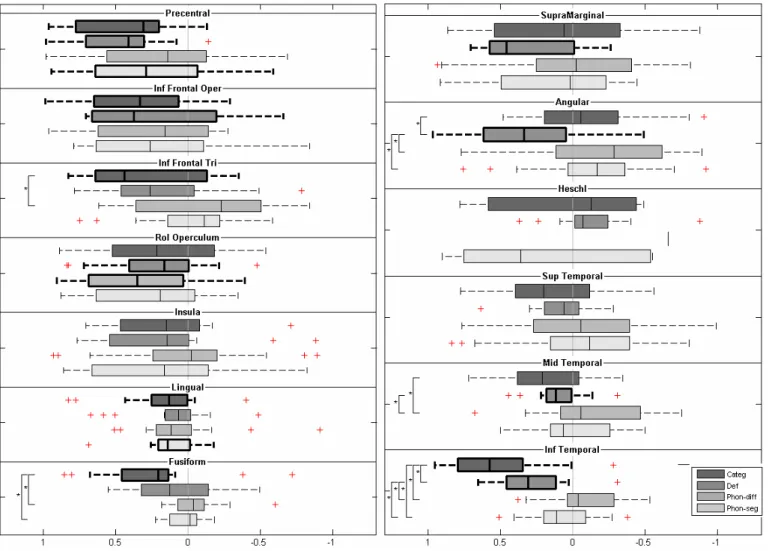
Boxplots based on these values were created. Similarly weighted mean activations within a ROI were estimated using the Marsbar toolbox (Brett et al., 2002) for each smoothed ROI. Wilcoxon signed rank tests (p<0.05) were performed on each ROI to determine significant activations (for all subjects) and group lateralization (i.e. left if LI significantly greater than zero, right if LI significantly smaller than zero, otherwise bilateral). To highlight laterality differences between paradigms, the Kruskal-Wallis test was performed on each ROI. A post-hoc non-parametric Mann-Whitney U-test (p<0.05) was performed to determine between-paradigm differences when the Kruskal-Wallis test was significant (p<0.05).
Results
We first carried out group analysis including statistical comparisons and conjunctions of the tasks within each category (i.e. reference auditory tasks; new visual tasks). Then, we carried out a comparison of the two categories as well as conjunction and union analysis of all tasks (i.e. whole panel). The statistical comparisons aimed to assess the specificity of each task or category. The conjunction (intersection) aimed to reveal more language-specific activations, while the union analysis aimed to combine the results of all tasks and to improve the sensitivity of the procedure.
Reference auditory tasks
These two lexico-semantic tasks with auditory stimulation were chosen from the literature to highlight a predominant activation of the left IFG (for the category task) or the
left STG (for the definition task). The data from one subject had to be discarded because of a technical problem with sound delivery.
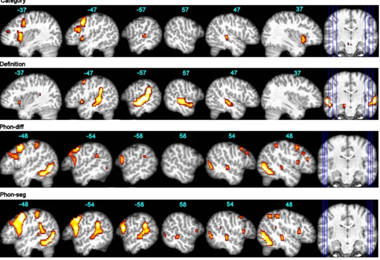
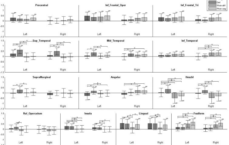
According to group analysis (Table 1; Fig. 1), the category task elicited a left-only activation in the caudal and dorsal IFG-triangularis, extending into the IFG-opercularis, and a bilateral activation of the insula extending only on the left in the ventral IFG-opercularis. Only a small and bilateral activation was seen in the middle part of the superior temporal sulcus (STS), and other activations appeared in the left precentral and lingual gyri. ROI analysis and LIs (Fig. 3 and 4) confirmed the significant left-lateralized activation of the IFG-triangularis and opercularis, as well as of the precentral and lingual gyri, while the activation of the STG did not appear to be significantly left lateralized.
The definition task yielded a strong bilateral activation along the STS, with a large and left-only sub-cluster in the posterior STG and adjacent supramarginal gyrus. Small left-only clusters occurred in the insula / ventral IFG-opercularis and in the precentral gyrus, and other clusters were located in the left and right lingual and parahippocampus region. ROI analysis and LIs confirmed a significant and left-lateralized activation of the MTG as well as the supramarginal and angular gyri, along with the IFG-opercularis and precentral gyrus.
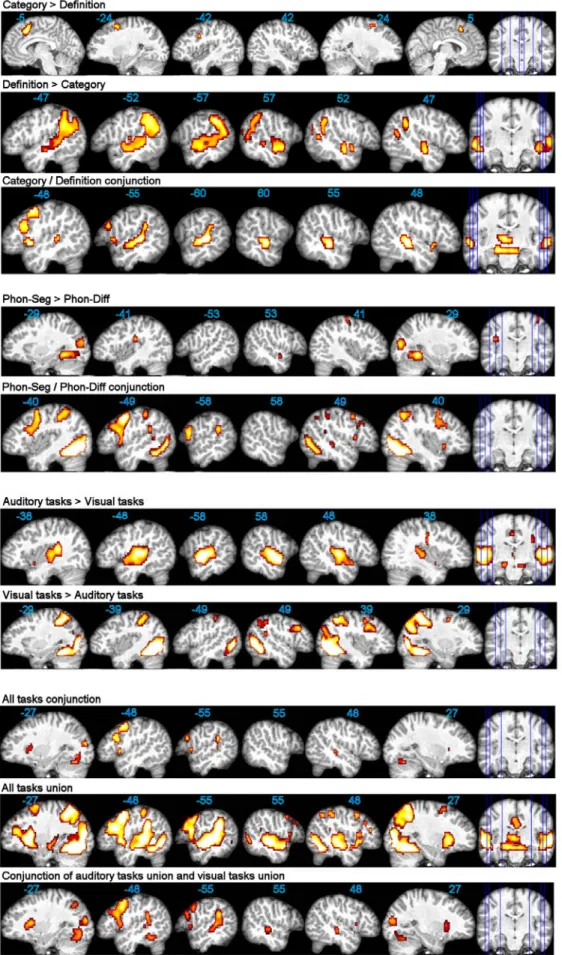
The statistical comparison of these two tasks (Table 2; Fig. 2) showed specific activations in the left precentral gyrus / IFG-opercularis junction for the category task compared to the definition task. The reverse comparison (definition > category) highlighted a strong left-dominant activation extending from the ventral inferior parietal cortex to the anterior STS. This significant difference is corroborated by ROI analysis.
The conjunction analysis between the two tasks (Table 2; Fig. 2) revealed common activations along the left STS, extending posteriorly in the STG / supramarginal junction, and in the right middle STS. Left-only common activations were also found in the dorsal IFG-opercularis and the insula, extending into the adjacent ventral IFG.
Therefore, as expected, the category task elicited a predominant activation of the left IFG compared to the left STG, while results from the definition task showed the inverse pattern. More precisely, in the group analysis, the category task highlighted the caudal and dorsal left IFG-triangularis, although the location is distinct in the statistical comparison. By contrast, the definition task specifically involved the left posterior STG/supramarginal region. Furthermore, the conjunction of both tasks detected both left temporal and frontal core language areas.
New visual tasks
These new phonological tasks with visual stimulation were designed to focus on the phonological brain network without any requirement for either reading or metalinguistic skills. In addition to occipital and inferotemporal activation due to the picture-naming condition, we expected both tasks to elicit an activation of the left posterior STG. Furthermore, in the case of the task involving a phonological change of segmentation, we expected a higher activation of the left IFG. For the phon-seg task, the last paradigm of the session, the data from two subjects had to be discarded because of excessive movement.
According to the group analysis (Table 1), the phon-diff task elicited activations centred on the left and right fusiform, the left precentral and the right angular gyri (p<0.05 FWE-corr.). At the threshold of p<0.001 (unc.) (Fig. 1), large clusters appeared in the left precentral gyrus and IFG (opercularis and triangularis), with smaller similar clusters on the right; a small cluster is located at the left posterior STG / supramarginal junction. ROI analysis (Fig. 3) confirmed a significant and left-dominant activation for the precentral gyrus and the IFG-opercularis, but this did not reach significance for lateralization. LIs (Fig. 4)
showed a significant left lateralization for the rolandic operculum, a region displaying a significant deactivation during the task (p<0.001 unc.).
The phon-seg task elicited a strong bilateral activation centred on the lingual gyri, and a left activation along the precentral gyrus / IFG-opercularis junction; two small left clusters appeared in the dorsal IFG-opercularis and posterior STG (p<0.05 FWE-corr.). At the threshold of p<0.001 (unc.) (Fig. 1), results showed a large cluster encompassing the precentral gyrus and the ventral and dorsal IFG-opercularis, as well as a large cluster centred on the posterior STG / supramarginal junction; only small counterparts were seen on the right. ROI analysis confirmed a significant activation of the left and right IFG and MTG, and LIs revealed a significant left lateralization for the precentral and lingual gyri.
The statistical comparison (Table 2; Fig. 2) showed that the phon-diff task did not elicit specific activation compared to the phon-seg task. The inverse comparison (phon-seg > phon-diff) highlighted the left lingual gyrus, as confirmed by ROI analysis, as well as the left and right fusiform gyri, the left rolandic operculum and a small cluster on the right STS.
By analysing the conjunction of the two tasks (Table 2; Fig. 2), we found a significant common activation in the left precentral gyrus, as well as in the adjacent IFG-opercularis into its more ventral and caudal part, associated with smaller counterparts in the right hemisphere. Another left cluster appeared in the posterior STG.
Therefore, these two new tasks, taken individually, were able to activate core language areas, namely the left IFG-opercularis and the posterior temporal / supramarginal region, even though the LIs did not show significant lateralization. In the group analyses, the phon-seg task compared to the other task activated more the left IFG, as expected. But, contrary to expectations, it also elicited higher activation in the left posterior temporal/supramarginal region. This may reflect an overall superiority of the phon-seg task for the language regions, which nevertheless did not appear in the statistical comparisons. Finally, the conjunction of
the two tasks, as observed with previous auditory tasks, was able to detect both left temporal and frontal core language areas.
Whole panel analysis
When statistically comparing the auditory reference tasks and the new visual tasks (Table 2; Fig. 2), the former elicited specific strong and bilateral activations centred on the middle STG and the Heschl’s gyri, with both areas also showing significant interparadigm differences in the ROI analysis (Fig. 3). Another specific activation was found bilaterally in the lingual gyri. The reverse comparison (visual tasks > auditory tasks) revealed specific clusters in the right middle frontal gyrus, in the right dorsal IFG-opercularis, and in the right posterior and medial STG. In addition, strong specific activations appeared in the fusiform gyri, as also confirmed by significant interparadigm differences in the ROI analysis (Fig. 3).
The conjunction analysis of the four tasks (Table 2; Fig. 2) highlighted the dorsal and ventral parts of the left IFG-opercularis, as well as the left posterior STG/supramarginal junction; on the right, only one small cluster appeared in the middle MTG. Additional common clusters were located in the left precentral gyrus, and, bilaterally, in the lingual gyri and the anterior insula. The conjunction analysis of the unions of auditory and visual tasks (Fig. 2) showed an enlargement of the same clusters with an extension of the left temporal cluster onto the middle STS.
The union analysis of all tasks (Fig. 2) highlighted, on the left, a large cluster encompassing the precentral gyrus, the dorsal IFG-opercularis/triangularis junction, the insula and the immediately adjacent ventral IFG-opercularis, as well as a strong activation extending from the supramarginal gyrus to the anterior STS. Smaller counterparts were seen on the right.
Therefore, the auditory lexico-semantic tasks showed more activation of the auditory receptive language region (i.e. middle STG, Heschl’s gyrus), whereas the visual phonological tasks involved more the occipitotemporal visual region (i.e. fusiform gyrus). For these latter tasks, the stronger contribution of the right IFG and STG, compared to the reference tasks, reflects a less left-lateralized activation of these regions, as suggested also by the LIs. Finally, the conjunction of all tasks highlighted more focused activations in the left language areas, while their union showed enlarged activations, especially for the posterior STG/supramarginal region.
Discussion
FMRI has an expanding role in the localization and lateralization of language in children, which is an important issue in clinical and research applications such as presurgical mapping (Anderson et al., 2006; O'Shaughnessy et al., 2008; Wilke et al., 2006) and the investigation of development language disorders (Dick et al., 2008; Friederici, 2006; Rapin et al., 2003). A major issue of language fMRI is due to the complexity of language, which means that it is crucial to choose a panel of language activation tasks able to detect all, but only, language brain areas (e.g. Tie et al., 2008). In the pediatric context, another specific issue is the need for child-adapted tasks (O’Shaughnessy et al., 2008; Wilke et al., 2003a, 2006), which is even more important with impaired children whose achievement of the task may be compromised by high attentional, reading or metalinguistic requirements.
In this study, we tested with a group of healthy children a panel of four fMRI language tasks that could be used with young impaired children. One important constraint was to avoid reading and metalinguistic requirements, with the aim of increasing the feasibility and efficiency of the procedure, while reducing the attentional and academic demands. Two
reference auditory lexico-semantic tasks were chosen from the literature, and two new tasks with visual stimulation were designed to focus on spontaneous (i.e. non-metalinguistic) phonological processing. When taken individually, the tasks of each modality aimed to stress distinct language areas, and the whole panel aimed to be sensitive and specific in the detection of the language network.
Methodological issues
FMRI in children implies special methodological precautions due to the risk of movement, attentional constraints, task design, task preparation and achievement, as well as appropriate reference brain data (Gaillard et al., 2001a; O'Shaughnessy et al., 2008; Wilke et al., 2003a).
To optimize the feasibility of the procedure for young disordered children, and minimize the movement artefacts and the attentional complications, we implemented four identical block-designed paradigms of equal periods, without control active tasks or motor responses required from the child. These choices reduced the heterogeneity and complexity of the protocol, as the child did not have to understand and achieve distinct control tasks, or give motor responses in addition to understanding and achieving the four language tasks themselves. This may be particularly crucial for disordered children with lower attentional, cognitive or language abilities. Furthermore, while requiring motor responses is well suited for metalinguistic tasks (i.e. judgment tasks), this is precluded in the investigation of more natural condition such as word production, whether covert or overt. Finally, the simplicity and identity of the paradigm parameters for all tasks facilitated the comparison and combination of the results.
Nevertheless, these choices have some drawbacks. First, the achievement of the tasks could not be directly checked. Requiring overt responses would have allowed online performance monitoring, but aloud speech increases the risk of movement, which is crucial with children and all the more with disordered children (O'Shaughnessy et al., 2008). Therefore, children were intensively prepared before the scanner session using the same order of tasks and stimuli, allowing to check that they understood and were able to achieve successfully the task, and they were questioned after the session. Secondly, the use of a low-level control condition (listening to the noise of the scanner and fixing a cross) may involve more non-language-specific coactivations than a more specified control task (e.g. Holland et al., 2007; Wilke et al., 2006). Therefore, we used conjunction analysis between the various tasks to highlight specific language activations.
Moreover, to select a sample representative of the general population and close to the clinical context for disordered children, we did not only recruit right-handed children. In this study, the proportion of left-handed children (11%), is within the normal range estimate (8-15%). To investigate the effect of left-handedness on our results, we carried out supplementary ROIs and LIs analysis of the data from the sixteen right-handed children only, focusing on the IFG, STG and supramarginal gyrus. For the four tasks, results showed no difference concerning the pattern of activation and lateralization within these ROIs compared to the whole group.
In data processing, to avoid distortions due to normalisation of the children’s data on an adult standard template (Wilke et al., 2002, 2003b), we used a recent tool dedicated to the creation of pair- and group-matched normalized templates based on normative brain data (Wilke et al., 2008). This enabled to avoid an age-related bias in the normalization steps by using a customized pediatric template based on the age and sex of our 18 subjects. To carry out local analyses, ROIs were based on the non-linear deformation of the AAL atlas (Tzourio
et al., 2002) on our customized template, as suggested by Wilke et al. (2003c). An inherent limitation of ROIs generated from an atlas is that they cannot adequately model subject variability, and hence do not allow to focus on sub-regions that might be of interest. Furthermore, activations may overlap several ROIs (e.g. the posterior STG and the supramarginal gyrus), so that the actual activations and lateralizations may, in fact, be minimized within each separated ROI. However, given the context and our objectives, this approach remains instructive by confirming significant activation and lateralization when they are located within the ROIs.
Reference lexico-semantic auditory tasks
As expected, the words generation from category task (category), when contrasted with rest, highlighted left frontal rather than temporal language areas. More precisely, the paradigm elicited a left activation of the dorsal and caudal IFG-triangularis, at the junction with the pars opercularis, with another cluster of interest extending from the insula to the adjacent ventral IFG-opercularis. By contrast, there was a weaker and bilateral activation of the middle STS, without activation in the posterior STG. Thus, our study confirms that this paradigm, easier than other verbal fluency paradigms (e.g. Riva et al., 2000; Warburton et al., 1996), is able to induce a relatively specific activation of Broca’s area in children.
This result is in line with the study by Gaillard et al. (2003), who used this paradigm with a group of 16 children (mean age: 10.2) and reported a left activation of the IFG without consistent activation of the STG. However, the location within the IFG is somewhat different from the present results, as these authors reported a main activation in the ventral pars orbitalis and a weaker activation in the anterior pars triangularis. Furthermore, no activation in the middle STS was reported. The task used in our study included three categories in each
27-s active block wherea27-s Gaillard et al.’27-s ta27-sk delivered one category within each 32-27-s block. Therefore, it is likely that our task results in a weaker executive demand and a greater receptive component. Moreover, in adults, the location of semantic processing within the left IFG remains to be specified. In their systematic review of studies using words generation from category, Costafreda et al. (2006) reported a ventral location. However, other studies of semantic processing have reported a dorsal location (e.g. Wagner et al., 2001), and Vigneau et al. (2006), in their meta-analysis of studies using various semantic contrasts, reported semantic clusters in the dorsal pars opercularis.
In contrast with the category task, the auditory-responsive task (definition) elicited a strong and left-dominant activation of the temporal language regions without significant activation in the left IFG. Left and right activations appeared along the middle and anterior STS, extending specifically on the left in the posterior STG and adjacent supramarginal gyrus. Again, one may note a left-only cluster extending from the insula into the adjacent ventral IFG-opercularis.
This in line with the study by Balsamo et al. (2002), who used a similar paradigm with a group of 11 children (mean age: 8.5) and reported a left-dominant activation centred on the middle STG and MTG, and including the primary auditory cortex. Furthermore, these results are similar to those obtained in other studies with children using different language comprehension tasks such as picture/verbal-description matching (Wilke et al., 2006), story listening (Ahmad et al., 2003), or sentence listening with correction judgement (Brauer and Friederici, 2007). Interestingly, a reading variant of the definition task (i.e. read response naming) has been shown to elicit activations in the left middle MTG without activation in the posterior STG (Gaillard et al., 2001b). Thus, the definition task appears to be able to cause a relatively specific activation in the left temporal region and especially in Wernicke’s area. In line with current knowledge (Hickok and Poeppel, 2007; Vigneau et al., 2006), the bilateral
activation of the middle STS may reflect the phonological-level processing of auditory speech (or written text) input, whereas the activation of the posterior STG / supramarginal gyrus may reflect the mapping of phonemes onto articulatory representation.
Our statistical comparison between these two reference tasks confirmed the specificity of the definition task and highlighted a specific activation for the category task in the left precentral/IFG junction. Moreover, the conjunction of both tasks revealed specific core language areas on the left, focusing on the posterior STG, the middle STS, the insula along with the adjacent ventral IFG-opercularis, and the dorsal IFG-opercularis.
New phonological visual tasks
The two new tasks with visual stimulation were designed to investigate spontaneous phonological processings. In contrast with most studies using metalinguistic tasks (e.g. rhyme judgments), these tasks are based on a picture-naming condition in which familiar objects to be named exhibit a close phonological composition.
The first new task (phon-diff) was designed to assess the brain basis of spontaneous production of minimal phonological differences (e.g. /pul/–/bul/–/mul/), and was expected to induce activations in the left IFG and temporo-parietal areas, as suggested by current knowledge about the brain areas involved in phonological production (e.g. Vigneau et al., 2006). Interestingly, at the threshold of p<0.001 (unc.), this task, when compared to rest, yielded left-dominant activations in the whole dorsal and ventral IFG-opercularis along the precentral sulcus, in a dorsal and more medial part of the IFG-triangularis, and, to a lesser extent, in the posterior STG.
The second new task (phon-seg) was designed to assess the brain basis of spontaneous phonological change of segmentation involved in the subtraction or addition of phonemes
(e.g. /kar/–/kart/–/kartõ/). Compared to the previous task, a higher activation of the left IFG was expected according to current hypotheses about the role of this structure in phonological segmentation (Burton, 2001). In fact, this paradigm elicited an interesting activation not only in the left IFG-opercularis, lying along the precentral sulcus, but also an equivalent activation in the left posterior STG. At the threshold of p<0.001 (unc.), our results showed an enlargement of these clusters, with an extension of the posterior temporal activation into the middle STS.
Statistical comparisons between the two tasks did not show task-specific activations in language areas, reflecting the similarity of their global activation patterns. Promising results are provided by the conjunction analysis, which showed left-dominant common activations in core language areas, namely the dorsal and ventral IFG-opercularis, lying along the precentral sulcus, and the posterior STG.
Thus, both new visual tasks yielded an interesting and similar activation pattern of core left language areas, in agreement with current knowledge about the brain basis of phonological processing (Buchsbaum et al., 2001; Burton, 2001; Heim et al., 2003; Vigneau et al., 2006). The location within the left IFG, along the precentral sulcus, is convergent with previous studies of phonological tasks involving phonemes isolation or sequencing (e.g. Vigneau et al., 2006). In addition, according to the group analysis, the task involving phonological a change of segmentation showed as expected, a greater activation of the IFG than the other task, in agreement with current hypotheses (Burton, 2001). Nevertheless, this task also yielded a higher activation of the left STG, which requires further explanation (for discussion, see also Gandour et al., 2003; Heim et al., 2009). The next step of the work needs to use these new tasks for disordered children population investigation. In particular, although the phon-diff task appeared to be less efficient in this study of healthy children, further study may show more efficiency with disordered children.
Whole panel
When statistically contrasted with the new visual tasks, the two auditory reference tasks yielded specific activations of the left and right middle STG, including the primary auditory cortex, and lingual gyri. The involvement of the middle STG may reflect auditory verbal processing. The recruitment of the lingual gyri is consistent with the involvement of ventromedial temporo-occipital regions during semantic processing, even in non-visual tasks, suggesting mental imagery or visualization strategies (see e.g. Abel et al., 2009; Sachs et al., 2008; Vitali et al., 2005; Wise et al., 2000).
Compared to the reference tasks, the two new visual tasks activated slightly more the right IFG-opercularis and posterior STG, which suggests less lateralization associated with the new tasks in these regions. Moreover, these new tasks yielded greater activation of the bilateral fusiform gyrus, whose function has been the subject of much debate and which has been shown to be involved in a number of tasks, such as picture naming, object processing, reading and amodal conceptual processing (for discussion, see Cohen and Dehaene, 2004; Price and Devlin, 2003; Hillis et al., 2005; Karnath et al., 2009).
Although this contrast between the auditory and visual tasks did not show differences in the left IFG and posterior STG / supramarginal gyrus, the separated conjunction analyses of the two groups of tasks provide distinctive results, with the former leading to a wider activation in the left posterior STG and the latter in the left IFG.
The dissociation between the lingual and fusiform regions, which are more intensely activated by the auditory and visual language tasks, respectively, may be surprising. Using a picture-naming task with verbal semantic distracters (interference paradigm), Abel et al. (2009) reported an activation of the left and right lingual gyri. By contrast, Balsamo and
Gaillard (2006) reported an activation of the left fusiform gyrus in children during an auditory semantic decision task. Further work is needed to clarify the respective contributions of these two regions in object and language processing.
The conjunction analysis across all four tasks was assumed to reveal more specific and essential language areas. Interestingly, it highlighted left-only common and focal activations in core language regions, i.e. the dorsal and ventral IFG-opercularis and the posterior STG. Furthermore, the union analysis of all the paradigms, assumed to be more sensitive for detecting a more comprehensive language network, showed clusters of slightly distinct but close locations in the left IFG, and an extended left-dominant parietotemporal activation from the supramarginal gyrus to the anterior STS. The conjunction of the auditory tasks union and the visual tasks union showed an intermediate picture more informative than the conjunction and more specific than the union. Thus, in agreement with previous authors (Gaillard et al., 2004; Ramsey et al., 2001; Roux et al., 2003; Tie et al., 2008; Wilke et al., 2006), our study confirms the usefulness of using a number of language tasks in a fMRI procedure.
In conclusion, out of the four language tasks in our panel, the two reference tasks (category and definition) demonstrated good abilities to yield selective left activations in the IFG and STG, respectively. The two new tasks studied here (phon-diff and phon-seg), which targeted phonological processing without requiring any metalinguistic or reading abilities, also yielded left-dominant activations in the dorsal and ventral IFG-opercularis, as well as the posterior STG, with an overall superiority of the phon-seg task. Compared to the reference tasks, the new tasks activated simultaneously both left frontal and temporal language regions, but less strongly and more bilaterally than the category task for the left IFG and than the definition task for the left posterior STG. When all tasks are taken together, conjunction and union analyses yielded interesting delineations of similar core language regions, with greater sensitivity being obtained from union analysis. This study confirms that a combination of
several tasks tapping different aspects of language is useful for language brain mapping in children, and provides new tasks for the investigation of the brain basis of spontaneous phonological processing. We believe that such an fMRI panel could be efficient and useful with young children in the context of presurgical mapping as well as the investigation of acquired or developmental childhood disorders.
Acknowledgments
We would like to thank the children who participated in the study and their parents. This work was supported by a “Projet Hospitalier de Recherche Clinique” (PHRC–2007; University Hospital, Pontchaillou, Rennes, France), and the “Association pour la Recherche Clinique sur l’Epilepsie”. This work benefited from a INRIA (Institut National de Recherche en Informatique et Automatique) research delegation for CdG. M.S.N. Carpenter post-edited the English style.
References
Abel, S., Dressel, K., Bitzer, R., Kümmerer, D., Mader, I., Weiller, C., Huber, W., 2009. The separation of processing stages in a lexical interference fMRI-paradigm. NeuroImage 44, 1113-24.
Ahmad, Z., Balsamo, L.M., Sachs, B.C., Xu, B., Gaillard, W.D., 2003. Auditory comprehension of language in young children: neural networks identified with fMRI. Neurology 60, 1598-605. Anderson, D.P., Harvey, A.S., Saling, M.M., Anderson, V., Kean, M., Abbott, D.F., Wellard, R.M.,
Jackson, G.D., 2006. FMRI lateralization of expressive language in children with cerebral lesions. Epilepsia 47, 998-1008.
Ashburner, J., Friston, K. J., 2005. Unified segmentation. NeuroImage 26, 839-51.
Balsamo, L.M., Xu, B., Gaillard, W.D., 2006. Language lateralization and the role of the fusiform gyrus in semantic processing in young children. NeuroImage 31, 1306-14.
Balsamo, L.M., Xu, B., Grandin, C.B., Petrella, J.R., Braniecki, S.H., Elliott, T.K., Gaillard, W.D., 2002. A functional magnetic resonance imaging study of left hemisphere language dominance in children. Arch. Neurol. 59, 1168-74.
Barlow, J.A., Gierut, J.A., 2002. Minimal pair approaches to phonological remediation. Semin. Speech Lang. 23, 57-68.
Berl, M.M., Vaidya, C.J., Gaillard, W.D., 2006. Functional imaging of developmental and adaptive changes in neurocognition. NeuroImage 30, 679-91.
Binder, J., Swanson, S., Hammeke, T., Morris, G., Mueller, W., Fisher, M., Benbadis, S., Frost, J., Rao, S., Haughton, V., 1996. Determination of language dominance using functional MRI: a comparison with the Wada test. Neurology 46, 978-84.
Brauer, J., Friederici, A.D., 2007. Functional neural networks of semantic and syntactic processes in the developing brain. J. Cogn. Neurosci. 19, 1609-23.
Brett, M., Anton, J.L., Valbregue, R., Poline, J.B., 2002. Region of interest analysis using an SPM toolbox. Presented at the 8th International Conference on Functional Mapping of the Human Brain, June 2–6, 2002, Sendai, Japan. Available on CD-Rom in NeuroImage 16.
Buchsbaum, B.R., Hickok, G., Humphries, C., 2001. Role of left posterior superior temporal gyrus in phonological processing for speech perception and production. Cogn. Sci. 25, 663-78.
Burton, M.W., 2001. The role of inferior frontal cortex in phonological processing, Cogn. Sci. 25, 695-709.
Burton, M.W., Small, S.L., 2006. Functional neuroanatomy of segmenting speech and nonspeech. Cortex 42, 644-51.
Burton, M.W., Small, S.L., Blumstein, S.E., 2000. The role of segmentation in phonological processing: an fMRI investigation. J. Cogn. Neurosci., 12, 679-90.
Chevrie-Muller, C., Simon, A.-M., Le Normand, M.-T., Fournier, S., 1985. Batterie d’évaluation psycholinguistique, Editions du Centre de Psychologie Appliquée, Paris.
Chomsky, N., Halle, M., 1968. The sound pattern of English, Harper & Row, New York.
Cohen, L., Dehaene, S., 2004. Specialization within the ventral stream: the case for the visual word form area. NeuroImage 22, 466-76.
Costafreda, S.G., Fu, C.H., Lee, L., Everitt, B., Brammer, M.J., David, A.S., 2006. A systematic review and quantitative appraisal of fMRI studies of verbal fluency: role of the left inferior frontal gyrus. Hum. Brain Mapp. 27, 799-810.
Crinion, J.T., Lambon-Ralph, M.A., Warburton, E.A., Howard, D., Wise, R.J., 2003. Temporal lobe regions engaged during normal speech comprehension. Brain 126, 1193-201.
Deblaere K., Backes W.H., Hofman P., Vandemaele P., Boon P.A., Vonck K., Boon P., Troost J., Vermeulen J., Wilmink J., Achten E., Aldenkamp A., 2002. Developing a comprehensive presurgical functional MRI protocol for patients with intractable temporal lobe epilepsy: a pilot study. Neuroradiology 44, 667-73.
DeLeon, J., Gottesman, R.F., Kleinman, J.T., Newhart, M., Davis, C., Heidler-Gary, J., Lee, A., Hillis, A.E., 2007. Neural regions essential for distinct cognitive processes underlying picture naming. Brain 130, 1408-22.
Démonet, J.F., Thierry, G., Cardebat, D., 2005. Renewal of the neurophysiology of language: functional neuroimaging. Physiol. Rev. 85, 49-95.
Dick, F., Richardson, F., Saccuman, M.C., 2008. Using functional magnetic resonance imaging to investigate developmental language disorders. In: Norbury, C.F., Tomblin, J.B., Bishop, D.V.M. (Eds.), Understanding developmental language disorders. Psychology Press, Hove, pp. 53-66. Durston, S., Casey, B.J., 2006. What have we learned about cognitive development from
neuroimaging? Neuropsychologia 44, 2149-57.
Friederici, A.D., 2006. The neural basis of language development and its impairment. Neuron 52, 941-52.
Friston, K.J, Penny, W.D., Glaser, D.E., 2005. Conjunction revisited. NeuroImage 25, 661-7.
Friston, K.J., Fletcher, P., Josephs, O., Holmes, A., Rugg, M.D., Turner, R., 1998. Event-related fMRI: characterizing differential responses. NeuroImage 7, 30-40.
Friston, K.J., Holmes, A.P., Worsley, K.J., Poline, J.B., Frith, C.D., Frackowiack, R.S., 1995. Statistical parametric maps in functional imaging: a general linear approach. Hum. Brain Mapp. 2, 189-210.
Frith, C.D., 2006. The value of brain imaging in the study of development and its disorders. J. Child Psychol. Psychiatry 47, 979-82.
Gaillard, W.D., Balsamo, L., Xu, B., Grandin, C.B., Braniecki, S.H., Papero, P.H., Weinstein, S., Conry, J., Pearl, P.L., Sachs, B., Sato, S., Jabbari, B., Vezina, L.G., Frattali, C., Theodore, W.H., 2002. Language dominance in partial epilepsy patients identified with an fMRI reading task. Neurology 59, 256-65.
Gaillard, W.D., Balsamo, L., Xu, B., McKinney, C., Papero, P.H., Weinstein, S., Conry, J., Pearl, P.L., Sachs, B., Sato, S., Vezina, L.G., Frattali, C., Theodore, W.H., 2004. FMRI language task panel improves determination of language dominance. Neurology 63, 1403-8.
Gaillard, W.D., Grandin, C.B., Xu, B., 2001a. Developmental aspects of pediatric fMRI: considerations for images acquisition, analysis and interpretation. NeuroImage 13, 239-49. Gaillard, W.D., Pugliese, M., Grandin, C.B., Braniecki, S.H., Kondapaneni, P., Hunter, K., Xu, B.,
Petrella, J.R., Balsamo, L., Basso, G., 2001b. Cortical localization of reading in normal children: an fMRI language study. Neurology 57, 47-54.
Gaillard, W.D., Hertz-Pannier, L., Mott, S.H., Barnett, A.S., Le Bihan, D., Theodore, W.H., 2000. Functional anatomy of cognitive development: fMRI of verbal fluency in children and adults. Neurology 54, 180-85.
Gaillard, W.D., Moore, E.N., Weber, D.A., Ritzl, E.K., Berl, M.M., 2006. Functional magnetic resonance imaging in normal and pathological language development. In: Riva, D., Rapin, I.,
Zardini, G. (Eds.), Language: normal and pathological development. John Libbey Eurotext, pp. 105-20.
Gaillard, W.D., Sachs, B.C., Whitnah, J.R., Ahmad, Z., Balsamo, L.M., Petrella, J.R., Braniecki, S.H., McKinney, C.M., Hunter, K., Xu, B., Grandin, C.B., 2003. Developmental aspects of language processing: fMRI of verbal fluency in children and adults. Hum. Brain Mapp. 18, 176-85. Gandour, J., Xu, Y., Wong, D., Dzemidzic, M., Lowe, M., Li, X., Tong, Y., 2003. Neural correlates of
segmental and tonal information in speech perception. Hum. Brain Mapp. 20, 185-200. Hardyck, C., Petrinovich, L. F., 1977. Left-handedness. Psychol. Bull. 84, 385-404.
Heim, S., Opitz, B., Müller, K., Friederici, A.D., 2003. Phonological processing during language production: fMRI evidence for a shared production-comprehension network. Brain Res. Cogn. Brain Res. 16, 285-96.
Heim, S., Eickhoff, S.B., Friederici, A.D., Amunts, K., 2009. Left cytoarchitectonic area 44 supports selection in the mental lexicon during language production. Brain Struct. Funct. 213, 441–456. Hertz-Pannier, L., Gaillard, W.D., Mott, S.H., et al., 1997. Noninvasive assessment of language
dominance in children and adolescents with functional MRI: a preliminary study. Neurology 48, 1003-12.
Hickok, G., Poeppel, D., 2007. The cortical organization of speech processing. Nat. Rev. Neurosci. 8, 393-402.
Hillis, A.E., Newhart, M., Heidler, J., Barker, P., Herskovits, E., Degaonkar, M., 2005. The roles of the "visual word form area" in reading. NeuroImage 24, 548-59.
Holland, S.K., Plante, E., Byars, A.W., Strawsburg, R.H., Schmithorst, V.J., Ball, W.S., 2001. Normal fMRI brain activation patterns in children performing a verb generation task. NeuroImage 14, 837-43.
Holland, S.K., Vannest, J., Mecoli, M., Jacola, L.M., Tillema, J.M., Karunanayaka, P.R., Schmithorst, V.J., Yuan, W., Plante, E., Byars, A.W., 2007. Functional MRI of language lateralization during development in children. Int. J. Audiol. 46, 533-51.
Indefrey, P., Levelt, W.J., 2004. The spatial and temporal signatures of word production components. Cognition 92, 101-44.
Jakobson, R., Fant, C.G.M., Halle, M., 1951. Preliminaries to speech analysis: the distinctive features and their correlates. MIT Press, Cambridge.
Karnath, H.O., Rüter, J., Mandler, A., Himmelbach, M., 2009. The anatomy of object recognition– visual form agnosia caused by medial occipitotemporal stroke. J. Neurosci. 29, 5854-62.
Lambert, E., Chesnet, D. 2001. Novlex: une base de données lexicales pour les élèves de primaire. Année Psychol. 101, 277-288.
Medina, L.S., Bernal, B., Ruiz, J., 2007. Role of functional MR in determining language dominance in epilepsy and nonepilepsy populations: a Bayesian analysis. Radiology 242, 94-100.
Moore, D.R., Rosenberg, J.F., Coleman, J.S., 2005. Discrimination training of phonemic contrasts enhances phonological processing in mainstream school children. Brain Lang. 94, 72-85.
Nichols, T., Brett, M., Andersson, J., Wager, T., Poline, J.B., 2005. Valid conjunction inference with the minimum statistic. NeuroImage 25, 653-60.
Oldfield, R.C., 1971. The assessment and analysis of handedness: the Edinburgh inventory. Neuropsychologia 9, 97-113.
O'Shaughnessy, E.S., Berl, M.M., Moore, E.N., Gaillard, W.D., 2008. Pediatric functional magnetic resonance imaging (fMRI): issues and applications. J. Child Neurol. 23, 791-801.
Paulesu, E., Frith, C.D., Frackowiak, R.S., 1993. The neural correlates of the verbal component of working memory. Nature 362, 342-5.
Piérart, B., Comblain, A., Grégoire, J., Mousty, P., 2005. Isadyle : Instruments pour le screening et l’approfondissement des dysfonctionnements du langage chez l’enfant, TEMA, Bruxelles. Price, C.J., Devlin, J.T., 2003. The myth of the visual word form area. NeuroImage 19, 473-81.
Ramsey, N.F., Sommer, I., Rutten, G.J., Kahn, R., 2001. Combined analysis of language tasks in fMRI improves assessment of hemispheric dominance for language functions in individual subjects. NeuroImage 13, 719-33.
Rapin, I., Dunn, M., Allen D.A., 2003. Developmental language disorders. In: Segalowitz, S.J. (Ed.), Handbooks of neuropsychology, Elsevier, Amsterdam, pp. 593-630.
Riva, D., Nichelli, F., Devoti, M., 2000. Developmental aspects of verbal fluency and confrontation naming in children. Brain Lang 71, 267-84.
Roux, F.E., Boulanouar, K., Lotterie, J.A., Mejdoubi, M., Le Sage, J.P., Berry, I., 2003. Language functional magnetic resonance imaging in preoperative assessment of language areas: correlation with direct cortical stimulation. Neurosurgery 52, 1335-47.
Rutten, G.J., Ramsey, N.F., van Rijen, P.C., Noordmans, H.J., van Veelen, C.W., 2002. Development of a functional magnetic resonance imaging protocol for intraoperative localization of critical temporoparietal language areas. Ann. Neurol. 51, 350-60.
Sachs, B.C., Gaillard, W.D., 2003. Organisation of language networks in children: functional resonance imaging studies. Current Neurology and Neuroscience Reports 3, 157-62.
Sachs, O., Weis, S., Krings, T., Huber, W., Kircher, T., 2008. Categorical and thematic knowledge representation in the brain: neural correlates of taxonomic and thematic conceptual relations. Neuropsychologia 46, 409-18.
Tie, Y., Suarez, R.O., Whalen, S., Radmanesh, A., Norton, I.H., Golby, A.J., 2009. Comparison of blocked and event-related fMRI designs for pre-surgical language mapping, NeuroImage 47, T107-15.
Tie, Y., Whalen, S., Suarez, R.O., Golby, A.J., 2008. Group independent component analysis of language fMRI from word generation tasks. NeuroImage 42, 1214-25.
Tzourio-Mazoyer, N., Landeau, B., Papathanassiou, D., Crivello, F., Etard, O., Delcroix, N., Mazoyer, B., Joliot, M., 2002. Automated anatomical labeling of activations in SPM using a macroscopic anatomical parcellation of the MNI MRI single-subject brain. NeuroImage 15, 273-89.
Vigneau, M., Beaucousin, V., Hervé, P.Y., Duffau, H., Crivello, F., Houdé, O., Mazoyer, B., Tzourio-Mazoyer, N., 2006. Meta-analyzing left hemisphere language areas: phonology, semantics, and sentence processing. NeuroImage 30, 1414-32.
Vitali, P., Abutalebi, J., Tettamanti, M., Rowe, J., Scifo, P., Fazio, F., et al., 2005. Generating animal and tool names: an fMRI study of effective connectivity. Brain Lang. 93, 32-45.
Wagner, A.D., Pare-Blagoev, E.J., Clark, J., Poldrack, R.A., 2001. Recovering meaning: left prefrontal cortex guides controlled semantic retrieval. Neuron 31, 329-38.
Warburton, E., Wise, R.J., Price, C.J., Weiller, C., Hadar, U., Ramsay, S., Frackowiak, R.S., 1996. Noun and verb retrieval by normal subjects. Studies with PET. Brain 119, 159-79.
Wilke, M., Schmithorst, V.J., Holland, S.K., 2002. Assessment of spatial normalization of whole-brain magnetic resonance images in children. Hum. Brain Mapp. 17, 48-60.
Wilke, M., Holland, S.K., Altaye, M., Gaser, C., 2008. Template-O-Matic: a toolbox for creating customized pediatric templates. NeuroImage 41, 903-13.
Wilke, M., Holland, S.K., Myseros, J.S., Schmithorst, V.J., Ball, W.S., 2003a. Functional magnetic resonance imaging in pediatrics. Neuropediatrics 34, 225-33.
Wilke, M., Schmithorst, V.J., Holland, S.K., 2003b. Normative pediatric brain data for spatial normalization and segmentation differs from standard adult data. Magn. Reson. Med. 50, 749-57.
Wilke, M., Sohn, J.-H., Byars, A.W., Holland, S.K., 2003c. Bright spots: correlations of gray matter volume with IQ in a normal pediatric population. NeuroImage 20, 202-15.
Wilke, M., Lidzba, K.J., 2007. LI-tool: a new toolbox to assess lateralization in functional MR-data. Neurosci. Methods 163, 128-36.
Wilke, M., Lidzba, K.J., Staudt, M., Buchenau, K., Grodd, W., Krägeloh-Mann, I., 2005. Comprehensive language mapping in children, using functional magnetic resonance imaging: what’s missing counts. NeuroReport 16, 915–19.
Wilke, M., Lidzba, K.J., Staudt, M., Buchenau, K., Grodd, W., Krägeloh-Mann, I., 2006. An fMRI task battery for assessing hemispheric language dominance in children. NeuroImage 32, 400-10. Wise, R.J., Howard, D., Mummery, C.J., Fletcher, P., Leff, A., Büchel, C., Scott, S.K., 2000. Noun
imageability and the temporal lobes. Neuropsychologia 38, 985-94.
Zatorre, R., Meyer, E., Gjedde, A., Evans, A., 1996. PET studies of phonetic processing of speech: review, replication and reanalysis. Cerebr. Cortex 6, 21-30.