LUND UNIVERSITY PO Box 117 221 00 Lund
Development of the gastrointestinal tract in young mammals
Effects of enteral provocation with protease or phytohaemagglutinin in neonatal rats
Arevalo Sureda, Ester
2017
Document Version:
Publisher's PDF, also known as Version of record
Link to publication
Citation for published version (APA):
Arevalo Sureda, E. (2017). Development of the gastrointestinal tract in young mammals: Effects of enteral provocation with protease or phytohaemagglutinin in neonatal rats. Lund: Lund University, Faculty of Science, Department of Biology.
General rights
Copyright and moral rights for the publications made accessible in the public portal are retained by the authors and/or other copyright owners and it is a condition of accessing publications that users recognise and abide by the legal requirements associated with these rights.
• Users may download and print one copy of any publication from the public portal for the purpose of private study or research.
• You may not further distribute the material or use it for any profit-making activity or commercial gain • You may freely distribute the URL identifying the publication in the public portal
Take down policy
If you believe that this document breaches copyright please contact us providing details, and we will remove access to the work immediately and investigate your claim.
Est Er A ré v A lo s u r Ed A D ev elo pm en t o f t he g ast ro in te sti na l t ra ct i n y ou ng m am m als 20 9 789177 531821 Faculty of Science Department of Biology
Development of the gastrointestinal
tract in young mammals
Effects of enteral provocation with protease
or phytohaemagglutinin in neonatal rats
EstEr ArévAlo surEdA
Development of the gastrointestinal tract
in young mammals
Development of the gastrointestinal
tract in young mammals
Effects of enteral provocation with
protease or phytohaemagglutinin
in neonatal rats
Ester Arévalo Sureda
DOCTORAL DISSERTATION
by due permission of the Faculty of Science, Lund University, Sweden. To be defended at Föreläsningssalen, Biologihus A, Sölvegatan 35
on the 31st of March 2017, 13.00.
Faculty opponent
Ass. Professor Francisco José Pérez-Cano
Organization LUND UNIVERSITY Document name DOCTORAL DISSERTATION Faculty of Science Department of Biology Date of issue 2017-03-31
Author(s): Ester Arévalo Sureda Sponsoring organization Title and subtitle: Development of the gastrointestinal tract in young mammals. Effects of enteral provocation with protease or phytohaemagglutinin in neonatal rats Abstract
The rat, as an altritial species, is born with an immature gastrointestinal tract and intestinal barrier function, which is highly absorptive to milk-borne bioactive molecules that can pass undigested and reach the general circulation of the suckling newborn. This passage occurs by the neonatal-Fc-receptor (FcRn) binding and trancytosis of immunoglobulin G in the proximal small intestine (SI) and by the highly endocytic vacuolated enterocytes non-selectively in the distal SI. Postnatal gut maturation accelerates at weaning, around postnatal day 21, coincident with the dietary transition from milk to solid food. Maturation of the gut can also be precociously induced by provocation with a lectin, phytohaemagglutinin (PHA), mimicking the naturally occurring changes in gut structure and function. The changes occurring during natural or induced gut maturation include stimulation of pancreatic function and cessation of the SI absorptive capacity to macromolecules (gut closure). Intestinal epithelial maturation has been related to the gut immune system and is suggested to depend on T-lymphocytes activation. Recently, the transcription factor B-lymphocyte-induced maturation-protein-1 (Blimp-1) has been proposed to be a key regulator of intestinal maturation in mice. Hence, the present study investigated the events occurring during gut development and the cues initiating the process. The study especially focused on changes in the barrier function and macromolecular permeability, pancreatic function, and the relation to gut immune factors. A novel animal model of pancreatic and pancreatic-like protease-induced precocious gut maturation was established in neonatal rats, and was used in comparison to the existing PHA-induced model, as well as natural gut development. The gut maturational changes observed during natural or induced maturation, by both protease or PHA, included the transition of foetal- to adult- type SI epithelium, with reduced FcRn expression in the proximal part and disappearance of vacuolated enterocytes in the distal part, associated with a similar change in intestinal epithelial Blimp1 expression. The early effects after exposure to the provocative agents, PHA and protease, revealed that both agents hampered macromolecular permeability and only protease also caused an increase in epithelial leakiness of the distal SI. These results indicated that protease and PHA affected the intestinal barrier function differently. Furthermore, the provocative agents were also tested in neonatal athymic nude rats, T-cell immunodeficient, and they appeared to be susceptible to induced precocious gut maturation. These results suggested that gut maturation is independent of thymus-derived T-celsl, but the involvement of other immune cells types, possibly innate immune cells, should be further investigated.
Thus, the findings of the present thesis will contribute to an increased understanding of initiating cues and the mechanisms of maturation of the intestinal barrier in young mammals. The knowledge obtained could be applied to improve strategies for the treatment of gut-related complications, often affecting premature infants.
Key words: gut, intestine, pancreas, development, precocious, protease, PHA, enterocytes, permeability, endocytosis, IgG, FcRn, Blimp1, T-lymphocyte, passive immunity, altricial, neonatal, suckling, athymic, rat, immunohistochemistry
Classification system and/or index terms (if any)
Supplementary bibliographical information Language English
ISSN and key title ISBN 978-91-7753-182-1 (print)
ISBN 978-91-7753-183-8 (electronic) Recipient’s notes Number of pages 182 Price
Security classification
I, the undersigned, being the copyright owner of the abstract of the above-mentioned dissertation, hereby grant to all reference sources permission to publish and disseminate the abstract of the above-mentioned dissertation.
Development of the gastrointestinal
tract in young mammals
Effects of enteral provocation with
protease or phytohaemagglutinin
in neonatal rats
Coverphoto by Marc Pallarès Carrera Copyright Ester Arévalo Sureda Faculty of Science
Department of Biology
ISBN 978-91-7753-182-1 (print) ISBN 978-91-7753-183-8 (electronic)
Printed in Sweden by Media-Tryck, Lund University Lund 2017
To you
(not only, but also)Qui fa tot el que pot, no esta obligat a més
Table of contents
List of Publications ... 10
Author Contributions ... 11
Conference abstracts ... 12
Publications not included in the thesis ... 13
Abbreviations ... 14
Abstract ... 17
Introduction ... 19
The digestive system ... 19
Gut maturation ... 21
The gut immune system ... 22
The gut immune system in the young ... 24
The intestinal barrier ... 25
The intestinal barrier in the young ... 27
Gut microbiota in the young... 31
Digestive and immune systems – developmental connections... 32
The suckling rat model ... 32
Scientific aims ... 35
Methodology ... 37
Animals and ethics statement ... 37
Experiments ... 37
Animal euthanasia and samples collection ... 40
In vivo intestinal permeability ... 42
Results and Discussion ... 49
Protease–induced precocious gut maturation ... 49
Maturation of the intestinal epithelium ... 51
Exocrine pancreatic function during development ... 55
Early effects of the provocative agents ... 59
Involvement of the immune system in gut maturation ... 62
Possible mechanisms for initiating gut maturation ... 67
Conclusions ... 70
Future studies and perspectives ... 71
Popular summary ... 73
Populärvetenskaplig sammanfattning ... 75
Resum de divulgació científica ... 77
Acknowledgements ... 79
List of Publications
Paper I
Prykhodko, O., Pierzynowski, S. G., Nikpey, E., Arévalo Sureda, E., Fedkiv, O., Weström, B. R. (2015).
Pancreatic and pancreatic-like microbial proteases accelerate gut maturation in neonatal rats.
PLoS ONE, 10(2), e0116947. doi: 10.1371/journal.pone.0116947.
Paper II
Arévalo Sureda E, Weström B, Pierzynowski S, Prykhodko O. (2016).
Maturation of the Intestinal Epithelial Barrier in Neonatal Rats Coincides with Decreased FcRn Expression, Replacement of Vacuolated Enterocytes and Changed Blimp1 Expression.
PLoS ONE, 11 (10), e0164775. doi: 10.1371/journal.pone.0169724.
Correction in PLoS ONE, 12(1), e0169724. doi:10.1371/journal.pone.0169724.
Paper III
Arévalo Sureda E, Gidlund C, Weström B, Prykhodko O.
Intestinal precocious maturation can be induced in athymic (nude) neonatal rats despite their T-cell deficiency.
Manuscript submitted to the American Journal of Physiology – Regulatory, Integrative and Comparative Physiology (2017).
Paper IV
Arévalo Sureda E, Prykhodko O., Weström B.
Early effects on the gut and the intestinal permeability after enteral provocation with protease or phytohaemagglutinin in neonatal rats.
Author Contributions
Paper I
Created the hypothesis: OP SGP
Conceived and designed of the work: OP SGP BRW Data collection: OP EN
Analysed the data: OP SGP EN EAS OF Data interpretation: OP SGP EN EAS OF BW Writing – original draft: OP
Wrote the paper: OP SGP EN EAS OF BW
Paper II
Conception and design of the work: BW OP Data collection and analysis: EAS OP Data interpretation: EAS BW OP Writing – original draft: EAS
Critical revision of the article: BW SGP OP Writing – review& editing: EAS BW SGP OP
Paper III
Conception and design of the work: BW OP Data collection and analysis: EAS OP CG Data interpretation: EAS CG BW OP Writing – original draft: EAS
Critical revision of the article: OP BW
Paper IV (Manuscript)
Conception and design of the work: EAS BW Data collection and analysis: EAS BW Data interpretation: EAS OP BW Writing – original draft: EAS
Conference abstracts
During the course of this PhD project preliminary results, some of them included in this thesis as additional unpublished data have been presented at international scientific conferences in poster format by the author, or as specified.
Role and expression of neonatal-Fc-receptor in the small intestine during normal and precociously induced maturation in the neonatal rat.
Arévalo Sureda E, Prykhodko O, Pierzynowski SG and Weström B
In: Abstracts from the 26th Meeting of the European Intestinal Transport Group (EITG), 2–5 October 2014. Acta Physiologica. 2015;214 (Supplement S701):1-16. doi: 10.1111/apha.12498. Poster.
In vivo macromolecule absorption in wild-type and immunodeficient athymic
neonatal rats during lectin-induced maturation.
Prykhodko O, Arévalo Sureda E, Zhou J, Chopek A, Fedkiv O, Pierzynowski SG and Weström B
In: Abstracts from the 26th Meeting of the European Intestinal Transport Group (EITG), 2–5 October 2014. Acta Physiologica. 2015;214 (Supplement S701):1-16. doi: 10.1111/apha.12498. Oral presentation by Olena Prykhodko.
Expression of the transcription repressor Blimp1 correlates to the disappearance of FcRn expression in proximal and vacuolated cells in distal small intestine during development in neonatal rats.
Arévalo Sureda E, Prykhodko O, Pierzynowski SG and Weström B
In: 48th Annual meeting of the European Society for Pediatric Gatroenterology, Hepatology and Nutrition (ESPGHAN): 2015; Amsterdam, The Netherlands. Poster. PO-G-0009
Increased pancreatic protease activity in relation to PAR2 receptor expression during intestinal postnatal development in rats.
Arévalo Sureda E, Weström B, Pierzynowski SG and Prykhodko O
In: 48th Annual meeting of the European Society for Pediatric Gastroenterology, Hepatology and Nutrition (ESPGHAN): 2015; Amsterdam, The Netherlands. Poster. PO-G-0014.
Microbial changes and TLR4 expression during natural and induced intestinal maturation in neonatal rats.
Prykhodko O, Arévalo Sureda E, Gacon AL, Fedkiv O, Pierzynowski SG and Weström B
In: 48th Annual meeting of the European Society for Pediatric Gastroenterology, Hepatology and Nutrition (ESPGHAN): 2015; Amsterdam, The Netherlands. Poster. PO-G-0016
Precocious gastrointestinal maturation can be induced in T-cell deficient athymic (nude) suckling rats
Arévalo Sureda E, Prykhodko O, Zhou J, Weström B
In: 49th Annual meeting of the European Society for Pediatric Gastroenterology, Hepatology and Nutrition (ESPGHAN): 2016; Athens, Greece. Poster. G-P-002.
Publications not included in the thesis
Lozinska, L., Prykhodko, O., Sureda, E. A., Szwiec, K., Podgurniak, P., Pierzynowski, S., & Weström, B. (2015). Monitoring changes in plasma levels
of pancreatic and intestinal enzymes in a model of pancreatic exocrine insufficiency - induced by pancreatic duct-ligation - in young pigs. Advances
Abbreviations
BIgG Bovine Immunoglobulin G
Blimp1 B-Lymphocyte Induced Maturation Protein – 1 BSA Bovine Serum Albumin
CCK Cholecystokinin CD Cluster of Differentiation DAO Diamine Oxidase DCs Dendritic Cells FcRn Neonatal-Fc Receptor FD4 FITC-dextran 4kDa FD70 FITC-dextran 70kDa FITC Fluorescein isothyanate GI Gastrointestinal g bwt grams body weight
HRP Horse Radish Peroxidase
HSA Human Serum Albumin IECs Intestinal Epithelial Cells
IELs Intraepithelial Lymphocytes Ig Immunoglobulin
IL Interleukin ILCs Innate Lymphocytes kDa kilo Dalton
Lac Lactulose
MHC Major Histocompatibility Complex Man Mannitol
Nude Athymic T-Cell Deficient Nude Rats PARs Protease Activated Receptors PBS Phosphate Buffered Saline PHA Phytohaemagglutinin PPs Peyer’s Patches Prot Protease
RIgG Rat Immunoglobulin G RT Room Temperature SD Sprague Dawley SI Small Intestine SPF Specific Pathogen Free TGF Transforming Growth Factor Th T-helper lymphocyte TLR Toll-Like Receptors TNF Tumour Necrosis Factor
Treg Regulatory T-lymphocyte Å Ångstrom
Abstract
The rat, as an altritial species, is born with an immature gastrointestinal tract and intestinal barrier function, which is highly absorptive to milk-borne bioactive molecules that can pass undigested and reach the general circulation of the suckling newborn. This passage occurs by the neonatal-Fc-receptor (FcRn) binding and trancytosis of immunoglobulin G in the proximal small intestine (SI) and by the highly endocytic vacuolated enterocytes non-selectively in the distal SI. Postnatal gut maturation accelerates at weaning, around postnatal day 21, coincident with the dietary transition from milk to solid food. Maturation of the gut can also be precociously induced by provocation with a lectin, phytohaemagglutinin (PHA), mimicking the naturally occurring changes in gut structure and function. The changes occurring during natural or induced gut maturation include stimulation of pancreatic function and cessation of the SI absorptive capacity to macromolecules (gut closure). Intestinal epithelial maturation has been related to the gut immune system and is suggested to depend on T-lymphocytes activation. Recently, the transcription factor B-lymphocyte-induced maturation-protein-1 (Blimp-1) has been proposed to be a key regulator of intestinal maturation in mice. Hence, the present study investigated the events occurring during gut development and the cues initiating the process. The study especially focused on changes in the barrier function and macromolecular permeability, pancreatic function, and the relation to gut immune factors. A novel animal model of pancreatic and pancreatic-like protease-induced precocious gut maturation was established in neonatal rats, and was used in comparison to the existing PHA-induced model, as well as natural gut development. The gut maturational changes observed during natural or induced maturation, by both protease or PHA, included the transition of foetal- to adult- type SI epithelium, with reduced FcRn expression in the proximal part and disappearance of vacuolated enterocytes in the distal part, associated with a similar change in intestinal epithelial Blimp1 expression. The early effects after exposure to the provocative agents, PHA and protease, revealed that both agents hampered macromolecular permeability and only protease also caused an increase in epithelial leakiness of the distal SI. These results indicated that protease and PHA affected the intestinal barrier function differently. Furthermore, the provocative agents were also tested in neonatal athymic nude rats, T-cell immunodeficient, and they appeared to be susceptible to induced precocious gut maturation. These results suggested that gut maturation is independent of thymus-derived T-cells, but the involvement of other immune cells types, possibly innate immune cells, should be further investigated.
Thus, the findings of the present thesis will contribute to an increased understanding of initiating cues and the mechanisms of maturation of the intestinal barrier in young mammals. The knowledge obtained could be applied to improve strategies for the treatment of gut-related complications, often affecting premature infants.
Introduction
The digestive system
The digestive system consists of the gastrointestinal (GI) tract and the accessory organs; including the liver and the pancreas. All the organs within the digestive system form a complex system which functions in the digestion and absorption of nutrients, the elimination of food components that are not absorbed or excreted and the protection of the organism. The GI tract is a tube extending from the mouth to the anus, and its inner surface comes into continuous contact with exogenous substances that are consumed via eating. Due to the variety and complexity of the functions of the digestive system, it has become clear that the enteric nervous system (ENS) and the immune system in the gut are of utmost importance. Along the GI tract the following can be found: the oral cavity and the oesophagus, the stomach, the small intestine (or the gut), the caecum, the colon, the rectum and the anus. To facilitate the functions of digestion and absorption it comprises an enormous surface area, which is about 200 m2 in adult humans 1. However, at the same time it is a tightly sealed barrier that limits the internal milieu from the external environment.
The small intestine
The small intestine (SI) can be divided into three sections: the duodenum, which begins just after the stomach; the jejunum, and the ileum, which is connected to the colon via the cecum. In the duodenum, decomposition of ingested food by pancreatic and bile secretions takes place, whereas the jejunum and ileum are the main sites of absorption 1.
The architecture of the SI consists of concentric layers, in order from the innermost to outermost (or luminal) layers: serosa, layer of mesothelium;
muscularis, with two smooth muscle layers (longitudinal and circular);
submucosa, connective tissue with blood and lymph vessels, nerve plexus and submucosal glands; and the mucosa. The mucosal layer is organized into the
lamina muscularis, a smooth muscle layer and the lamina propria, a loose
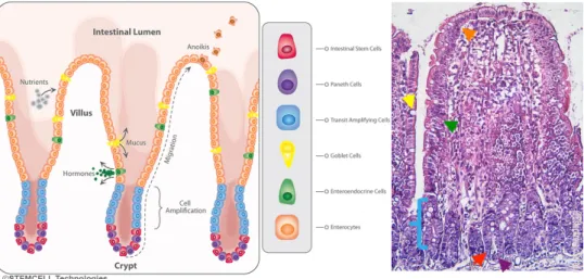
finally, the epithelium. The SI epithelium is formed by a monolayer of cells organized in finger-like structures towards the lumen (villi) and invaginations into the submucosa (crypt) (Figure 1). Hence, the functional unit of the SI epithelia is the crypt-villus complex. The cellular types that can be found at the intestinal mucosa are enterocytes (absorptive cells), goblet cells (mucous secreting cells), and endocrine cells (responsible for the secretion of regulatory hormones) distributed in the villi region, and Paneth’s cells (responsible for the secretion of antibacterial factors into the lumen of crypts cells) and intestinal stem cells in the crypt region.
All cells in the mucosal epithelium originate from the crypt region, from intestinal stem cells. Two kinds of stem cells have been described in the intestine: crypt base columnar cells (CBCs) 2, which are distributed between Paneth’s cells in the bottom of the crypts and can be identified by the marker Lgr5 (leucine group repeated 5) 3. The second type of stem cells are the transit-amplifying (TA) cells or “+4 cells” 4, which give origin to the other cells of the epithelia and undergo rapid cell proliferation. The intestinal epithelium is in a continuous state of cell turnover, which is completed within 3-5 days 5,6. Renewal of cells occurs by proliferation, migration and differentiation into the different epithelial cell types from crypts up to the villus and apoptosis and shedding of “old” cells at the tip of villi. Each villus is built on several proliferating crypts.
Figure 1. Intestinal epithelium structure.
Diagram of the small intestinal epithelium indicating the distribution of the main cell types (left). Photomicrograph of H&E staining representative of the small intestine epithelium (200x) (right) with the different cell types indicated with coloured arrows of the corresponding cell-type colour in the legend: orange – enterocytes, green – enteroendocrine cells, yellow – goblet cells, blue – transit amplifying cells, red – stem cells and purple – paneth cells. The small intestinal epithelium diagram on the left is used with permission, courtesy of STEMCELL technologies.
The pancreas
The pancreas is a diffuse, lobulated, glandular, accessory organ situated in the abdominal cavity, in direct contact with the stomach, the small intestine and colon portions of the GI tract, as well as the spleen. As reviewed by Wathall (2005), the pancreas has a dual function: the endocrine function, confined to the cells of the islets of Langerhans, and the exocrine function, performed by the acinar cells. The exocrine function involves the production and secretion of the digestive enzymes, as well as the chyme neutralising secretions containing bicarbonate ions into the duodenum of the GI tract, via the joint pancreatic-biliary duct in rats. The main digestive enzymes contained within the pancreatic secretions include lipase, amylase, protease and nucleases. These enzymes participate in the hydrolysis ofdietary fat, carbohydrates and proteins, as well as DNA and RNA 1.
Gut maturation
At birth, the GI system is immature and adapted to the strict milk-based diet of the young. The immature stomach secretes low levels of chymosin, the milk-clotting enzyme, and hydrochloric acid, the levels of which increase at weaning together with the secretions of pepsin, intrinsic factor and gastrin 1. In this thesis, however, the main focus will be the small intestine and pancreas.
The small intestine
The SI intestine is immature at birth with reduced crypts, slow cell turnover, finger-shaped villi and enhanced permeability to macromolecules. At birth, the high permeability to macromolecules guarantees the absorption of bioactive molecules from maternal milk. The enterocytes in the distal SI are characterised by the presence of large supranucleolar vacuoles with high absorptive and digestive capacities 7, while in the proximal SI the enterocytes express neonatal Fc receptors. Functionally, the enzymatic activity of brush border disaccharidases is predominantly lactase.
At weaning, by the 3rd week of life in rats, the SI epithelium changes into the mature type, with the tongue-shaped villi and developed crypt structure in the mucosa and the permeability to macromolecules is drastically reduced (intestinal closure). The vacuolated enterocytes in the distal part of the SI disappear and neonatal-Fc-receptor (FcRn) expression in the proximal SI enterocytes decreases 8. Besides, the enzymatic brush border activities switch from lactase dominance to that of maltase and sucrase. In conclusion, during weaning the digestive organs
undergo changes for the adjustment to the new solid food diet and a more selective absorption.
Differentiation to other specialised epithelial cell-types, such as goblet cells also gradually appear in the mucosal epithelium occurs with age with a drastic increase from weaning at 21 days old 9,10. Paneth’s cells are also absent at birth and with age the crypts develop and organize and ultimately Paneth cells also appear 11,12.
Pancreatic function
The pancreas, a gut accessory organ, is functionally immature at birth, and during the first few postnatal weeks, especially at weaning, undergoes a process of maturation, which includes growth and an increase in exocrine enzyme production and secretion 1. Pancreas growth and development in foetal and postnatal rats is under the hormonal control of thyroxine and corticosteroids, with low responsiveness to secretagogues in the young, as reviewed by Morisset (2008) 13. However, during the first few days of life, a decrease of pancreatic weight has been observed in rats, which has been suggested to be due to the release of pancreatic enzymes 14. The secretion of pancreatic enzymes has also been stimulated in 10 day-old suckling rats with a combination of caerulein, a cholecystokinin (CCK) analogue, and secretin 15. The increase in proteolytic enzymes in the pancreas has also been induced in suckling rats by oral administration of a protease inhibitor, camostate, in an endogenous CCK independent manner 16. Thus, the pancreas grows exponentially during the first few postnatal weeks and is completely mature by the 4th week of age 17, with adaptation to the solid food diet, characterized by changes in the pancreatic content and enzyme composition, with a remarkable increase in proteolytic activity at weaning 18. Also, the endocrine function of the pancreas, including the production and secretion of insulin is developed postnatally, triggered by the change in diet at weaning 19.
The gut immune system
The immune system consists of a collection of organs, scattered cells and molecules that collectively mediate the physiological function of preventing and/or eradicating infections in the organism. Immunity, or the resistance to infectious disease, in mammals can be innate, with native immunity present in all healthy individuals; or adaptive, acquired immunity depending on the life history of the individuals and the infectious agents encountered. Thus, the immune system is a network of specialised cells in continuous transit across the body via the blood and
lymphatic vessels, passing through the different organs, providing surveillance and protection, and activating the defence action mechanisms when necessary.
The immune system is composed of different cellular types originating from the bone marrow and thymus, the primary lymphoid organs. Innate immune cells, or leukocytes, originate from a common myeloid precursor in the bone marrow that further differentiate into the different cell types in circulation or in the tissues including: mast cells, dendritic cells (DCs), neutrophils, macrophages, etc. The cell lineage of the adaptive immunity originate instead from a common lymphoid precursor also in the bone marrow, where the development of B-lymphocytes takes place, with the exception of bird species where this takes place in the bursa of Fabricius. Otherwise, T-lymphocytes precursors transfer to the thymus for further development, a bilobed organ situated above the heart that it is at its maximum size per body weight at birth and involutes with age. Afterwards, all immunocompetent lymphocytes can populate the secondary lymphoid organs, including the spleen, which is the largest. The spleen is a solid organ situated in the peritoneal cavity that filters blood and creates an environment favourable for the immune cells, especially B-lymphocytes, to encounter antigens and become activated; otherwise, the activation of lymphocytes takes place in the lymph nodes spread throughout the body connected by the lymphatic vessels.
The first line of defence of an organism is the surface in immediate contact with the environment, thus the skin as well as the respiratory, the genitourinary and the intestinal mucosa. The mucosal surfaces, due to their structure represent a much larger surface than that of the skin and due to their major role in recognition of non-pathogenic and pathogenic antigens, have been defined as mucosa-associated lymphoid tissues (MALT).
In the gut mucosa, the intestine is not just a monolayer of intestinal epithelial cells (IECs), there are also members of the immune system distributed in organised structures along the GI tract. The immune system along the GI tract consists of organized immune cells or aggregates of immune cells distributed along the intestinal mucosa, the so-called gut-associated-lymphoid tissue (GALT) and can be categorized as effective or inductive. At first, solitary immune cells can be directly associated to the epithelia, intraepithelial lymphocytes (IELs); loose lymph tissue with immune cells in the lamina propria, and sometimes there are lymphocyte-filled villus spread along the length of the SI and intestinal lymphoid follicles (ILFs) can be found in the submucosa. All the previous have in common their function as effector sites. At last, the Peyer’s patches would be the inductive site conformed by the highest organization of the aggregate lymphoid tissue in the gut 20.
The gut immune system in the young
Similarly to the digestive system, the degree of development of the immune system has been linked to gestation length in mammals 21, i.e. rodents vs. humans 22, with a shorter gestation associated to the a more immature immune system at birth.
Due to the low antigenic environment in utero, the immune system at birth is functionally naïve 23. During the suckling period, maternal milk provides protection of the young by milk-borne immunoglobulins (Ig), lactoferrin and immune cells 24,25. Thus, passive immunity transfer from the mother to the young is crucial for providing protection to the young until their own adaptive immune system is fully functional at the time of weaning 23,24,26,27. The activation of the immune system is enhanced during the weaning period after stimulation by dietary antigens. Strikingly, the immune system of a 10 day-old rat pup is immature, but that of a 21 day-old rat is comparable to that of an adult 22 evidencing the parallelism in the activation of the immune system of the young at weaning 28-30. The development of the immune system in the young depends on the passage of antigens across the intestinal epithelia and the stimulation of inflammatory or tolerogenic responses. In early life, during the suckling period, development of oral tolerance is of great importance for individuals not to be harmed by oral antigens, dietary or commensal microbes. Impairment in establishing a tolerogenic immune status is associated with pathologies such as allergies. The major immune cell type involved in this process are regulatory T-lymphocytes (Treg), which have been shown to be dependent on interleukin (IL) – 2 for maintenance and peripheral homeostasis 31,32.
At weaning, the maturation of the immune system is characterised by the recruitment of an increased number of immune cells and subsequent epithelial crypt hyperplasia. Such activation is first associated with the up-regulation of several pro-inflammatory cytokines, i.e., IL1β, IL6 and tumour necrosis factor (TNF) – α, as observed in rats and piglets 33-35 and an exponential increase in the number of lymphocytes in the intestinal wall and an increase in the mesenteric lymph nodes (MLN) 36-38. This has been referred to as “physiological” inflammation at weaning, which is caused by the antigenic stimulation by new solid food components and microbial flora 29. Hence, activation of the immune system and gut maturation has been described as processes that occur in parallel.
The intestinal barrier
Despite the digestive functions of the GI tract, it is also the most extensive surface in contact with the environment and hence is responsible of protecting the body and keeping it separated from the external environment, and at the same time supporting constant bidirectional communication. The barrier function has been defined as “The ability to control uptake across the mucosa and protect from damage of harmful substances from the lumen” by Keita and Söderholm (2010) 39. The barrier functions of the gut are of physical, mechanical and chemical nature, and include the excretion of undigested macromolecules, protection from pathogenic bacteria, tolerance to the commensal microflora and dietary antigens, and biosensor of the environment. Furthermore, the loss of the intestinal barrier function has been associated with several pathologies, including inflammatory bowel disease (IBD), coeliac disease, intestinal ischemia, food intolerance, allergy and malnutrition, etc. 40-43.
Routes across the intestinal barrier
The SI epithelium is formed by a monolayer of cells constituting a tightly sealed physical barrier, which is however, permeable. This property of intestinal permeability has been defined as “a functional feature of the intestine, measurable by analysing flux rates across the intestinal wall as a whole or across wall components of defined molecules that can be adequately measured” by Bischoff et al. (2014) 44.
The uptake of macromolecules in the SI can occur by pino/endocytic vesicles, either via a selective or a non-selective pathway (Figure 2) 45,46. The non-selective pathway consists of non-specific trancytosis within the cytoplasmic vesicular system of IECs. The selective pathway, also referred to as a specific receptor-mediated trancytosis, transports intact macromolecules bound to their receptor across the epithelia ensuring its functional arrival into the circulation 47. Abrahamson and Rodewald (1981) showed that, both pathways of macromolecule uptake, specific and non-specific, probably occur within the same endocytic vesicles and the content is sorted during their intracellular transport. Macromolecules taken up into vesicles that are not bound to receptors are probably digested by lysosomal hydrolases in the digestive vesicles. However, receptor-bound molecules would bud off in independent coated vesicles that cross the cells, fuse with the basolateral membrane and release their content, preventing the degradation of the bound molecules 46,48.
Passage of luminal content trough the epithelium can also occur through the tight junctions, allowing water, ions and small molecules to pass. However, the paracellular route is often called leakiness, which is associated with dysfunctional
permeability under inflammatory circumstances, with loosening of the tight junctions allowing the passage of bigger molecules from the luminal content between the IECs, considered pathological.
Figure 2. Routes of transport across the small intestinal epithelium.
Depending on the properties of the luminal content the transport can occur via different pathways across the intestinal epithelial cells (transcellular transport) or between the epithelial cells (paracellular transport). The transport of molecules through the epithelial cells can also be differentiated between transcellular diffusion and vesicular trancytosis. The uptake via vesicular trancytosis can be ahieved by unspecific or receptor-mediated endocytosis.
Measurement of the barrier properties
The permeability properties of the gut can be measured by the use of marker molecules of different molecular size and properties (overview in table 4, methodology section p42), i.e. protein markers, such as serum albumin from different species (~70kDa, bovine and human serum albumin, BSA and HSA respectively) and bovine immunoglobulin (BIgG, 150kDa), used for the evaluation of the macromolecules uptake via vesicular trancytosis. Albumin is considered to be a non-specific absorption marker whereas BIgG is considered to be a marker for the specific pathway, receptor-mediated trancytosis 49. Therefore, BIgG could also be considered as a marker for FcRn expression and function. In vivo permeability studies are often susceptible to the possible digestion of the marker molecules after oral administration and thus, non-degradable markers are also available, i.e. fluorescein isothiocyanates (FITC) in combination with dextrans of different molecular sizes (FD). These molecules are recommended for comparison to protein markers, i.e. FD70 (70kDa) would be an equivalent to albumin. The paracellular transport across the intestinal epithelium can be assessed by the
permeability of small molecular weight marker molecules, i.e. FD4 (4kDa). Furthermore, the combination of mannitol (monosaccharide, 0.18kDa) with lactulose (disaccharide, 0.36kDa) can be used as a non-invasive method of assessment of the intestinal permeability by oral administration of the markers and subsequent measurement of their excretion in urine. The lactulose:mannitol ratio (Lac:Man ratio) is a permeability test that evaluates the relative importance of two different routes, since lactulose is believed to pass via the paracellular route, hence more prone to show changes in permeability, whereas mannitol is believed to pass by transcellular diffusion, passing through the epithelium in a more steady manner independent of any permeability changes. However, the Lac:Man ratio is recommended for use as a measurement of the health of the epithelia, rather than a marker for the specific transport routes 50.
Hence the combination of different permeability tests as well as using different cocktails containing different types of marker molecules allows for the monitoring of intestinal permeability and integrity, contributing to the evaluation of gut health and disease.
The intestinal barrier in the young
The luminal milieu
Maternal milk is not only a source of nutrition for offspring; it also provides bioactive molecules essential for offspring protection and development, which include antibodies, hormones, antibacterial compounds, etc. during the suckling period. Thus, maternal milk provides the offspring with a beneficial luminal environment within a GI system adjusted to effectively digest milk nutrients and permit milk-born bioactive macromolecules to pass undigested through the GI tract, allowing them to reach the SI intact and be absorbed 24,51-56.
Moreover, milk is not the only luminal component which confers protection to the young gut, but the gastrointestinal secretions also contribute. For instance, the stomach pH is kept less acidic allowing the bioactive molecules to pass intact. Also, the pancreatic exocrine secretions are low at first and increase within the second postnatal week, with not only digestive functions but also antibacterial. Nonetheless, the intestinal epithelium itself can be a source of protective secretions. It has been shown that enterocytes, besides being mainly absorptive cells, can also secrete antimicrobial factors, cytokines, etc. 57. The small intestine has a loose, unattached mucus layer which together with liquid secretions and motor activity, limit the organism to bacterial exposure by flushing 58. However,
the mucus secreting specialised IECs, goblet cells, increase in numbers with age in the intestinal epithelia, especially in the crypt region, in newborn rats 10, and thus increasing the mucus layer above the epithelium participating in the defence from microbial colonisation. The Paneth’s cells, which are also specialised IECs localised at the bottom of the crypt region that secrete antimicrobial factors such as lysozyme and defensins, also increase in numbers with age 59 in the proximal-distal axis 12,60,61.
The epithelium
Absorptive capacity
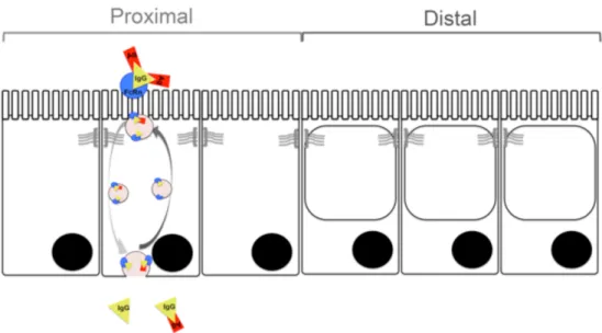
During the suckling period the distal half of the SI is mostly constituted by vacuolated enterocytes 62, which are foetal-type cells with high endocytic activity and a large supranuclear vacuole with intracellular digestive function occupying most of the cytoplasm (Figure 3)7. These distal absorptive cells may contribute to the uptake and transfer of intact macromolecules in a non-specific manner 63.
Figure 3. The proximal – distal axis in the small intestine during the suckling period in rats.
In the proximal SI the neonatal-Fc-Receptor (FcRn) binds maternal antibodies to the offspring via receptor-mediated trancytosis across the intestinal epithelial cell barrier. In the distal SI the dominant cell type are the vacuolated enterocytes with high endocytic capacity with their cytoplasma occupied almost entirely by large supranuclear vacuoles.
The transport of immunoglobulins has long been studied and attributed to a highly saturable membrane receptor with pH-dependent binding 48. Later studies showed that binding of immunoglobulin occurs at a slightly acidic pH in absorptive
intestinal cells 47. Finally, the IgG receptor was purified revealing a major histocompatibility complex (MHC)-class-I-related heavy chain molecule non-covalently associated to a stabilizing α-2-microglobulin forming a heterodimer, the FcRn 64-66. FcRn is expressed by the SI epithelium and has the physiological role of transferring adaptive passive immunity from the mother to the offspring, i.e. protecting milk-borne IgG from proteolytic degradation during trancytosis 67. At weaning, FcRn expression is drastically reduced in the SI epithelium and the localization pattern is altered once the development of both the gut and the immune system is complete. Additionally, it is known that the switch in FcRn expression with age is related to functional changes. The functions of FcRn in adults, as reviewed by Rath et al. (2012) 68, include e.g. extending IgG and albumin half-lives, and thus becoming the most abundant with the longest lifespan molecules in blood circulation 65,69.
Environmental sensors
In the epithelial cells, there is a general repertoire of receptors expressed in contact with the luminal contents, including dietary, microbial and secretions from the host, that act as sentinels of the intestinal milieu. These sensors mediate the crosstalk between the environment and the host and include innate immune receptors or pathogen-recognition receptors (PRRs) that recognise conserved structures termed pathogen associated molecular patterns (PAMPs). There are several types of PRRs grouped according to the type of PAMPs they recognise, such as the toll-like receptors (TLRs) and lectin-binding receptors (LBRs), among others. TLRs are a family of transmembrane glycoproteins that recognise bacterial antigens, i.e. TLR4 recognises the endotoxin characteristic of gram-negative bacteria, lipopolysaccharide (LPS) 70, and its activation often leads to signalling via the NFκB pathway which is suggested to be dependent on endocytosis of the receptor bound to the ligand 71,72. The LBRs often act as endocytic receptors but may also be capable of signalling, possibly by direct signalling through the MAP kinase and NFκB pathways or may act indirectly by modulating TLRs.
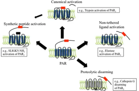
Proteinase – activated – receptors (PARs) are a family of seven transmembrane domain receptors coupled to G proteins that are activated by proteolytic cleavage of the extracellular domain in the amino terminus, which acts as a tethered ligand. The proteases that activate this family of receptors act as signalling molecules that regulate other cellular processes, and thus, PARs have been proposed as biosensors. There are four known types of PARs, however the target of our studies is PAR2. This receptor is expressed in organs of the GI tract such are the pancreas, stomach and intestine, in the IECs 73. The activation of PAR2 can occur by different mechanisms (Figure 4), such as canonical activation, which occurs by proteolytic cleavage, unmasking the tethered ligand domain. Non-tethered ligand activation of PAR2 can also occur by proteolytic cleavage in a different site than
that of canonical cleavage, possibly unmasking a new tethered ligand. The complete cleavage of tethered ligands can cause a proteolytic disarming of the receptor. Activation of the receptor can also be achieved with the binding of synthetic agonists to the receptor, independent of proteolytic cleavage, by binding at the site of the tethered ligand 74-76.
Figure 4. Protease Activated Receptors (PAR) activation mechanisms: PAR2 as an example.
Canonical activation occurs by proteolytic cleavage unmasking the tethered ligand domain. Non-tethered ligand activation occurs by proteolytic cleavage in a different site than that of canonical cleavage, possibly unmasking a new tethered ligand. The complete cleavage of the tethered ligand causes the proteolytic disarming of PAR2. Activation of the receptor can also be achieved with binding of synthetic agonists to the activation site. Adapted from Zhao P, Metcalf M, Bunnett NW. Biased Signaling of Protease-activated Receptors. Frontiers in Endocrinology. 2014;5. doi: 10.3389/fendo.2014.00067 74.
Immune barrier function
At birth, the newborn immune system is immature and the defence of the offspring depends on the maternal transfer of passive immunity, which occurs during the suckling period in rats. Thus, from birth there is a competing balance between hostile stimuli, pro-inflammatory factors, activators of the naïve adaptive immune system and the protective mechanisms, anti-inflammatory factors, to prevent an overwhelming and dysfunctional activation of the immune system. Thus, the induction of tolerance and the activation of the immune system occur synchronously.
The sampling of antigens from the luminal content, their uptake, and the site of the encounter with antigen presenting cells (APCs) is important for the appropriate activation of the immune system. FcRn has been reported to be capable of
bidirectional transport of immune complexes (antigen-IgG-FcRn) 77,78, and therefore contributes to the activation of the naïve neonatal immune system by antigen retrieval from the luminal side as well as immune-complexes antigen-presentation to APCs, DCs and T-lymphocytes 77,79. Maternal milk contains secretory IgA (sIgA), which restricts exposure to luminal antigens and hence prevents the maturation of intestinal T-lymphocytes 80. It has also been proposed that sIgA-antigen immune complexes could also pass across the barrier and contribute to antigen presentation 81.
Gut microbiota in the young
Bacterial colonization is also an important change occurring at birth, when mammals go from the low antigenic milieu in utero to a hostile environment82. It has been reported that the first colonizing community is of urogenital maternal origin when a vaginal delivery occurs or from the skin and environment community, when delivery occurs via C-section 83,84.
During the suckling period, the milk diet favours Gram-positive bacteria, such as
Lactobacilli spp. and Bifidobacteria spp., and there is a low diversity of species.
At weaning, postnatal changes in microbiota have been described to undergo a community maturation process with an increase in gram-negative bacteria, due to the cessation of milk consumption, and also an increase in diversity of species, from Firmicutes and Proteobacteria towards a Bacteroidetes dominated community, as reviewed by Jain and Walker (2015) 85.
Noteworthy, the changes in the microbial community also contribute to intestinal health via their involvement in metabolic processes and modulation of the barrier function, although not being a barrier property itself 44. At first, tolerance must be established for the commensal community, which compete with the opportunistic and pathogenic colonizers contributing to maintaining homeostasis within the microbial ecosystem.
This change in microbiota with an increasing proportion of Gram-negative bacteria at weaning makes Toll-like receptor 4 (TLR4) an interesting candidate to be studied during gut maturation. The role of this receptor in the regulation of mucus-producing goblet cells and its importance for the development of necrotizing enterocolitis in immature mice has been shown 86,87 as well as its expression in the GI tract and accessory organs in adult rats 88. In suckling rats the expression of TLR4 has been reported without any marked changes during intestinal development (unpublished data, AL Gacon).
Digestive and immune systems – developmental
connections
Natural development of the gut at weaning has been suggested to be dependent on T-cell activation in rats 89-91. Alternatively, previous studies trying to relate gut and immune system maturation in mice showed that in absence of adaptive immunity the maturation progress was temporarily delayed and that passive immunity affected the enterocytes gene expression especially in those mice with defective adaptive immunity 92.
Recently it was reported that the transcription factor Blimp1 is strongly expressed throughout the epithelium of the embryonic gut but its expression is down-regulated in mice after birth 93,94. Among the discoveries it was described the loss of Blimp1 expression at weaning and pointed it as a key genetic program participant of gut maturation regulating element with alternative tissue-specific promoters and there could also be differences between species 95. Moreover, the Blimp1 knockout mice were observed to have severe consequences such as postnatal mortality, reduced growth, requirement for adaptation of enterocytes to suckling, increased epithelial turnover and accelerated Paneth’s cells development 93,94. However, the physiological role of Blimp1 expressed by the immature gut epithelium remains unclear.
The suckling rat model
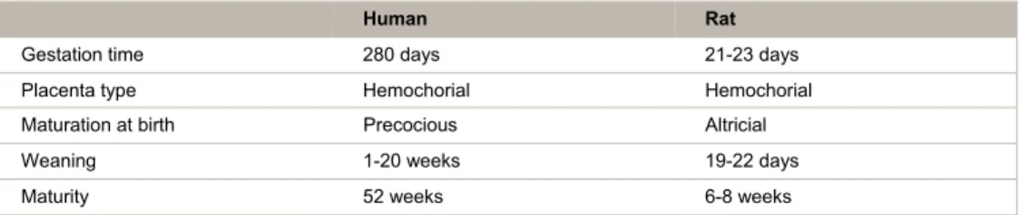
In mammals, the stage of intestinal maturation at birth varies among different species 45 and it correlates to the length of gestational period. Species with longer gestation periods are born more mature, such as precocious species including ungulates (pigs) and humans. Rodents, however, are altricial species, which due to the shorter gestational periods are born furless, blind, deaf, and dependent on maternal support for thermoregulation, nutrition, locomotion and the emptying of their bowels. The antenatal development of the GI tract between the third and the fifth months of pregnancy is considered to be comparable to that occurring in neonatal rats during the first three weeks of life, specifically with regards of antibody transfer (passive immunity) 96. Thus, as an altricial species, the suckling rat is a suitable model for the study of the gut in early life as well as for biomarker identification, pathogenesis, and mechanisms; as well as for immunological studies 53,97,98. The suitability of the suckling rat as a study model relies on general practical advantages provided by the animal models such as those related to ethical
issues, availability, handling, etc. over and above the biological similarities as well as the accumulated knowledge for their characterization.
Table 1. Speceies, rats vs humans, comparison of placentation and development timing status.
Human Rat
Gestation time 280 days 21-23 days Placenta type Hemochorial Hemochorial Maturation at birth Precocious Altricial Weaning 1-20 weeks 19-22 days Maturity 52 weeks 6-8 weeks
Functional and structural changes in the digestive system occur in parallel to the changes in diet that occur from birth. At first, the neonates ingest colostrum and then milk. In rats, the suckling period lasts for the first 18-21 days of life, with a 2-week period of strict milk consumption in the beginning. Afterwards, the transition from a milk-based diet to solid food starts during the third postnatal week, the weaning period, which correlates with the gut maturation process 1.
Figure 5. Timeline for the natural development of rats.
Date of birth established as day 0. Intervals of 7d (days) were studied. Diet indication at each stage of age.
Precocious induced maturation – a research tool
Despite being considered a pre-programmed process during ontogeny, the mechanisms and regulating factors involved in the gut maturation process have not yet been fully defined. Gut maturation during normal development follows a series of changes that culminate during weaning. These changes are not only structural, but also functional.
It has been shown that maturation of the SI can be induced precociously by i.e. early weaning 99, hormonal control (exogenous corticosteroids and thyroxin) 100, enteral administration of polyamines 101 and the lectin phytohaemagglutinin (PHA) 102. Recently, luminal exposure to pancreatic and pancreatic-like enzymes with proteolytic activity has been shown to accelerate gut maturation in our research group (paper I).
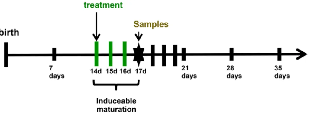
Experimental models of precocious induced gut maturation have been developed in our group. These models of experimental gut provocation also performed in a splitted-litter manner, so the distribution of littermates between treatment and control groups contributes to reduce the variability between different litters. The studies on PHA-induced precocious maturation revealed the importance of oral administration since enteral PHA induced gut maturation and also affected the immune organs spleen and thymus, however, when parenterally administered PHA only had effects on the immune organs but gut maturation was not induced 103. The original model consists of a moderate dose of 0.05 mg/g bwt of PHA and 0.6 mg/g bwt of microbial protease administered orally once per day for three days on days 14, 15 and 16 after birth, with sample collection and permeability test on day 17, 72 hours after the first treatment administration. Hence considering that natural changes are not evident until day 21 after birth.
Figure 6. Experimental model of precocious induced maturation.
Birth is established as day 0. On day 14 after birth treatment by gavage with PHA or protease starts. It can either be a single dose (provocation) or 3 days once a day (14, 15 and 16 days after birth) with a lower dose. 72 hours after treatment initiation permeability test and sample collection are performed.
Furthermore, studies on induced precocious maturation revealed that it could also be achieved by a single administration of a higher dose 0.1 mg/g bwt of PHA and 0.8 mg/g bwt of microbial protease on day 14 and sample collection on day 17, 72 hours after the provocation (unpublished data). Thus, precociously induced maturation of the gut in the suckling rat provides a useful research tool to investigate the mechanisms behind gut development in a more defined time frame with a better control on the circumstances.
Scientific aims
Paper I – ‘Involvement of pancreatic enzymes in gut maturation’
Previous studies on PHA-induced maturation of the gut in suckling rats showed that the pancreatic function was stimulated concomitantly. Consequently, the study aimed to investigate if the pancreatic enzymes themselves were involved in gut maturation. Thus, the main pancreatic enzyme activities were studied as well as their possible role and contribution to the maturational changes of GI tract in the suckling rat model.
Paper II – ‘Characterisation of the developmental changes in the intestinal
epithelial barrier’
Monitoring of a combination of changes occurring during postnatal development in the SI epithelium of suckling rats during natural and precociously induced maturation, emphasising the intestinal epithelial barrier properties, was done. The targeted parameters of the study included the expression of the FcRn receptor and the presence of endocytic vacuolated cells in the intestinal epithelium in relation to the transcriptional repressor Blimp1.
Paper III – ‘Importance of the T-lymphocytes during gut maturation’
Previous studies on gut development in suckling rats had suggested that it was dependent on T-lymphocytes. Hence, a hypothesis on the requirement of T-lymphocytes activation during the neonatal period for the initiation of gut maturation was studied in the athymic nude animal model. Therefore, the effects of luminal provocation on precocious gut maturation in thymus-derived cells T-lymphocytes deficient suckling pups were studied.
Paper IV – ‘Early effects of enteral provocation resulting in gut maturation’
Previous studies lead to the establishment of the protease- and PHA- induced precocious gut maturation experimental models in suckling rats by mimicking what occurs at weaning. However, only the effects of PHA had been investigated over time. In this study, the objective was to investigate the early effects after gut provocation with protease in comparison to PHA, with especial focus on intestinal permeability as well as exocrine pancreatic secretion stimulation. The main aim was to get a better understanding on what triggers the gut maturation process.
Methodology
Animals and ethics statement
The experiment was approved by the local Malmö-Lund Ethical Review Committee for Animal Experimentation and conducted in accordance with the European Community regulation concerning the protection of experimental animals (2010/63/EU) and the Swedish Animal Welfare Act (SFS 1988:539). The studies were carried out using rats (Rattus norvegicus) of the Sprague-Dawley strain (Mol: SPRD Han; Taconic M&B, Denmark) and of the athymic T-cell-deficient (nude) strain (NIH-Foxn1rnu, Charles River Laboratories International Inc.) that were bred and kept under pathogen-free conditions in the Department Animal facility at Lund University (20±1°C, 50±10 RH%, 12:12 h light-dark cycle). Before parturition, the pregnant dams were moved to separate cages (polycarbonate) with aspen wood bedding (Beekay B & K Universal AB), enriched with paper-nesting material (Sizzle-pet, Lillicobiotech). Parturition date was denominated as day 0 and litters were restricted to 10–12 pups for the study. All rat pups were kept with their dams during the experiments. The rat dams had free access to water and a rodent laboratory chow (R3, Lactamin) placed on the lid of cages. In order to prevent the pups from eating the solid chow, the cage height was increased using a 7 cm wall extender.
Experiments
The experiments were performed within experimental sets in a split-litter mode with random distribution of the rat pups from the same litter in the different groups to minimize variation. The pups were kept with their dam during the experiments or until postnatal day 21, after which the dam was separated from her litter.
– Paper I –
Induced precocious maturation was studied in groups of 14 day-old suckling rats gavaged by a soft stomach tube once a day for three days (14-16 days of age) with Creon 10000 (Abbott Products GmbH); microbial derived enzymes (Sigma-Aldrich): proteinase from Aspergillius melleus (type XXIII), a lipase from
Burkholderia cepacia (Amano Lipase PS) and an α-amylase from Aspergillius oryzae; dissolved in water, while the control group received the vehicle (water). – Paper II –
Natural development was studied in five age groups of litter-mates: suckling rats 7 day-old (7d) and 14 day-old (14d), on day 21 after birth, coinciding with the day of separation from their dam (21d), and two post-weaning groups at 28 day-old (28d) and 35 day-old (35d). Induced precocious gut maturation was studied on 17 old suckling rats that were gavaged once a day for three days (14-15-16 day-old) with microbial derived proteinase from Aspergillius melleus (type XXIII) or the purified lectin PHA from red kidney beans (Phaseolus vulgaris) 102,104, while the control group received the vehicle (water).
– Paper III –
The experiments were performed on the suckling nude pups in two different nursing sets. The first experimental set consisted of nude rats nursed by their natural dam (Nude/Nude). A second set consisted of nude pups reared by foster SD mothers (Nude/SD) from the 3rd post-natal day. Crossfostering of the nude pups was included to improve their survival and nutrition. Nude suckling rats were gavaged once a day (14 day-old) or once a day for three days (14-15-16 day-old) with the protease porcine pancreatic trypsin (Prot and Protx3) or purified PHA dissolved in water, while the control group received the vehicle (water). The experimental effects were studied at 17 day-old.
– Paper IV –
The early effects of gut provocation were studied in 14 day-old suckling rats at different time points, 1, 4, 8 and 24 hours, after the treatment. Each experimental litter included one of the treatments and controls at two different time points. The treatments administered were the protease porcine pancreatic trypsin or the purified lectin PHA and the control group received the vehicle, water.
39 e2. Ov er vi ew of the agents , dos e and exposur e per iod used fo r enter al pr ov ocati on to i
nduce gut matur
ati on w ithi n the s tudi es. nt Origin Source Dose (mg/g b.w t) Rat Strai n Exposur e peri od (da ys of age) Time of sacrifi ce after tre atmen t Stud y 0000 Sus scro fa (pancr ea tic po rc ine) Abbott Produ cts GmbH 1.5 SD 14-16 72 h Paper I ro te ase Asper gillius m elleus (t ype X X III) Sigma-Al drich 0.5, 0.2 5, 0.125, 0.062 5 SD 14-16 72 h Paper I Bur kholder ia cepacia Amano Lipas e PS 0.06 SD 14-16 72 h Paper I -A m yl ase Asper gillius or yz ae Sigma-Al drich 3.33 SD 14-16 72 h Paper I ro te ase Asper gillius m elleus (t ype X X III) Sigma-Al drich 0.237 SD 14-16 72 h Unpubli shed Car ica papaya (papaya f ruit) Sigma-Al drich 0.237 SD 14-16 72 h Unpubli shed in Ananas com osus (p ineapple f rui t) Sigma-Al drich 0.237 SD 14-16 72 h Unpubli shed yp sin Sus Scr of a (pancr ea tic po rc ine) No va 0.237 SD 14-16 72 h Unpubli shed A e m ag glutin in Phaseolus vulgar is (r ed kidney beans ) Purified i n ho use 0.05 SD 14-16 72 h Paper II ro te ase Asper gillius m elleus (t ype X X III) Sigma-Al drich 0.4 SD 14-16 72 h Paper II A e m ag glutin in Phaseolus vulgar is (r ed kidney beans ) Purified i n ho use 0.1 NIH-Foxn 1 rnu 14 72 h Paper III A (PH A x 3) e m ag glutin in Phaseolus vulgar is (r ed kidney beans ) Purified i n ho use 0.05 NIH-Foxn 1 rnu 14-16 72 h Paper III yp sin Sus scro fa (pancr ea tic po rc ine) No vo 1 NIH-Foxn 1 rnu 14 72 h Paper III yp sin ( Protx 3) Sus scro fa (pancr ea tic po rc ine) No vo 0.6 NIH-Foxn 1 rnu 14-16 72 h Paper III A e m ag glutin in Phaseolus vulgar is (r ed kidney beans ) Purified i n ho use 0.1 SD 14 1, 4, 8, 24 h Paper I V yp sin ( Prote ase) Sus scro fa (pancr ea tic po rc ine) No vo 0.8 SD 14 1, 4, 8, 24 h Paper I V ev iat ions: m g /g bw t: m g pe r g body w eig ht ; SD: Spr ag ue -Daw le y, NIH-Fox n1 rn u: at hy m ic nud e r at
Animal euthanasia and samples collection
On the designated collection day, the animals were anesthetized by a subcutaneous injection of a mixture of ketamine (Ketalar®, Pfizer) and azaperone (Stresnil®, Janssen Pharmaceutica) or isoflurane (Abbott) inhalation. At first, urine was collected during administration of the anaesthesia or directly from the bladder after laparotomy. Thereafter, the abdomen and thorax were opened; 1ml of blood was collected by direct heart-puncture into a syringe containing a mixture of anticoagulant and protease inhibitor. Plasma was obtained by blood centrifugation at 3000xg for 15 min at +4ºC and stored at -20ºC. Next, the SI was dissected from the pylorus to the ileo-caecal junction, and divided into a proximal and a distal half. The luminal content was flushed out. Intestinal samples, approximately 1cm long, were taken from the middle of each half and fixed in 10% neutral buffered formalin for 24 hours at room temperature and then kept in 70% ethanol until paraffin embedding, standard procedure. The rest of SI tissue was stored at -70ºC until further analyses.
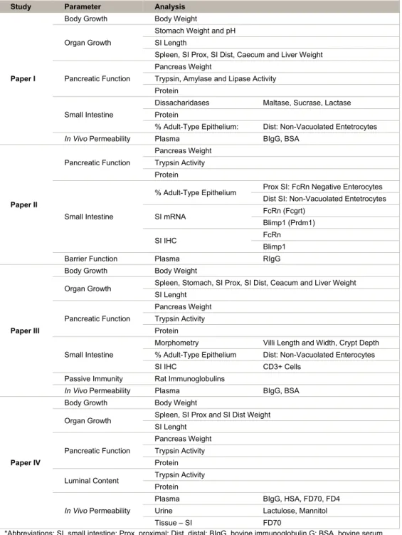
Table 3. Overview of the analysis performed within each study. Study Parameter Analysis
Paper I
Body Growth Body Weight Organ Growth
Stomach Weight and pH SI Length
Spleen, SI Prox, SI Dist, Caecum and Liver Weight Pancreatic Function
Pancreas Weight
Trypsin, Amylase and Lipase Activity Protein
Small Intestine
Dissacharidases Maltase, Sucrase, Lactase Protein
% Adult-Type Epithelium: Dist: Non-Vacuolated Entetrocytes
In Vivo Permeability Plasma BIgG, BSA
Paper II Pancreatic Function Pancreas Weight Trypsin Activity Protein Small Intestine
% Adult-Type Epithelium Prox SI: FcRn Negative Enterocytes Dist SI: Non-Vacuolated Entetrocytes SI mRNA FcRn (Fcgrt)
Blimp1 (Prdm1) SI IHC FcRn
Blimp1 Barrier Function Plasma RIgG
Paper III
Body Growth Body Weight
Organ Growth Spleen, Stomach, SI Prox, SI Dist, Ceacum and Liver Weight SI Lenght Pancreatic Function Pancreas Weight Trypsin Activity Protein Small Intestine
Morphometry Villi Length and Width, Crypt Depth % Adult-Type Epithelium Dist: Non-Vacuolated Enterocytes SI IHC CD3+ Cells
Passive Immunity Rat Immunoglobulins
In Vivo Permeability Plasma BIgG, BSA
Paper IV
Body Growth Body Weight
Organ Growth Spleen, SI Prox and SI Dist Weight SI Lenght
Pancreatic Function
Pancreas Weight Trypsin Activity Protein Luminal Content Trypsin Activity
Protein
In Vivo Permeability
Plasma BIgG, HSA, FD70, FD4
Urine Lactulose, Mannitol Tissue – SI FD70
*Abbreviations: SI, small intestine; Prox, proximal; Dist, distal; BIgG, bovine immunoglobulin G; BSA, bovine serum albumin; FcRn, neonatal – Fc – receptor; Blimp1, B – lymphocyte induced maturation protein – 1; IHC,
immunohistochemistry; RIgG, rat immunoglobulin G; HSA, human serum albumin; FD70, FITC – Dextran 70kDa; FD4, FITC – Dextran 4kDa