Université de Montréal
Cloning and characterization
ofxerC
gene of
Streptococcus suis
par
Fuli Jia
Département de microbiologie et immunologie
Faculté de Médecine
Mémoire présenté à la Faculté des études supérieures
en vue de l’obtention du grade de Maître ès sciences (M. Sc.)
en microbiologie et immunologie
Decembre, 2005 coe0>\
\‘ ‘i
“ç
de Montréal
Direction des bibliothèques
AVIS
L’auteur a autorisé l’Université de Montréal à reproduire et diffuser, en totalité ou en partie, par quelque moyen que ce soit et sur quelque support que ce soit, et exclusivement à des fins non lucratives d’enseignement et de recherche, des copies de ce mémoire ou de cette thèse.
L’auteur et les coauteurs le cas échéant conservent la propriété du droit d’auteur et des droits moraux qui protègent ce document. Ni la thèse ou le mémoire, ni des extraits substantiels de ce document, ne doivent être imprimés ou autrement reproduits sans l’autorisation de l’auteur.
Afin de se conformer à la Loi canadienne sur la protection des renseignements personnels, quelques formulaires secondaires, coordonnées ou signatures intégrées au texte ont pu être enlevés de ce document. Bien que cela ait pu affecter la pagination, il n’y a aucun contenu manquant. NOTICE
The author of this thesis or dissertation has granted a nonexclusive license allowing Université de Montréal to reproduce and publish the document, in part or in whole, and in any format, solely for noncommercial educational and research purposes.
The author and co-authors if applicable tetain copyright ownership and moral rights in this document. Neither the whole thesis or dissertation, nor substantial extracts from it, may be printed or otherwise reproduced without the author’s permission.
In compliance with the Canadian Privacy Act some supporting forms, contact information or signatures may have been removed from the document. While this may affect the document page count, it does flot represent any loss of content from the document.
Ce mémoire intitulée
Cloning and characterization ofxerC gene of $treptococctts suis
présentée par:
Fuli lia
a été évalué par un jury composé des personnes suivantes:
Dr France Daigle
président-rapporteur
Dr George Szatmari
Directeur de recherche
Dr Josée HareT
Membre du jury
XerC et XerD, deux recombinases impliquées dans la recombinaison site spécifique, résolvent des plasmides multimères en monomères. Les multimères générés lors de la réplication du chromosome circulaire chez les bactéries, sont en général très instables et peuvent engendrer des pertes de matériel génétique. Le rôle important de XerC et de XerD est donc de veiller à la stabilité chromosomique chez les bactéries. Membres de la famille des tyrosines recombinases, ont retrouvent plusieurs homologues de ces protéines chez les bactéries. Chez Staphylococcus aureus. $treptococcus pneumoniae et Pseudornonas fluorescens, des mutants Xer atténuent l’infection et la pathogénicité chez la souris. Chez Streptococcus suis, une bactérie gram positive impliquée dans de nombreuses maladies chez l’animal et l’humain, une forte homologie de séquences a été trouvée entre un gène et celui de xer chez les bactéries $treptococcus. Pour déterminer l’implication de Xer dans la pathogénicité chez Streptococcus suis, nous avons cloné, surexprimé et purifié XerC dans le but de réaliser des études de fonctionnalité avec cette protéine. Comme c’est le cas chez Bacitius subtilis, les résultats montrent que XerC s’attache sur l’ADN au site dif D’autre part, nous montrons qu’une mutation dans XerC affecte la croissance et cause d’importants changements morphologiques.
Mots-clés Recombinasionspécifique de site /tyrosine recombinase/XerC/dif’ Streptococcus suis
$ummary
XerC and XerD mediated site-specific recombination contributes to the stabiÏity of circular chromosomes in bacteria by resolving plasmid mutiimers and chromosome dimers to monomers prior to ceil division. The XerC and XerD proteins are members of the tyrosine recombinase family. Homologues of xerC/xerD genes have been found in many bacteria. Recently, xer mutants in Staphylococcus aureus, Streptococcus pneumoniae and Pseudomonas fluorescens have demonstrated reduced pathogencity suggesting a possible relationship between Xer proteins and the pathogenicity of these bacteria. Streptococcus suis is a Gram-positive bacterium, which is a leading cause of a wide range of diseases in animais and is also implicated in human diseases. The analysis of the S. suis genomic sequence demonstrated the presence of an open reading frarne (ORF) that shows strong homology to the xer genes of streptococcal bacteria. In our project, we cloned, overexpressed and purified the xerC gene and its product as a maltose binding protein fusion. The function of XerC protein was characterized and showed DNA binding activity at dif site of Bacillus subtiÏis. The df site of S. suis was aiso discovered during this work and the XerC protein of S. suis showed specific binding to this site. A S. suis xerC mutant showed a slower growth rate and displayed significant morphological differences.
TABLE 0F CONTENTS
Titie page Identification ofjury ii Résumé Summary iv Table of content y Listoftables x List of figures xiList of abbreviations xiii
CHAPTER 1: INTRODUCTION 1
1. Site-specific recombination 1
1.1. The resolvase/invertase family 3
1.2. Lambda integrase family 6
1.2.1. Generalities 6
1.2.2. The recombination reaction 6
1.2.3. The conservative motif 9
2. Xer site-specific recombination 10
2.1. Generalities 10
2.2. XerCandXerD 13
2.2.1. The conserved genes 13
2.2.2.1. Generalities .14 2.2.2.2. Function 15 2.2.3 XerD 16 2.2.3.1. Generalities 16 2.2.3.2. Function 17 2.2.3.3 Structure 18
2.2.4 The catalytic mechanism ofXerC and XerD 20
2.3 The site of action ofthe Xer recombinases 25
2.3.1. Chromosome recombination site 27
2.3.1.1 Escherichia cou df 27
2.3.1.1.1. Position and polarity 27
2.3.1.1.2. Structure 29
2.3.1.2 Bacillus subtiÏis df 31
2.3.2. Plasmid recombination site 32
2.3.2.1 Co1EY cer 32 2.3.2.2 p$C101psi 33 2.4 Accessory factors 35 2.4.1 ArgR’PepA 36 2.4.1.1 ArgR 36 2.4.1.2PepA 38
2.4.2 ftsK .43
2.5 Regulation of Xer recombination 47
2.5.1 DAZ and FtsK control 47
2.5.2 Homologus recombination control 51
2.6 Xer system and pathogenicity 53
2.6.1 Pseudomonasfiuorescens 53
2.6.2 StaphyÏococcus aureus 54
2.6.3 Streptococcus pneumoniae 55
3. Streptococcus suis 55
4. The master’s project 57
CHAPTER 2: ARTICLE
59Abstract 60
1. Introduction 61
2. Materisis and methods 63
2.1. Bacterial strains and pÏasmids 63
2.2. Growth conditions and DNA manipulations 64
2.3. PCR conditions 64
2.4. Protein overexpression and purification 65
2.5. DNA-binding assay 66
2.6. Preparation of S. suis Electrocompetent celi 67
2.8. Phenotypic analysis/microscopy .69
3. Resuits 70
3.1. Sequence analysis 70
3.2. The $.suis XerC proteins bind to the 3.subtilis difsite 72
3.3. Cooperative Binding Studies 73
3.4. Discovery ofa putative $.sïtis dif site 75
3.5. Inactivation ofthe S.suis xerC gene 76
3.6. Phenotypic analysis of xerC mutant ofS.suis 77
4. Discussion 79
Acknowledgements $4
References $5
CHAPTER 3: DISCUSSION 91
1. xerC gene of Streptococcus suis 91
2. DNA binding activity ofXerCSs-MBP 92
3. Cooperative binding studies 94
4. Phenotypic assay ofxerCmutant 95
5. ht vitro recombination reaction 96
6. In vivo recombination 9$
REFERENCES
.100APPENDIX I.
xivAPPENDIX II:
Nucleotide and amino acid sequence of xerC gene xvLI$T 0F TABLES
CHAPTER 1: INTRODUCTIONTABLE 1: The recombinases and FtsK homologues
in eubacteria and archaeabacteria 12
TABLE 2: Alignment of df sites from different bacteria and core
sequences ofplasmid-borne Xer sites 26
CHAPTER2: ARTICLE
TABLE 1: Chain types comparative between xerC wild-type
LIST 0F FIGURES
INTRODUCTION
fIGURE 1: Outcomes from site-specffic recombination 2
FIGURE 2: Mode! ofthe action of the reso!vases/invertases 5 FIGURE 3: Sequential strand exchange by the X Int family
site-specific recombinases 8
FIGURE 4: Overail structure of the XerD protein 19 f IGURE 5: Comparison ofthe structures ofthe C-terminal
domains ofXerD, X Int and HP1 Int 20
FIGURE 6: Control ofcatalysis in Xer recombination 23
FIGURE 7: Map ofthe E. cou chromosome 2$
FIGURE 8: Hierarchy of specificity determinants in the XerC
and XerD binding sites ofdf 30
FIGURE 9: The structure ofthe ArgR C-terminal domain in E. cou
and the mode! of B. stearothermophilus ArgR binding to DNA 38
FIGURE 10: Model for complex formed between two cersites in
the presence of PepA and ArgR and model for the Xer complex 42
FIGURE 11: FtsK-dependent and independent pathways ofXer
recombination at df 46
FIGURE 12: Mode! for segregation of the Ter dornains and chromosome
dimer resolution 50
ARTICLE
FIGURE 1: Alignment ofXerC protein of S. suis,
S. pneumoniae and B. subtilis 71
FIGURE 2: DNA binding assays with Xer proteins
from S. suis and E. cou sequences 73
FIGURE 3: XerC S. suis stirnulates XerDEc binding to dUEc 74
FIGURE 4: Aligmnent of chromosomal recombination
site sequences 75
FIGURE 5: Southern blot analysis ofwild type and
mutant 8. suis 77
FIGURE 6: Growth curves and morphology of S. suis xerC mutant 78 APPENDIX
List of Symbols and Abreviations
AMINO ACIDS MEASUREMENT UNITS
A: alanini
À:
angstrom unitC: cystein bp: base pair
D: aspartic acid cm: centimetre
E: glutamic acid Da: Dalton
F: phenylalanine g: gram
G: glycine h: hour
H: histidine kb: kilobase
I: isoleucine kDa: kilodalton
K: lysine tg: microgram L: leucine tl: microlitre M: methionine tM: micromole N: asparagine mm: minute P: proline ml: millilitre
Q:
glutamine mM : millimole R: arginine ng: nanogram S: serine s: secondT: tbreonin xg: centrifugation speed
V: valine °C: Degree celsius
OTHERS
Ap: ampicillinATPase: adenosine triphosphatase BsArgR: ArgR of Bacillus subtiÏis C-terminal: carboxyl-terminal clUBs: df site of Bacillus subtiÏis dUSs: df site of Streptococcus suis DNA: deoxyribonucleic acid HJ: Holliday junction
LB: Luria-Bertani NaC1: sodium chloride
OD600nm: optical density at 600 nanometre PAGE: polyacrylamide gel electroporesis PCR: polymerase chain reaction
TBE: tris-borate EDIA buffer Ts: thermosensitive
THY: Todd-Hewitt broth with 1% yeast extract UV: ultraviolet
XerDEc: XerD of Escherichia cou Y: tyrosine
3 ‘OH: three prime hydroxyl
ATP: adenosine triphosphate BSA: bovine serum albumin CodVBs: CodV of Bacillus subtiÏis DAPI: 4’, 6-diamidino-2-phenylindole dUEc: df site of Escherichia cou
DIG: digoxygenine
EDTA: ethylenedinitrolotetraacetic acid IPTG: isopropyl B-D thigalactoside MBP: maltose binding protein
NEB: New England Biolabs ORF: open reading frame
PB S: phosphate bufferred saline $D$: sodium dodecyl sulfate THA: Todd-Hewitt broth with agar
cL: alpha : beta X: lambda
XerCEc: XerC of Escherichia cou XerC$s: XerC of Streptococcus suis
X Int: X phage integrase 3’P04: three prime phosphate
Introduction
1. Site-Specific Recombination
Recombination is a ubiquitous process where organisms reshuffle their genetic infoniiation. This genetic exchange occurs between DNA molecules from the two parents
or between two DNA segments within the same molecule. Such recombination may be general, occurring between two DNA substrates with extensive homology, which is called
general homologous recombination, or site-specific, occurring between two specific, relatively short DNA targets, which is designated site-specific recombination.
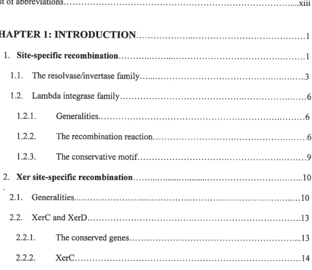
In DNA rearrangements mediated by site-specific recombination, four DNA strands are broken, exchanged and resealed at specific positions of two separate recombination sites (Stark et al., 1992; Landy, 1993; Nash, 1996; Jayaram, 1994). Outcomes of a
recombination event depend on the relative disposition of the two sites. Intramolecular recombination between inverted or directly repeated sites will invert or excise respectively
the intervening DNA segment. Recombination between sites on separate DNA molecules will integrate one molecule into the other (Figure 1).
These different structural consequences of site-specific recombination lead to various biological functions. It includes integration and excision of bacteriophages into and out of bacterial chromosomes, inversion gene switches that provides alternative gene expression of bacterial celi surface proteins and of pliage tau fibers, conversion of initial products of intermolecular genetic transposition into transposition end products, copy number control
and stable inheritance of microbial plasmids, and normal partition of the Escherichia cou
n
lb
Inversion
Excision/resolution
Figure I Outcomes from site-specific recombination. Triangles show the orientation ofthe
recombination sites. a and b indicate the position of distinct genetic markers and the recombination loci. ‘Excision’ and ‘integration’ refer to recombination events involving genetic entities of different size and /or function (e.g., the bacterial chromosome and a pliage genome), whereas ‘resolution’ and ‘fusion’ apply to equivalent DNA molecules,
(e.g., two pÏasmids) (Adapted from Hallet and $herratt, 1997).
Site-specific recombinases utilise a topoismerase 1-like mechanism, cleaving and rejoining one strand of DNA per promoter. A complete recombination event therefore requires at Ïeast four molecules of the recombinase, two on each DNA recombination partner. DNA strand exchange is conservative in two ways: there are no deletions or additions of nucleotides at the site of exchange and there is no need for high- energy factors. A transient 3’-phosphotyrosine/phosphoserine linkage between protein and DNA conserves the energy ofthe cleaved phosphodiester bond (Nunes-DUby et aï., 199$).
Site-specific recombinases fail into one of two unrelated families, the resolvase/DNA invertase family and the lambda integrase family (Hatfull and Grindley, 198$; Argos et aï., 1986; Sadowski, 1986; $tark et al., 1992). Enzymes of both families catalyze conservative DNA break-j oin reactions that proceed by two-step transesterifications in which protein phosphodiesters act as reaction intermediates.
1.1 The Resolvaseflnvertase Family
The resolvase/invertase family, of which there are cunently approximately 40 different members, forms a rather homogenous group of related proteins in which a conserved serine residue plays a key catalytic role (Hatfull et al., 1988; Leschziner et aï., 1995). The best-characterized recombinases of this family are the invertases Gin from bacteriophage Mu and Hin from $almoneÏÏa sp. and the resolvases of Tn3 and y transposons ($tark et al., 1992; Van de Putte et al., 1992; Grindley et al., 1994; $tark et al., 1995; Johnson, 1995; Johnson, 1991).
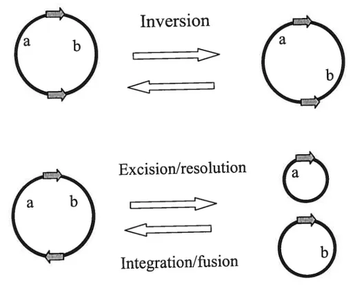
In a recombination catalyzed by resolvases or invertases, double strand breaks
staggered by 2 bp occur at the rniddle of the two paired core sites, giving rise to recessed 5’ ends and 3’-OH overhangs. One recombinase subunit is linked to each of the 5’ ends
through the conserved the serine residue of the family (Reed et aï., 1984; Kiippel et aï.,
1988). This serine presumably provides the primary nucleophile hydroxyl group in the
cleavage reaction (Leschziner et al., 1995). The ligation step that follows strand exchange
can be viewed as the converse of the cleavage: the protein-DNA phosphoseryl bond of one
strand is attacked by the 3 ‘-OH end of the partner to release the enzyme and reseal the
DNA backbone in the recombinant configuration (Figure 2). Thus, recombination by a
resolvase/invertase family occurs by a mechanism in which four DNA strands are broken
$2—
3a7
(ZI?,
.4LJZtZZE
3 5Figure 2 Model of the action of the resolvases/invertases. The subunit rotation model is shown. The oyaIs represent recombinase subunits with the conserved catalytic serine’S’.
Thick and thin unes are the top and bottom strands of the recombination sites, respectively.
The short vertical bars are the 2 bp of the overlap region between the two cleavage points.
Black arrows represent the nucleophilic attacks of phosphates (black dots) by hydroxyl groups (arrowheads). The four DNA strands are cleaved (a), exchanged by 1$00rotation of
the haif-site bound subunits (b) and religated in the recombinant configuration (c) (Hallet and Sherratt, 1997).
3*
1.2 Lambda Integrase Family 1.2.1 Generalities
The lambda integrase or ‘tyrosine recombinase’ family inc[udes over 130 members
identffied according to sequence similarity (Nunes-Dtiby et ai, 199$). Most biochemical studies of this family of enzymes have focused on the integrase from bacteria phage X (Int) (Landy, 1989), Flp recombinase from yeast 2 plasmid ($adowski, 1995), Cre recombinase
from bacteria phage Pi (Hoess et al., 1985) and the XerC and XerD recombinases from
Escherichia cou ($herratt et al., 1995). These proteins share only limited sequence
similarity and are rnuch more divergent, with only four completely invariant residues intimately involved in catalysis: the RHRY tetrad (Argos et al., 1986; Abremski et al.,
1992; Blakely et al., 1996). However, these recombinases carry out site-specific
recombination using a common mechanism that involves the formation of a Holliday junction (HJ) intermediate (Craig, 198$). Moreover, unlike the recombinases of the resolvase/invertase family, site-specffic recombinases related to X Int exchange the two pairs of DNA strands separately and sequentially.
1.2.2 The Recombination Reaction
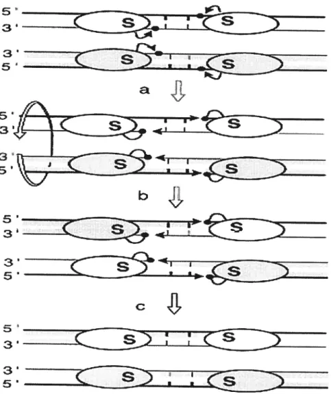
To initiate the first strand exchange, the tyrosine residue of the conserved catalytic
motif RHRY attacks a specific scissile phosphate in one strand (defined here afier as the
top strand) of each recombination core sites, thereby forming a 3 ‘phosphotyrosyl-linked
this cleavage reaction is thus reversed when compared to that of the resolvase/invertase mediated cleavages. In a second step, the recombinase-DNA phosphotyrosyl bond is attacked by the 5’-OH end from the partner duplex to generate a four-way branched structure, or ‘Holiday junction’ intermediate, in which only two DNA strands have recombined. To resolve this intermediate and complete the recombination reaction, the two other (bottom) strands are exchanged by repeating the cleavage/religation process 6-8 bp downstream ofthe first strand cleavage position (Hallet and Sherratt, 1997).
I.,
—-r.
— 1— -— ‘11
I
SFigure 3. Sequential strand exchange by the 2 Int family site-specffic recombinases. The DNA strand swapping !isomerisation model is presented. The letter ‘Y’ refers to the
conserved catalytic tyrosine. The oyais represent recombinase subunits. Thick and thin
unes are the top and bottom strands of the recombination sites, respectively. Black arrows represent the nucleophulic attacks of phosphates (black dots) by hydroxyl groups (anowheads). The top strands (thick unes) are cleaved first ta), swapped between the two partners (b), and then religated (c). The branch point of the generated Holliday junction intermediate is positioned at the middle of the (6-bp) overlap region and the top strands are
crossed. Isomerisation of the Holliday junction to a recombination configuration in which the bottom strands are crossed requires the reorganization of the DNA helices and the four
haif-sites-bound recombinase subunits within the complex (d). The resulting Holliday
junction isoform is resolved by repeating steps a to c in order to exchange the bottom
strands (e) (Hallet and Sherratt. 1997).
\)
3
1.2.3 The Conserved Motifs
The proteins of the tyrosine recombinase family are very divergent and share limted similarity in the amino acid sequence, but how they can carry out site-specific recombination using a common mechanism? Alignments of this integrase family of proteins identffied some conserved motifs, which are related to their catalytic function. Ail proteins harbor two conserved regions, Box I and Box II, with marked sequence similarity, originaiiy identified from the alignment of only eight recombinases (Argos et aÏ., 1986). Box I includes the fourth conserved residue R, and Box II contains other three conserved residues, the triad H-R-Y, which includes the active site tyrosine (Abrernski and Hoess, 1992; Nuiies Dtiby, 199$). The conservation of Box I is striking in prokaryotic recombinases and it
extends with some variation to eukaryotic recombinases. Box II is also relatively strongly conserved among the prokaryotic recombinases, but Ïess so between prokaryotic and eukaryotic proteins. Whereas the active tyrosine is absoiutely conserved, the surrounding residues are rather divergent, allowing for quite different secondary structures. Furthermore, the crystal structure of the 2 Int catalytic domain revealed an additional pattem of conserved hydrophobic residues that forms the core of the globular structure. It suggests that ail members of the integrase famiÏy adopt similar folds for the region spanning Box I, the interval region and Box II (Nunes-DUby et aÏ., 1998).
In addition to the highly conserved Box I and Box II motifs and the pattern of core hydrophobic residues, three patches of conserved sequence were identified in the extensive aÏignment ofthe prokaryotic recombinases (Nunes-DUby et aÏ., 1997). Patch lis tocated within the
short N-terminal region tipstream of Box I, consensus sequence LT-EEV—LL. Patch 11 contains a lysine (K235) flanked on both sides by serine or threonine in one subgroup of proteins and by glycine or methionine in another subgroup. For example, Lambda integrase (S234, K235, T236) belongs to the first subgroup, whereas XerD (G234, K235, G236) belongs to the second one. Patch III consists of a hydrophobic cluster rich in phenylalanines, preceded by acidic and followed by
polar residues in the majority of proteins: [D, E]-[F, Y, W, V, L, I, A]3—6 [S, T]. It is located in the
divergent region between Box II and I, and is an important stabilizer oftÏie native folds of integrase farnily recotnbinases (Nunes-DUby et al., 1998).
2. Xer Site-Specific Recombination
2.1 Generalities
The physical state of circular chromosomes, unlike linear chromosomes, can be
changed by homologous recombination. Odd numbers of homologous recombination events between circular replicons during or afier replication, produce dimers that need to be converted to monomers before they can be segregated normally at ceil division (Austin et aï., 1981; 31akely et al., 1991; Kuempel et al., 1991). Plasmid dimers can also arise as a
consequence of rolling circle replication during conjugal transfer (Warren and Clark, 1980; Erickson and Meyer, 1993). The Xer site-specific recombination system, initially discovered for its role in plasmid Co1E1 stable inheritance, also functions in the normal inherjtance of the Escherichia cou chromosome and the stable inheritance of other multicopy plasmids. h is encoded by the circular chromosomes of many bacteria and functions to ensure that both circular chromosomes and multicopy plasmids are monomeric
before their segregation to daughter ceils at ce!! division (reviewed in Shenatt etal., 1995).
Xer recombination is mediated by enzymes that be!ong to the lambda integrase family of site-specific recombinases (the ‘tyrosine recombinases’), which are structura!ly and mechanistically related to the type lB topoisomerases of eukaryotes (reviewed in Sherratt
and Wigley, 199$).
However, Xer site-specific recombination exhibits three features that distinguisli it
from other well-characterized members of the lambda integrase farnily. First, it uses two re!ated recombinases, XerC and XerD, each of which catalyses one specific pair of strand
exchanges (Blakely et aÏ., 1993, 1997; Arciszewska and Sherratt, 1995; Col!oms et al.,
1996, 1997; Arciszewska et al., 1997). The use of two recombinases potentia!ly allows each pair of strand exchanges to occur separate!y and could direct the order of strand
exchanges (Co!!oms et al. 1996). Second, the recombination reaction has different requirements and outcomes depending on whether it occurs at plasmid or chromosomal recombination sites. Recombination at natural plasmid sites is preferentia!!y intramolecu!ar
and requires the two recombinases and the 28-30 bp recombination core site, as we!l as
additional accessory proteins and adjacent accessory DNA sequences. Interaction of the accessory proteins and accessory sequences promotes the formation of a synaptic complex ofprecise topology, that can form efficiently on direct!y repeated recombination sites in the
same molecu!e (Colloms et al., 1996, 1997). In contrast, the recombination at the E. cou
chromosome site, d7 requires only a 2$ bp recombination core site at which the two recombinases act. Recombination in vivo at df present in multicopy p!asmids, occurs
Tecklenberg et aÏ., 1995). Third, despite the sequence divergence of integrase family recombinases, Xer-like recombinase sequences are present in the majority of eubacteria, suggesting that the mechanism of dimer resolution used by E. cou is highly conserved (Table 1) (Recchia and Sherratt, 1999).
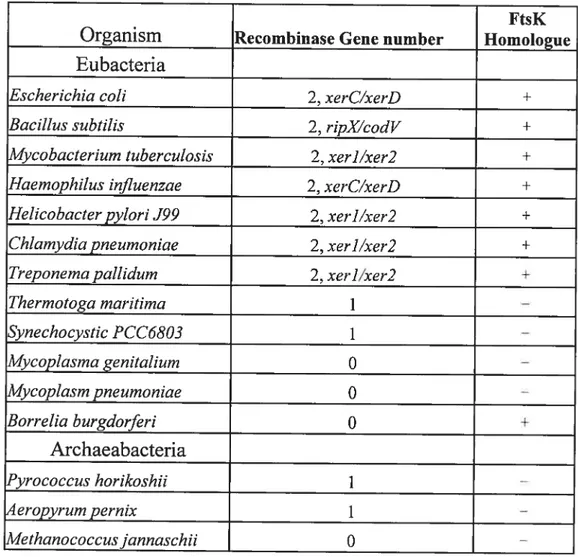
Table 1 The Xer recombinases and ftsK homologues in eubacteria and archaeabacteria.
for FtsK, ‘+‘ means having homologues.
‘—‘ means no homologue found [adapted from
Recchia and $herratt, 1999].
FtsK
Organism Recombinase Gene number Homologue Eubacteria
Escherichia cou 2, xerC/xerD +
BaciÏlus subtitis 2, ripX/codV +
Mycobacterium tuberculosis 2, xer]/xer2 +
HaernophiÏus influenzae 2, xerC/xerD +
Heticobacter pylori J99 2, xer]/xer2 +
ChÏamydiapneumoniae 2, xer]/xer2 +
Treponema pallidum 2, xer]/xer2 +
Thermotoga maritirna 1 — Synechocystic PCC6803 1 — MycopÏasma genitalium O — Mycoplasm pneumoniae O — Borrelia burgdorferi O + Archaeabacteria Pyrococctts horikoshii 1 — 4eropyrurn pernix 1 — Methanococcusjannaschii O —
2.2 XerC and XerD
XerC and XerD are encoded at 4024 kb and 3050 kb on the E.coti chromosome respectively. Each recombinase is expressed with at Ieast two other proteins that don’t appear to have a role in Xer recombination (Colloms et al., 1990; Blakely et aÏ., 1993). XerC and XerD belong to the large tyrosine recombinase family and possess the characteristic RHRY signature of active site residues of this family (Esposito et al., 1997; $henatt et al., 199$). They show 37% identity and bind to separate halves of the recombination site (Blakely et aÏ., 1993).
2.2.1 The Conserved Genes
The alignment of the amino acid sequence of the tyrosine recombinases firstly reveals that the complete genomes of 16 eubacteria and 5 archaebacteria contain proteins homologous to XerC and XerD in bacteria with circular chromosome as shown in Table 1 (Recchia and $herratt, 1999). Now, There are more Xer recombinase have been found such as Caulobacter crescentus (Jouan and $zatmari, 2003), LactobacilÏus leishrnanniiBecker and Brendel, 1996), Proteus mirabilis (Manuela and Szatmari, 199$) $treptococcus pneumoniae(Reichmann et al., 2002), Vibrio cholerea( Huber and Waldor, 2002). Moreover, most eubacteria with only partial genome sequences are also possess two Xer protein including Pseudomonas, Vibrio, Bordetella, Neisseria, Staphylococcus and Enterococcus species (Recchia et al., unpubUshed). Secondly. the majority of eubacteria possess two putative Xer recombinases, suggesting that the mechanism of dimer resolution
used by E. cou is highly conserved. However, two eubacterial species appear to contain only one Xer homologue. In these cases, either one Xer protein lias been lost or, assuming
that xerC and xerD genes arose from a single ancestral gene, these organisms diverged
from other bacterial lineages prior to this duplication. Likewise, organisms in which no
Xer-like sequences were identified may have either lost both sequences or separated from other bacterial lineages prior to the evolution of Xer. Thirdly, the bacteria with a linear chromosome such as the spirocheaete Borrelia burgdrnferi lack any identifiable Xer homologues, which may indicate the Xer recombination only occurs in bacteria with a circular chromosome. Furthermore, species such as Mycoplasma genitaÏium and Mycoplasmapneurnoniae are deficient in homologous recombination genes and also lack in
the Xer genes. These correlations are consistent with the functional inter-relationship
between homologous recombination and Xer recombination. Finally, most eubacteria that possess Xer recombinases also possess ftsK homologues, whereas M genitaflïtm and M pneurnoniae, which lack identifiable Xer homologues, also appear to lack an ftsK
homologue (Table 1). This suggests that the functional interaction between Xer and FtsK proteins in controlling chromosome dimer resolution is highly conserved (Recchia and
Sherratt, 1999).
2.2.2 XerC
2.2.2.1 Generalitics
By sequence analogy, XerC appears to be a member of the bacteriophage lambda
maps close to the E. cou origin ofreplication, oriC, at 85 min (3700 kb). It is expressed as
the third gene of a four-gene multicistronic unit that contains dapF, oij235, xerC and orJ238. The oij235 and oij238 are unknown in their function but appear to be translated at
levels similar to those of dapf and xerC (Kohara et al., 1987; Richaud et al., 1987;
Richaud and Printz, 1988; Colloms et al., 1990).
The xerC gene encodes a protein with a calculated molecular mass of 33.8 kDa. The translated protein sequence of XerC contains two regions, whieh are homologous to the two conserved domains of the lambda integrase family of site-specific recombinases (Argos et al., 1986; Colloms et al. 1990). Domain 2 of the XerC sequence has three totally
conserved amino acids, histidine (H), arginine (R), and tyrosine (Y), as well as other less conserved amino acids. The XerC sequence has 32% amino acid identity to the E. cou proteins FimB and FimE in an alignment covering about 160 amino acids. These two proteins are involved in inverting a segment of the E. cou chromosome to switch fimbrial antigen (Klemm et al., 1986). Within conserved domain 2, the XerC sequence shows considerable similarity (66% identity) to an integrase-like inferred protein sequence from plasmid R46 (Hall et al., 1927).
2.2.2.2 Function
The stable inheritance of natural muticopy plasmids related to Co1E1 requires the
function of the Xer site-specific recombination system (for example, cer in CoYE 1; Summers and Sherratt, 1984). The recombination occurs only intramolecularly and resolves plasmid multimers, which arise by intermolecular homologous recombination, to
monomers. Three unlinked E.coli genes whose products are required for recombination at cer and its natural plasmid homologs have already been discribed and characterized. XerC
has been shown to bind to recombination sites (Colloms et al., 1990). ArgR (originally
XerA) and PepA (originally XerB) are required for recombination at cer, arid have an accessory role participating in the resolution selecticity process (Stirling et al., 1988, 1989; Summers, 1989).
In addition to its role in converting multimers ofplasmid Co1E1 to monomers, XerC also has a role in the segregation of replicated chromosome at celi division. xerC mutants
form filaments with aberrant nucleoids that appear unable to partition properly. A DNA segment (dU) from the replication terminus region of the E. cou binds XerC and acts as a substrate for Xer-mediated site-specific recombination when inserted into multicopy plasmids. This df segment contains a region of 28 bp with sequence similarity to the cross over region of CoÏE1 cer (Blakely et aÏ., 1991). Therefore, XerC not only functions in maintaining CoYE1-like plasrnids in the monomeric state, but also has a role in normal E.
cou chromosomal metabolism, which resolve chromosome dimers to monomers prior to celi division.
2.2.3 XerD
2.2.3.1 Generalities
During the characterization of the RecJ exonuclease of E. cou, an open reading frame was reported and showed sequence similarity to the integrase family of site-specific recombination (Lovett and Kolodner, 1991). This open reading frame was designated xerD
(Blakeiy et al., 1993). The predicted amino acid sequence of the XerD protein showed a
37% amino acid identity to XerC. Both XerC and XerD are predicted to have 29$ amino acids. A high degree of sequence conservation between XerC and XerD is present in
domain I and II, regions highly conserved in ail integrase famiiy recombinases (Biakeiy et
al., 1993). Note the presence ofthe invariant four amino acids (R.. .H.. .R...Y): Mutations
at each of these four positions lead to ioss of normal recombination activity in FLP
recombinase (Lee et al., 1992; Chen et aÏ., 1992). The conserved tyrosine in domain II of
FLP is required for the nucleophilic attack that initiates the first strand exchange (Prasad et aÏ., 1987; Pargeilis et al., 198$). It has been proposed that the three other conserved
residues of f LP forrn part of the active site that is involved in activation of the phosphodiester targets prior to nucieophilic attack during each of the transesterification
steps (Lee et al., 1992).
2.2.3.2 Function
XerD is also required in addition to XerC for site-specific recombination at cer and df(B1ake1y et al., 1993). The xerD gene is cotranscribed with two other genes, xprA and
reci Insertion of Tn]O-9 into xprA and recJ did flot generate a Xer phenotype. In
contrast, an insertion at xerD gene gave a Xer phenotype suggested XerD is transcribed
from its own promoter. A plasmid containing a deletion that removes most of the xerD gene fail to complemented the xerD2 mutation, whereas a piasmid deleted for more than
haif of the xprA gene complemented the xerD2 defect. Therefore, the two related recombinases XerC and XerD are required for site-specific recombination at cer and dzfand
the two genes that are coexpressed with xerD (xprA and recJ) have no apparent role in Xer
site-specific recombination. The putative catalytic active sites of both XerC and XerD are required for normal Xer site-specific recombination in vivo. XerC and XerD bind
separately and cooperatively to the c4f and cer sites in vitro. XerC binds the djflefi-haif
site and XerD binds the dfright-ha1f site (Blakely et al., 1993).
2.2.3.3 Structure
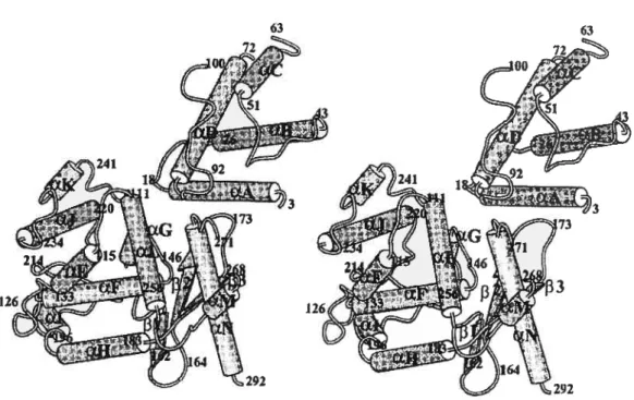
The structure of XerD bas been solved at 2.5À resolution and reveals that the protein comprises two domains ($ubramanya et aÏ., 1997). Domain 1 consists ofresidues
1-107, while domain 2 comprises residues 108-29$. Domain 1 contains four a-helices,
arranged such that there are two parallel helix hairpins arranged at 90° to each other. Domain 2 is also mainly a-helical, but with a tbree—stranded antiparalled f3-sheet along one
edge (Figure 4). The fold of this domain is similar to that determined for X and HP1
integrase (Hickman et al., 1997; Kwon et aÏ., 1997). Domain 1 and Domain 2 of XerD correspond to domains ofXInt, HP I Int and FLP identified by limited proteolysis (Moitoso de Vargas et al., 198$; Evans et aÏ., 1990; Chen et al., 1991; Pan and Sadowski, 1993;
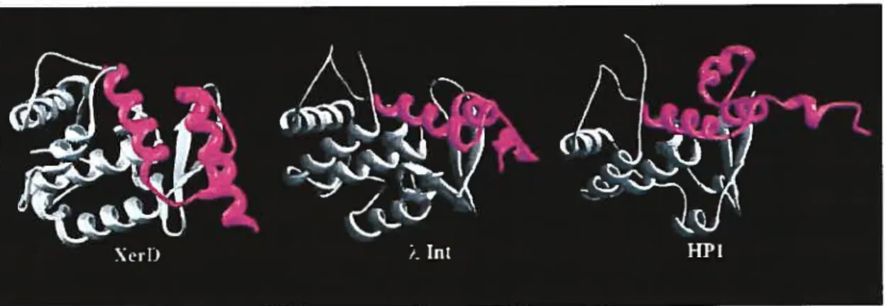
The region of structural homology within the C-terminal domains of XerD, 2 Int and HP1 Int spans 170 residues (figure 5). Two conserved sequence motifs are located in
domain 2 of XerD. The locations of motif I and the N-terminal portion of motif II are similar in the structure of XerD (residues 145—159 and 244—2$ 1, respectively) and those of
2 and HP1 integrases (Hickman et al., 1997; Kwon et al., 1997). However, the extreme
C-terminal portions of these proteins, which include the C-C-terminal portion of motif II, are
quite different (Figure 5). In ?\. Int, these C-terminal residues (334—356) form a flexible
Ïoop that is disordered in one of the two molecules in the asymmetric unit, but is more
Figure 4. Overail structure of the XerD protein. The numbering refers to the beginning and
end of secondary structural elements. Residues that are not defined are located at the N and C-termini and in three disordered loops (residues 64—70, 101—110 and 269-270).
ordered in the other, where the final 15 residues form two additional f3-strands along one edge ofthe antiparallel sheet. By contrast, in XerD, this region (residues 271—29$) forms a turn followed by a long a-helix, containing the active site tyrosine, which extends almost to the C-terminus) (Subramanya et al., 1997).
Figure 5. Comparison of the structures of the C-terminal domains of XerD. Int and HP 1 Int. Regions of the C-terminal domains of the proteins that show the greatest structural similarity are shown in grey. The major structural differences (shown in magenta) are located in the polypepfide segments that extend from conserved motif II (Argos et al.,
1986) to the C-terminus ofthe proteins. (Adapted from Subramanya et al., 1997).
2.2.4 The Catalytic Mechanism of XerC and XerD
XerC and XerD are related 298-amino-acid site-specific recombinases, each of which is responsible for the exchange of one pair of strands in Xer recombination. Both recombinases encode functions necessary for sequence-specific DNA-binding, co-operative XerC/XerD interactions. synapsis and catalysis.
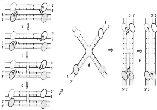
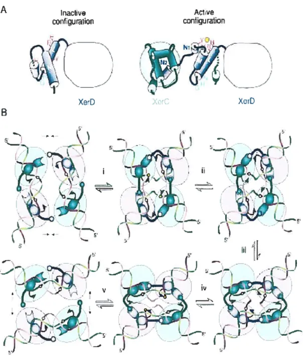
In recombination mediated by XerC and XerD, DNA strands are cieaved and rej oined through the formation of a transient DNA—protein covalent intermediate invoiving a conserved tyrosine as the catalytic nucleophile. The same mechanism is used by the related type lB topoisomerases (reviewed in Sherratt and Wigiey, 199$). However, type lB topoisomerases break and reseal the same phosphodiester bond to remove supercoils in DNA, whereas XerC and XerD catalyze two consecutive pairs of strand exchanges, with the formation of a Hoiliday junction (HJ) as a recombination intermediate. Each reciprocal strand exchange reaction is a concerted two-step process in which the 3’ phosphotyrosyl DNA—protein bonds generated by cleavage of one DNA strand in each recombination site are subsequentiy attacked by the free 5’ OH ends of the partner sites. DNA strands are exchanged by swapping of a few central region nucleotides (Nunes-DUby et al., 1995; reviewed in Guo et ai, 1999). This mechanism impiies that specific pairs of active sites are sequentiaiiy switched on and off in the recombinase tetramer to ensure that appropriate DNA strands wili be exchanged at both reaction steps. It has been demonstrated that the catalytic activity of XerC and XerD is controiled by an interaction involving the extreme C-terminal donor region of each protein and an internai acceptor region adjacent to the active site [figure 6]. The donor—acceptor region interactions between adjacent recombinase molecules act as moiecular springs in the switch that leads to sequentiai and synchronized activationlinactivation of pairs of recombinase subunits during recombination (Hailet et aL, 1999).
XerC and XerD cleave DNA by providing ail catalytic residues in cis (Arciszewska and Sherratt, 1995; Blakely et aÏ., 1997). Consistent with this, the crystai structure ofXerD
shows that tyrosine and the other active site residues are clustered together (Subramanya et
aÏ., 1997). The integrases of phages lambda and HP1 and the recombinase Cre from
bacteriophage Pi can also cleave DMA in cis (Nunes-DUby et aÏ., 1994; Guo et al., 1997; Hickman et al., 1997; Kwon et ai, 1997). However, the yeast recombinase FLP cleaves DMA in trans (Chen et al., 1992; Lee et al., 1999).
Inactive corUguraiion
N,
—
Xe-D
Figure 6. Control of Catalysis in Xer Recombination. (A) Proposed reconfiguration of XerD C-terminus upon assembly of the recombination complex on DNA. (B) A mode! for the reciproca! contro! of cata!ysis by XerC and XerD
A
B
Ar. v coniiguration
• -• XerD
“t
r s,___ i-z--nE, I’Color code is as in (A). The bail-and-socket joint depicts the interaction between the donor and acceptor regions of adjacent subunits. Step i to step y j the recombination pathway in which XerC strand exchange occurs first. (i) Interactions between XerC and XerD molecules bound on a same duplex, possibly coupled with additional interpromoter interactions across the synapse, force the DNA to bend in a configuration where the top (green) strand of the recombination site central region is exposed toward the outside of the duplex. The torsion energy stored in the bent DNA may act on the XerC—XerD donor— acceptor interaction so as to activate XerC catalysis by repositioning of the tyrosine nucleophile (arrowhead), and possibly other catalytic residues with respect to the DNA target phosphate (circle). DNA torsion strains released upon cleavage may also promote the unwinding and extrusion of the cleaved strands in order to orient the 5’ OH ends for the rejoining step. (ii) Completion ofthe strand exchange reaction generates a 2-fold symmetric HI intermediate in which the top strands are crossing. (iii) Coupled protein and DNA conformation changes convert the complex into a configuration in which the bottom strands (purpie) are crossing. (iv) This leads to synchronized inactivation of the XerC subunits and concomitant activation of the XerD subunits. (y) The recombinant duplexes are bent in the opposite direction to that of the initial recombination sites. This inversion of the DNA bending strains may promote the restacking of the DNA helices and the dissociation of the resealed molecules from the complex (Hallet et al., 1999).
2.3 The Site of Action of the Xer Recombinases
Xer recombinase mediated recombination occurs in two different recombination substrates and has different biological functions. One is at chromosome recombination sites called dÇ originally found in Escherichia cou. The Xer site-specific recombination ensures that dimeric chromosomes are converted monomers prior to ceil division (Blakely et al., 1991; Kuempel et al., 1991). Another is at plasmid sites such as Co1E1 cer and pSC1O1 psi. The Xer site-specific recombination system is involved in the stability of naturally occurring plasmids by enstiring that plasmid multimers are converted to monomers ($ummers and Sherratt, 1984; Cornet et al., 1994). The Xer site-specific recombination is conserved in most eubacteria (Recchia and $herratt, 1999). The alignment of 19 naturally occurring plasmids and some eubacterial chromosomes revealed that the wide existence of the homologues of Xer recombination core site (Table 2) (Hayes et al., 1997; Lesterlin et aÏ., 2004). XerC binding sites are more variable whereas XerD binding sites are well conserved. The central region of the Xer sites, which displays no consensus and separates XerCD binding sites by a 6 (chromosome site) or $ bp (plasmid site) spacer, is a key determinant of the Xer recombination pathway. It determines the requirements for accessory proteins and accessory sequences on the plasmid recombination site (e.g. Co1E1 cer site or pSCÏOl psi site). It also determines the presence of ftsK in chromosome dimer resolution (Barre et al., 2001). $everal sets of data, obtained on the Xer systems and other tyrosine recombinase system, indicated that this region is an important determinant of the comformation of the recombinase-core sequence complexes (Azaro and
Landy, 1997; Gopaul et aï., 199$; Arciszewska et aÏ., 2000; Lee and Sadowski, 2001;
Capiaux et al., 2002).
Table 2 Alignment of df sites from different bacteria and core sequences ofplasrnid-borne Xer sites (Adapted from Rayes et aÏ., 1997 and Lesterin et aÏ., 2004)
XerC binding site Central region XerD binding site Origin
Plasmids Sites
Co1E1 cer GGTGCGTACAA TTAAGGGA TTATGGTAAAT
ColA car GGTGCGTACAA ----CGGATG TTATGGTAAAT
C1oDFY3 parB GGTACCGATAA ----GGGATG TTATGGTAAAT
CoÏK ckr GGTGCGTACAA TTAAGGGA TTATGGTAAAT
CoiN GGTGCGTACAA --TAAGGGA TTATGGTAAAT
NPT16 GGTGCGCGTAA --TGAGACG TTATGGTAAAT
pMB1 GGTGCGTACAA TTAAGGGA TTATGGTAAAT
pSC1O1 psi GGTGCGCGCAA ----GATCCA TTATGTTAAAT
CoIE2 GGGGCGTACAA ----CGGGAG TTATGGTAAAT
Co1E3 GGTGCGTACAA ----CGGGAG TIATGGTAAAT
Co1E4-CT9 GGTGCGTACAA ----CGGGAA TTATGGTAAAT
Co1E5-099 GGTACGTACAA ----CGGGAG ITATGGTAAAT
Co1E6-CT14 GGTGCGTACAA ----CGGGAG TTATGGTAAAT
Co1E7-K317 GGTGCGTACAA ----CGGGAG TTATGGTAAAT
CoYE8-J GGTACGTACAA ----CGGGAA TTATGGTAAAT
Co1E9-J GGTACGTACAA ----CGGGAG TTATGGTAAAT ChromosomeSites(dt)
E. cou GGTGCGCATAA TGTATA TTATGTTAAAT
S. typhirnurium GGTGCGCATAA TGTATA TTATGGTAAAT
S. typi GGTGCGCATAA TGTATA TTATGGTAAAT
V choÏerae chrl ATGGCGCATTA TGTATG TTATGGTAAAT
V. cholerae chrll AATGCGCATTA CGTGCG TTATGGTAAAT
H. infiuenzae ATTTCGCATAA TATAAA TTATGGTAAAT
2.3.1 Chromosome Recombination Site 2.3.1.1 Escherichia cou dif
2.3.1.1.1 Position and Polarity
Two main sites of the circular E. cou chromosome are implicated in the ceil cycle:
oriC, where replisomes are assembled for bidirectional replication (Messer et al., 1996),
and the diametricalÏy opposite
c4f
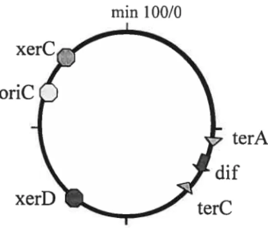
site, where chromosome dimers are resolved (Blakely et aï., 1991; De Massy et al., 1987). The dfis located in the replication terminus region atmin 33.6 of the genetic map, kilobase 1608 of the physical map, between the innermost terminators terA and terC (figure 7) (Kuempel et al., 1991). Recently, it has been discovered that this position is crucial for dimer resolution (Leslie and Sherratt, 1995; Tecklenburg et al., 1995; Cornet et al., 1996; Kuempel et al., 1996). To be active, df must
be inserted within a nanow zone around its natural position, the DAZ (d/ activity zone). The DAZ is the scene of specific recombination between dif sites that occurs only in celis that are able to form chromosome dimers (i.e. proficient for homologous recombination)
oriC
min 100/0
terA
Figure 7. Map ofthe E. cou chromosome, showing the position of the xerC and xerD
genes, the position of oriC and dtyÇ and the position of two replication terminator sites [from Barre and Sherrat, 2002]
The sequences sunounding df appear to be intrinsically polarized along the oriC-c4f axis and their relative orientation is the main determinant of DAZ positioning. Notably, the
deletion of sequences surrounding dfis harmless, whereas inversion of the same sequences inhibits dimer resolution (Tecklenburg et al., 1995; Cornet et al., 1996; Pérals et aÏ., 2000).
The data suggest that the polarization determinants are present throughout a large terminal dornain (more than 200 kb around dU) and are highly repeated. Chromosome sequences are
oriented following the oriC/ter axis, defining the two replichores (Blattner et aÏ., 1997).
Several types of short-sequence elements showing a strongly biased orientation following
the oriC-df axis exist. This resuits from the intrinsic biased orientation of chromosome sequences that define its replichore organization: strongly expressed genes, G/C skew, Chi
sites and numerous other oligomers ($alzberg et ai., 1998; Lobry and Louarn, 2003). Among these, short degenerate motifs, termed RAG, have been proposed as good candidate based on their highly biased orientation (Lobry and Louai-n, 2003). However, previous attempts to show that the RAG motif controls another FtsK activity or colocalized other active elements were unfruitful (Pci-ais et ai, 2000; Massey et ai., 2004; Saleh et ai., 2004). However, DNA motifs, named ftsK orienting polar sequences (KOPS), have been identifted which direct the movement of the E. cou FtsK transiocase (Levy et aï., 2005; Bigot et ai., 2005). Levy group (2005) identffied the GNGNAGGG motif, its complement,
or both as the best candidate to specify ftsK directionality. They found that a
GNGNAGGG sequence efficiently reverses FtsK transiocation. Bigot group (2005) used a functional approach and also identified this motif, displaying a high biased orientation and over-represented on the whole chromosome. In vitro, these motifs display KOPS activity: they inhibit Xer recombination activation by ftsK in an orientation-dependent manner; they also stop FtsK from dissociating branched DNA structures depending on their orientation; additionally, single molecule data suggest that they block FtsK transiocation. Their effect on ftsK transiocation is stochastic; the presence of two or three motifs is required to observe a strong effect.
2.3.1.1.2 Structure
The minimal df site sufficient for chromosome monomerization activity and for recombination in a plasrnid substrate is 28 bp in Iength (Leslie & Sherratt, 1995; Tecklenburg et aï.,
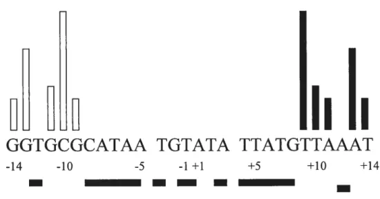
binding sites separated by a 6 bp central region at the boundaries of which strand cleavage and exchange occur. The XerC and XerD binding sites are partial palindromes, but thetwo halves ofdf are recognized specifically by the XerC and XerD recombinases, which themselves share 59% sirnilarity (Blakely et aÏ., 1993). The XerC and XerD binding sites are partial palindromes at six of li positions, but other five positions are neyer palindromes which determine the specifically binding ofXerC and XerD, as shown in figure 8.
u
h
Ii
GGTGCGCATAA TGTATA TTATGTTAAAT
-14 -10 -5 -1 +1 +5 +10 +14
—
— —
—
Figure 8. Hierarchy of specfficity determinants in the XerC and XerD binding sites of df Shaded boxes below the sequence denote positions that are palindromic between the XerC and XerD binding sites. Bars above the sequence indicate the relative contributions of particular nucleotides to XerC or XerD binding specificity. The longest bars identif’ bases that are most significant for specificity and the shortest bars denote nucleotides whose contribution is least critical. Bars of intermediate length indicate positions of intermediate importance. Note that, while the T- C substitution at position +9 had a strong affect on
XerD binding and recombination in vivo, ail piasmid sites examined to date (exceptpsi) have a G nucieotide at this position (Adapted from Rayes et aÏ. 1997).
2.3.1.2 Bacitltts subtitis dif
Homologues of E. cou cflf site have found in other bacterial chromosomes (Table 2). The Bsdtf site is located at approximately 166° on the 3. subtilis chromosome, 6° counterclockwise from the B. subtilis terminus of replication (Kunst et al., 1997). There are six different nucleotides in the CodV (XerC) binding region when compared to the df site, which may be required for the specific binding of CodV. The RipX (XerD) binding site is more conserved and has only one divergent nucleotide. Experimental evidence has been provided to substantiate the authenticity of this site. First, integration of nonautonomously replicating plasmids carrying either cloned Bsdf DNA or a synthesized 3sdfo1igomer occurs at a high frequency in recA backgrounds. The integration of Bsdf containing plasmids was dependent on the presence of RipX, CodV, and the chromosomal df site. Second, deletion of the Bsdf site from the chromosome resulted in the development of a subpopulation of cefls with aberrantly partitioned nucleoids that closely resembled in appearance and frequency those seen in ripX mutants. Third, the RipX and CodV proteins demonstrated specific binding to, and cleavage of, synthetic 3sdfDNA in vitro. Therefore, Bsdtf is utilized by the CodV and RipX recombinases to ensure that normal chromosome partitioning occurs in advance of the completion of ceil division (Sciochetti et al., 2001).
2.3.2 Plasmid Recombination Site
Unlike the 28 bp df site, which does flot require accessory sequences, the plasmid borne sites contain accessory sequences and require additional accessory proteins. They mclude a core site, to which XerC and XerD bind, and 1 $0 bp of adjacent upstream accessory sequence to which additional accessory proteins bind (Stirling et al., 198$; Colloms et al., 1997). The function of accessory proteins and accessory DNA sequences impose a ‘topological filter’ on Xer recombination, which ensures that the recombination is preferentially intramolecular (Alen et aÏ., 1997; Colloms et al., 1997).
2.3.2.1 Co1E1 cer
The cer locus of the Co1E1 plasmid is the archetype of sites displaying a strong bias towards intramolecular exchanges and multimer resolution (Summers and $henatt, 1924). Its presence improves plasmid stability in E. cou by maximizing the number of segregation units at division (Summers et al., 1993; Summers and Sherratt, 1984). Importantly, the core sequence alone is inactive, and the presence of accessory sequences and factors is required flot only for directionality of exchanges but also for the overali recombination activity of the site. The core 30 bp sequence, which is recognized by XerC and XerD (Blakely et al., 1993) and contains the site of strand exchange (Summers et al., 1985; Summers, 1989), is embedded in a longer sequence of 280 bp (Summers and Sherratt, 198$) which binds accessory factors required for full activity. These accessory factors are ArgR and PepA. The structure of the cer site is different from the df site of E. cou in two aspects: one is containing the accessory sequences; another is that the binding sites of
XerC/D are separated by $ bp spacer. Recombination at cer sites is preferentially intramolecular. This selectivity is correlated with the requirement for accessory protein and 18O bp of accessory sequences (Summers and Sherratt 1984; Summers, 1989; Sherratt et al., 1995). What determines whether recombination will be preferentially intramolecular and require accessory factors, or will be both intermolecular and intramolecular, requiring only recombinases and a recombination core site? Summers (1989) demonstrated that the central region size difference could determine recombination requirements and outcomes. Moreover, Blakely and $herratt (1996) set up a model system to explore the selectivity for intramolecular recombination. They found that the requirement for accessory factors could arise by increasing the spacing between XerC- and XerD-binding sites from 6 to 8 bp. This reduces the affinity of the recombinases for the core site and changes the geometry of the recombinase/DNA complex. These changes are conelated with the altered interactions of the recombinases with the core site and a reduced efficiency of the XerC-mediated cleavage. The accessory sequences and proteins compensate for these changes and provide a nucleoprotein structure of fixed geometry that can only form and function effectively on circular molecules containing directly repeated sites (Blakely and Sherratt, 1996).
2.3.2.2 pSC 101 psi
In pSC1O1, the cer/dfhomolog, psi, is located between positions 6783 and 6810 (Bemardi et al., 1984), downstream from the essential replication gene repA and just beyond an unknown open reading frame orj’X The psi and d/sequence are very similar in the two 11-bp flanking elements, especially the right-hand one. The XerC-binding site has
a two-nucleotide difference, whereas the XerD-binding site only has one divergent nucleotide. The central region is also 6 bp like the
c4f
site but the sequence is different (Cornet et al., 1994). Deletions of psi and its surrounding region resuÏted in the reduction of stability compared with that of the parental pSC1O1 plasmid. The role of the psi sequence in site-specific recombination lias been explored in two contexts. It was cloned in a derivative of plasmid pi 5A and inserted into the chromosome in place ofc4f
In the first situation, psi activity required accessory sequences and resulted in multimer resolution and recombination was intramolecular; in the second situation, it suppressed the effects of thedjf deletion and promoted intermolecular exchanges. Thus, psi is a site whose
recombination activity (intramolecular or intermolecular) depends on the context, the first in the cer/dffamiIy known to exhibit such flexibility (Cornet et ai., 1994).
Although the psi recombination site is similar to the cer site, differences between cer and psi site recombination in
vivo
and in vitro have been observed. first, recombination between psi sites in vivo requires PepA, XerC and XerD, but not ArgR, whereas cer sites recombination requires both PepA and ArgR. Second, in in vitro reactions, recombination at psi occurs by XerC-mediated top-strand exchange followed by XerD-mediated bottom strand exchange, to produce a fully recombinant product via a Holliday junction intermediate. However, recombination at cer stops after XerC-rnediated top-strand exchange, producing a Holliday junction-containing product. Third, in vitro, cer produces Holliday junctions whereas psi produces catenanes. In vivo, cer also produces Holliday junctions early in the reaction which persist for quite some tirne, but recombination at psi in vivo goes by XerC-mediated Holliday junction formation followed by XerD-mediatedHolliday junction resolution (Colloms et al., 1996). Moreover, in addition to PepA as an accessory protein for recombination at the psi site, another protein, ArcA, is required for effecient recombination in vivo at psi. The DNA-binding protein ArcA and the sensor kinase ArcB constitute a two-component regulatory system that regulates gene expression in E.coli in response to anaerobic growth conditions. ArcA is an accessory protein for recombination at psi in that ArcA-P binds to the accessory sequences of psi and stimulates recombination. ArcB is flot absolutely required for recombination in vivo. ArcA plays a sirnilar role atpsi to that played by ArgR at cer(Stirling et aï., 1988; Colloms et al., 1998).
2.4 Accessory Factors
In the Xer recombination system, accessory factors are required to complete the recombination reaction. For example, a complete dimer resolution reaction during recombination at dfrequires the action of the C-terminal domain of FtsK (FtsKc). The cer and psi sites require accessory factors (ArgR/PepA, ArcAIPepA) to convert multimers to monomers. These accessory factors don’t directly participate in the strand exchange reaction, but are thought to activate (FtsK) or bring sites together in the correct conformation (ArgR, ArcA and PepA).
Firstly. for the FtsK accessory factor, ftsK has been implicated in positioning the terminus regions of chromosome dimers at mid-cell and synapsing their df sites (Capiaux et al., 2002; Corre and Louam, 2002). Moreover, FtsK is directly involved in Xer recombination and in locally promoting XerD strand exchanges after synapse formation (Aussel et aï., 2002). Secondly, recombination at cer is exclusively intramolecular and
C
occurs only between directiy repeated sites, so that it resolves but does flot generate plasrnid multimers. ArgR, PepA and the accessory sequences of the ce,’ have been implicated in ensuring this resolution selectivity. Evidence for this cornes from the study of
a number of conditionally constrained ce,’ variants which recombine exclusively
intramolecularly in the presence of ArgR, PepA and the accessory sequences, but recombine inter- and intra- rnolecularly when any one of these factors is rernoved
(Summers, 1989; Guhathakurta and Summers, 1995; Guhathakurta et al., 1996).
2.4.1 ArgRIPepA
2.4.1.1 ArgR
ArgR, originally identffied as a repressor of genes for arginine biosynthesis, is also
essential for cer-rnediated multimer resolution (Stirling et aï., 1988). The ArgR protein is 156 amino acids long and is a 100 kDa hexarner of identical 17 kDa subunits (Lim et aï., 1987; Lu et aï., 1992). The polypeptide forms a very stable hexamer in the presence of arginine.
ArgR possesses at least two functions. firstly, ArgR represses transcription of the chrornosomal arg regulon by binding to two 18 bp inverted repeated sequence (ARG boxes) separated by 2 or 3 bp (Cunin et al., 1986). Binding appears to be co-operative, as the affinity for binding two boxes is about 100-fold higher than binding to a single box.
ArgR binding introduces a bend of about 70-90° in the DNA helix axis (Tian et aï., 1992:
C
single binding box for ArgR approximately 100 bases to the lefi of the XerC binding site.
ArgR binds to the single ARO box within cer, 110 bp from the point of strand exchange, and induces a bend of -65° (Burke etal., 1994). It seems likely that during recombination at cer, a single ArgR hexamer binds to one ARG box from each participating
cer site,
helping to synapse two cer sites and/or introducing a structurally important bend within the accessory sequences. Miller group (1997) have proposed that AhrC, the Baciltus subtilis homologue of ArgR, binds to a single ARG box bending the DNA around itself so that one sepecific and one non-specific set of protein-DNA interactions are made. This model might also be appropriate for the ArgR-cer interaction (Hodgman et al., 1998). It is interesting to note that AhrC can substitute for ArgR in cer recombination (Smith et aï.,
1989).
Mutagenesis resuits have shown that the ArgR subunit is made up of two functional regions: a basic N-terminal haif responsible for DNA binding and an acidic C-terminal haif responsible for oligomerization and arginine binding (Burke et aÏ., 1994; Tian and Maas.
1994). The N-terminal domain (residues 1—70) is a member of the winged helix-tum-helix
family and adopts the same fold as shown for this region of BaciÏÏus stearothermophilus
(Ni et aÏ., 1999). The X-ray structure ofthe hexameric C-terminal oligomerization domain
shows that ArgR forms a 32-symmetric hexamer in which the subunits are organized into two trirners, each with tightly packed hydrophobic cores. Each subunit has a Œ/f3 foÏd composed of a four-stranded antiparallel -sheet and two antiparalleÏ a-helices. 3-strands 3
and 4 from each of tbree subunits contribute side-chains to form the hydrophobic core of a
al., 1996). The X-ray structure of the entire ArgR protein from B. stearothermophilus has also been solved. It proposed a model, in which the arginine-bound ArgR interacts with ArgR box (figure 9B) (Ni et al., 1999).
-,
L)
-r J -; :‘ ..: - çS -— __J. A -. L’ •I. t*,’ ‘%Figure 9 (A) the structure ofthe ArgR C-terminal domain in E. cou [Adapted from van Duyne et al., 19961. (B) The model of B. stearothermophilus ArgR binding to DNA (Adapted from Ni et al., 1999).
2.4.1.2 PepA
PepA, originally designated as XerB, is an aminopeptidase and has strong similarity to bovine lens leucine aminopeptidase (LAP) (Vogt, 1970; Stirling et al., 1989). It is a hexamer in solution, consisting of six identical 55 kDa monomers, each comprising 503 amino acids. It is an Mn2*dependent aminopeptidase (McCulloch et al., 1994). The structure of PepA has been determined at 2.5À resolution. PepA comprises two domains, which have simjlar folds to the two domains of LAP. The smaller N-terminal domain
o
(residues 1-166) probably plays a significant role in DNA binding and is rotated by 190 compared with its position in LAP. The larger C-terminal domain (residues 93-503) contains the aminopeptidase active site. Both domains have a mixed Œ/t3 structure. A long
Œ-helix Iinks the N-terminal and C-terminal domain ($trteret al., 1999).
PepA is a multifunctional protein. firstly, it is an aminopeptidase and cleaves a broad range of peptide substrates. It belongs to the widespread family of leucine aminopeptidases, which are present in mammals, plants and bacteria (Cuypers et al., 1982; Bartiing and Weiler, 1992; Burley et al., 1992; Wood et aÏ., 1993). Secondly, it is also involved in pyrimidine-specific transcriptional regulation of the carAB operon.
This
operon encodes the genes for carbamoylphosphate synthetase, which catalyses a comrnon step in the liiosynthesis of arginine and pyrimidines (Charlier et aÏ., 1995). Thirdly, it has been found that PepA is required for Xer site-specific recombination(Stirling et al., 1989).
It might play a structural role and could involve direct interactions between PepA and the
recombination site DNA andlor protein-protein interactions with ArgR and recombinases (Guhathakurta et aÏ., 1995). It has been dernonstrated that the peptidase activity is not required in the pyrimidine-specific regulation of carAB or in Xer site-specific recombination (McCulloch et aÏ., 1994; Charlier et aÏ., 1995). PepA appears to act as an architectural protein, bending and wrapping DNA so as to allow interaction between other proteins bound at distant sites on the DNA.
C
2. 4. 1. 3 Xer Svnaptic Complex
Since PepA and ArgR are required for Xer site-specific recombination at CoYEÏ cer
site, how they can assemble into a specific structure for completing the recombination
reaction. Iwo alternative models have been proposed for the Xer complex, in which either one or two PepA molecuies, ArgR and the recombinases interact with the two
cer sites
(figure 1OA) (Alén et al., 1997). Both types of complex are proposed to contain 2-fold molecular axis, such that each cer site makes equivalent interactions with PepA and ArgR. PepA makes contacts with cer adjacent to the recombinase-bindingsites and adjacent to the ARG box distal to the recombinases-binding sites. It also shows a highly curved 60 bp loop of DNA bePveen the ARG box and the recombinase-binding sites. Furthermore, based on the structural and biochemical data of PepA, a model for the cer synaptic complex was presented (Figure 1 OB) (Striter et al., 1999). The most striking feature of this type of molecular sandwich is that the presumed DNA-binding grooves of PepA form right-handed helical paths, about which two cer sites couid be interwrapped to form a —3 synapse. Two cer sites are wrapped around the common 3-fold axis ofPepA and ArgRand PepA again by way of the PEP 1, ARG and PEP2 sequences. This leaves two vacant DNA-binding grooves, which can bind to the third sequence (PEP3) of each cer site inorder to juxtapose the two recombination core sites and allow Xer recombination. Each cer site therefore interacts with the proteins in the order PEP1-ARG-PEP2-6Obp LOOP-PEP3-XERC-XERD.
The model proposed by Striter et al is two hexamers of PepA and one hexamer of ArgR are aligned alone their threefoid axes. The DNA is bound by the C-terminal grooves of PepA and does not contact the N-terminai domains extensicvely. Ail three grooves are
C
occupied by DNA in both PepA hexamers. However, Reijns et al. (2005) proposed different model according to their resuits of mutagenesis of PepA. They selected PepA mutants that were unable to support efficient Xer recombination. These mutants were defective in DNA-binding and in transcriptional regulation of carAB, but had normal peptidase activity. The mutations define extended patches of basic residues on the surface of the N-terminal domain of PepA that flank a previouslyproposed DNA-binding groove in the C-terminal domain of PepA. Based on their data, they propose a new model for the Xer synaptic complex, in which two recombination sites are wrapped around a single hexamer of PepA, bringing the cross-over sites together for strand exchange by the Xer recombinases. In this model, PepA stabilizes negative plectonemic interwrapping between two segments of DNA by passing one segment through the C-terminal groove while the other is heÏd in place in a Ïoop over the groove. In this new model for the synaptic complex, two DNA crossings are trapped on two faces of the triangularPepA hexamer, and the recombination core complex occupies the third face. ArgR and ArcA serve only to bend the DNA in the overpassing loops and it is easy to see how PepA alone could define the entire structure and topology of the synapse. PepA is the major determinant of the
intervvrapped synapse structure. whereas in Strater model, muchof the interwrapping of the
two sites is around ArgR rather than PepA.
The accessory sequences of both cer and psi are thought to form a specific
interwrapped synaptic complex with the accessory proteins before strand exchange at these sites. This complex can only be formed easily between two sites in directly repeat on a
supercoiled molecule (Alén et al., 1997; Colloms et al., 1997). The rcquirement for this complex ensures that recombination occurs only between directly repeated sites on the same molecule. This ensures the biologically important directionality of the recombination, so that multimers are converted to monomers and flot vice versa.
A
«
1Figure 10 (A) Model for complex formed between two cer sites in the presence of PepA and ArgR (Adapted from Alén et al., 1997). (B) Mode! for the Xer Complex. PepA and ArgR are represented by their mo!ecu!ar surfaces co!oured in b!ue and green, respective!y. The two-cer sites are co!oured in ye!!ow and red (Strâter et al. 1999).