Université de Montréal
Molecular characterization of XerS/difSL site-specific recombination
system in Streptococcus suis
Par
Fabio Andres Castillo Martinez
Département de microbiologie et immunologie Faculté de Médecine
Mémoire présenté en vue de l’obtention du grade de Maîtrise en microbiologie et immunologie
Janvier, 2020
Université de Montréal
Faculté des études supérieures et postdoctorales
Ce mémoire intitulé
Molecular characterization of XerS/difSL site-specific recombination system in Streptococcus
suis
Présenté par Fabio Castillo Martinez
A été évalué par un jury composé des personnes suivantes
Marylise Duperthuy Président-rapporteur George Szatmari Directeur de recherche Josée Harel Membre du jury
Résumé
L'état circulaire du chromosome bactérien pose un problème particulier lors de la réplication. Un nombre impair d'événements de recombinaison homologue donne des chromosomes dimères concaténés qui ne peuvent pas être divisés en cellules filles. Pour résoudre ce problème, les bactéries ont mis au point un mécanisme de résolution des dimères basé sur un système de recombinaison spécifique au site.
Ceci est effectué par le système Xer/dif. Dans ce système, les protéines Xer effectuent une réaction de recombinaison dans le site dif au niveau du septum cellulaire immédiatement avant la division cellulaire. Dans la plupart des bactéries, cette réaction est effectuée par deux recombinases, XerC et XerD. Cependant, Streptococcus suis, un agent pathogène zoonotique important utilise un système de recombinaison différent, constitué d'une seule enzyme recombinase appelée XerS, qui catalyse la réaction de recombinaison dans un site dif non conventionnel. Pour caractériser le mode de clivage de XerS, des expériences EMSA ont été réalisées en utilisant des fragments de PCR marqués par HEX et des "suicide substrates". Nos données suggèrent que 1.) XerS est capable de lier la séquence entière de difSL; 2.) XerS lie plus efficacement le côté gauche des mutants difSL incomplets que le côté droit; 3.) XerS coupe les brins supérieur et inférieur du site difSL, avec une réaction plus efficace au bas. 4.) Modifications des nucléotides de la région la plus externe ou de la région centrale changent les préférences de clivage. 5.) XerS n'a montré aucune activité spécifique sur un autre site dif non conventionnel des Firmicutes, 6.) XerS interagit avec la sous-unité FtsK-.
L'ensemble des résultats présentés permet de mieux comprendre le fonctionnement de la recombinaison XerS dans le système de recombinase unique de Streptococcus et comment cette recombinaison est régulée par des facteurs de l'hôte.
Mots-clés: Recombinaison specifique du site, XerS, Tyrosine recombinases, XerC/XerD, S. suis, site dif, dimérisation.
Abstract
The circular state of the bacterial chromosome presents a specific problem during replication. An odd number of homologous recombination events results in concatenated dimer chromosomes that cannot be partitioned into daughter cells. To solve this problem, bacteria have developed a mechanism of dimer resolution based on site-specific recombination system.
This is performed by the Xer/dif system. In this system, the Xer proteins perform a recombination reaction in the dif site at the cell septum immediately prior to cell division. In most bacteria this reaction is performed by two recombinases, XerC and XerD. However, an important zoonotic pathogen; Streptococcus suis harbors a different recombination system, composed by a single recombinase enzyme called XerS, that catalyzes the recombination reaction in an unconventional
dif site; difSL. A region characterized by two imperfect inverted repeat regions that flank a central
region of 11 bp.To characterize the mode of cleavage of XerS, EMSA experiments were performed by using HEX-labelled PCR fragments and “nicked suicide substrates”. Our data suggests that; 1.) XerS is able to bind the entire difSL sequence; 2.) XerS binds more efficiently the left half side on incomplete difSL mutants than the right half side; 3.) XerS cleaves both the top and bottom strands of the difSL site, with a more efficient reaction at the bottom strand; 4.) Nucleotides at the outermost region of a T rich region seem to be determinant for binding selectivity and modifications of the extra spacing between the inverted repeat arms as well as length modifications of the central region change cleavage preference. 5.) XerS did not show any specific activity on another unconventional dif site in Firmicutes, as tested on difH. 6.) XerS interacts with FtsK- subunit.
This research aims to understand how XerS recombination works in the single recombinase system of Streptococcus and how this recombination is regulated by host factors. Exploration of these recombinases will provide a better understanding of the mechanisms of DNA exchange and genome stability in bacteria. It can also increase our knowledge of the evolution and speciation of recombinogenic bacteria.
Keywords: Site-specific recombination, XerS, Tyrosine recombinases, XerC/XerD, S. suis, dif sites, dimerization.
Table des matières
Résumé ... 5
Abstract ... 7
Table des matières ... 9
List of tables ... 11 List of figures ... 13 List of abbreviations ... 15 Acknowledgments ... 21 Chapter I ... 23 1. Introduction ... 26
2. The disadvantage of having circular DNA ... 26
3. Avoiding dimer formation ... 30
4. Coping with dimers ... 31
5. Establishing rules for dimer resolution ... 35
6. Alternative dif/Xer resolution in prokaryotes ... 38
6.1 Plasmid resolution: Multicopy plasmid ColE1 and accessory proteins ... 38
6.2 The Bacillus subtilis model and the effect of two translocases ... 41
6.3 Multichromosome bacteria and IMEX ... 44
6.4 The difSL/XerS model ... 48
6.5 The Helicobacter and Campylobacter (difH/XerH) model ... 50
6.6 The Archaea dif/XerA model ... 51
7. Future directions ... 52 8. Literature cited ... 53 Chapter II ... 55 Article ... 56 Abstract ... 57 1. Introduction... 58
2. Material and methods ... 60
2.2 Protein Production and purification ... 61
2.3 DNA-binding and cleavage assays ... 61
2.4 Pull-down assay ... 62
3. Results and discussion ... 63
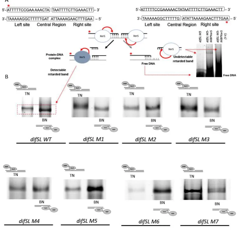
3.1 Characterization of essential nucleotides in difSL involved in binding and recombination reactions. ... 63
3.2 The subdomain of FtsKc interacts with XerS ... 69
4. Perspectives ... 71
5. Literautre cited ... 71
Figure Legends... 80
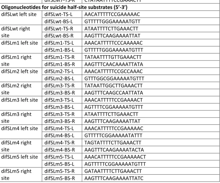
Figure 1. Titration of a 339 bp DNA fragment containing difSL wild type and difSL mutants (M1 to M7) ... 80
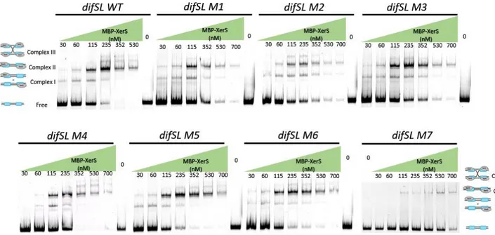
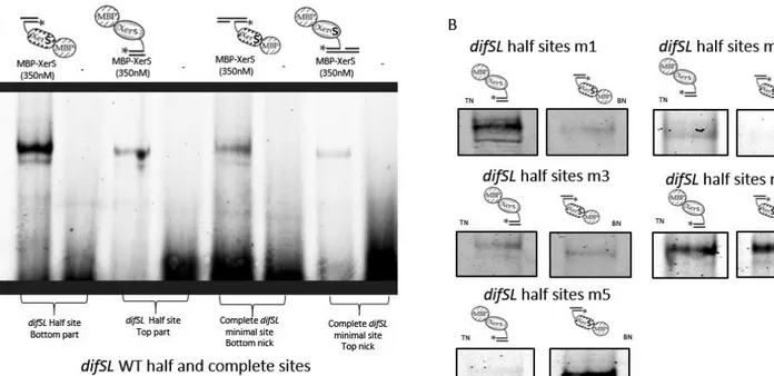
Figure 2. Titration of a 315 bp DNA fragment containing difSL-M7 half left site, M7-LHS (A) and half right site, M7-RHS ... 80
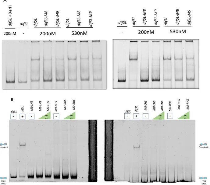
Figure 3. A. Duplicate of titration of a 339 bp DNA fragment containing difSL, difSL mutants M8 and M9 ... 80
Figure 4. XerS activity on suicide substrates. ... 80
Figure 5. XerS activity on half suicide substrates ... 81
Figure 6. Pull-down assay between immobilized MBP-XerS and His-FtsK. ... 81
Chapter III ... 91
General discussion... 91
1. Results ... 91
2. XerS/difSL system ... 91
Conclusions and perspectives ... 96
List of tables
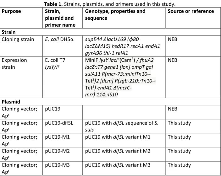
Table 1. Strains, plasmids, and primers used in this study………72
List of figures
Chapter I: Review paper
Figure 1. The two primary and most generalized pathways to solve chromosomal dimers
generated by RecA-dependent repair or stalled replication forks. ... 29
Figure 2. Segregation of the sister chromatids during chromosome dimer resolution in E. coli. 34 Figure 3. Sequence alignment of (A) dif, (B) cer, (C) psi, (D) Bsdif, (E) dif1, (F) attPCTX, (G) attPVGJ, (H)attPTLC, (I) difSL and (J) difH. ... 40
Chapter II: Article
Figure 1. Titration of a 339 bp DNA fragment containing difSL wild type and difSL mutants (M1 to M7) by increasing concentrations of MBP-XerS. ... 83Figure 2. Titration of a 315 bp DNA fragment containing difSL-M7 half left site, M7-LHS (A) and half right site, M7-RHS (C). ... 84
Figure 3. Titration of a 339 bp DNA fragment containing difSL, complete difSL mutants M8 and M9 and half sites. ... 85
Figure 4. XerS activity on suicide substrates. ... 86
Figure 5. XerS activity on half suicide substrates. ... 87
List of abbreviations
AMINO ACIDS D: acid aspartic E: acid glutamic A: alanine R: arginine N : asparagine C: cysteine Q: glutamine G: glycine H: histidine I: isoleucine L: leucine K: lysine M: methionine F: phenylalanine P: proline S: serine T: threonine W: tryptophan Y : tyrosine NUCLEIC ACIDS A: Adenine C: Cytosine G: Guanine N: A, G, C ou T R: Purine T: Thymine P: Pyrimidine UNITS OF MEASUREMENT Å: angstrom bp: base pair cm: centimeter Da: Dalton °C: degree Celsius g: gram h: hour kb: kilobase kDa: kiloDalton μg: microgram μl: microliter μΜ: micromolar ml : milliliter mM : millimolar min: minute ng: nanogramrpm: revolutions per minute s: second
v/cm: volt per centimeter GREEK LETTERS : alpha : beta : gamma : delta : epsilon :phi : lambda OTHERS
AAA+: ATPase associated with various activities 5’OH : 5 prime hydroxyl end
6-HEX: 6-carboxy-2’,4,4’,5’,7,7’-hexachlorofluorescine AIMS: Architecture Imparting Sequences
ASPS: Archaea Short Polarized Sequences ATP: adenosine triphosphate
ATPase: adenosine triphosphatase BN: Bottom nick
BSA: Bovine Serum Albumin C-terminal (e): carboxy-terminal DNA: Deoxyribonucleic acid
DAZ: dif activity zone
EDTA: Ethylenediaminotetraacetic acid EMSA: Electrophoretic mobility shift assay FRET: Fluorescence resonance energy transfer GGI: gonococcal genomic island
HEX: Single isomer fluorescein dye
IPTG: isopropyl B-D thiogalactopyranoside KOPS: FtsK-orienting polar sequences LB: Luria-Bertani
MBP: maltose binding protein N-terminal (e): amino-terminal NEB: New England Biolabs PCR: Polymerase chain reaction RNA: Ribonucleic acid
SDS: sodium dodecyl sulfate SPR: Surface plasmon resonance SRS: SpoIIIE Recognition Sequence TBE: tris-borate EDTA buffer
TDMNG: Tris DTT MgCl2 NaCl Glycerol
TENG: Tris EDTA NaCl Glycerol THA: Todd-Hewitt agar
THY: Todd-Hewitt + Yeast extract TN: Top nick
Acknowledgments
I am really grateful for the opportunity of having met such wonderful souls during this moment of my life. I would like to say thank you to Professor George Szatmari for all his support and valuable lessons professional and life wise. And thank you Amal Benmohamed and Asmaâ Agoussar for being there, always bringing a wonderful and positive energy to the laboratory, I will always remember your thoughtful ideas to improve the harmony in the lab. I also want to thank Ali Farrokhi for all his support and professionalism.
Thank you to the members of the jury for your time and valuable input for the completion of this thesis.
A big thank you to all members of the Department of Microbiology, professors, students, technicians, administration staff, thank you Montreal and montrealais, I have learned so much from all of you!
And a special dedication to my family, to my strong father Jorge Armando Castillo, my beautiful mother Sonia Martinez Ayala, my creative brother David Castillo Martinez, my powerful sister Luisa Castillo Martinez, my brave grand-mother Virginia Ayala and to my love and best friend Debora Parrine Sant’Ana.
Chapter I
Review paper
This published review paper focuses on the action of different site-specific recombinases to solve dimerization problems during DNA replication and cellular division in bacteria. It initially addresses causes and effects of such events, two important aspects vaguely connected by most papers. With this review, we intended to offer a broader idea of the cause of such events, the reasoning for a certain outcome and the complexity to solve them. After reviewing all the mechanisms involved in crossing over events and dimer formation, we proceeded to explain the mechanisms involved in their repair and in the acquisition of new genomic material from viruses by presenting a detailed overview of all current known site-specific recombinase systems in bacteria.
I wrote the majority of this paper. George Szatmari and Amal Benmohamed provided valuable input, precise information and very helpful comments and corrections before and during the publication process.
Xer site specific recombination: double and single
recombinase systems
Fabio Castillo Martinez, Amal Benmohamed, George Szatmari* Département de microbiologie, infectiologie et immunologie
Université de Montréal
Keywords: Site-specific recombination, tyrosine recombinases, single recombinases, XerS, XerH, XerA, IMEX.
Emails: [email protected], [email protected], [email protected]
Abstract
The separation and segregation of newly replicated bacterial chromosomes can be constrained by the formation of circular chromosome dimers caused by crossing over during homologous recombination events. In E. coli and most bacteria, dimers are resolved to monomers by site-specific recombination, a process performed by two Chromosomally Encoded tyrosine Recombinases (XerC and XerD). XerCD recombinases act at a 28 bp recombination site dif, which is located at the replication terminus region of the chromosome. The septal protein FtsK controls the initiation of the dimer resolution reaction, so that recombination occurs at the right time (immediately prior to cell division) and at the right place (cell division septum). XerCD and FtsK have been detected in nearly all sequenced eubacterial genomes including Proteobacteria, Archaea, and Firmicutes. However, in Streptococci and Lactococci, an alternative system has been found, composed of a single recombinase (XerS) genetically linked to an atypical 31 bp recombination site (difSL). A similar recombination system has also been found in ε-proteobacteria such as Campylobacter and Helicobacter, where a single recombinase (XerH) acts at a resolution site called difH. Most Archaea contain a recombinase called XerA that acts on a highly conserved 28 bp sequence dif, which appears to act independently of FtsK. Additionally, several mobile elements have been found to exploit the dif/Xer system to integrate their genomes into the host chromosome in Vibrio cholerae, Neisseria gonorrhoeae and Enterobacter cloacae. This review highlights the versatility of dif/Xer recombinase systems in prokaryotes and summarizes our current understanding of homologs of dif/Xer machineries.
1. Introduction
Bacteria and archaea have developed a variety of well-regulated and coordinated mechanisms of replication and segregation of their genomes that ensure the genetic material is transmitted faithfully to the daughter cells, despite the absence of temporal separation between DNA synthesis, chromosome separation and cell division 1. However, the circular state of their
chromosomes and plasmids constitutes a constant threat to genome stability and proper segregation because of dimer formation during recombinational exchanges between sister chromatids. These rearrangements can combine their genomes into larger molecules, compromising an equal distribution of the genetic material to the daughter cells 2–5. This
topological problem was fully addressed in 1981, when Austin et al. (1981) demonstrated that the stable inheritance of the prophage P1 was due to site-specific recombination (SSR), a specialized system that catalyzes DNA exchange between two defined DNA sequences, and which plays a major role in dimer resolution by converting multimeric forms to the monomeric forms. Later studies performed with the plasmid ColE1 connected SSR with plasmid monomerization and stability 7. In 1990, the site-specific recombinase (XerC) was identified as the first protein
responsible for SSR on cer 8, followed by the identification of a second recombinase, XerD
required for this reaction 9.
2. The disadvantage of having circular DNA
In most bacteria and some archaea, replication begins at a single origin of replication oriC at which DnaA binds and stimulates the assembly of the replisome 10. Replication forks then proceed
bi-directionally until the two replication forks meet in an antipodal terminus region flanked by ter sequences. These sequences in conjunction with the replication terminator protein (Tus) stop the replication forks to synchronize their arrival at the same time and place 11–13. However,
chromosome replication is not a continuous process and is continuously halted by different types of DNA lesions such as UV irradiation, free radicals, genotoxic agents, DNA replication errors, transcription-replication conflicts, tightly bound protein-DNA complexes, or RNA secondary structures 12,14–17. To maintain their genomic integrity, bacteria have developed several and
sophisticated mechanisms to minimize the frequency of these DNA lesions before the occurrence of replication. The initial barrier against deleterious DNA modifications is carried out by specialized mechanisms, each one required for a given type of lesion, such as proofreading, direct reversal of DNA damage, base excision repair, nucleotide excision repair and mismatch repair 18– 21. Additional groups of mechanisms are responsible for avoiding transcription-replication
encounters, equally lethal for bacteria, such as the coordination of temporal and spatial gene activation and co-orientation, modulators of RNA polymerases (RNAPs) and replicative accessory helicases 16,22–24. Nonetheless, it is unavoidable that some of this DNA damage or conflicts will
escape the initial barrier and interfere with replication fork migration, leading to the eventual inactivation of the replication machinery and formation of double-strand breaks (DSBs), interstrand cross-links and single-stranded gaps (SSG). These represent critical forms of DNA damage that must be removed for chromosome replication and transcription to proceed 25,26.
Therefore, a second barrier of repair is called into play to cope with these “evasive” damages. This second barrier is preferentially carried out by the homologous recombination repair system (HR). Estimates indicate that HR repair is required in almost every cycle of replication 27–29. In fact,
the HR system is now not only considered as a functional mechanism for generating genetic diversity but also as a decisive factor in DNA repair, the latter being the primary role of this system in the maintenance of the genome and the main source of dimer events 30. Thus, HR plays a
central role in removal and/or repair of DNA damage and rescue and/or re-assembling of replication forks that have been broken or stalled 26,31. In the traditional HR system in E. coli, its
mode of action consists of a multistep process of breakage and rejoining of homologous sequences (one old and one newly synthesized DNA strand). It initially involves 1) recognition of the DNA lesion by the complexes RecBCD or RecFOR, depending on the type of DNA lesion; 2) formation of 3’-ssDNA overhangs processed by the exo and endonuclease activity of the Rec proteins, and subsequent coating by RecA; 3) strand invasion of the 3-terminal ssDNA into the homologous duplex DNA molecule and search of the complementary strand; 4) formation of a D-loop intermediate, transformation into a branched intermediate and Holliday junction (HJ) formation and 5) completion of the recombination process by resolution of the HJ, catalyzed by the systems RuvABC or RecG 32–34. HJ resolution can result in two alternative products;
1. ‘crossover’ or spliced products; where reassortment of the flanking genes of the cleavage site has occurred, obtaining one different genotype at one side compared to the former DNA duplex, and therefore, long range of genetic exchange.
2. ‘non-crossover’ or patch products where the flanking regions were not exchanged, and instead, the resulting DNA duplex contain a ‘patch’ of hybrid DNA with a shorter range of genetic exchange 35,36.
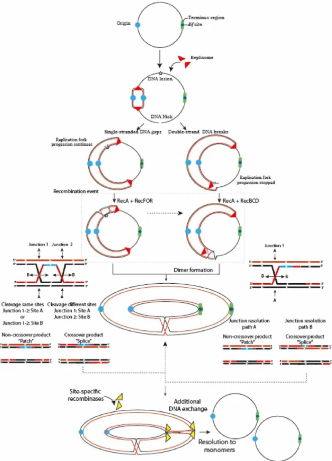
Because circular chromosomes do not have “ends”, they are vulnerable to concatenation during formation of an odd number of crossover events. Thus, swapping DNA flanking regions tangles the sister chromatids and forms larger ring chromosomes that compromise cellular division (Figure 1) 2,4,37. To ensure proper chromosomal segregation, bacteria and archaea have overcome
these major threats by two broad mechanisms. One is to minimize the formation of crossing-over events, and the other is to solve dimer formation by performing an additional DNA exchange, immediately prior to cell division, at a specific region called dif (Deletion-Induced Filamentation).
Figure 1. The two primary and most generalized pathways to solve chromosomal dimers generated by RecA-dependent repair or stalled replication forks.
If the fork encounters a non-coding lesion (oxidative damage, pyrimidine dimer or an abasic site) depicted with a yellow star, it usually generates SSG (left path), although it can also lead to DSBs ends during repair by the RecFOR system. The pathways can also diverge on the location of the DNA lesion. If the lesion is located on the lagging strand template, the replisome will be able to bypass the lesion by blocking ‘Discontinuous DNA synthesis’ and then resume it downstream of the lesion, leaving a gap that would be repaired by the RecFOR system. On the other hand, a lesion on the leading strand template might transiently stop the replisome, cause its dissociation and then, bind further downstream to a new leading-strand primer, although these mechanisms are still under debate 25,31. Alternatively, when the fork encounters a nick in the template strand (unrepaired
SSG) or some cases of replication fork collapse, a DSB is generated (Right path). The DSBs are processed by the RecBCD complex that catalyzes the reattachment of the damaged DNA to the sister DNA duplex, forming a D-loop structure and eventual recognition by the replication-restart PriA protein that directs replisome assembly and resumption of the replication process in an origin-independent manner. However, odd numbers of crossover events generate dimer products 38–40. Thus, if the resolution of the HJ occurs in the same sites, it will
generate monomeric chromosome (Non-crossover products). In contrast, if the resolution takes place in different sites, it will generate chromosome dimers (Crossover products) 15,41.
3. Avoiding dimer formation
One way to avoid dimer formation, as simple as it sounds, is to decrease the likelihood of dimer formation. This, however, is an intricate process of coordination and selection of the right enzymes at the right moment. Therefore, if the resolution of HJ intermediates by endonucleolytic cleavage can only result in crossover or non-crossover products, the likelihood of obtaining one or another is 50%. However, minimizing crossover events during homologous recombination repair seems to be the rule rather than the exception in organisms with circular chromosomes 42.
In E. coli, homologous recombination repair is processed by two predominant recombinational pathways; The RecBCD pathway associated with DSB repair, replication fork collapse, replication fork reversal and replication fork arrest, and the RecF pathway, which is mostly involved in the repair of SSG, and under certain conditions, can also repair DSBs 43,44. Both mechanisms lead to
the formation of HJs that are mostly resolved by the RuvABC complex in E. coli or RecU in Firmicutes and Mollicutes 41. Deletion of the genes of the RuvABC complex eliminates
non-crossover formation bias, supporting the idea that bias formation mostly depends on the action of Ruv proteins more than Rec proteins 35. This idea was initially discussed by Van Gool et al.
(1999) who demonstrated that crossover and non-crossover products are not random and, conversely, they are influenced by the positioning and orientation of the resolvasome on the HJ
intermediate, which in turn directs RuvC strand cleavage direction. Additionally, topological conditions such as DNA supercoiling, DNA catenation, adjacent HJ intermediates or the presence of double HJ intermediates can also influence assembly of the RuvABC complex on the HJ and indirectly affect resolution 45. Subsequently, Cromie and Leach (2000) showed that RuvABC
positioning may depend on the nature of the substrate caused by the type of DNA lesion, thus DSBs are more likely to result in crossover products (Frequently processed by RecBCD) whereas SSG are more likely to result in non-crossover products (frequently processed by RecF). Although, some fractions of SSG can cause DSBs 46. However, the specific causes of crossover or
non-crossover formation are still under debate, and different reactions cannot be completely dismissed. A clear example of this is the fact that RecBCD as well as RecF are not restricted to DSBs and SSG respectively and, on the contrary, the both may have interchangeable functions 47.
That would explain why some replication fork arrests generate non-crossover products even if they are mostly processed by the RecBCD pathway 48, or why RecF contributes almost equally to
dimeric chromosome formation in E. coli despite the fact that it is responsible for SSG resolution
4.
Interestingly, the fact that DNA lesions and transcription-replication conflicts are more abundant in the leading strand than in the lagging strand in E. coli, and that these lesions usually generate non-crossover products, reinforces the idea that organisms with circular chromosomes favored a system that minimizes dimer formation during HR repair completion 16,35,49. These biased
reactions have also been detected in other microorganisms such as B. subtilis where the resolvase RecU biases homologous recombination towards non-crossover products 41. Despite this
non-crossover preference by HR system in recA+ cells, dimer formation still occurs reaching 10 to 15% of the growing cells 50.
4. Coping with dimers
It is clear that dimer formation is regarded as a negative outcome that must be solved. Despite this, Mazin et al. (1996) proposed that under certain conditions of selective stress, plasmid dimerization could confer an advantage for the selection of adaptive mutations due to rapid accumulation and selection of plasmids carrying a specific mutation and subsequent segregation
to the daughter cells. Berza et al. (2013) also reported that plasmid dimerization greatly increased synthesis of a foreign protein and that plasmid content is unaffected by dimer formation showing some advantages for transcriptional events. However, these benefits were only considered for plasmids. Regarding circular chromosomes, dimerization must be resolved by the action of site-specific recombinases (SSRs), which are enzymes that are responsible for breaking and rejoining specific sites without requiring DNA synthesis or high energy cofactors 53. The relevance of this
system for proper chromosome segregation is supported by the high degree of conservation in Bacteria and Archaea. The Xer complex is considered one of the most conserved structural features in cells containing circular chromosomes, as well as RecA and FtsK enzymes 50,54,55. The
SSRs act on specific short DNA sequences, called recombination sites, where DNA exchange occurs in three different types of DNA rearrangements; deletion (divided into excision or resolution), insertion, or inversion. All these processes depend on the orientation and direction of the two recombination sites 56 (Figure 1). All known site-specific recombinases are classified
into two unrelated families, tyrosine–type or serine-type recombinases (Tyr or Ser) based on the amino acid residue that forms a covalent linkage between the protein and a phosphate at the DNA cleavage site 57. Serine recombinases, often referred to as the resolvase/invertase family, act
on a recombination site with just 2 bp separating the cleavage sites on top and bottom strands and the cleavages occur simultaneously to create a double strand break, while tyrosine recombinases, often referred to as the λ integrase family enzymes perform a two-step cleavage and rejoining process where the cleavage sites are separated by 6-11 bp. Each recombinase family possesses a distinct mechanism. Tyrosine recombinases are divided according to the recombination directionality; unidirectional or bidirectional recombinases. Whereas serine recombinases are divided according to their size; small or large recombinases 56,58.
The chromosome dimer resolution (CDR) process and heritable stability were originally elucidated in E. coli 8,9,59–61, where two paralogous site-specific tyrosine recombinases, XerC (298 aa) and
XerD (298 aa) (Chromosomally Encoded Recombinases) were shown to act on a 28 bp DNA sequence (dif site), located in the ter region. The synaptic XerCD/dif complex consists of two XerC and two XerD subunits respectively bound to two dif sites (Figure 2b). Limited structural information of some tyrosine recombinases have revealed a conserved catalytic domain fold 62,
facilitating the analysis of experimental data and allowing the development of a general model for Xer recombinases 63 consisting of; XerD 64, XerA 65, XerH 66 and other related tyrosine
recombinases like Cre 67,68, HP1 integrase 69, FLP 70 and λ integrase 71,72. The E. coli dif site is divided
into two 11 bp half-sites that share partial dyad symmetry linked by a 6 bp central region that defines the positions of strand cleavage and exchange 73. The initial step of site-specific
recombination during dimer resolution requires the formation of a synaptic complex consisting of a tetrameric protein/DNA complex (four protomers of tyrosine recombinases and two recombination site duplexes). Once the synaptic complex is formed, two opposing and activated protomers cleave the DNA strand of each recombination site duplex. This occurs when the hydroxyl group of the nucleophilic tyrosine attacks the scissile phosphate in the central region to form a 3′ phosphotyrosyl intermediate and a 5’-hydroxyl end. This intermediate conserves the energy from the phosphodiester bond cleavage to perform the first strand exchange. The recently formed 5′- hydroxyl attacks the 3′ phosphotyrosyl linkage on the partner site to reseal the strand breaks creating a HJ intermediate 53. HJ formation and isomerization activates the second pair of
subunits bound to the other half of the recombination sites and inactivates the first pair of subunits. The second pair of subunits then cleaves, exchanges and rejoins the second pair of strands by the same mechanism just described; this second cleavage allows the resolution of HJ-intermediate and results in the recombinant DNA (Figure 2B) 63,74. This process implies that the
specific pairs of recombinases and/or active sites are continuously switched on and off to synchronize when and how recombination occurs, this coordination depends on allosteric interactions between the recombinases and external factors imposed on the synaptic complex 75– 77. In the XerCD/dif system, XerC normally initiates catalysis of one pair of DNA strands to form
the HJ-intermediate without a subsequent resolution by XerD. Therefore, the HJs are rapidly converted back to the original DNA rearrangement. This XerC-first interaction is functionally active during the integration of certain bacteriophages that utilize Xer recombination to integrate their genomes into their host dif sites or in the resolution of plasmid multimers. In contrast, during chromosomal dimer resolution, pre-synapsed XerCD/dif complexes favor XerD activation by the FtsK protein to mediate the first strand exchange, generating a transient (XerD-HJ) intermediate,
subsequent isomerization forms a XerC-HJ intermediate that is rapidly resolved to recombinant DNA by XerC, see below and Figure 2A, 3A 54,78.
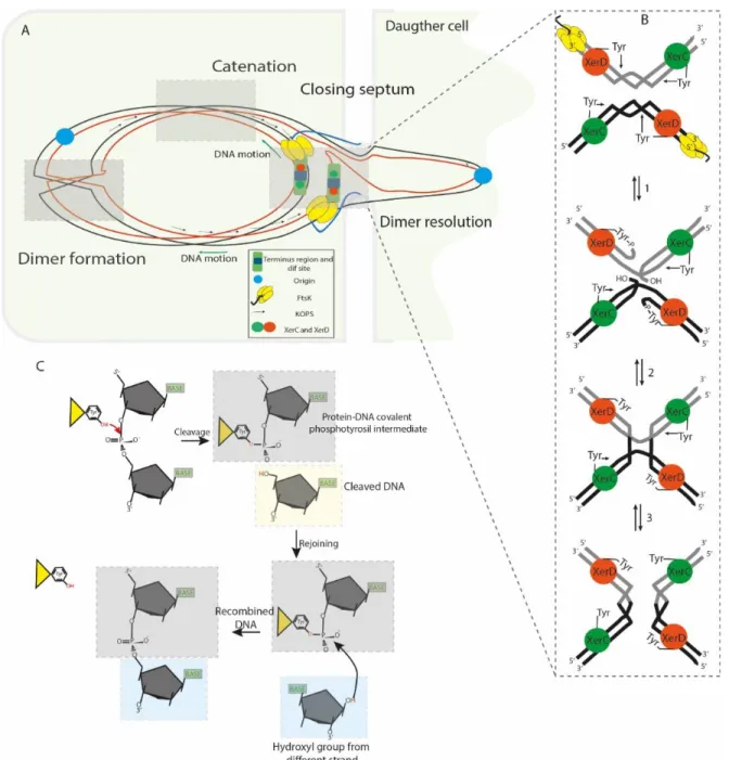
Figure 2. Segregation of the sister chromatids during chromosome dimer resolution in E. coli.
The illustration depicts the central part of a dividing cell in the final steps of chromosome segregation. The closing division septum, the motor domain αβ of FtsKC (yellow hexameric ring), the unstructured linker domain
FtsKL (Blue ribbon), the KOPS sequences and the XerCD/dif synaptic complex are indicated. Concatenation
prevents proper migration of the nascent chain of DNA; the origin regions move toward their respective cell poles, but the rest of the knotted DNA is stretched across and behind the septum. (B) FtsKC loads onto the KOPS
to reach the XerCD/dif complexes and bring them into proximity; as a consequence, the γ-subdomain of the FtsKC region activates XerD (Orange sphere) to perform the first strand cleavage. Then, XerC (Green sphere)
mediates the second strand cleavage, allowing separation of the sister chromatids from each other. (C) Illustration of the SSR mechanism used by tyrosine recombinases: The OH group of the active residue tyrosine attacks the scissile phosphate forming a 3′-covalent phosphotyrosyl enzyme–DNA covalent intermediate and a free 5′-hydroxyl end. The covalent intermediate is attacked in turn by the other 5’- end to reverse the cleavage reaction and obtain a recombinant product.
5. Establishing rules for dimer resolution
One of the fundamental questions about site-specific recombination at dif concerns how the system is controlled to ensure a proper chromosome dimer resolution (CDR) into monomers in the right place and at the right time without promoting the reverse reaction, which would generate dimers from monomers. It is understood that Xer-mediated recombination mostly depends on an active HR system because it is the major process that provides concatenated chromosomes. However, catenation problems caused by replication may require Xer-recombination system as well 3,79,80. This reaction occurs at two polarized and specific regions of
~10kb at either side of dif called DAZ (dif Activation Zone), where oppositely oriented KOPS (FtsK Orienting Polar Sequences) converge and guide FtsK DNA translocation towards the dif locus 81.
This directional control is achieved by the interaction between the Xer recombinase system and the C-terminal domain of FtsK (Filamentous Temperature-Sensitive cell division protein K), a large division septum-associated DNA translocase, which coordinates chromosome segregation and cell division when chromosome organization has been affected (e.g. chromosome dimer formation, decatenation or delayed replication) 54,82–87. FtsK was initially documented in 1995 due
to observations in E. coli TOE44 (AB2497 ftsK44) mutant cells and their ability to form long chains of cells due to a single substitution of one amino acid in the N-terminal domain (FtsKN), by then,
FtsK was thought to participate in septum formation as a peptidoglycan-modifying enzyme. Then, Yu et al. (1998) demonstrated that inactivation of the C-terminal region of FtsK affected normal chromosome segregation due to the formation of long chains of cells and detected abnormal DNA distribution in some ftsK1::cat minB double mutants of E. coli minicells. Finally, in 1999 Steiner et al. (1999) discovered that site-specific recombination at dif requires FtsKC and thus, chromosome
dimer resolution only occurs in its presence. The ~1329aa FtsK protein can be divided into three domains; The ~279aa N-terminal domain (FtsKN) is responsible for attachment of the protein to
the membrane by four transmembrane segments and interaction with other proteins of the division septum such as FtsZ 90. The linker domain FtsK
L, not commonly conserved in FtsK
homologs, is primarily composed of proline-glutamine residues 91. Its length and composition
varies between species, being ~650 amino acids long in E. coli and most of Proteobacteria, ~200aa long in Vibrios 92 or ~125aa in Pseudomonas 93. Experiments performed by Bigot et al. (2004)
demonstrated that ftsKL mutations increased filamentation phenotypes even higher than xer
mutants and that this filamentous formation did not correspond to problems in chromosome dimer resolution. On the contrary, it was thought to be due to a deficient positioning of the protein, reducing the possibility of contact between FtsKC and the DAZ region of the chromosome.
Subsequently, Dubarry et al. (2010) revealed that different parts of the linker domain interact with other proteins of the divisome such as FtsQ, L, I and Z and these interactions help to stabilize the whole divisome at the site of septation. Interestingly, they also suggested that FtsKL domain
may stop or slow down cell division during dimer resolution because of the destabilization of the divisome components when FtsKC has been pulled by the DNA during translocation, this force can
separate FtsZ and delay septum constriction. There is also a proportional relation between the glutamine-proline concentration and its length, where the longest linkers are usually richer in these residues 95. Following the linker, the highly conserved ~500aa C-terminal domain (FtsK
C),
usually referred as the motor of the FtsK, is comprised of three separated subdomains called α, β and γ. Structural studies of the translocation module FtsKαβ of P. aeruginosa demonstrated that
it assembles as a hexameric ring around double-stranded DNA forming a central channel of 30Å in diameter, where double stranded DNA (dsDNA) passes through 93,96. Later structural studies of
the orientation module FtsKγ of P. aeruginosa and E. coli demonstrated that six γ subdomains are
loosely attached by a short linker of 10 aa to the hexameric ring FtsKαβ84,97. The FtsKαβ subdomains
are responsible for the ATP hydrolysis-dependent DNA translocation of the protein. The 68aa FtsKγ subdomain is a helix-wing-helix domain that performs two main functions. The first role of
this subdomain is to recognize the 8 bp KOPS sites and then directs FtsK translocation toward the
brought together to form the synaptic complex XerCD/dif (Figure 2A). KOPS are over-represented on the leading strand of replication where their concentration gradually increases as dif is reached; indeed, more than 90% of KOPS sequences nearby dif are located on the leading strand
82; giving a possible estimate of 34 KOPS motifs located in the DAZ region 98 with a frequency of 1
motif every 13Kb 83. The second main function of the FtsK
γ subdomain is to activate the XerD
catalytic function to generate the first HJ and subsequent dimer resolution 85,98,99. How FtsK
locates and assembles to initiate translocation in the correct KOPS sequence is still arguable, for this reason two models have been proposed; the loading model and the target search model, recently reviewed by Besprozvannaya & Burton (2014). New evidence strongly suggests that FtsK acts in a 350-kb region around dif that covers 7% of the genome where monomers of FtsKC
assemble exclusively at KOPS motifs as described by the loading model. An initial interaction of a single monomer of FtsKγ will trigger a rapid and stepwise formation of the hexameric ring under
high concentration of FtsK 100,101. It is likely that FtsKγ assembles quickly and binds to KOPS as a
trimer initially, with three FtsKγ modules interacting with consecutive GGG, NA, and GGG bases and then it hexamerizes gradually 84 Once a KOPS motif is detected, allosteric modifications occur
leading to hexamerization of FtsKαβ, which alters the angular conformation of FtsKγ on the DNA
affecting KOPS recognition, and activates FtsKαβ ATP hydrolysis. As a consequence, FtsK is no
longer able to recognize subsequent KOPS motifs during translocation, unless FtsK migration is impaired, and KOPS recognition is obligated to restart 84,97,98,102. FtsK has been demonstrated to
be the fastest known DNA translocase, reaching levels of 17.5 ± 3.5 kb/s at 37 °C or even faster with a striking stall force and a slight supercoiling induction, 1 positive supercoil per every 150 bp translocated 86,103,104. It has also demonstrated a striking capacity to displace, evict or bypass
different obstacles, especially proteins bound to the DNA such as RNA polymerases 98. However,
FtsK acts differently upon collision with RecBCD and XerD-XerCD/dif complex proteins. When FtsK collides with XerCD/dif, in a synapsed form, it activates XerD to create the XerD-HJ transient intermediate (structural rearrangements increase the distance between dif sites from about 53 to 67 A˚) 66,78 followed by a rapid dissociation from the DNA (dissociation takes to 0.5 to 1
seconds). Cleverly, May et al. (2015) demonstrated that recombination of the synaptic complex XerCD/dif takes 1 second longer than the FtsK dissociation time. Therefore, they suggested that
this time span can provide a regulatory control for dimer resolution because concatenated chromosomes will reform XerCD/dif synaptic complexes every time that resolution failed. Thus, multiple sets of FtsK hexamers colliding multiple times against XerCD/dif synaptic complexes will increase the likelihood of generating recombinant products. This regulatory mechanism ensures monomeric products are formed during translocation of impaired DNA.
6. Alternative dif/Xer resolution in prokaryotes
6.1 Plasmid resolution: Multicopy plasmid ColE1 and accessory proteins
Plasmid dimerization and eventual multimerization has been termed as the “Dimer catastrophe” due to its deleterious effect in cell populations 105,106. Dimer catastrophe represents two major
problems in bacteria; 1) unequal plasmid distribution among populations, in particular, multicopy plasmids that are more vulnerable to plasmid loss and 2) metabolic burden caused by the rapid accumulation of dimers into the host 106,107. As mentioned previously, dimer resolution was
originally elucidated in ColE1, resulting in the first functional characterization of XerC and subsequent identification of XerD by sequence homology to XerC 9. These discoveries constituted
a new approach for site-specific recombinases and their role in dimer resolution. Subsequent investigations led to the identification of SSR enzymes involved in dimer resolution in other plasmids of E. coli, and other bacteria. Current estimates have identified more than 1300 tyrosine recombinases where many of them are associated with other host proteins to regulate their activity, directionality, or processivity 63. Large plasmids usually carry their own recombinase
machineries adjacent to the recombination site. Whereas, small plasmids, like those in the ColE1 family, use the chromosomally encoded dimer resolution system of their host 108,109. For ColE1
resolution, XerC/D proteins act on a specific site called cer (Figure 3B), a non-codifying region of 280 bp where two additional proteins act with XerCD to catalyze SSR reactions: the arginine repressor (ArgR) (an arginine-dependent DNA binding protein originally called XerA) 59,110, and
aminopeptidase A (PepA) (a bifunctional transcriptional regulatory protein that reacts to environmental signals, which was originally called XerB) 111. SSR in cer is catalyzed by XerC within
a sequence of 30 bp composed of two 11 bp half sides and a central region of 8 bp. XerC and XerD bind to the left and right halves cooperatively and respectively. Strand exchanges are catalyzed
by XerC to form a HJ intermediate that is eventually resolved by an uncharacterized cellular HJ resolvase to generate a recombinant product 112,113. The cer site is comprised of a 30 bp core
recombination site and two accessory DNA sequences of ~180 bp in length, in which one or two hexamers of PepA and one hexamer of ArgR control the reaction 114. The two accessory proteins
are necessary for recombination, since in their absence, plasmid dimer resolution cannot be completed. However, at abnormal high concentrations of PepA, recombination in vitro can proceed without the help of ArgR 115,116. This is also seen at the recombination site psi of plasmid
pSC101 which requires XerC, XerD and PepA but not ArgR (psi dimer resolution requires another accessory protein called ArcA instead of ArgR, and the cleavage reaction is performed by XerC and XerD) 113,117. In cer, PepA and ArgR control recombination directionality so that dimers can
only be converted into monomers and not the opposite reaction. Dimer resolution directionality caused by these two proteins involves the formation of two directly repeated cer sequences positioned in an antiparallel direction; this conformation is favored by negative supercoiling where the cer sequences are interwrapped three times around the proteins resulting in the formation of a right-handed synapse structure that brings the XerCD binding sites together. Sites in an inverted repeat position prevent right-handed formation; this ensures only dimer resolution occurs. Thus, XerC and XerD bind to the 30 bp cer synapse region and may interact with the N-terminal domains of the PepA hexamers. Whereas the ArgR protein, which is flanked by one or two PepA hexamers, might be involved in bending the DNA, tightening it and activating cer SSR by possible interaction with the C-terminal region of the Xer recombinases. Another possible function is to bring the two cer sites together and to allow PepA loading to form a nucleoprotein complex that promotes XerCD binding and recombination 114–116. Additionally, cer also encodes
for a 70 nt RNA fragment called Rcd that is only transcribed during dimer formation by the Pcer
promoter and regulated in a sequence-specific manner by the FIS protein 118. Rcd binds to the
enzyme tryptophanase and induces a quiescent state by increasing indole production within the cell. The quiescent state permits the cell to arrest cell division and chromosomal replication but still be active metabolically. This process is thought to be part of a dimer formation checkpoint that allows the XerCD/cer system to resolve dimer formation during this pause 106.
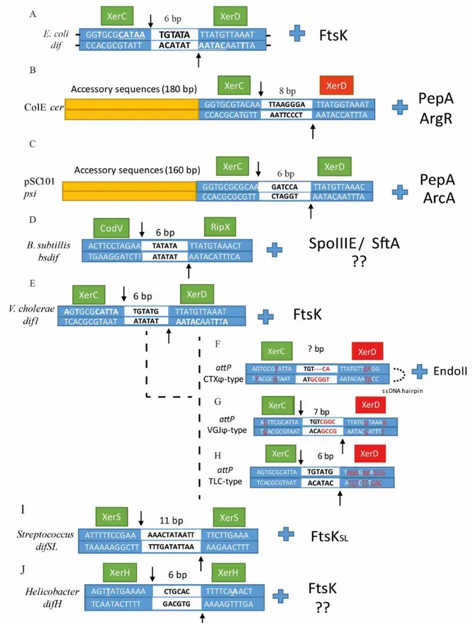
Figure 3. Sequence alignment of (A) dif, (B) cer, (C) psi, (D) Bsdif, (E) dif1, (F) attPCTX, (G) attPVGJ,
(H)attPTLC, (I) difSL and (J) difH.
Dyad bases in each arm are underlined in dif. XerC and XerD cleavage points are indicated by arrows, the central region is depicted in the middle of the sequence as a white box with the number of base pair corresponding to
each dif site above. Left and right arms are depicted as blue boxes with their corresponding sequences for each dif site. The catalytic unit is depicted as a green box, whereas the inactivated unit is depicted as a red box. For (E), (F), (G) and (H), bases that differ from dif1 in V. cholerae are underlined and colored in red. Next to each dif alignment is the corresponding accessory protein that coordinates/activates dimer resolution.
6.2 The Bacillus subtilis model and the effect of two translocases
The capacity to perform site-specific recombination to resolve chromosome dimers is highly distributed among bacteria and archaea. Thus, homologs of XerC and XerD have been sequenced in a variety of species 108,119. In B. subtilis, two homologs of XerC and XerD called CodV and RipX
perform dimer resolution at a 28 bp dif site (Bsdif) close to the terminus region (Figure 3D). The
Bsdif region is comprised of two 11 bp half-sites with imperfect dyad symmetry where CodV and
RipX bind simultaneously and a 6 bp central region where DNA exchange occurs. Both CodV and RipX share a 37 and 44% identity with the XerC and XerD respectively, and 39% between them
120. CodV binds preferentially to the left half-site and preferentially cleaves the top strand whereas
RipX is able to bind to both sides with preferential binding to the right-half-site and preferential cleavage of the bottom strand. Cleavage by CodV is more efficient than cleavage by RipX, which suggests that CodV performs the first strand cleavage followed by RipX in in vitro experiments 121.
Sciochetti et al. (1999) also demonstrated that RipX could interact effectively with the E. coli dif site, unlike CodV which showed a weaker interaction with this substrate. However, addition of XerC to RipX/difE. coli or XerD to CodV/difE.coli generated larger complex formation in gel retardation
analysis, demonstrating protein-protein interactions between these four proteins, which confirms some conserved features of tyrosine recombinases among bacteria. This is supported by the fact that the right half-site presents highly conserved features with respect to other dif sites among some bacteria, whereas the left-half site is less conserved, which could explain why RipX can bind difE.coli 121. In contrast to E. coli, the synaptic complex can be brought together by
the action of two DNA translocases: the membrane-associated SpoIIIE protein (Stage III Sporulation Protein E) and the soluble SftA protein (Septum-associated FtsK-like Translocase of DNA). Both translocases harbor AAA+ATPase and C-terminal domains with 56% of sequence similarity between them. SftA exhibits 50% identity with respect to the E. coli FtsKγ domain
FtsKγ subdomain of E. coli 122. The N-terminal domains of these proteins are more divergent;
SpoIIIE and FtsK share 36% of identity to respect to the four transmembrane helix whereas SftA lacks the transmembrane spanning domain 123,124. N-terminal domain variations coincide with
their different location and activation in the genome. FtsK and SpoIIIE share a similar mechanism to anchor to the inner membrane of the dividing cell by their N-terminal regions; this transmembrane interaction is possibly reinforced by interactions with FtsZ or other cell division components such as FtsA or ZapA 91. During vegetative growth, SpoIIIE shows two predominant
states: a static phase, where SpoIIIE is assembled close to future sites of cellular septation, and a mobile phase, where SpoIIIE does not occupy a specific position. Once cellular division begins, the static phase takes place when SpoIIIE is recruited by FtsZ and other division machinery proteins and is escorted to the center of the division septum 125. SpoIIIE remains in the invaginating septum
and hexamerizes independently of the cell division stages (Vegetative, division and sporulation stages) and independently of DNA interaction 126, suggesting that SpoIIIE assembly may not be
restricted to the presence of impaired DNA and on the contrary, may be involved in normal DNA segregation as demonstrated by the following paper, 125. Experiments using high-resolution
microscopy revealed that under formation of asymmetric (sporulation) or symmetric (vegetative growth) septa, the SpoIIIE concentration increased 2.5-fold around the constricting septa, even without evident formation of the septa, indicating close interaction with other components of the division machinery, that in turn regulates its activity under specific conditions 125. SftA can be
localized either to the cell center or more frequently, to the forming division septum. Although SftA lacks an integral membrane domain, the FtsZ ring recruits the enzyme and attaches it to the division septum during the initiation of cellular division, which explains its localization through the cell cycle 123,127,128. These patterns of localization suggest that both translocases (SftA and
SpoIIIE) are present at the septum at various times of segregation and that they perform DNA migration independently of each other, although SftA is only involved in DNA cytokinesis in contrast to SpoIIIE that may be involved in cytokinesis and cell division processes 127. DNA
translocation is initially carried out by SftA during septation, probably by recognition of the 8-nucleotide SRS motifs (SpoIIIE Recognition Sequences) which are similar to the E. coli KOPS sequences. The SRS motifs are mostly located on the leading strand (up to 85%), and direct
translocation towards the Bsdif site 83. Therefore, the primary function of SftA consists of moving
chromosomal DNA until the ter regions are positioned at midcell and the origin regions migrate to each pole of the cells. The SftA may also be required for other proteins involved in cytokinesis and FtsZ positioning 129. SpoIIIE, the second translocase in B. subtilis, may take over DNA
translocation working synergistically but not interchangeably with SftA, it can also function as a DNA segregation checkpoint preventing membrane fusion until chromosome segregation is completed 123,125. Cattoni et al. (2014) suggested that SpoIIIE binds non-specifically to the DNA in
a pre-formed hexameric open ring conformation and then searches for SRS motifs without hydrolysis of ATP. Similarly to FtsK proteins, SRS recognition by the SpoIIIEy domain triggers
allosteric modifications that activate the ATPase activity of SpoIIIEαβ and therefore, DNA
translocation 130. Once it encounters SRS motifs, the hexameric ring changes to the closed and
active form pumping the chromosome in an oriented manner by recognizing further SRS motifs and translocating it towards Bsdif 126. As mentioned before, SpoIIIE is actively expressed in all
growing cells and is essential during sporulation to translocate the remaining DNA from the mother cell into the forespore compartment, and during vegetative growth to guarantee that concatenate formation or disrupted genomes will not affect normal cellular division. Moreover, SpoIIIE is also required for septal membrane fusion after completion of chromosome translocation. During sporulation, asymmetric septation encloses the DNA and traps 25–30% of one chromosome into the forespore. SpoIIIE pumps the remaining 70-75% by an analogous mechanism used by FtsK; the reaction only takes 20 minutes demonstrating its incredible speed
96,131. The mechanism of SpoIIIE DNA translocation through the membrane is still unclear, since
recent single molecule-imaging experiments still provide valid information for two main models; the paired DNA conducting channel model 132,133 and the aqueous channel model 125. This system
of both translocases has been also detected in Staphylococcus aureus termed FtsK and SpoIIIE because of their amino acid homology to SpoIIIE and FtsK from B. subtilis and E. coli respectively. However, in contrast to B. subtilis system, in S. aureus both enzymes seem to present a redundant, although independent role in DNA segregation. Individual deletions of either FtsK or SpoIIIE did not exhibit major changes in chromosome segregation for S. aureus, however when combined together they represented a major threat for S. aureus genome stability 134.
Consistent with their different roles, SftA and SpoIIIE do not colocalize during vegetative-replicative stages or sporulation. Thus, SftA in concert with FtsZ and division proteins moves chromosomal DNA away from the closing division septum. Then, upon septum closure, entrapped DNA is translocated through the SpoIIIE pore or channels into the correct compartment (either a forespore or a daughter cell). However, unlike FtsK that activates XerCD recombination reactions, neither SftA nor SpoIIIE directly activate CodV or RipX recombinases. In this case, SftA and SpoIIIE affect the CodV/RipX reaction by proper positioning of the ter region, but there is no evidence of direct interaction between these enzymes to date 123,129.
6.3 Multichromosome bacteria and IMEX
Vibrio cholerae, as well as 10% of sequenced bacteria to date, possess a very distinct property
among bacteria; it harbors more than one chromosome 135. One ancestral chromosome I (chrI) of
2.96 Mbp and one plasmid-derived chromosome II (chrII) or ‘chromid’ of 1.072 Mbp, encode 2,775 and 1,115 ORFs, respectively. ChrI contains most of the housekeeping genes whereas chrII contains essential genes specialized in adaptation to new environments or pathogenicity 136–139.
Harboring two or more chromosomes have shown to be highly heritable among these bacteria, which suggests that multiple chromosomes offer a positive selective pressure to maintain them. One possible explanation is that multiple chromosomes might offer an advantageous feature against dimer formation. Val et al. (2008) showed that dimer formation increases exponentially in relation to the size of the replicons, thus, dividing a single replicon into two or more replicons may reduce this topological problem. However, genome size might not be relevant for the presence or absence of Xer/dif recombination machinery. Some large chromosomes do not require Xer/dif recombination machinery as in some Legionellales (genome size ranging from 2 Mb to 5 Mb) whereas some small-sized chromosomes still require Xer/dif recombination machinery as demonstrated by some Rickettsiales (genome ranging from 0.85 to 1.52 Mb in size)
50.
Homologs of XerC/XerD and FtsK have been characterized on chrI, referred as XerCVC and XerDVC
with 53% and 68% of amino acid similarity to E. coli XerC and XerD, respectively 140,141. Whereas
chrII does not encode any Xer recombinase involved in dimer resolution. dif-like sequences are present in both chromosomes (dif1 & dif2) located near GC skew shift-points 55,92. Interestingly,
both dif sites differ from each other in their sequences, dif2 harbors five different nucleotides compared to dif1 and most α-proteobacterial dif sites, four of them in the central region, resembling dif-like plasmid composition 55. Dimer resolution in V. cholerae requires FtsK
VC
translocation by recognition of KOPS-like motifs (GGGNAGGG) in a similar way to that found in E.
coli. Once the dif sites are brought together nearby, FtsKVC activates XerDVC, which is positioned
to cleave the bottom strand, and perform the first strand cleavage. Then XerCVC cleaves the top
strand and performs the second strand cleavage; these reactions are carried out on both chromosomes at their respective dif sites (Figure 3E) 92. Additional studies demonstrated that E.
coli FtsK was able to activate 50% of the XerCDVC synaptic complexes at dif1 whereas only 20% of
XerCDVC were activated at dif2, suggesting that the dif2 recombination process requires more
accurate interactions between the FtsK proteins and the XerCD complex 92. An additional feature
of multiple chromosomes is their capacity to synchronize replication termination at the same time despite their different sizes 136. This capacity may confer an additional regulatory control against
dimer formation due to the time-lapse between the replicated chromosomes and cellular division. Demarre et al. (2014) showed that terII sites (chrII) separate earlier than terI and that this early separation keeps terII sites at midcell by the macro domain MatP/matS organization system. This restriction during concatenation induces several collisions at midcell between terII sites, increasing the number of recombinational events and the likelihood of dimer resolution. It also ensures that ter sites of bacterial chromosomes remain exclusively in mid-cell to be processed by FtsK.
Although XerC and XerD recombinases normally perform dimer resolution, they are also exploited by other replicons such as plasmids, bacteriophages, and other integrative elements. Indeed, initial studies on plasmid stability in ColE1 and phage integration of bacteriophage λ led to the discovery of XerC and the mechanistic insights of the tyrosine family 63. In V. cholerae, the
causative agent of the potentially fatal human disease cholera, XerCVC and XerDVC are hijacked by
some vibriophages to integrate their genomes into the chromosome. They are usually referred to as IMEX (Integrative Mobile Elements Exploiting Xer), and the best-known ones are VGJϕ (Vibrio Guillermo Javier filamentous phage), TLCϕ (Toxic Linked Cryptic), and CTXϕ (Cholera Toxin Phage). CTXϕ is a lysogenic [(+)ssDNA] filamentous bacteriophage that encodes the A-B type
enterotoxin CT in V. cholerae 143. These three vibriophages harbor a particular attachment site
(attP), a dif-like site that serves to classify the three different groups of IMEX, (CTXϕ-type, VGJϕ-type & TLCϕ-VGJϕ-type) 144. Although the components to integrate their genomes are very similar, their
mechanisms of integration differ from one to the other and from their host strains. Direct ssDNA integration by CTXϕ-type phages is characterized by the formation of a ~150bp folded structure created by the intra-strand base pairing interaction between two palindromic attP sites (attP1 &
attP2) separated by 90 nt on the ssDNA sequence (Figure 3F) 143. The two overlap regions attP1
and attP2 reassemble the XerCVC side of dif1 and dif2 regions but differ from the XerD-side. This
lack of homology between XerDVC recognition site and attPCTXϕ limits the catalytic reaction to
XerCVC that catalyzes the complete reaction. An additional host factor called EndoIII participates
in the directionality of the reaction, which blocks further rounds of strand cleavage by XerCVC
causing its dissociation and therefore preventing CTXϕ excision 145. Although XerD
VC is not
involved in the catalytic reaction, it is still necessary for a successful integration, probably by its role in synaptic complex formation 146. Once the integration is completed, host DNA replication
proteins resolve the formed HJ intermediate and convert it to dsDNA. Prophage CTXϕ cannot be excised from its host since it loses the capacity to fold itself, which in turn prevents further base-pairing interactions between the attP sites, which ultimately abolishes the XerCVC catalytic
reaction 143. Interestingly, CTXϕ integration in El Tor strains is only found in chrI, and it is generally
associated with two other vibriophages, TLCϕ and RS1 that enable CTXϕ integration in V. cholerae genome by reconstituting a functional dif site, and by promoting CTXϕ replication and transmission 147. In the classical biotype strains, CTXϕ usually targets both chromosomes 148.
Similarly, to CTXϕ, VGJϕ integration uses the XerCVC catalytic reaction at the dif1 site, but unlike
CTXϕ, it only harbors one dif-like attachment site (attPVGJϕ) of 29 bp that allows its integration
into the chromosome as a dsDNA. The attP central region contains four different nucleotides close to the XerD binding side with respect to the central region of the dif1 site (Figure 3G). The lack of homology at the XerDVC central region side prevents XerDVC participation in the catalytic
reaction. Once integrated, prophage VGJϕ acquires two attP sites (attPL and attPR), equally functional for the XerCVC excision reaction, in contrast to CTXϕ, where Xer recombinases can
recombinases for its integration. Its attPTLCϕ site possesses high homology with the XerCVC binding
side and central region of dif1 whereas it is highly divergent from the XerDVC binding site (Figure
3H). The prophage form of TLCϕ is almost always linked to CTXϕ integration confirming the regular synergistic interactions found in most IMEX. Paradoxically, despite the lack of homology between the XerD binding sites of dif1 and attPTLCϕ, TLCϕ integration/excision is mediated by
XerDVC and then completed by XerCVC resembling dimer resolution in bacteria, but independently
of FtsK participation 79.
IMEX are recombination platforms that permit bacteria to evolve and adapt through the acquisition and reordering of relevant genes. They have strengthened bacterial evolution, playing an important role in the rise of multidrug resistance, gene transfer mechanisms and virulence factors among clinically relevant bacteria 149,150. Besides the vibriophages just described above,
some other relevant IMEX have been found; the gonococcal genomic island (GGI) related to pathogenic Neisseria species 151 and the EludIMEX-1 found in Enterobacter ludwigii 152. GGI is an
unusually long IMEX (57 kb long) found in almost 80% of N. gonorrheae strains and is involved in the expression of type IV secretion system (T4SS) genes 153. GGI carries a degenerate dif site called
difGGI of 28 bp with a XerC-binding site and a central region homologous to the conserved
Neisseria dif site (difNg) and a divergent XerD-binding site. GGI insertion into the Neisseria genome
follows a chromosome dimer resolution-like process where FtsK activates XerD to perform the first strand cleavage between difNg and difGGI followed by isomerisation of the synaptic complex
and activation of XerC to perform the second strand cleavage, creating a GGI integrated form with two active Xer sites. Interestingly, the GGI synapse has given important clues about how IMEX might remain integrated in the host genome despite the presence of dif sites. Fournes et al. (2016) revealed by experiments in vitro that a trimeric form of the E. coli FtsK protein (t-FtsKαβγEc) was
unable to activate XerCD recombination at one of the two dif sites (the difGGI site), in fact, the
XerCD/difGGI complex was unable to stop t-FtsKαβγEC translocation. As a consequence, the XerCD
complex is dissociated from difGGI and the excision process is inhibited.
EludIMEX-1 is a 29.1-kb IMEX found in E. ludwigii (ECAA-01) that carries the blaNMC-A gene that
encodes for a serine carbapenemase. It was first characterized by Antonelli et al. (2015) when they sequenced the whole genome of a NMC-A-positive isolate of E. ludwigii. The results indicated