DNA-BINDING SPECIFICITY OF THE E2F GENE FAMILY, AND CLONING AND CHARACTERIZATION
OF A NOVEL FAMILY MEMBER by
Brian A. Fairchild
B.S., Cellular and Molecular Biology University of Michigan, 1992
Submitted to the Department of Biology
in partial fulfillment of the Requirements for the Degree of Doctor of Philosophy in Biology
at the
Massachusetts Institute of Technology February 1999
© 1999 Massachusetts Institute of Technology All rights reserved
Signature of A uthor ... . .... ... Brian A. Fairchild Department of Biology January 7, 1999 Certified Accepted b y ... ... Jacqueline A. Lees Assistant Professor of Biology Thesis Supervisor
b y . . . .. .. ... ... ....... .. ..
J
Tenry Orr-WeaverProfessor of Biology Co-Chair, Department Committee on Graduate Students
ASSACHUSETTS INSTITUTE OFTECHNO 0
~R
1 999
.1DNA-BINDING SPECIFICITY OF THE E2F GENE FAMILY, AND CLONING AND CHARACTERIZATION
OF A NOVEL FAMILY MEMBER by
Brian A. Fairchild
Submitted to the Department of Biology on January 7, 1999 in partial fulfillment of the requirements for the degree of Doctor of Philosophy in
Biology
ABSTRACT
The E2F transcription factors have been implicated in the regulation of cell cycle progression and apoptosis. The E2F proteins are differentially regulated, and have distinct functions in addition to any shared ones. This work describes the identification of a novel E2F protein, E2F-6. Unlike the previously characterized E2F proteins, E2F-6 lacks a transcriptional
activation domain and does not associate with the tumor suppressor pRB or its homologs p107 or p130. E2F-6 must therefore function in ways different from those of the previously identified E2F proteins. The identification of FIP- 1, a novel zinc-finger protein which specifically interacts with E2F-6, is also described.
The distinct functions of the E2F proteins are presumed to result from the differential regulation of E2F-responsive target genes by individual E2F proteins. This work explores the hypothesis that this specificity of target gene regulation could be mediated by differences in the preferred DNA binding sequences. A PCR-based selection to determine the preferred DNA binding sequences for different E2F complexes identified substantially similar sequences for each E2F complex tested. Furthermore, these sequences were able to bind to each complex with similar affinities. The sequences also showed very little differential specificity when used in
transcriptional activation assays with different E2F proteins. Specificity in target gene activation by particular E2F proteins is therefore more likely to be mediated by differences in promoter context than by differences in the sequences of the E2F sites.
Thesis Supervisor: Jackie Lees Title: Assistant Professor of Biology
TABLE OF CONTENTS
Abstract ...ract...3
Chapter I: Introduction A . O verview ... ... .. 7
B. Adenovirus: a tool to identify mechanisms of cellular proliferation control ... 8
B l. El A deregulates cellular proliferation ... 9
B2. p300 and CBP are bound, and possibly inactivated, by ElA...12
C. ElA inactivates the tumor and apoptosis suppressor pRB...15
Cl. RB is frequently inactivated in human tumors ... 15
C2. RB participates in restriction point control ... 16
C3. Cyclin-dependent kinases hyperphosphorylate pRB at the restriction point... 17
C4. RB suppresses apoptosis...23
C5. RB inactivation is insufficient to promote transformation ... 24
C6. ElA also binds the pRB homologs p107 and p130 ... 25
C7. pRB suppresses growth through its "large pocket" ... 27
D. E2F function is antagonized by pRB, p107, and p130...29
DI. The E2F and DP gene families... 30
D2. pRB-mediated repression...31
D3. p107 and p130 also bind E2F and can repress transcription ... 33
E. E2F promotes proliferation and apoptosis ... 35
El. E2F promotes progression through GI...35
E2. E2F promotes transformation and apoptosis ... 37
E3. E2F as an oncogene, and as a tumor suppressor ... 39
F. E2F genes: specificity in function and in regulation ... 41
F1. Individual E2F genes have specific functions...41
F2. The E2F genes are regulated in overlapping but distinct ways...43
G. E2F genes: transcriptional targets... 47
G1. Some E2F targets are regulated by repression, some by activation ... 49
G2. Individual E2F genes may regulate specific subsets of transcriptional targets ... 50
H . S u m m ary ... 5 1
Chapter II: The individual E2F family members bind a common DNA recognition sequence
A . A b stract ... . .. 69
B . Introduction ... 70
C. Results ... ... ... 74
C1. Production of recombinant proteins... 74
C2. E2F site selection...77
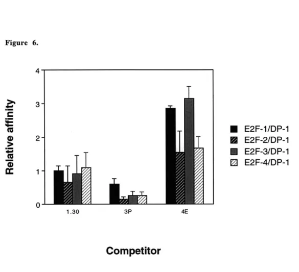
C3. Relative affinities of E2F complexes for selected sites... 85
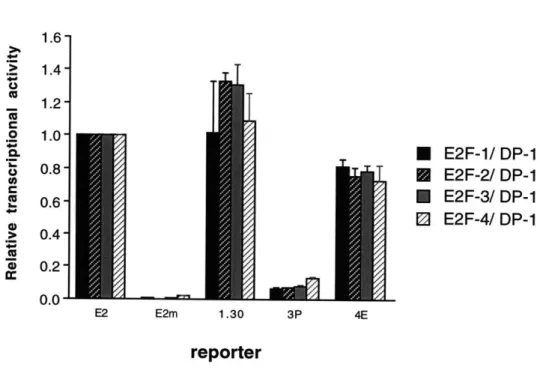
C4. Sequence variations do not confer E2F complex specificity in transactivation...85
D . D iscu ssion ... 90
E. Materials and Methods... 94
F. Acknowledgements ... 97
G. References ... 98
Chapter III: E2F-6, a member of the E2F family that can behave as a transcriptional repressor A . A b stract...10 2 B . Introdu ction ... 10 3 C . R esu lts ... 10 6 C1. Isolation of cDNAs encoding an E2F family member...106
C2. E2F-6 is widely expressed in vivo...107
C3. E2F-6 displays low-affinity E2F DNA-binding activity ... 113
C4. E2F-6 is unable to activate the transcription of E2F-responsive genes...116
D . D iscu ssio n ... 120
E. Materials and Methods ... 123
F. Acknowledgements ... 126
G . R eferences ... 127
Chapter IV: FIP-1, a zinc-finger protein, interacts specifically with E2F-6 A . A b stract...13 0 B . Introduction ... 13 1 C . R e su lts ... 13 5 C1. Isolation of FIP-l...135
C2. Expression pattern of FIP-1 ... 140
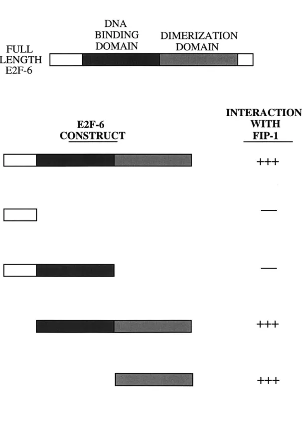
C4. Specific interaction of FIP-1 with E2F-6 ... 146
D . D iscu ssio n ... 14 9 E. Materials and Methods ... 152
F. Acknowledgements ... 154
G . R eferences ... 15 5 Chapter V: Conclusion A. Cloning and characterization of novel E2F genes ... 161
Al. What are the biological functions of E2F-6?...161
A2. Additional E2F genes must exist...164
B. Toward an understanding of the mechanisms of E2F target specificity...166
C. Broadening our understanding of E2F: tumor genetics and genetic screens ... 170
Cl. E2F and cancer genetics...170
C2. Genetic screens could uncover addditional E2F functions or regulators ... 173
C3. Genetics, molecular biology and biochemistry in the study of E2F ... 174
D . R eferen ces...17 6 Acknowledgements ... 178
CHAPTER I
Introduction
A. Overview
The transcription factor E2F confers a GI/S-specific pattern of transcription on a variety of cellular genes. This is accomplished in part through the association of E2F with members of the pRB family of transcriptional repressors during early in the G1 phase of the cell cycle. This association may be critical to the well-documented tumor-suppressive and apoptosis-suppressive activities of pRB. Similarly, the adenoviral protein ElA dissociates E2F from pRB family members, and this function of the ElA protein contributes to its transforming and pro-apoptotic activities. This chapter discusses the biological and biochemical activities of E A, pRB, and E2F, with special attention to the genetic and biochemical heterogeneity of the E2F transcription factor.
B. Adenovirus: a tool to identify mechanisms of cellular proliferation control The regulation of cell division and of programmed cell death is critical to the growth, development and reproduction of multicellular organisms. Deregulated cell growth and division is the hallmark and perhaps the definition of cancer. Cancer cells also demonstrate distinct changes in their susceptibility to programmed cell death. The proper regulation of these processes is therefore a study of enormous medical interest. Some viruses induce cells to enter the DNA synthesis phase (S phase) of the cell cycle, which promotes replication of the viral DNA. In addition to promoting re-entry of the cell cycle and, hence, cellular proliferation, these viruses also manipulate the
regulation of apoptotic control pathways to prevent the premature death of an infected cell. Viruses have therefore been used extensively by cancer biologists as tools to identify key components of the cellular regulatory pathways controlling proliferation and apoptosis. One such virus is the adenovirus.
The adenoviruses are a family of double-stranded DNA viruses that infect humans and other animals. They have been the subjects of intensive study, in part because of their role in human disease: of the more than forty human-specific adenoviruses described so far, some are associated with respiratory infections, others with ocular disease and still others with
gastrointestinal infections (see Lucher, 1995 for a review). Of particular interest to cancer
biologists, however, is the observation that adenoviral infection can induce cellular transformation. They are also known to evade mechanisms inducing programmed cell death (apoptosis) (reviewed in White, 1995). The mechanisms through which adenoviruses override the normal regulation of cellular growth, proliferation, and death are being carefully investigated, primarily as a tool to improve our understanding of those normal regulatory mechanisms and how tumor cells might escape them.
Adenoviruses can infect either dividing or quiescent cells. Upon infection by adenovirus, quiescent cells enter the DNA synthesis phase (S phase) of the cell cycle, with a concomitant induction of the expression of dihydrofolate reductase (DHFR), thymidine kinase, and other genes associated with cellular DNA replication. This induction of reentry of quiescent cells into the cell
cycle is believed to be mediated by the products of the adenoviral gene first expressed upon infection, the ElA gene.
B1. EtA deregulates cellular proliferation
The adenoviral El A gene is expressed as at least five different alternatively spliced messenger RNAs (mRNAs), of which the two most abundant are the two largest, known as 12S
and 13S (reviewed in Zantema and van der Eb, 1995). The 12S and 13S mRNAs of adenovirus type 5 (Ad5), one of the most studied species of adenovirus, encode proteins of 243 and 289 amino acids, respectively. The ElA proteins from the various species of adenovirus are
homologous to each other. This homology is concentrated in three conserved domains, referred to as CR1, CR2, and CR3. The 12S mRNA encodes both CR1 and CR2 as well as intervening and flanking sequences; the 13S mRNA encodes a nearly identical gene product, differing only by the additional presence of CR3.
ElA drives proliferation and apoptosis
Adenoviral strains bearing small deletions in their genome have been generated to study the roles of particular genes in the life cycle of the virus. The E2A and E2B genes, for example, are required for the replication of the viral genome, whereas the EIB gene products are required to
prevent apoptosis of the host cell during infection (reviewed in Philipson, 1995). Viruses that lack the El A region are deficient in multiple biological activities. The El A gene is the first viral gene to be expressed after infection; expression of the 13S mRNA is essential for efficient transcription of the remaining viral genes and, therefore, for the execution of the remainder of the viral life cycle. Internal deletions within the ElA gene have revealed that this function requires CR3, consistent with the ability of the 13S but not the 12S splice forms to mediate this transactivation (reviewed in Jones, 1995). As mentioned above, adenoviruses that lack a functional EIB region induce apoptosis upon infection; this apoptosis is induced by the ElA gene products (White, 1995, and references therein).
The El A gene products are also involved in the stimulation of cell cycle progression of the host cell. Whereas normal viruses will rapidly induce the host cell to enter S phase, viruses that lack the EIA region are unable to do so (Howe et al., 1990). This does not seem to result from the requirement for this region in the transactivation of other viral genes. CR3, which is necessary and sufficient to induce the expression of the viral genes, is not required for S phase induction: the 12S mRNA species, which lacks CR3, is competent to promote the reentry of the cell into the cell cycle (Lillie et al., 1987; Zerler et al., 1987; Bellett et al., 1989; Howe et al., 1990), and expression of EIA by plasmid microinjection is sufficient to induce S phase in the absence of viral infection
(Lillie et al., 1987).
In addition to promoting re-entry of the host cell into the cell cycle, ElA can also promote the transformation of primary cells. Adenoviral infection of cells that do not support the complete viral life cycle can lead to cellular transformation (reviewed in Lucher, 1995); transformation is rarely observed in other cell types, presumably as a result of cellular lysis at the end of the infection cycle. A combination of deletion studies and transfection experiments have determined that the ElA gene is capable of mediating cellular transformation (reviewed in Williams et al., 1995). Expression of EIA in the absence of viral infection causes focus formation in primary baby rat kidney (BRK) cells, although the foci degenerate as a result of ElA-induced apoptosis (White,
1995, and references therein). Coexpression of ElA with another gene that prevents ElA-induced apoptosis, such as E IB, bcl-2, or activated ras, is sufficient to induce complete and stable
The effects of EJA are mediated by specific domains of the protein
Two distinct regions of the El A protein contribute to its ability to promote cell
proliferation. The first is the amino-terminal region of the protein, including CR1 and a non-conserved region at the extreme amino-terminus, and the second is CR2. Mutation of either of these regions impairs the ability of ElA to induce DNA synthesis in most assays, and mutation of both regions completely abrogates S phase induction by ElA (Lillie et al., 1987; Zerler et al.,
1987; Bellett et al., 1989; Howe et al., 1990). The same two regions are required for the transforming activity of ElA (Lillie et al., 1986; Moran et al., 1986; Lillie et al., 1987; Whyte et
al., 1988b). The ability of ElA to induce S phase and its ability to induce cellular transformation are therefore genetically inseparable. Interestingly, the same regions are also required for ElA-induced apoptosis (Samuelson and Lowe, 1997).
The EIA proteins do not seem to possess an intrinsic DNA-binding activity. It has therefore long been assumed that the cellular effects of El A expression in the absence of viral infection are mediated by protein-protein interactions. Consequently, E lA-associated proteins have been intensively studied in an attempt to determine the mechanisms through which ElA promotes cellular transformation and apoptosis. Immunoprecipitation of ElA from an adenovirus-transformed cell line revealed that a number of proteins can be found in a stable complex with EIA; the most abundant of these have molecular weights of approximately 105, 107 and 300 kD (Yee and Branton, 1985; Harlow et al., 1986). These proteins are cellular in origin: they are also present in a variety of cells that have not been exposed to adenoviral infection (Harlow et al.,
1986). The 300 kD protein, known as p300, requires both CR1 and the amino-terminus of ElA for efficient binding to that protein (Whyte et al., 1989; Eckner et al., 1994). Efficient binding of ElA to p107 requires the presence of CR2; maximal binding to p105 also requires part of CR1
(Whyte et al., 1989). Given that these same regions of ElA are known to mediate its effects on cellular proliferation and apoptosis, much study has been focused on p300, p105 and p107 as potential effectors of ElA function.
B2. p300 and CBP are bound, and possibly inactivated, by ElA
The p300 gene was identified by screening an expression library with an antiserum to p300 protein purified through its association through ElA (Eckner et al., 1994). p300 binds to the amino-terminus of E l A; mutations in CR1 or in the extreme amino-terminus of E lA are capable of disrupting the interaction between ElA and p300, which would be consistent with a role for p300 in ElA-induced cell proliferation or apoptosis. The p300 protein is homologous to the CREB-binding protein CBP (Eckner et al., 1994; Lundblad et al., 1995). This homology includes the region used by p300 to bind to ElA, and in vitro studies have found that CBP and p300 bind to ElA with similar affinities (Lundblad et al., 1995). Thus, both p300 and CBP are bound by ElA
and one or both may participate in the deregulation of cellular proliferation by ElA.
Like CBP, p300 also binds to many other transcription factors in the cell, suggesting a role as a transcriptional adaptor. Both have been reported to bind a wide variety of transcripton factors and to have histone acetyltransferase (HAT) activity (Giles et al., 1998), indicating that these proteins probably also have a role in chromatin remodeling at promoters that are being actively transcribed. Studies on ElA-mediated repression of the SV40 enhancer suggest that p300 and/or CBP may have a required role in the activation of some promoters and that ElA may sequester or otherwise inactivate p300 and CBP, preventing this activation. In addition, they are consistent with p300 and/or CBP mediation of at least some of the in vivo effects of ElA.
p300, CBP, and cellular proliferation
The regions of EIA required for binding to p300 and CBP also mediate EIA's promotion of cellular proliferation and apoptosis. In fact, a point mutation abrogating the ability of ElA to bind to p300 in a co-immunoprecipitation assay, but not its ability to bind p105 or p107, also
abrogates the transforming activity of E1A(Whyte et al., 1988b; Whyte et al., 1989).
Furthermore, no El A mutation has been described in which the transforming activity is retained but p300-binding is not, and all tested mutations in ElA that retain p300-binding activity also retain
the ability to promote S phase entry and apoptosis. Nevertheless, no direct evidence currently indicates a role for either p300 or CBP in either the promotion of S phase or of apoptosis.
Studies of the effects of EIA on p300-mediated transcriptional modulation suggest that E l A neutralizes p300 function, at least in some contexts, perhaps through sequestration of the protein (see above). If inactivation of p300 or CBP by ElA does indeed promote cellular transformation, their inactivation might also be expected at some frequency in naturally occuring
human tumors. To date, there has also been one report of somatic mutation in p300 in a colorectal carcinoma and a gastric carcinoma; these are associated with loss of heterozygosity at the locus, suggesting that the mutations may be more likely to be hypomorphic than neomorphic (Muraoka et al., 1996). Patients with Rubinstein-Taybi syndrome, a disease caused by inheritance of an inactivated allele of the CBP gene, have been reported to bear a mildly increased predisposition to certain tumor types, particularly to certain tumors of the nervous system (Miller and Rubinstein,
1995). CBP may, therefore, act as a tumor suppressor in some cellular contexts, although no studies have yet addressed the fate of the second CBP allele in Rubinstein-Taybi tumors.
In addition, both p300 and CBP have been found in translocations with MLL, a homolog of the Drosophila Trithorax gene, in leukemia patients (reviewed in Giles et al., 1998). CBP has also been found in translocations with MOZ, a putative acetyltransferase, in patients with acute myelocytic leukemia (Borrow et al., 1996). The fusion products of these translocations are believed to be expressed, and are believed to be tumorigenic as a result of a gain-of-function, in contrast to the hypomorphic mutations observed in Rubenstein-Taybi and the presumably hypomorphic mutations in the two carcinomas mentioned above, suggesting that the mechanism
for their tumorigenicity may be unrelated to the mechanism(s) in the loss-of-function scenarios. In summary, the sequestration of p300 or CBP is required for some of the transcriptional effects of E lA. Sequestration or other modification of their activity might also play a role in the deregulation of cell proliferation or apoptosis by ElA, although no direct evidence supports this hypothesis. The alterations in p300 and CBP in some human tumors does suggest that these proteins may participate in the regulation of cellular proliferation in some contexts. As
transcriptional adaptors, however, both p300 and CBP have been reported to bind a wide variety of transcription factors. p300 and CBP are therefore potentially implicated in an immense number of signaling pathways. Perhaps only the discovery of a tumorigenic mutation specifically
affecting only a limited subset of these pathways will permit cancer biologists to identify which functions of p300 and CBP are relevant to proliferation control and most urgently deserving of further study.
C. ElA inactivates the tumor and apoptosis suppressor pRB
Like p300, the 105 kD ElA-associated protein, p105, binds to regions of ElA that mediate its effects on cellular proliferation and apoptosis; in the case of p105, optimal binding to ElA requires both CR1 and CR2. A combination of immunological methods and protease digestion experiments revealed that p105 is identical to pRB, the product of the retinoblastoma-susceptibility gene, RB (Whyte et al., 1988a).
C1. RB is frequently inactivated in human tumors
The RB gene was originally identified based on its frequent mutation, often homozygous deletion, in human retinoblastomas (Friend et al., 1986; Friend et al., 1987; Lee et al., 1987). Predisposition to retinoblastomas is inherited as an autosomal dominant trait, and usually results in in bilateral retinoblastomas, whereas unilateral retinoblastomas are more often the result of sporadic mutations (Knudson, 1971). Statistical analyses on patients with unilateral or bilateral
retinoblastomas suggested a requirement for two mutational events with similar mutation rates in the genesis of retinoblastomas (Knudson, 1971). The frequent loss of heterozygosity observed in retinoblastomas on the long arm of chromosome 13 in the vicinity of the retinoblastoma
susceptibility locus is consistent with a tumor-suppressive function for this locus (Cavenee et al., 1983; Dryja et al., 1984). Although gross genomic deletions of RB, the
retinoblastoma-susceptibility gene, are found in only about 30% of retinoblastoma cell lines studied (Friend et al., 1986; Friend et al., 1987; Lee et al., 1987), gross alterations in the transcript are seen in almost all retinoblastoma cells (Friend et al., 1987; Lee et al., 1987) . In one study the protein was
undetectable in all eighteen retinoblastoma cultures tested, including thirteen short-term cultures (Horowitz et al., 1990). This locus is believed to be inactivated in all human retinoblastomas, and this inactivation is rate-limiting for tumor formation.
Humans who inherit a one defective (and one functional) copy of the RB gene develop retinoblastomas with approximately 95% penetrance, and most of these retinoblastomas are bilateral. Patients who survive retinoblastoma often develop other tumors, including sarcomas
such as osteosarcomas. Indeed, mutations of the RB gene have been detected in primary tumors and/or tumor-derived cell lines from osteosarcomas, synovial sarcomas, small cell lung
carcinomas, breast carcinomas, prostate carcinomas, cervical carcinomas and other tumors in addition to retinoblastomas (Harbour et al., 1988; Lee et al., 1988; T'Ang et al., 1988; Yokota et al., 1988; Bookstein et al., 1989; Varley et al., 1989; Horowitz et al., 1990). These mutations are often biallelic deletions, although missense and nonsense mutations have also been described. Thus, the RB gene clearly plays an important role in tumor suppression in a wide variety of human cell types.
A role for RB in the prevention of tumorigenesis is also supported by mouse studies. Mice that inherit an inactivated allele of RB develop pituitary tumors, and less frequently cancer of the thyroid (Jacks et al., 1992; Williams et al., 1994). The cause(s) of the differences in the tissue
specificity of tumorigenesis between mice and humans in the case of RB mutation is (are) not known; nevertheless, the RB gene does play an essential role in the prevention of tumorigenesis in both species.
C2. RB participates in restriction point control
The commitment of a mammalian cell to replicate or to remain quiescent is usually determined during the GI phase of the cell cycle. During early- to mid-GI the growth of mammalian cells in tissue culture is dependent on the continuous presence of growth factors, usually provided by supplementation of the growth medium with serum; removal of the serum during this period of GI induces the cells to enter a quiescent state (reviewed in Pardee, 1989). Later, during late G1, S or G2, progression through the cell cycle becomes serum-independent. The point at which the cell switches from serum-dependence to serum-independence is termed the restriction point (Pardee, 1989). The period of serum-dependence is also characterized by a marked sensitivity to the protein synthesis inhibitor cycloheximide: fibroblasts treated with cycloheximide will arrest in early G 1, but once they have passed the restriction point they become much less sensitive to cycloheximide levels (reviewed in Pardee, 1989).
The RB gene contributes to control over passage through the restriction point. Mouse embryonic fibroblasts (MEFs) in which both alleles of the RB gene have been mutated lose some, but not all, aspects of restriction point control. RB-deficient MEFs are smaller than wild-type
MEFs and spend less time in GI (Herrera et al., 1996; Lukas et al., 1995). RB-deficient MEFs lack the extreme sensitivity of wild-type MEFs to cycloheximide during early- and mid-GI
(Herrera et al., 1996). Serum-dependence is also markedly reduced in RB-deficient MEFs (Lukas et al., 1995; Brugarolas et al., 1998), although not eliminated: complete serum withdrawal before late GI will still arrest RB-deficient MEFs (Herrera et al., 1996). Given that RB-deficient MEFs retain some degree of serum-dependence, loss of RB does not abrogate all forms of restriction point control, but RB is nevertheless required for complete enforcement of the checkpoint.
RB mutation also leads to a relaxation of control over the GUS transition in some tissues of the developing mouse. Specifically, aberrant S phase entry is observed in RB-deficient mice in the lens and both the central and peripheral nervous systems (Morgenbesser et al., 1994; Macleod et
al., 1996). Inactivation of the RB gene is therefore sufficient to permit inappropriate entry into the cell cycle of some cell types that are normally quiescent. As noted above, the regions used by El A to bind the RB-encoded protein, pRB, are involved in the promotion of S phase by ElA. Given
that ElA expression and RB inactivation share this phenotype, binding of ElA to pRB may inactivate a pRB function and thus prevent (full) enforcement of normal controls on the GUS transition.
C3. Cyclin-dependent kinases hyperphosphorylate pRB at the restriction point Concomitant with passage of the cell through the restriction point, the majority of the RB protein, pRB, becomes hyperphosporylated (DeCaprio et al., 1989). This hyperphosphorylation is mediated by the cyclin-dependent kinases (CDKs). CDKs have been demonstrated to play a central role in promoting progression through the cell cycle in a wide variety of organisms, including fusion and fission yeast, fruit flies, frogs, and mammals, among others. CDK activity
requires association of the CDK subunit with a cyclin subunit; modulation of cyclin protein levels during the cell cycle seems to be one mechanism regulating CDK activity during the cell cycle.
Cyclins D and E promote passage through GJ
The first CDK complexes known to be active in the GI phase of the mammalian cell cycle are CDK4 and CDK6 in association with one of the "D-type" cyclins, DI, D2, and D3. Treatment of mammalian cells in tissue culture with mitogens induces expression of D-type cyclins at the RNA and protein levels, but both disappear rapidly upon withdrawal of the mitogens (Matsushime et al., 1991). Overexpression of D-type cyclins leads to a shortening of GI and smaller cell size (Jiang et al., 1993; Quelle et al., 1993; Resnitzky et al., 1994) Rodent fibroblast cells
overexpressing D-type cyclins also show a reduced (but not eliminated) dependency on serum for cell cycle progression (Quelle et al., 1993; Resnitzky et al., 1994). Microinjection of a monoclonal
antibody to cyclin DI during early GI prevents S phase entry, whereas no effect is observed closer to the GI/S transition (Baldin et al., 1993; Quelle et al., 1993). Similar results are observed for antibodies to cyclin D2 (Lukas et al., 1995b) or cyclin D3 (Bartkova et al., 1998) in cells
expressing these cyclins. Thus, the D-type cyclins, presumably in combination with their partners CDK4 and CDK6, play an essential role in promoting passage through GI, and this role is
arguably preceding or concomitant with passage through the restriction point.
Like the D-type cyclins in conjunction with CDK4 and CDK6, cyclin E/ CDK2 plays an essential role in the GI/S transition. Ectopic expression of cyclin E shortens the GI phase of the cell cycle; coexpression of cyclin Dl with cyclin E shortens GI even further (Resnitzky et al.,
1994; Resnitzky and Reed, 1995). Microinjection of an antibody to cyclin E during early GI prevents passage through the GI/S transition, whereas microinjection later has no effect (Ohtsubo et al., 1995). Similarly, overexpression of dominant negative CDK2 arrests cells in GI (Hofmann and Livingston, 1996).
Cyclins D and E participate in the hyperphosphorylation of pRB
Multiple lines of evidence indicate that pRB is phosphorylated by CDK complexes with D-type cyclin subunits. The D-D-type cyclins can bind directly to pRB (Dowdy et al., 1993; Ewen et
al., 1993) and, in combination with CDK4 or CDK6, will phosphorylate pRB in vitro (Ewen et al., 1993; Kato and Sherr, 1993) on sites that are also phosphorylated in vivo (Kato and Sherr, 1993). Cyclin D/CDK4 and cyclin D/CDK6 complexes also phosphorylate pRB more efficiently than they phosphorylate histone HI; they are the only CDKs known to have this subtrate
specificity. Co-expression of cyclin DI with CDK4 induces hyperphosphorylation of pRB in transient transfections (Lundberg and Weinberg, 1998). Furthermore, pRB is maintained in a primarily hypophosphorylated state in cells overexpressing p16, a specific inhibitor of CDK4 and CDK6, indicating that these kinase activities are required for full phosphorylation of the protein (Lundberg and Weinberg, 1998).
pRB is also phosphorylated by cyclin E/ CDK2 complexes. Overexpression of cyclin E is sufficient to induce hyperphosporylation of pRB (Hinds et al., 1992; Lundberg and Weinberg, 1998), and cyclin E/ CDK2 complexes can phosphorylate pRB directly in vitro (Sherr, 1994). Overexpression of a dominant negative form of CDK2 prevents pRB from reaching a fully phosphorylated state (Lundberg and Weinberg, 1998). Cyclin E protein levels and cyclin E-associated kinase activity both peak in late GI (Ohtsubo et al., 1995), at the time when pRB first becomes hyperphosphorylated. The phosphorylation of specific residues of pRB is significantly restricted in irradiated wild-type MEFs, in which the CDK inhibitor p21 is induced and cyclin E-associated kinase activity is inhibited, but not in p21-deficient MEFs, suggesting that specific pRB phosphorylation events require active cyclin E/ CDK2 complexes (Moberg, 1998). The
phosphorylation of pRB on serine 780, in contrast, is mediated specifically by cyclin D (Kitagawa et al., 1996).
Inactivation of pRB, in part by D-type cyclins, is required for passage through GJ
Both D-type cyclins and cyclin E, together with their cognate CDK subunits, can therefore phosphorylate pRB in vitro, are required for full pRB hyperphosphorylation in vivo, and promote passage through the GUS transition. In the case of the D-type cyclins, the inactivation of pRB seems to be the only function required for cell cycle progression. Microinjection of an antibody to cyclin DI can only induce a GI arrest in cells with wild-type pRB: cells without functional pRB are unaffected, although sensitivity can be restored by ectopic expression of wild-type pRB in these cells (Lukas et al., 1994; Lukas et al., 1995a). Similarly, only cells with wild-type pRB can be arrested in GI by ectopic expression of the CDK4/ CDK6 inhibitor p16 (Lukas et al., 1995).
Because overexpression of D-type cyclins and mutations inactivating pRB both promote S-phase progression and because inactivation of pRB relieves the requirement for D-type cyclin-associated kinase activity, it seems clear that 1) pRB is a critical target for this kinase activity in
vivo and 2) that this activity is required for the hyperphosphorylation and inactivation of pRB and
for the subsequent passage through the GUS transition. This must, furthermore, be the only requisite target for this activity in cells grown in tissue culture, given that the activity is dispensable in cells that lack wild-type pRB. Cyclin E, in contrast, must have additional critical targets in vivo: microinjection of an antibody to cyclin E inhibits cell cycle progression even in cells that lack functional pRB (Ohtsubo et al., 1995).
HyperphosphorylatedpRB no longer interacts with a viral oncoprotein
It is interesting to note that ElA is not the only viral oncoprotein that is known to bind pRB. SV40 large T antigen and the E7 protein of human papilloma virus (HPV) are among the best known transforming viral proteins (other than ElA) that are known to interact with pRB through regions required for their transforming functions (DeCaprio et al., 1988; Dyson et al.,
1989; Dyson et al., 1992). At least SV40 large T seems to interact specifically with the
underphosphorylated form of pRB (DeCaprio et al., 1988), and the same is suspected to be true of the other viral oncoproteins (Weinberg, 1995). Hyperphosphorylated pRB has therefore lost at
least two functions: inhibition of S phase progression and binding to viral oncoproteins. This also indicates that any functions of pRB that must be abrogated by the oncoproteins are absent in the hyperphosphorylated protein. Oncoprotein binding and CDK-mediated hyperphosphorylation interfere with the same function of pRB, and this function is required for progression to S phase.
Genes regulating the hyperphosphorylation of pRB are also mutated in human tumors
This requirement for the inactivation of pRB prior to passage through the GI/S transition is likely to be the reason RB is often mutated in human tumors. If this is the case, extragenic
mutations that also have the effect of inactivating pRB should also be observed in some tumors. Indeed, mutations that cause elevated levels of D-type cyclin-associated kinase activity are frequently observed in human tumors. Cyclin DI was originally identified through its
rearrangement in some B-cell lymphomas (Motokura et al., 1991), and cyclin DI levels have also been shown to be elevated in a number of other tumor types, in some cases as a result of
amplification of the locus (Herwig and Strauss, 1997). Cyclin D2 has also been shown to be a proto-oncogene in the mouse (Hanna et al., 1993). Amplification of CDK4 has also been observed in some gliomas and glioblastomas (He et al., 1994; Schmidt et al., 1994). Similarly, mutation of p16 is observed in familial melanoma (Hussussian et al., 1994; Kamb et al., 1994), and deletion of p16, often in combination with its homolog p15, another specific inhibitor of
CDK4 and CDK6, is observed in a wide variety of tumor types (Weinberg, 1995). Although the interpretation of the deletion studies is complicated by the recent identification of another tumor suppressor, p19-ARF, present at the same locus (Quelle et al., 1995; Kamijo et al., 1997), the identification of mutations affecting only p16 prove that p16 is clearly a tumor suppressor in its own right (Quelle et al., 1997). Furthermore, the observation that small cell lung carcinomas almost invariably demonstrate mutations affecting either pRB expression or p16 expression, but not both, argues strongly that pRB and p16 are components of the same oft-targeted pathway in the tumorigenesis of these tissues (Otterson et al., 1994). Thus, the inactivation of pRB by CDK complexes promotes passage through the restriction point, and the mutagenesis of either pRB,
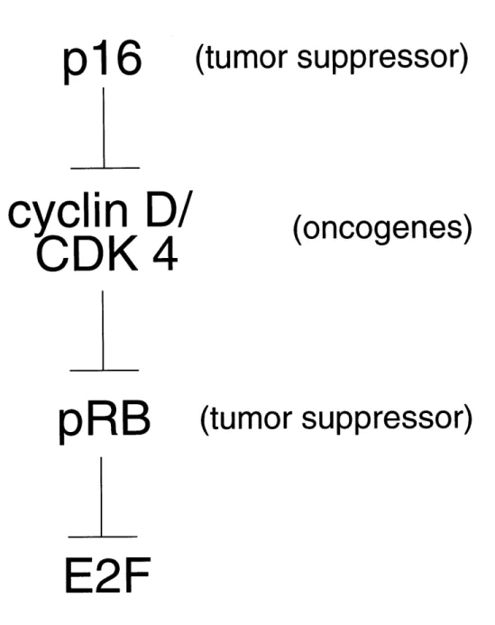
Figure 1.
p16
cyclin D/
CDK
4
pRB
(tumor suppressor)
(oncogenes)
(tumor suppressor)
E2F
Figure 1. Inactivation of pRB is associated with tumorigenesis. Inhibitory interactions are depicted by inverted "T" symbols.
CDK complexes, or CDK inhibitors can lead to a loss of proliferative control and to tumorigenesis (Figure 1).
C4. RB suppresses apoptosis
Some of the tissues in which unscheduled S phase entry is observed in RB-deficient mice are also prone to apoptosis (Morgenbesser et al., 1994; Macleod et al., 1996). The abnormal levels of apoptosis observed in the RB-deficient lens or central nervous system (CNS) are
dependent on the presence of wild-type p53, a tumor suppressor and an inducer of apoptosis: mice that lack both pRB and p53 do not exhibit elevated levels of apoptosis in either the lens or CNS, although aberrant S phase entry is still observed (Morgenbesser et al., 1994; Macleod et al., 1996). Aberrant apoptotic figures are also observed in the peripheral nervous systems (PNS) of deficient mice, but this apoptosis is apparently p53-independent (Macleod et al., 1996). RB-deficient MEFs are also sensitized to apoptosis-inducing drugs such as methotrexate (Almasan et al., 1995).
Overexpression studies also suggest pRB can act to inhibit apoptosis. HeLa cells are a human cervical carcinoma-derived cell line expressing low levels of HPV oncoproteins,
inactivating their endogenous pRB and p53; overexpression of pRB can protect HeLa cells from apoptosis induced by overexpressed p53 (Haupt et al., 1995). Expression of wild-type pRB in SAOS-2 cells, a tumor cell line lacking functional pRB or p53, promotes cell survival following treatment with ionizing radiation (Haas-Kogan et al., 1995). Wild-type pRB can therefore inhibit
either p53-dependent or -independent apoptosis in certain contexts.
The anti-apoptotic effects of pRB are consistent with the observed pro-apoptotic effects of ElA proteins that can bind and inactivate pRB. They are also consistent with the known pro-apoptotic effects of cyclin DI. Overexpression of cyclin Dl can induce apoptosis in multiple cell lines (Kranenburg et al., 1996; Sofer-Levi and Resnitzky, 1996). Elevated levels of cyclin DI have been observed in neuronal cells undergoing apoptosis in reponse to growth factor
Overexpression of the CDK4/6 inhibitor p16 has been shown to inhibit apoptosis during myocyte differentiation (Wang and Walsh, 1996). Thus, either the loss of pRB function through
mutagenesis or its inactivation through hyperphosphorylation can promote apoptosis in a variety of tissues.
C5. RB inactivation is insufficient to promote transformation
pRB is a plausible target for E lA-mediated S phase entry and apoptosis and mutations that inactivate pRB promote tumorigenesis. In addition, inactivation of pRB may eventually prove to be required for cellular transformation in vitro. For example, mutants of SV40 large T antigen that have lost the ability to bind to pRB have also lost the ability to transform wild-type MEFs
(Christensen and Imperiale, 1995; Zalvide and DeCaprio, 1995). There is, however, no evidence that the loss of pRB is sufficient to promote cellular transformation . pRB-deficient MEFs are not transformed; like wild-type MEFs, pRB-deficient MEFs require functional oncogenes to induce transformation. The large T mutants that cannot bind pRB are also unable to transform RB-deficient MEFs (Christensen and Imperiale, 1995; Zalvide and DeCaprio, 1995). Similar results were obtained with studies using the HPV E7 protein (Zalvide and DeCaprio, 1995). Thus, the inactivation of pRB does not, on its own, significantly increase the efficiency of cellular
transformation. Furthermore, the pRB binding domain of the oncoprotein is required even in cells lacking pRB; inactivation of this region must also compromise additional functions required for transformation. In fact, the regions of ElA required for binding to pRB are also involved in binding to the ElA-associated proteins p107 and p130 (Whyte et al., 1989; Dyson et al., 1992), raising the possibility that they, alone or in combination with pRB, may mediate the effects of ElA on cellular transformation.
Indeed, although RB loss promotes tumorigenesis, S phase entry, and apoptosis, mouse embryos without a functional RB gene survive until embryonic day 13.5; death is most likely the result of a hematopoietic defect (Jacks et al., 1992). Although some tissues exhibit abnormally high levels of S phase entry and apoptosis, most embryonic tissues appear entirely normal, having
undergone many normal cell cycles and appropriate differentiation. In chimeric mouse embryos carrying some percentage of RB-deficient cells, those cells can contribute to most, if not all, tissues (Williams et al., 1994). In conclusion, despite the clear importance of pRB in preventing
tumorigenesis and its contribution to the prevention of inappropriate S phase entry and apoptosis, there are clearly additional safeguards in most cells working in concert with the pRB pathway to prevent the deregulation of these processes.
C6. ElA also binds the pRB homologs p107 and p130
The EA-associated proteins p107 and p130 have been cloned and are homologs of pRB (Ewen et al., 1991; Hannon et al., 1993; Li et al., 1993; Mayol et al., 1993). p107 and p130 are more homologous to each other than either is to pRB; nevertheless, they share noticeable
homology to pRB throughout their length, particularly in the carboxy-terminal half of the protein. They also share an insertion between two domains highly conserved among pRB, p107, and p130. These two domains are referred to collectively as the "small pocket"; the insertion is referred to as the "spacer", and contains a motif with homology to the CDK inhibitors p21, p27, and p57.
Indeed, both p107 and p130 are able to stably interact with cyclin A/ CDK2 or cyclin E/ CDK2 and to inhibit their kinase activity (Woo et al., 1997; Castano et al., 1998). Like the overexpression of p21 or p27, which arrests cells in GI (Xiong et al., 1993; Toyoshima and Hunter, 1994),
overexpression of p107 in some cells induces a GI arrest, and this arrest is dependent on the integrity of the cyclin/ CDK binding regions of p107.
Neither p107 nor p130 is frequently mutated in human tumors
In contrast to the frequent mutations of RB in human tumors, the evidence potentially implicating p107 or p130 in tumorigenesis is sparse. Chromosomal alterations in the p107 gene have been described in one cell line derived from a T-cell leukemia, one line derived from a B-cell
lymphoma, and one primary tumor from an acute lymphoblastic leukemia (Takimoto et al., 1998). Full-length p107 protein was no longer detectable in either cell line. One point mutation at the
p130 locus has been described in a cell line derived from a small cell lung cancer (Helin et al., 1997). The possibility remains that the cell line mutations could have arisen during tissue culture; furthermore, the lack of tumor-free controls from the affected patients allow the possibility that the mutations may be unrelated to the genesis of the cancer. It is also possible that the inactivation of p107 or p130 may indeed contribute to tumorigenesis, if infrequently. Mutation of p107 in the mouse has variable consequences depending on the genetic background (Lee et al., 1996; LeCouter et al., 1998a). In no background has the loss of p107 been reported to promote tumorigenesis, although loss of p107 does induce hyperplasia in certain myeloid tissues of one mouse strain (LeCouter et al., 1998a). Chimeric mice with cells deficient in both pRB and p107 develop retinoblastoma at high frequency, suggesting that p107 may share a tumor-suppressive function with pRB, at least in this system (Lee et al., 1996). As seen for p107 mutations, mutations inactivating p130 have dramatically differing consequences in different genetic backgrounds, but these consequences do not seem to include an increased frequency of tumorigenesis (Cobrinik et al., 1996; LeCouter et al., 1998b).
p107 and p130 in apoptosis, transformation, and cell cycle progression
p107 and p130 associate with the same domains of El A required for interaction of the latter with pRB (Dyson et al., 1992), raising the possibility that they might participate in the regulation of apoptosis, transformation, or the GUS transition. Although mice lacking p107 are healthy and fertile, mice lacking both p107 and pRB demonstrate increased levels of apoptosis in the liver and CNS than mice only lacking pRB, indicating that they may share an apoptosis-suppressing
function (Lee et al., 1996). Some mice in which the p130 gene has been mutated show extensive apoptosis in a variety of tissues, but this apoptosis is only observed in certain genetic backgrounds (LeCouter et al., 1998b).
In contrast, no direct evidence implicates either protein in the regulation of cellular
transformation. p107 and p130, like pRB, do associate with other transforming viral oncoproteins such as SV40 large T antigen and HPV E7, and the inability of a T antigen mutant that has lost the
ability to bind pRB, p107 or p130 to transform wild-type or pRB-deficient MEFs may point to a
transformation-suppressive function for p107 and/or p130. Interestingly, an additional domain of T antigen that promotes the degradation of p130 and changes in the phosphorylation state of p107 and p130 is also required to induce MEFs to grow to high density or in low serum; this domain is dispensable in MEFs deficient in both p107 and p130 (Zalvide et al., 1998).
The nature of the role played by these proteins in the regulation of the GUS transition remains somewhat controversial. p130 is expressed primarily in GO cells, although it is still detectable in GI and S phase (Cobrinik et al., 1993). The expression of p107, in contrast, is strongly induced upon reentry of cells into the cell cycle (Moberg et al., 1996). Like pRB, p107 and p130 can be phosphorylated by GI-specific CDK complexes (Li et al., 1993; Beijersbergen et al., 1995). As mentioned above, overexpression of p107 can induce a GI arrest. The effects of p107 or p130 loss on cellular proliferation in mice are strongly dependent on the genetic strain of the mice. In one genetic background, both MEFs and peripheral lymphocytes from these mice retain restriction point controls, although lymphocytes from mice lacking both p107 and p130 are hypersensitive to stimulation by concanavalin A and MEFs deficient in both proteins have been reported to enter S phase slightly earlier than wild-type cells following release from serum
starvation (Cobrinik et al., 1996; Mulligan et al., 1998). In another background, loss of p107 leads to a doubling of the proliferation rate of MEF cultures, and loss of p130 in the mouse leads to extensive aberrant S phase entry in neural tissues. There exist, therefore, biological contexts in
which p107 and p130 can play critical roles in proliferation control, although the genetic modifer or modifiers that must also contribute to proper cellular regulation remain unknown.
C7. pRB suppresses growth through its "large pocket"
pRB, pl07, and p130 bind to viral oncoproteins through their conserved "small pocket" (Hu et al., 1990; Huang et al., 1990; Kaelin et al., 1990; Ewen et al., 1991; Hannon et al., 1993). Mutations in this region of pRB that prevent its interaction with viral oncoproteins also inhibit the ability of ectopically expressed pRB to suppress the growth of RB-deficient tumor-derived cell
lines (Qian et al., 1992; Qin et al., 1992; Hiebert, 1993). Similarly, although overexpression of wild-type pRB can inhibit apoptosis in SAOS-2 cells, expression of a mutant pRB that is unable to bind E l A is also unable to inhibit apoptosis in these cells (Haas-Kogan et al., 1995). This
suggests a model in which ElA interferes with a function of pRB through direct binding to the domain of the protein required for that function.
While the small pocket is necessary for pRB-mediated growth suppression, it is not
sufficient (Qian et al., 1992; Qin et al., 1992; Hiebert, 1993). The minimal region of pRB required to suppress growth includes additional sequences immediately to the carboxy-terminus of the small pocket (Qin et al., 1992; Hiebert, 1993); together with the small pocket, this region is referred to as the "large pocket", and is somewhat conserved with p107 and p130. All known tumor-derived mutations of RB disrupt the large pocket through deletion, truncation or point mutation (Sellers and Kaelin, 1996). This region, then, appears central to the function of pRB both in vitro and in vivo.
Like ElA, pRB is believed to carry out its biological activities through interactions with other proteins. Phosphorylation of the protein by CDK complexes, for example, seems to regulate the inactivation of the protein as the cell prepares to traverse the GI/S transition. While this reveals much about the proper regulation of the protein and the importance of the timing of its biological activity, it does not reveal how pRB exerts its activity on the cell. Although many proteins have been reported to bind to pRB in vitro, one particular cellular factor has been shown to stably interact with pRB both in vivo and in vitro in a manner specifically dependent on an intact, underphosphorylated large pocket: the transcription factor E2F.
D. E2F function is antagonized by pRB, p107, and p130
pRB binds to E2F and inhibits cell proliferation
The E2F transcription factor was originally identified as an El A-responsive cellular
transcription factor with a sequence-specific DNA binding activity (Kovesdi et al., 1986b; Kovesdi et al., 1986a; Kovesdi et al., 1987; Yee et al., 1987). In the context of adenoviral infection, E2F binds to two sites in the promoter of the viral E2 gene; these sites are essential for efficient
expression of the gene (Kovesdi et al., 1986b). Not surprisingly, E2F sites have also been identified in a number of endogenous cellular genes, and have been shown to mediate the upregulation of these genes at the GUS transition. Most of these genes are regulatory genes required in GI or S, such as RB (Shan et al., 1994) or B-myb (Lam and Watson, 1993; Lam et
al., 1994; Lam et al., 1995), or genes required for the machinery of DNA synthesis, such as dihydrofolate reductase (Means et al., 1992; Wade et al., 1992; Slansky et al., 1993; Wade et al., 1995) or DNA polymerase alpha (Pearson et al., 1991).
In cycling cells, a significant percentage of E2F is associated with pRB (Bandara and La Thangue, 1991; Chellappan et al., 1991). This interaction is disrupted by wild-type ElA, but not by mutant forms that have lost the ability to bind pRB. Furthermore, all known tumor-derived mutations of pRB disrupt its ability to bind E2F (Sellers and Kaelin, 1996). The inactivation of pRB function, either in tumor-derived mutations or in association with viral oncogenes, frees E2F from its interactions with pRB.
In normal cells, the association between pRB and E2F is regulated during the cell cycle. Hypophosphorylated pRB, which predominates during cell cycle arrest, can be found in
association with E2F. pRB that has been hyperphosphorylated by cyclin/ CDK complexes following passage through the restriction point (DeCaprio et al., 1989), in contrast, has lost its affinity for E2F (Chellappan et al., 1991; Knudsen and Wang, 1997) Mutations of the
phosphorylation sites in pRB enhance the abilility of the protein to induce a cell cycle arrest when overexpressed, but this arrest can be overcome by the overexpression of an E2F subunit (Qin et
al., 1995). Thus, hypophosphorylated pRB binds E2F and inhibits cell proliferation, whereas the hyperphosphorylation of pRB leads to the dissociation of E2F and passage through the GUS transition.
D1. The E2F and DP gene families
Although E2F was originally described as a DNA-binding activity, it is in fact rather heterogeneous, resulting from the proteins encoded by at least seven distinct genes in mammals (Helin et al., 1992; Kaelin et al., 1992; Shan et al., 1992; Girling et al., 1993; Ivey-Hoyle et al.,
1993; Lees et al., 1993; Beijersbergen et al., 1994; Ginsberg et al., 1994; Hijmans et al., 1995; Ormondroyd et al., 1995; Sardet et al., 1995; Wu et al., 1995; Zhang and Chellappan, 1995; Rogers et al., 1996). These are grouped into two families of genes, referred to as E2F (E2F-1 through -5) and DP (DP-1 and -2). Either of the DP polypeptides are capable of heterodimerizing with any of the E2F polypeptides; each of the possible ten resulting pairs are observed in cell extracts. Heterodimerization is essential for efficient binding to DNA and transcriptional activation by the complex (Bandara et al., 1993; Helin et al., 1993; Krek et al., 1993).
Whereas the sequence conservation between the two DP genes is high throughout the length of their coding sequences, the sequence homology among the E2F genes is concentrated in a few key domains. The domain encoding the DNA-binding function is the most highly conserved, and does not resemble DNA binding domains from other transcription factors. This domain is followed immediately by a domain required for efficient heterodimerization with the DP proteins. The transcriptional activation function of the complex is encoded by a conserved region in the 3' end of the E2F genes. Based on sequence conservation and gene structure, E2F-4 and -5 form a subfamily of E2F proteins distinct from the subfamily including E2F-1, -2, and -3. This latter group contains an amino-terminal region that includes a conserved nuclear localization signal (Verona et al., 1997); this region is absent in E2F-4 and -5.
Domains required for DNA binding and dimerization are also found in the DP proteins, and these regions show limited homology to the corresponding regions of the E2F proteins. No
domain in the DP proteins contributing to transcriptional activation by the complex (other than the dimerization and DNA-binding domains) has been identified; rather, presence of the DP moiety in the E2F/DP complex is essential for DNA binding and the potentiation of the transcriptional activation function of E2F. The DP polypeptide also promotes the interaction of the complex with pRB, although this binding occurs primarily through the transcriptional activation domain of the E2F subunit.
D2. pRB-mediated repression
The association of pRB with E2F is regulated during the cell cycle. This regulation is believed to occur both through the specific phosphorylation of pRB at the GUS transition by CDK
complexes and through transcriptional regulation of the RB gene. pRB/ E2F complexes cannot be detected in quiescent cells, but can be detected in late G 1 as these cells reenter the cell cycle and the transcription of pRB is activated (Ikeda et al., 1996; Moberg et al., 1996). They can also be detected in S phase. This is believed to result from newly synthesized pRB, not yet
hyperphosphorylated by CDK complexes, binding to E2F. Importantly, when cells are induced to arrest and differentiate, pRB is no longer present in a primarily hyperphosphorylated form, and a much greater percentage of E2F complexes associate with pRB (Ikeda et al., 1996).
When bound to E2F, pRB inhibits E2F-mediated transcription. This was first indicated by studies of the E2 promoter: overexpression of pRB inhibits the expression of the promoter in a manner dependent on the presence of the E2F sites, and this effect is only observed with alleles of pRB that are competent to bind E2F (Hiebert et al., 1992). The repression of the E2 promoter can be alleviated by co-expression of EtA or HPV E7 (Hiebert et al., 1992). This derepression can be explained by the in vitro and in vivo observations that EIA induces the dissociation of pRB from E2F complexes (Bagchi et al., 1990). pRB does not prevent DNA binding by the complex; endogenous pRB-E2F complexes are easily detectable in gel shift assays (Chellappan et al., 1991; Moberg et al., 1996). The inhibitory activity of pRB is instead specifically a repression of
pRB represses basal transcription
The presence of pRB, however, does not merely mask the transcriptional activation domain of the E2F moiety, but also represses the basal level of transcription when present at a promoter. This was first suggested by the observation that the overexpression of pRB inhibits the E2 promoter more than the simple deletion of the E2F sites does, and this additional inhibition is dependent on the integrity of the E2F sites (Hiebert et al., 1992). Similarly, it has been shown that the insertion of E2F sites can repress artificial promoters in cells with wild-type pRB, whereas the same insertion activates the expression of those promoters in cells without functional pRB, or in cells expressing wild-type ElA (Weintraub et al., 1992). That this repression is the direct result of the action of pRB on the promoter has been demonstrated with fusion proteins between pRB and the DNA-binding domain of the yeast GAL4 protein: this fusion can repress the expression of
artificial promoters in a manner dependent on the presence of either a GAL4 site (to which GAL4-pRB could directly bind) or an E2F site (to which GAL4-GAL4-pRB could bind through an E2F
intermediary) (Adnane et al., 1995; Bremner et al., 1995; Weintraub et al., 1995). Thus, although "free E2F", E2F/DP complexes not associated with pRB, can activate transcription, pRB-E2F complexes repress basal levels of transcription.
The mechanisms of pRB-mediated repression are under active investigation. Repression requires only the "small pocket" of pRB, and can be alleviated by overexpression of viral
oncoproteins or of cyclins A or E. Repression may be effected by interactions between pRB and other promoter elements (Dyson, 1998) and/or by recruitment of histone deacetylase activity to the promoter by pRB (Brehm et al., 1998; Luo et al., 1998; Magnaghi-Jaulin et al., 1998).
D3. p107 and p130 also bind E2F and can repress transcription
pRB, p107, and p130 bind E2F at distinct times during the cell cycle
Although "free E2F" and pRB-associated E2F account for a significant percentage of the E2F in the cell, other E2F complexes exist as well. Like the pRB association, these complexes can be disrupted by ElA (Bagchi et al., 1990). The cloning of the ElA-associated proteins p107 and p130 and their significant homology to pRB led rapidly to their identification as components of these "other" E2F complexes (Ewen et al., 1991; Cao et al., 1992; Shirodkar et al., 1992; Cobrinik et al., 1993).
E2F complexes containing p107 and those containing p130 show distinctly different patterns of appearance and disappearance during the cell cycle. p130/ E2F complexes are present at their highest levels in quiescent cells, and become increasingly less abundant as cells reenter the cell cycle, although they can still be detected as cells enter S phase (Cobrinik et al., 1993; Ikeda et al., 1996; Moberg et al., 1996). p107/ E2F complexes, in contrast, are not detectable in quiescent cells, first appearing in late GI and reaching their highest levels in S phase (Moberg et al., 1996). Binding of p107 or p130 to CDK complexes and E2F is not mutually exclusive; the five
polypeptide complex p107 or p130/ cyclin A or E/ CDK 2/ E2F/ DP complex can be observed bound to DNA in a gel shift assay, although the potential biological function of a presumably inactive kinase in this complex remains unknown. Like pRB, p107 and p130 can repress E2F activity (Bandara et al., 1993; Zamanian and La Thangue, 1993; Bremner et al., 1995; Ferreira et al., 1998; Pierce et al., 1998b) and may be associated with a histone deacetylase (Ferreira et al.,
1998). p13 0 is therefore proposed to mediate the repression of E2F target genes in quiescent cells; the role p107/ E2F complexes might be playing, at times in the cell cycle when known E2F target
pRB, p107, and p130 bind to distinct forms of E2F
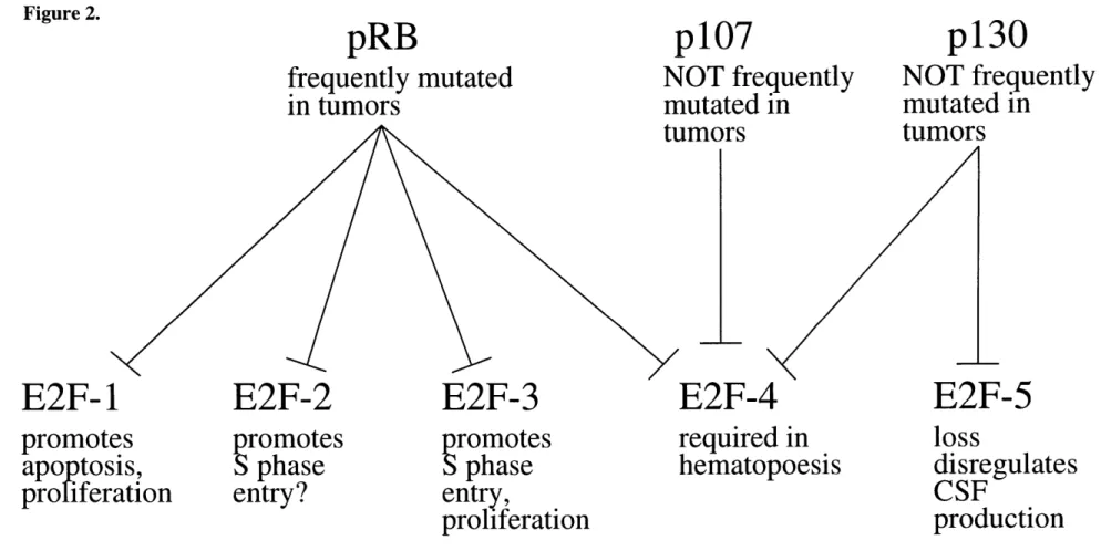
It has been proposed that the failure to detect specific roles for p107 or p130 in apoptosis or growth control in some contexts could result from extensive redundancy of function among pRB, p107, and p130. In this model, the three proteins would share a common suppressive function, such as transcriptional repression at E2F DNA-binding sites, and only the inactivation of all three genes could reveal their importance to the biology of the cell. Alternatively, the differences in the biology of pRB, p107, and p130 could result from their associations with biochemically distinct forms of E2F. Indeed, pRB can be found in association with E2F-1, -2, -3, or -4 (Moberg et al.,
1996), whereas p107 is associated only with E2F-4 (Moberg et al., 1996) and p130 with both E2F-4 and E2F-5 (Hijmans et al., 1995; Sardet et al., 1995); although the DP subunit is always present in these complexes, the identity of the subunit, DP- 1 or DP-2, does not seem to affect to the specificity of the interaction.
A different subset of E2F transcriptional targets is deregulated in MEFs lacking p107 and p130 than the subset deregulated in pRB-deficient MEFs. Cyclin E expression, for example, is upregulated in pRB-deficient MEFs, but not in p107/ p130-deficient cells (Herrera et al., 1996; Hurford et al., 1997). B-myb, in contrast, is derepressed in p107/p130-deficient MEFs, but
shows a wild-type pattern of expression in pRB-deficient cells (Hurford et al., 1997). These proteins therefore regulate different biochemical pathways in these cells. Given that pRB, p107
and p130 associate with different E2F proteins, this specific deregulation could result from differential promoter selection mediated either by the E2F subunit or by interactions between the pRB homolog and other promoter elements .