AVIS
Ce document a été numérisé par la Division de la gestion des documents et des archives de l’Université de Montréal.
L’auteur a autorisé l’Université de Montréal à reproduire et diffuser, en totalité ou en partie, par quelque moyen que ce soit et sur quelque support que ce soit, et exclusivement à des fins non lucratives d’enseignement et de recherche, des copies de ce mémoire ou de cette thèse.
L’auteur et les coauteurs le cas échéant conservent la propriété du droit d’auteur et des droits moraux qui protègent ce document. Ni la thèse ou le mémoire, ni des extraits substantiels de ce document, ne doivent être imprimés ou autrement reproduits sans l’autorisation de l’auteur.
Afin de se conformer à la Loi canadienne sur la protection des renseignements personnels, quelques formulaires secondaires, coordonnées ou signatures intégrées au texte ont pu être enlevés de ce document. Bien que cela ait pu affecter la pagination, il n’y a aucun contenu manquant.
NOTICE
This document was digitized by the Records Management & Archives Division of Université de Montréal.
The author of this thesis or dissertation has granted a nonexclusive license allowing Université de Montréal to reproduce and publish the document, in part or in whole, and in any format, solely for noncommercial educational and research purposes.
The author and co-authors if applicable retain copyright ownership and moral rights in this document. Neither the whole thesis or dissertation, nor substantial extracts from it, may be printed or otherwise reproduced without the author’s permission.
In compliance with the Canadian Privacy Act some supporting forms, contact information or signatures may have been removed from the document. While this may affect the document page count, it does not represent any loss of content from the document.
Identification and characterization of a new adhesin
involved in the binding of Streptococcus suis to the extracellular
matrix proteins
par
Miriam Esgleas Izquierdo
Département de pathologie et microbiologie
Faculté de médecine vétérinaire
Thèse présentée à
la
Faculté de médecine vétérinaire en vue de l'obtention du grade dePhilosophire Doctor (Ph.D.) en sciences vétérinaires
option microbiologie
Décembre, 2008
Faculté des études supérieures et postdoctorales
Cette thèse intitulée
IDENTIFICATION AND CHARACTERIZATION OF A NEW
ADHESIN INVOL VED IN THE BINDING OF
STREPTOCOCCUS SUIS
TO THE EXTRACELLULAR MATRIX PROTEINS
présentée par
MIRIAM ESGLEAS IZQUIERDO
a été évaluée par un jury composé des personnes suivantes:
Michaël Mourez, président-rapporteur
Marcelo Gottschalk, directeur de recherche
J. Daniel Dubreuil, codirecteur
Mariela A. Segura, membre du jury
Daniel Grenier, examinateur externe
John'M. Fairbrother, représentant du doyen de la FÉSP
RÉSUMÉ
Streptococcus suis est un important pathogène porcin bactérien causant principalement des septicémies, des méningites, des endocardites et l'arthrite. Des 35 sérotypes connus, le sérotype 2 est le plus fréquemment isolé et associé à la maladie. S. suis est aussi reconnue comme étant un agent de zoonose. Les humains peuvent être infectés via par des petites coupures ou abrasions aux mains par ce pathogène lors de manipulations avec de la viande ou des carcasses de porc infectées. L'infection humaine peut être sévère et causer des méningites, des septicémies, des endocartides, la surdité (consequence de la méningite) et éventuellement la mort.
La pathogénèse de l'infection causée par S. suis n'est pas encore bi~n définie et plusieurs étapes sont probablement impliquées. Les mécanismes qui permettent à S. suis de se disséminer à partir des amygdales (le réservoir chez l'animal) ne sont pas bien compris. Il est admis que l'aptitude d'une bactérie à adhérer aux tissus de l'hôte est une étape critique de la plupart des infections microbiennes. Cette adhésion est considerée comme la première étape à une colonisation des muqueuses et peut aussi être la première étape à l'invasion des cellules de l'hôte, processus qui peut mener aux bactérémies ou aux septicémies. Un des types d'adhésines bactériennes les plus connues et les plus étudiés sont les protéines liant la fibronectine. De plus, la plupart des protéines liant la fibronectine décrites pour différents pathogènes ont été suggérées comme cibles potentielles de vaccins afin de prévenir l'infection bactérienne. En effet, il a été décrit que les anticorps liant ces protéines de surface ont la double activité de bloquer l'adhérence et d'augmenter la opsono-phagocytose.
À ce jour, trois types de protéines liant la fibronectine ont été identifiés chez les streptocoques: i) la famille MSCRAMM, renfermant un motif LPXTG médiant la liaison de cette proteine à la surface des bactéries par des sortases, ii) les protéines liant la choline et qui n'ont pas de motif LPXTG mais qui possèdent des répétitions liant la choline qui interagissent avec la phosphorylcholine des acides lipotéichoiques des bactéries à Gram positif et finalement, iii) un nouveau groupe de protéines nommé « anchorless » qui ne possèdent pas de motif LPXTG ou des répétitions liant la choline. Les mécanismes utilisés par ce dernier type de protéine pour être exporté à la surface de la bactérie sont encore inconnus.
Comme S. suis est un pathogène extracellulaire qui adhère probablement aux composants de la matrice extracellulaire (ECM) lors des différents étapes de la pathogénèse, les principaux objectifs de ce travail étaient: i) d'étudier l'adhésion de S. suis sérotype 2 aux plus importants composants de la ECM, ii) d'identifier et de caractériser les adhésines impliquées dans l'adhésion de S. suis sérotype 2 aux protéines de l'ECM, plus particulièrement à la fibronectine, iii) de déterminer le rôle de ces adhésines dans la pathogénèse de l'infection causée par S. suis sérotype 2 et finalement, iv) d'étudier l'activité protectrice de ces adhésines.
Nous démontrons pour la première fois que S. suis sérotype 2 peut lier spécifiquement les composantes majeures de l'ECM, comme la fibronectine et différents types de collagène. Alors que S. suis ne peut lier directement le collagène de type IV des membranes basales, il a le potentiel d'adhérer à cette protéine par un nouveau mécanisme en utilisant lafibronectine liée à sa surface comme pont. Cette propriété pourrait s'avérer être un nouveau mécanisme de colonisation et d'évasion immune de S. suis. La forte inhibition de l'adhésion de S. suis à la fibronectine et au collagène lorsque traité aux protéases et à la chaleur indique que ces mécanismes de liaison sont médiées surtout par des protéines. De plus, ces travaux démontrent que l'adhésion de S. suis à la fibronectine et au collagène est principalement de type sortase-indépendant, indiquant que la plupart des adhésines impliquées dans ces adhésions, contrairement à ce qui a été préalablement décrit pour d'autres streptocoques, ne sont pas des adhésines de la famille MSCRAMM.
Lors de nos efforts pour mieux comprendre la pathogénèse de l'infection causée par S. suis, nous avons identifié et caractérisé une nouvelle protéine liant la fibronectine. Cette protéine a été identifiée comme étant une a-énolase (SsEno). La séquence en acides aminés de cette protéine démontre une absence de motif LPXTG ou de répétition liant la choline, classant cette protéine dans le nouveau groupe désigné protéines « anchorless » et attestant que l'activité des protéines liant la fibronectine de S. suis n'est pas reliée aux MSCRANIMs. À ce jour, la principale fonction décrite pour les énolases bactériennes de surface est une activité de liaison au plasminogène. Par contre, nous avons décrit pour la première fois que l'énolase de surface de S. suis n'a pas seulement une grande affinité pour le plasminogène mais aussi une affinité semblable pour la fibronectine. De plus, nous avons montré que cette protéine multifonctionnelle est aussi une protéine de choc thermique qui a aussi une activité de liaison aux IgG. Fonctionellement, nous avons prouvé que cette protéine est une nouvelle adhésine de S. suis qui participe à la capacité d'adhésion et d'invasion de cette bactérie via-à-vis les cellules endothéliales, indiquant que SsEno pourrait être une molécule clé dans la pathogénèse de l'infection causée par ce pathogène. Ces caractéristiques, incluant la grande immunogénicité de cette protéine chez le porc, fait de SsEno un candidat intéressant pour un vaccin contre les infections à S. suis. Par contre, les résultats obtenus dans ces travaux démontrent que, malgré que cette protéine induit une bonne réponse immunitaire, SsEno ne confère pas une protection contre les l'infection à S. suis.
En resumé, ces travaux ont permis d'identifier une nouvelle adhésine importante de S. suis qui pourrait participer dans différentes étapes de la pathogenèse de cette bactérie comme l'évasion par la bactérie du système immunitaire de l'hôte et l'entrée de la bactérie dans le système nerveux central par les cellules endothéliales formant la barrière hémato-encéphalique.
Mots clés: S. suis, MEC, fibronectine, enolase, IgG, HSP, adhésion, invasion, cellules endothéliales, vaccin
\
SUMMARY
Streptococcus suis is an important bacterial pathogen of pigs that mainly causes septicemia, meningitis, endocarditis, and arthritis. Of the 35 known serotypes, serotype 2 is the most frequently
isolated and associated with disease. S. suis is also a zoonotic agent. Humans can be infected with the pathogen while handling infected pig carcasses and meàt through exposed cuts and abrasions on
their hands. Human infections may be severe, with meningitis, septicemia, endocarditis, and deafness as possible outcomes of infection. Death can also occur.
The pathogenesis ofthe infection caused by S. suis is not clear and many steps are probably involved. The mechanisms that enable S. suis to disseminate from the tonsils (animal reservoir) throughout the animal are not weil understood. It is commonly believed that the ability of bacteria to adhere to host tissues is a critical step in the onset of most microbial infections. This adhesion is the first step for the colonization of mucosal surfaces and may also be the first step before the invasion of host cells, a process that may lead to bacteremia and sepsis. One of the most known and studied bacterial adhesins are the binding proteins. In addition, most of the fibronectin-binding proteins described up to now for different pathogens have been suggested as potential vaccine targets for preventing bacterial infections since antibodies against these surface proteins are believed to have the dual activity of both adherence blocking andopsonic function.
Up to now, three types of streptococcal fibronectin-binding proteins have been identified: i) the MSCRAMM family, containing an LPXTG motif that mediates bacterial surface binding by sortases, ii) the choline-binding proteins that lack the LPXTG motif but contained choline-binding repeats which interact with the phosphorylcholine ofthe lipoteichoic acid of Gram positive bacteria, and finally iii) the new denominated "anchorless" proteins which do not possess LPXTG motif or choline-binding repeats. The mechanism used by this type of proteins to be exposed to the surface of bacteria is still unknown.
As S. suis is primarily an extracellular pathogen and it presumably adheres to components of the extracellular matrix (ECM) to initiate an infection, the main objectives of this work were: i) to study the adhesion of S. suis serotype 2 to sorne of the most important ECM proteins, ii) to identify and characterize the adhesins involved in the adhesion of S. suis serotype 2 to the ECM proteins, specially to fibronectin, iii) to determine the role of these adhesins in the pathogenesis of the infection caused by S. suis serotype 2 andfinally, iv) to study the protective activity of these adhesins.
We have demonstrated for the first time that S. suis is able to specifically bind to major constituents of the ECM such as fibronectin and different types of collagen. Interestingly, while S. suis is not able to bind directly to basement membrane collagen type IV, it has the potential to
adhere to this protein via a surface-bound fibronectin mechanism. This property might represent a novel mechanism of colonization and immune evasion. The dramatic reduction in binding observed after protease and heat-treatment of S. suis to both, fibronectin and collagen, indicates a prote in-mediated binding mechanism for these adhesions. In addition, this work also demonstrates that the adhesion of S. suis to fibronectin and collagen is mostly sortase-independent, which indicates that most of the adhesins involved in these S. suis adhesions, in contrast with what it has been described early for other streptococci, are not adhesins from the MSCRAMM family.
In our continued effort to understand the pathogenesis of the infection caused by S. suis, we have identified and characterized a predominant S. suis fibronectin-binding prote in identified as an a-enolase (SsEno). Its amino acid sequence shows the absence of LPXTG motif or choline-binding repeats, including this prote in in the new denominated "anchorless proteins" and corroborating that the fibronectin-binding activity of S. suis are not related with MSCRAMMs. Up to now, the major function described for surface bacterial enolases is a strong plasminogen-binding activity. However, we have demonstrated for the first time that S. suis surface enolases have not only a high affinity for plasminogen but also a similar high affinity for fibronectin. In addition, the multifunctional SsEno is a heat-shock prote in that also has IgG-binding activity. We have also shown that this protein is a new S. suis adhesin that participates in the adhesion to and invasion ofthis bacterium to endothelial cells, indicating that SsEno cou Id be a key molecule in the pathogenesis of the infection caused by this pathogen. These features, including the high immunogenity of this protein in pigs, made of SsEno an interesting candidate for a vaccine against S. suis infections. However, results obtained in this work demonstrated that, although this prote in elicits a good immune response, SsEno did not confer protection against experimental S. suis infection.
In conclusion, this work has permitted to identify a new and important S. suis adhesin which can participate in the different steps of the pathogenesis of the infection caused by this bacteria including its evasion of the host immune system and its entry to the CNS through the microvascular endothelial ce Ils forming the blood brain barrier.
Key words: S. suis, ECM, fibronectin, enolase, IgG, HSP, adhesion, invasion, endothelial cells, vaccine
TABLE OF CONTENTS
JlTRY IDENTIFICATION ... ii
SOMMAIRE ... iii
SUMMARY ... v
TABLE OF CONTENTS ... vii
LIST OF TABLES ... xi
LIST OF FIGURES ... xii
LIST OF ABBREVIA TIONS ... xvi
DEDICA TION ... xxi
ACKNOWLEDGEMENTS ... xxii
1. INTRODUCTION ... 1
II. LITERA TURE REVIEW ... 4
1- Streptococcus suis ... 5 1.1. Introduction ... ; ... 5 1.2. History ... ~ ... 5 1.3. Distribution ... 6 1.4. S. suis infections ... 7 1.4.1. In swine ... 7 1.4.1.1. Clinical signs ... 7 1.4.1.2. Transmission ... 8 1.4.1.3. Diagnosis ... 8 1.4.1.4. Treatment ... ' ... 9 1.4.1.5. Prognosis ... 9 1.4.2. In humans ... : ... 9 1.4.2.1. Clinical signs ... 10 1.4.2.2. Transmission ... 10 1.4.2.3. Diagnosis ... Il 1.4.2.4. Treatment ... Il 1.4.2.5. Prognosis ... ; ... Il 1.4.3. In other species ... 12
1.5. Identification and detection ... 12
1.6. Virulence factors ... 13
1.6.1. The polysaccharide capsule (CPS) ... 14
1.6.2. The cell wall ... 15
1.6.4. Hemolysin (Suilysin) ... 16
1.6.5. Hemagglutinins ... 17
1.6.6. Albumin-binding potein or GAPDH ... 18 ..
1.6.7. IgG binding protein ... 19
1.6.8. FBPS ... 19
1.6.9. Serum opacity factor. ... 20
1.6.10. Sao ... 21 1.6.11. SalKiSalR ... 21 1.6.12. Enzymes ... 22 1.6.12.1. Hya1uronate lyase ... 22 1.6.12.2. Arginine deiminase ... 22 1.6.12.3. Superoxide dismutase ... 23 1.6.12.4. Proteases ... 24
1.7. The pathogenesis of S. suis infection ... 25
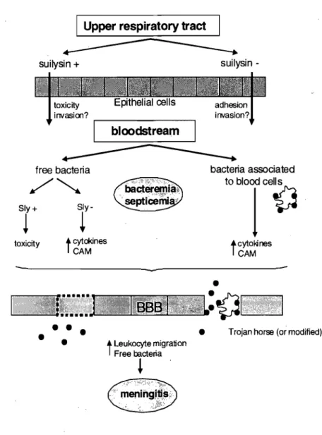
1.7.1. Colonisation and invasion of the respiratory epithelium ... 25
1.7.2. Blood dissemination and phagocytosis resistance ... 27
1.7.3. Septicemia ... 28
1.7.4. Meningitis ... 30
1.8. Vaccines against S. suis ... ; ... 31
2- Bacterial adhesion to Extracellular Matrix proteins ... 34
2.1. Extracellular matrix ... 34
2.1 .1. General compos ition ... 34
2.1.1.1. Structural ECM components ... 34
2.1.1.1.1. Collagens ... 34 2.1.1.1.2. Laminin ... 36 2.1.1..1.3. Elastin ... 37 2.1.1.2. Adhesive glycoproteins ... 38 2.1.1.2.1. Fibronectin ... 38 2.1.1.2.2. Vitronectin ... 40
2.1.1.2.3. Fibrinogen and fibrin ... 41
2.1.1.2.4. Thrombospondin ... 43
2.2. Bacterial adhesion to extracellular matrix proteins (ECM) ... .45
2.3. Bacterial fibronectin-bind ing proteins ... 46
2.3.1. MSCRAMMs ... 47
2.3.2. Choline-binding proteins ... 48
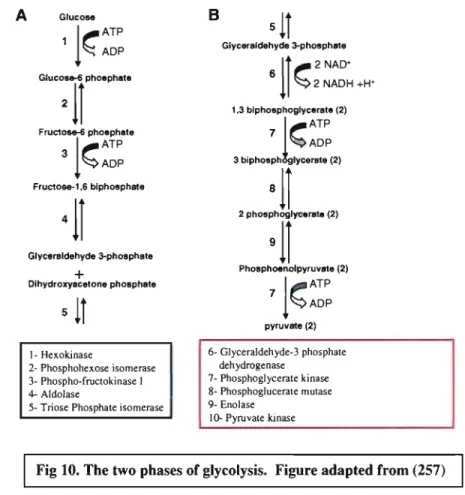
2.4. S. suis adhesion to ECM proteins ... .49 3- Enolase ... 51 3.1. Glycolysis ... 51 3.1.1. Introduction ... 51 3.1.2. History ... 51 3.1.3. Phases ... 52 3.2. Enolase ... 53 3.2.1. Introduction ... 53 3.2.2. Distribution ... : ... 53
3.2.3. Primary and secondary structure of a-eno1ase ... 54
3.2.4. Enolase activity ... 54
3.2.5. Mechanism of enolase activity ... 54
3.2.6. Enolase superfamily ... 55
3.2.7. Location diversity ... 56
3.2.7.1. Enolase as a cytosolic enzyme ... 56
3.2.7.2. Enolase as a surface protein 3.2.7.3. Enolase as a nuc\ear prote in ... 56
... 57
3.2.8. Multifunctional nature of enolase ... ;; ... 57
3.2.8.1. Enolase as a plasminogen-binding protein ... 58
3.2.8.2. Enolase as a laminin-binding protein ... 58
3.2.8.3. Enolase as an eye T-crystallin protein ... 58
3.2.8.4. Enolase as a Myc-binding protein (MBP-1) ... 59
3.2.8.5. Enolase as an endothelial hypoxic stress prote in ... 59
3.2.8.6. Enolase as a heat shock protein ... 59
3.2.8.7. Enolase as microtubule organizer ... 60
3.2.9. Enolase and disease ... 60
3.2.9.1. Anti-a-enolase antibodies in infectious diseases ... 60
3.2.9.2. Anti-a-enolase antibodies in systemic auto immune Disorders ... 61
3.2.9.3. Pathogenic role of anti~a-enolase antibodies ... 62
4-References ... 64
III. MA TERIAL, METHODS AND RESUL TS ... 93
Article I. Streptococcus suis serotype 2 binding to extracellular matrix proteins. 2005. FEMS Microbiology Letters. 244: 33-40 ... 94
Article II. Isolation and characterization of a-enolase, a new fibronectin binding protein from Streptococcus suis. 2008; Microbiology. 154:
2668-2679 ... III
Article III. Immunisation with SsEno fails to protect mice against challenge with
Streptococcus suis serotype 2. 2008. (FEMS Microbiology Letters
accepted 10 feb 2009) ... 137
IV. DISCUSSION ... 153
V. GENERAL CONCLUSIONS ... 179
VI. ANNEX ... xxiii
Annex 1. Purification of pig plasma fibronectin ... xxiv
Annex 2. SsEno is present in ail the 35 described serotypes for S. suis ... XXIX Annex 3. Characterisation of SsEno as a heat shock protein (HSP) ... xxxiii
Annex 4. Characterisation of SsEno as an IgG-binding prote in ... xxxvi Annex 5. Characterisation of SsEno as a colIagen-binding protein ... xl
Annex 6. Identification of genes associated with the colIagen-binding capacity of
, .
Streptococcus suis using random insertional mutagenesis ... xliii
Annex 7: Article IV. Immunization with recombinant Sao protein confers protection against Streptococcus suis infection. 2007. Clinical Vaccine Immunology.
14:937-47 ... xlviii
Annex 8: Article V. Disruption of srtA gene in Streptococcus suis results in decreased interaction with endothelial cells and extracellular matrix
LIST OF TABLES
MA TERIAL METHODS AND RESUL TS
Article 1
Table 1. Characteristics of the Streptococcus suis strains used in this study ... 99 Table 2. Adherence to ECM proteins of different S. suis serotype 2 field
strains ... 103
Annex6
Table 1. List of genes identified by plasmid rescue and Blast ... xlvii
Annexe 7: Article IV
Table 1. Bacteriological analysis of postmortem samples from pigs
LIST OF FIGURES
LITERATURE REVIEW
1- Streptococcus suis
Fig. 1. Summary of the knowledge and proposed hypotheses for the different
steps involved in the pathogenesis of meningitis of S. suis serotype 2 ... 29
2- Bacterial adhesion to ExtraceUular Matrix Proteins Fig. 2. Diagram of the laminin fork structure ... 36
Fig. 3. Diagram of the fibronectin structure ... 39
Fig. 4. Diagram of the vitronectin structure ... 40
Fig. 5. Diagram ofthe fibrinogenlfibrin structure ... 42
Fig. 6. Coagulation cascade of proteins ... 43
Fig. 7. Diagram of the thrombospondin structure ... 44
Fig. 8. Schematic model indicating how binding of multiple fibronectin molecules to FnBPs attached to the bacterial cell surface might result in integrin clustering on the host cell surface and subsequent uptake of bacteria ... 47
Fig. 9. Diagram of the MSCRAMMs structure ... 47
3- Enolase Fig. 10. The two phases of glycolysis ... 52
Fig. Il. Reactions of the enolase superfamily ... 56
MATERIAL METHODS AND RESULTS Article 1 Fig. 1. Effect of ECM protein concentrations and S. suis concentration on S. suis serotype 2 strain S735 binding to ECM proteins ... 100
Fig. 2. Time course of S. suis serotype 2 strain S735-binding to different ECM proteins ... 101
Fig. 3. Effect of incubation temperature on S. suis serotype 2 strain S735 -binding to different ECM proteins ... 102
Fig. 4. Effect of pre-incubating bacteria for 90 min with 150 Jlglml of soluble plasma fibronectin onS. suis serotype 2 strain S735-binding to plasma fibronectin, cellular fibronectin, collagen type l, collagen type III, collagen type V, and collagen type IV ... 104
Article II
Fig. 1. Isolation of a 52-kDa prote in by fibronectin-affinity chromatography ... 120
Fig. 2.' Expression and purification of recombinant SsEno ... 121
Fig. 3. Enolase activity assay of recombinant SsEno ... : ... 122
Fig. 4. Qualitative binding ofSsEno to fibronectin and plasminogen ... 123
Fig. 5. Lysine-dependent binding ofSsEno to fibronectin and plasminogen ... 123
Fig. 6. Quantitative binding ofSsEno to fibronectin and plasminogen ... 124
Fig. 7. Surface localization ofSsEno on S. suis ... 125
Fig. 8. Decrease of the adhesion and invasion of S. suis strain SS166 to PBMEC by antibodies against SsEno ... 125
Article III Fig. 1. ELISA detection of antibodies against SsEno in sera from pigs before and after challenge with S. suis serotype 2 ... 143
Fig. 2. Serum antibody responses in mice immunized with Qui! A or Qui! A plus recombinant SsEno ... .144
Fig. 3. Survival (a) and weight lost (b) of mice in the active protection assay ... 145
Fig. 4. Survival of mice in the passive protection assay ... 145
Fig. 5. Effect of antibodies on opsonophagocytic killing of S. suis strain 166' by mouse phagocytes ... 146
DISCUSSION Fig. 1. Important unresolved questions about the pathogenesis of the infection caused by S. suis ... 157
Fig. 2. Transwell system ... 167
Fig. 3. Role ofSsEno in the pathogenesis of the infection caused by S. suis ... 170
APPENDIX Annex 1 Fig. 1. Porcine plasma fibronectin purification ... xxvi
Fig. 2. Proteins recovered after the first step of the purification of porcine plasma fibronectin ... ~ ... xxvi
Fig. 3. HPLC porcine plasma fibronectin purification ... xxvii
Annex 2
Fig. 1. SsEno is present at the surface of ail the described S. suis serotypes ... xxxi
Annex3
Fig. 1. SsEno is a heat shock protein ... xxxv
Annex 4
Fig. 1. Representative ELISA analysis for the binding to 5 ~g/ml SsEno
binding to immobilized human IgG and IgY ... xviii
Annex 5
Fig. 1. Representative ELISA analysis for the binding to 5 ~g/ml SsEno
binding to immobilized collagen ... xlii
Annex 6
Fig. 1. Adhesion of Tn91 7 mutants to collagen ... xlv Fig. 2. Diagram of Plasmid rescue technique ... ~ ... xlvi
Annex 7. Article IV
Fig. 1. Serum antibody responses in mice immunized with Quil A or Quil A plus
recombinant Sao ... Ivi Fig. 2. Serum antibody responses in pigs immunized with Quil A or Quil A
plus recombinant Sao ... Ivii Fig. 3. Survival ofmice immunized with Quil A (open circles) or Quil A plus
recombinant Sao (solid circles) following challenge with S. suis
31533 ... ; ... Ivii Fig. 4. Protection ofpigs immunized with Quil A (open circles) or Quil A plus
recombinant Sao (sol id circles) following challenge with S. , suis 166 ... Iviii Fig. 5. Effect of antibodies on opsonophagocytic killing of S. suis by porcine
neutrophils ... Iix Fig. 6. Western blot showing the variation in Sao protein of S. suis ... lx Fig. 7. PCR amplification products of the full-Iength of saD gene of S. suis and the
Annex 8. Article V
Fig. 1. Adhesion and invasion to PBMEC by mutant SRT [}.A compared to the
wild-type strain NCTC 1 0234 ... Ixxv Fig. 2. Binding to different concentrations of plasma fibronectin and collagen type
1 by mutant SRT [}.A compared to the wild-type strain NCTC 10234 ... lxxvi Fig. 3. Kaplan-Meier survival analysis ofCDI mice infected with the
A: aa: Abs: AD: ADP: ADS: Ala: ANCA: Arg: Asn: Asp: ATP: BBB: BC: BMEC: bp: BSA: C.F.U.:
C:
CAM: CAR: CCF: Cd2+: CK: CNS: Co2"'": CPS: DPP IV: EACA: Eap: ECM: EDTA: LIST OF ABBREVIATIONS centigrade degrees adenine amino acid antibodies arginine deiminase adenosine 5'-biphosphate arginine deiminase system alanineanti-neutrophils cytoplasm antibodies arginine
asparagine aspartic acid
adenosine S'-triphosphate blood brain barrier before Christ
brain microvascular endothelial cells base pairs
bovine serum albumin colony forming units cytosine
cel! adhesion molecules cancer-associated retinopathy cerebrospinal fluid
cadmium
carbamate kinase central nervous system cobalt
polysaccharide capsule dipeptidyl peptidase IV e-amino-n-caproic acid extracellular adherence protein extracellular matrix
EF: EGF: ELISA: FBPS: Fe(IIi+: G: GAG: Gal: GAPDH: Gb03 Gb04: Gb05: GBS: gdh: Glc: Gin: Glu: Gly:
h:
H20: H202: HA: HAP: HE: HEPES: His: HSP: HSR: hyl: Hyp: lCAM: Ig: IL: lYS: ka: kb: extracellular factor epidermal growth factorenzyme-linked immunosorbent assay fibronectin-fibrinogen binding protein ferrous iron
guanine
glycosaminoglycan galactose
glyceraldehyde-3 -phosphate dehydrogenase trihexosy lceramide globoside Forssman glycolipid group B streptococcus glutamate dehydrogenase glucose glutamine glutamic acid glycine hours water hydrogen peroxide hyaluronic acid
cell associated stress prote in Hashimoto's encephalopathy
4-(2-hydroxyethyl)-I-piperazineethanesulfonic acid histidine
heat shock protein heat shock resistant hyaluronate lyase gene hydroxypro line
intercellular cell adhesion molecule immunoglobulin
interleukin
in vivo selected genes association constant kilobases
kd: kOa: L: LPXTG: LTA: Lys: MBP: MC: MCP: Mg +2: mg: MHC: MLE: mm: dissociation constant kiloOalton litre Leu-Pro-X-Thr-Gly lipoteichoic acid lysine Myc-binding protein mixed cryoglobulinemia monocyte chemotactic protein magnesium
milligram
major histocompatibility complex muconate lactonizing enzyme millimetres
matrix metalloproteinase manganese
MR: mande late racemase
MRP: muramidase-released prote in
MSCRAMM: microbial surface components recognizing adhesive matrix molecules NAD: NAOH: Ni2+: nm: O2 • OCT: OFS: ORF: p.i.: PAl: PBS: PBST: PCR: PEP: PGA: Phe:
nicotinamide adenine dinucleotide
nicotinamide adenine dinucleotide (reduced form) nickel
nanometres oxygen
omithine carbamoyl-transferase opacity factor of S. suis
open reading frame post-infection
plasminogen activator inhibitor phosphate-buffered saline
PBS containing 0.05% (v/v) Tween 20 polymerase chain reaction
phosphoenolpyruvate 2-phospho-O-glycerate phenylalanine
Pro: Pyr: RA: RGD: RNA: Sao: SOS-PAGE: SEN: Ser: SLE: sly: Sm3+: SNe: SOD: SOF: SPR: srt: SSc: SsEno: T: Tb3+: TGF: THB: Thr: TLR: TNF: tPA: TSP: UK: USA: v/v: Val: VIDO: ex: proline pyruvate rheumatoid arthritis arginine-glycine-asparagine ribonucleic acid
surface antigen one
sodium dodecyl sulfate-polyacrylamide gel electrophoresis Streptococcus pyogenes enolase
serine
systemic lupus erythematosus suilysin gene
samarium
système nerveux central superoxide dismutase serum opacity factqr surface plasmon resonance sortase
systemic sclerosis
Streptococcus suis enolase timine
terbium
transforming growth factor Todd-Hewitt broth
threonine toll-like receptor tumor necrosis factor tissue plasminogen activator thrombospondin
United Kingdom
United Stated of America volume/volume
valine
Vaccine and Infectious Disease Organization Zin.c
~: beta
ô:
delta E: epsilony:
gamma IJ,g: microgram 't: tauDEDICATION
Quiza porque mi niFiez sigue jugando en tu playa Yescondido tras las canas duerme mi primer amor
Llevo tu luz y tu olor por donde quiera que vaya Yamontonado en tu arena guardo amor, juegos y penas
Yo que en la piel tengo el sabor amargo dei IIanto eterno Que han vertido en ti cien pueblos, de Aigeciras a Estambul
Para que pintes de azul sus largas noches de invierno A fuerza de desventuras tu alma es profunda y oscura
A tus atardeceres rojos se acostumbraron mis ojos Como el recodo al camino
Soy cantor, soy embustero
Me gusta el juego y el vino, tengo alma de marinera
lQué le voy a hacer si yo naci en el Mediterraneo? Naci en el Mediterraneo
y te acercas, y te vas después de besar mi aldea Jugando con la marea te vas, pensando en volver
Eres como una mujer perfumadita de brea Que se anora y que se quiere, que se conoce y se teme
Ay ... si un dia para mi mal viene a buscarme la parca Empujad al mar mi barca con un levante otonal y dejad que el temporal desguace sus alas blancas Ya mi enterradme sin duelo entre la playa y el cielo
En la fadera de un monte, mas alto que el horizonte Qùiero tener buena vista
Mi cuerpo sera camino
Le daré verde a los pinos y amaril/o a la genista
Cerca dei mar, porque yo naci en el Mediterraneo Naci en el Mediterraneo
Naci en el Mediterraneo
ACKNOWLEDGEMENTS
• 1 express my gratitude to my director Dr. Marcelo Gottschalk, who gave me the opportunity to come to Canada, who encourage me to continue with my studies of 3rd cycle and who show me a lot about life. Without his help either in the scientific, economic and administrative subjects nothing wou Id have been possible.
• Also 1 want to thank Dr. J. Daniel Dubreuil for its codirection and help during aIl my studies.
• Special thanks to the member of my committee Dr. Josée Harel, Dr. Michaël Mourez and Dr. Sylvain Quessy who have ever encourage me and from which 1 have learned very much.
• 1 am grateful to aIl my colleagues of laboratory, those who are still there and those who have finished their studies (including those of serology), specially to Sonia Lacouture who in addition to show me at the beginning of my experience in St-Hyacinthe, maintained a pleasant working atmosphere and have been close to me everyday being nice colleague and friend.
• Special thanks to my friends from GREMIP and from the Faculty of Veterinary Medicine who, in addition to be there in good times, were also there in the less good times.
• 1 am especially happy to have known Nancy, Guillaume, Genevieve, Max and Verena. They have showed me that it is possible to do real friends in a foreign country and also leam what best friends means.
• Finally, 1 want to really thank aIl my family, especially my father, my mother and my brother, and my friends from Spain without whom 1 would never have been able to finish my studies in Canada. MOLTES GRÀCIES!!!!!
Streptococcus suis is a major swine pathogen that mainly causes sèpticemia, meningitis, endocarditis, and arthritis. Of the 35 known serotypes, serotype 2 is most frequently isolated and associated with disease. It has been proposed that two serotypes (serotypes 32 and 34) be excluded from S. suis species and re-designated as Streptococcus orisratti. S. suis, especially serotype 2, has also been described as an important zoonotic agent that affects people in close contact with infected pigs or pork-derived products. Indeed, an important number of cases of human disease with a high rate of mortality in China were directly linked to a concurr~nt outbreak of S. suis infection in pigs.
Little is known about S. suis virulence factors. The capsule polysaccharide (CPS) is a critical virulence factor given that unencapsulated isogenic mutants are completely avirulent and rapidly cleared from the circulation in pig and mouse infection models. However, non-virulent strains are also encapsulated, indicating that virulence of this pathogen is a multifàctorial process. Another critical S. suis virulence factor is the new described OFS (opacity factor serum), since experimental infections of piglets with an isogenic ofs mutant strain revealed that OFS is necessary for S. suis serotype 2 virulence. OFS carries the typical structural elements of MSCRAMMs, thus it has been speculated that OFS functions as an adhesin and, in particular, that the C-terminal repeats of OFS bind fibronectin. However, no detection of any fibronectin-binding activity was found for the recombinant OFS. Other potential virulence factors have also been described in S. suis, including a hemolysin (suilysin), a 136-kDa muramidase-released protein (MRP), a 110-kDa extrace\1ular factor (EF) protein, Sao, a hyaluronidase, a superoxide dismutase, various proteases and different adhesins.
The pathogenesis of S. suis infection is not fully understood and Iikely involves many steps. The pathogen is able to spread systemically from the palatine and pharyngeal tonsils, that are both potential portaIs of entry for S. suis, resulting either in general septicemia or infections of specifie organs (e.g. endocarditis, meningitis, arthritis), followed frequently by death. However, the mechanisms that enable the pathogen to disseminate throughout the animal and colonize different tissues are not weil understood. It has been demonstrated that S. suis is able to bind to and, in sorne cases, invade endothelial and epithelial cells of human and porcine origin. However, the S. suis adhesins and the host receptors involved in these interactions are still unknown.
Host extraceUular matrix (ECM) proteins are used as ceH receptors by many pathogens. The ECM is a stable macromolecular structure underlying epithelial and endothelial cells and surrounding connective tissue cells. Its composition varies among different organs, but the main components are fibronectin, collagen, elastin, laminin and glycosaminoglycans. Pathogen binding to these ECM proteins might have many consequences that influence their pathogenicity. On one hand these adhesions can mask the microbial surface and thereby interfere with antigen presentation and provide an overall immune evasion strategy. On the other, it can serve as a bridge b-etween the bacterium and host cel! surface wh en ECM proteins bind to their natural receptor on host cell
surfaces such as integrins. Adhesion of pathogen to these integrins via ECM proteins is usually the first step in colonization of tissues but also in penetrating into the body through activation of host cell cytoskeleton. In fact, this activation pennits bacterial invasion of host cells and can be used by pathogens to cross sorne host barriers. While it has been demonstrated that various streptococci specifically bind to host ECM and that these interactions play a role in disease pathogenesis, little is known about the ability of S. suis to bind to ECM proteins and the adhesins involved in these adhesions. Only a fibronectin-fibrinogen-binding (FBPS) protein has been proposed as a contributing factor in the colonization of organs due to its binding to ECM proteins of host cells. However, interactions between S. suis and ECM proteins have never been studied before.
The majority of pigs that have undergone infections caused by S. suis develop a solid immunity indicating that the immune response elicited by the infection is protective. This feature is of fundamental importance for the development of an efficient vaccine to eradicate the S. suis diseases around the world. Sorne of the S. suis virulence factors described above have been investigated in vitro and in vivo as suitable candidates for a S. suis vaccine. However, up to now, ail these efforts to prevent pigs and human infections have failed. As bacterial virulence is detennined by a wide variety of factors that influence bacterial attachment, penetration into tissue, and the escape from host, different bacterial ECM-binding proteins have been suggested as potential vaccine targets for preventing bacterial infeètions because antibodies against these surface proteins are believed to have the dual activity ofboth adherence blocking and opsonic function.
From this knowledge, our work hypothesis is that adhesion of S. suis type 2 to ECM proteins represent a key step in the pathogenesis of the infection caused by this pathogen. The result of this adhesion is bacterial dissemination from tonsils to bloodstream and bacterial penetration across sorne of the host barriers, such as the blood brain barrier (BBB), which is responsible, at least in part, for the development of S. suis infections. In addition, we hypothesize that antibodies against the adhesins involved in those adhesions protect host from S. suis infections by preventing bacterial attachment to cells and by functioning as opsonic antibodies.
General aim: To study S. suis serotype 2 interactions with ECM proteins Specifie objectives:
1. To study the adhesion of S. suis serotype 2 to sorne of the most important ECM proteins II. To identify and characterize the adhesins involved in the adhesion of S. suis serotype 2
to the ECM proteins
III. To detennine the role ofthese adhesins in the pathogenesis of the infection caused by S. suis serotype 2
1. Streptococcus suis
1.1. Introduction:
Streptococcus suis is a major porcine pathogen worldwide. It is one of the most important agents of swine meningitis (152). In addition, it is also responsible of other important diseases in pigs such as meningo-encephalitis, septicemia, arthritis, endocarditis, pericarditis, polyserositis, rhinitis, and abortion (152). S. suis. is also considered as an opportunistic pathogen or a secondary invader in pneumonia cases because it is commonly isolated from the respiratory tract of sick pigs in combination with other recognized respiratory pathogens (116, 151). Furthermore, S. suis is a zoonotic agent related with cases of human meningitis, endocarditis, septicemia and toxic-shock-like syndrome (152). S. suis infection in hum ans is considered as an occupational disease as most of the infected persons were in close contact with infected pigs or with infected carcasses (152).
Biologically, S. suis is a Gram-positive bacteria, motionless, ovoid coccus, that can stay singly, in pairs or, in some occasions, in short chains (124). Ail the known strains are a-hemolytic on sheep blood agar, and many strains can also produce ~-hemolysis on horse blood agar (353). Biochemically, S. suis is a chemo-organotroph microorganism, with a fermentative and facultative anerobic metabolism (184).
1.2. History:
The first case of S. suis infection was reported in Netherland in 1951 by Jansen and Van Dorssen (171). Bacterial isolation in the affected piglets (1-6 months old) demonstrated that these animaIs carried hemolytic streptococci bacteria in brainand other internaI organs (171). Some years later, in 1954, Field et al. observed a similar process in which the affected animais were not only . piglets but also adult pigs (103). Isolation of bacteria demonstrated that the pathogens responsible were also alpha-hemolytic streptococci. In 1963, De Moor described similar alpha-hemolytic streptococci, also isolated of septicemic pigs, which were different biochemically and serologically from the streptococcal species described at that moment (80). Erroneously, he placed the identified strains in the new serologic groups R, S, RS and T, following the nomenclature of Lancefield (80). Three years later, Elliott suggested that De Moor's group S was similar to his pyomiositis (PM)
Streptococcus which possessed the wall antigen of the group D of Lancefield, the lipoteicoic acid, and he proposed the name Streptococcus suis serotype 1 for this new species (92). In 1975 Windsor and Elliott isolated other porcine streptococci which corresponded to De Moor's group R. They named these bacteria S. suis serotype 2 (394). Isolates reacting with antisera against both serotypes 1 and 2 were designated as serotype 1/2, which corresponded to the originally denominated RS
group by De Moor. Between 1983 and 1995, 32 new serotypes based on capsular antigens were described, out ofa total number of35 serotypes (123,124, 149,284).
The first time that S. suis was officially described as a new species was in 1987 by Kilpper-Balz and Scheleifer (184). They demonstrated, using hybridation studies, that the serotypes of this new species have genetic homogeneity although they are genetically unrelated to other members of the group D of Lancefield.
The phyl6genetic diversity of S. suis serotypes was studied later by two inde pendent research groups using the comparison of 16 rRNA gene sequence (63) and a variable region of the chaperonin 60 gene (52). Results showed that 32 of 35 reference strains had a nucleotide sequence similarity which ranged between 93 and 100%, and fell into a major group comprising three clusters. Comparison with nucleotide sequence from other streptococci indicated that, with the exception of serotypes 32, 33 and 34, S. suis reference strains did not cluster with any other Streptococcus species in the genus. It has recently been proposed that serotypes 32 and 34 be excluded from S. suis species and re-designated as Streptococcus orisratti (154). However, there is no indication suggesting that members of serotype 33 should be transferred to another species.
The number of untypeable isolates is, in general, relatively low. Most of the times, these isolates are recovered from sporadic cases of disease and it seems that there is no justification at the present time for the characterisation of new capsular types (147).
1.3. Distribution
Among the 35 serotypes described, the serotype 2 has always been considered the most virulent and the most frequently isolated serotype from diseased animaIs (128). However, the situation may be different depending the geographical location and also, throughout time. For example, the percentage of S. suis serotype 2 strains isolated from diseased animais in Canada decreased in the last years and remained relatively low compared with those reported in. sorne European countries, such as France, Italy and Spain, where most of isolates recovered from diseased animais belong to serotype 2 (39, 396). Under specific circumstances, sorne strains belonging to other serotypes of S. suis appear highly virulent, as it is the case for serotype 14 in UK (145), serotypes 1/2 (unpublished observations M.Gottschalk) and 5 (71) in Canada and serotype 9 in central Europe (396).
Serotype 2 is considered the main cause of serious infections in humans, especially in people in close contact with swine or pork products. S. suis does not usually cause outbreaks of human infection. However, after the first S. suis human case described in Denmark in 1968 (20), sporadic cases have been reported in many countries that have intensive swine industry. To date, most of the human infections have occurred in northem Europe and Asia (223). The total number of cases worldwide is weIl over 400 (223). China, Thailand, and the Netherlands are responsible for
69%, Il % and 8% of the total cases reported, respectively (223). Mysteriously, only few cases have been reported in Canada and USA. This is probably the consequence of a serious diagnostic problem in laboratories working with human medicine. In fact, most of these laboratories would probably misidentifY an isolate of S. SUlS as enterococci, Streptococcus pneumoniae, Streptococcus
bovis, viridans group streptococci or even Listeria spp. (152). In many cases, the initial Gram stain diagnosis of the cerebrospinal fluid (CSF) specimen is considered as pneumococcal meningitis.
1.4. S. suis infections
Streptococcus suis is a well-recognized worldwide swine pathogen of emerging clinical significance in most countries with intensive swine industry. However, important cases of S. suis infections in humans have also been reported in the last years.
1.4.1: In swine
The first S. suis case reported in pigs was in Netherlands in the 50's (171). After that, it has been observed that S. suis is a worldwide cause of a variety of porcine infections being the most commons septicemia, arthritis, endocarditis, meningitis, pericarditis, rhinitis and abortion, among others (152).
Although S. suis is commonly isolated from the respiratory tract of pigs, it is unclear if it is also responsible of another illness like pneumonia because S. suis is usually isolated in sick pigs with other recognized respiratory pathogen like Pasteurella multocida, Actinobacil/us pleuropneumoniae, Haemophilus parasuis, Bordetella brochiseptica and others. Thus, it is commonly believed that S. suis could behave as an opportunistic pathogen when other pulmonary pathogens are found in lungs (384). However, sorne controversy appears when pure cultures of S.
suis are isolated in cases ofswine pneumonia (116, 151). In contrast, in cases ofmeningitis, S. suis is the only microorganism recovered From the brain, and thus is considered a primary pathogen (90).
1.4.1.1. Clinicat signs
The earliest sign of S. suis infection is usually a rise in rectal temperature to as high as 42.5°C (152). This may occur at the beginning without any other obvious clinical sign. This high temperature is accompanied by a detectable bacteremia or pr~:mounced septicemia (152). If pigs don't receive any treatment, these symptoms may persist for up to 3 weeks. During this period, there is usually fluctuation in body temperature and variable degrees of inappetence, depression, and shifting lameness (68). In sorne cases, pigs may be found dead without any premonitory signs (152). In cases of septicemia, S. suis is isolated from a great variety of organs like spleen, li ver, heart, lung and brain where there are inflammatory lesions, without a typical pattern (302). Infected pigs that resist septicemia generally have gross lesions in the central nervous or respiratory system, but not in both (302). In cases of meningitis, the most consistent clinical signs are the neurological
on es like lateral recumbence, paddling, convulsions, ataxia. and opisthotonus (353). The predominant lesions found in a pig infected with this pathogen are neutrophilic meningitis and chorioiditis, with hyperemic meningeal blood vessels, fibrinopurulent or suppurative epicarditis and suppurative bronchopneumonia(98, 302, 321). Evidence of encephalitis, oedema and congestion of the brain may also be present (353). In addition, the choroid plexus may have disruption of the plexus brush border and fibrin and inflammatory cell exudates may be present in the ventricles (353). These microscopic lesions do not seem to be serotype-associated (302).
Sorne of the pigs with the acute form of the disease survive, resulting in healthy carriers or alternatively the infection becomes chronic. In chronic disease lameness and residual nervous signs such as otÎtis interna might be evident, as weil as chronic arthritis signs (225, 302).
1.4.1.2. Transmission
The main S. suis reservoirs seems to be the swine (152). The most common tissues colonized by S. suis in healthy pigs are the tonsils and nasal cavities, from where it can disseminate throughout the animal without exhibiting any clinical feature (24, 71, 85, 155). Different carrier rates from 0 up to 100% in the upper respiratory tract have been reported (19,69,255). This carrier status in tonsils might persist ev en after treatment with penicillin (353). Sorne carrier animaIs will remain healthy carriers, whilst other, will sooner or later develop clinical signs (128). Pigs of any age can be infected with S. suis, but susç:eptibility generally decreases with age following weaning (152). Outbreaks are usually attributed to the introduction of a carrier into the herd (152). However, within a carrier herd, outbreaks can occur especially at young and stress predisposed animais (152).
Several ways for the transmission of S. suis between animais in a herd have been suggested: i) piglets born to sows with genital colonization (13), ii) respiratory transmission via aerosols by nose to nose contact (38), Hi) oral transmission by contact with a source of infection and, iv) transmission via the nasal, genital or alimentary tract (353).
It has been demonstrated that faeces, dust, water and feed may become secondary sources of infection in which bacteria can persist for short or long periods (353). In addition, vectors of S.
suis such as flies and mice can also play a role in disease transmission (97).
1.4.1.3. Diagnosis
Diagnosis of S. suis infections Îs generally based on i) clinical signs, ii) age of animaIs and Iii) macroscopic les ions (152). Confirmation of the S. suis infections is achieved by i) the isolation of the infectious agent and il) the typical microscopic lesions in tissues (152).
Since S. suis is a normal inhabitant of the upper respiratory tract of pigs and may be recovered from lungs of healthy pigs, it is important to isolate this pathogen in other tissues, overcoat in case of septicemia, because, as mentioned above, it is important to interpret wjth caution the presence of S. suis in the respiratory tissues,. Once isolated, identification of bacterial isolates as S. suis can be done by different tests (see point 1.5)
1.4.1.4. Treatment
The choice of the best antibacterial agent against S. suis infections must be based on several criteria such as i) the susceptibility of the organism, ii) the type of infection and iii) the mode of administration (152). In fact, it has been demonstrated that S. suis is highly susceptible to several antibiotics.
A study in which the antimicrobialsusceptibility of S. suis strains isolated from diseased pigs to 10 compounds Iicensed in veterinary medicine was carried out recently in Europe (398). The antimicrobial agents included in the tests were ceftiofur, cefquinome, enrofloxacin, florfenicol, gentamicin, penicillin, spectinomycin, tetracycline, tilmicosin and trimethoprim/sulphamethoxazole. The isolates that were tested were collected in seven European countries and represent the ma st common serotypes involved in clinical disease (396). Results showed that 100% of the strains tested were sensible ta ceftiofur, cefquinome, enrofloxacin, florfenicol and penicillin. Only 1.3% of the strains were found ta be resistant to gentamicin, 3.6% to spectinomycin and 6% ta trimethoprim-sulphamethoxazole. In contrast, high levels of resistance were observed for tetracycline (75.1 %) and tilmicosin (55.3%) (398). It is important ta remark that variations in antimicrobial usage from one country ta another, variations in methodology and differences in serotypes tested may contribute to apparent differences in antimicrobial susceptibility
wit~in the countries. These results suggest that we have ta be careful in the treatment of S. suis infections and that antibiotics have to be used only in cases where sensibility tests have shown that S. suis is sensitive (152).
Up ta now, the prompt recognition of the early clinical signs of streptococcal meningitis followed by immediate parenteral treatment of affected pigs with an appropriate antibiotlc is currently the best method to maximize pig survival (353). In addition, adjunctive therapy with an anti-inflammatory agent is recommended for treatment of S. suis meningitis in pigs (13). Treatment can also be administered via the drinking water or in medication feed. However, due ta the spread features of the disease, treatment needs to be started very quickly and as it has been explained, pigs in early stage of infection may be difficult ta detect (152).
1.4.1.5. Prognosis
Although morbidity rarely exceeds 5%, it can reach more than 50% in cases of poor hygiene and/orconcurrent disease (353). Once a herd is infected, mortality is usually low (0-5%) with . appropriate treatment at the beginning of the infection. However, it can approach 20% in untreated or wrongly treated herds (71).
1.4.2: In humans
The first cases of severe human meningitis and s~psis due to S. suis capsular type 2 occurred between 1968 in Denmark (20). Since these cases, many other cases have occurred around the world in the most diverse places like in the Netherlands, Denmark, Italy, the United Kingdom,
France, Spain, Portugal Sweden, Ireland, Austria, Hungary, Hong Kong, Japan, Argentina, Belgium, Germany, Taiwan, Canada, Singapore, Croatia, Thailand and China (152). Except for two human S. suis infection cases caused by serotype 1 (although not serologically confirmed), two cases of septicemia caused by §erotype 14 and one case caused by serotype 4 ail other human S. suis infections are attributed to serotype 2 (190, 389).
ln Hong Kong and Vietnam, S. suis has been identified as one of the most common causes of meningitis in adults (162, 229). In Canada, there are not any recorded cases earlier than 1982, and the first case in Quebec was diagnosed in 1996 (243). ln China, an outbreak of the disease in humans caused by S. suis serotype 2 with higher than usual human morbidity and mortality was reported in Sichuan province in 2005 (223). During the Sichuan outbreak, a total of 204 human cases, with 38 fatalities, have been reported (223).
1.4.2.1. Clinical signs
Human infections with S. suis are most frequently manifested as purulent meningitis, but cases of septic shock with multiple organ failure, endocarditis, pneumonia, arthritis, and peritonitis have also been reported (223). Differences in clinical signs among pa,tients infected with S. suis have been observed. In the acute form of meningitis, symptoms include high fever, headache, chili s, nausea, vomiting, and vertigo, followed by one or more of the following: hearing loss, walking ataxia, coma, neck stiffness, petechia, articular pain, peripheral and facial paralysis, severe myalgia, ecchymosis, rashes, and rhabdomyolysis (110, 233, 311, 362). In the acute form of toxie septic shock, besides high fever, chills, headache, vomiting, vertigo, and abdominal pain, other clinical signs were also observed, such as hypotension, tachycardia, liver dysfunction, subcutaneous hemorrhage (purpura fulminans), disseminated intravascular coagulation, acute renal failure, and acute respiratory distress syndrome (110,236,389). Hearing loss is the most common sequela after recovery from purulent meningitis, whereas death often follows septic shock (223).
1.4.2.2. Transmission
Most of the infected persons are adults in good health who have had direct contact with pigs and/or their excrements (113). That indicates that S. suis infection in human can be considered as an occupational disease (223). It has been proposed that high exposure to S. suis may lead to a colonization of the upper respiratory tract without producing any health consequences. Only in sorne cases, predisposing factors such as splenectomy, alcoholism, diabetes mellitus and malignancy may influence disease progression (l18, 161,389).
Although S. suis can colonise hurnan upper respiratory tract, the route of entry used for this pathogen in most of the cases is the skin (243). S. suis can use sorne smaU injury or minor abrasions as a route of entry and to disseminate to the different organs producing the associated diseases described above (243). However, other cases were reported where the route of entry is not designated because the person who contracted the infection had never been in direct contact with
pigs (152). There are sorne reported cases where the illness is related to raw pork or uncooked pig's blood consumption, and the route of entry could be the oral or respiratory ones, but this hypothesis has not been demonstrated yet (110).
1.4.2.3. Diagnosis
As for pigs, presumptive diagnosis ofhuman S. suis infection is based on clinical signs and microscopic les ions. Confirmation of infection is achieved by isolation of the infectious agent arid the recognition of microscopic les ions in tissues. Epidemiology history, such as information about direct contact with sick pigs, is very useful for final confirmation. In addition to clinical information, the initial routine laboratory examination is essential for diagnosis offhis disease (236, 311 ).
Patients infected with S. suis display elevated white blood cell counts at 13.8-26.6xl09fL
(81-95% neutrophils) and high C-reactive prote in concentrations of 130-236 mgIL (normal value <10 mg/L) (236, 311). In sorne cases, high activities of alanine aminotransferase and aspartate aminotransferase were detected because of liver damage (236). Examination of patient's CSF usually revealed turbidity, accompanied by polymorphonuclear pleocytosis (white blood cell count of 1.25-3.24 xl 09 /L), and very low concentrations of protein and glucose (236, 311). Gram stain of CSF, blood, and sometimesjoint fluid, can show pairs or short chains of Gram-positive coccoids.
1.4.2.4. Treatment
As explained before, S. suis is sensitive to many antibiotics. Once the infectious agent of S suis is verifie d, antibiotic treatment, accompanied with other associated treatments, is very effective in humans (223). Therapeutic treatment varies between patients, depending on the clinical signs. After confirmation of infection, patients are commonly treated with penicillin G, accompanied by one or more other antibiotics including ceftriaxone, gentamicin, chloramphenicol, and ampicillin (223).
Additional intensive supportive care and treatments, such as maintenance of blood glucose concentrations at 4--6 mmol/L, selective digestive-tract decontamination, strategies for prevention of iatrogenic infection, and intravenous immunoglobulin against shock were required (14).
1.4.2.5. Prognosis
In general, most of the cases in humans have a positive prognosis with an early diagnostic of the disease and the appropriate treatment. However, septic shock syndrome may le ad to severe damage of organs, including liver, kidneys, and circulatory system, and therefore, mortality can be high (more than 70%) despite adequate treatments (223). It is usual that, as occurs in swine infections, infected pers ons have a loss (total or partial) of hearing. The incidence of this sequel in this type of meningitis is higher than with others meningitides (20, 225).
1.4.3: In other species
S. suis has also been isolated from a wide range of other animal species such as cows, cats, dogs, horses and birds among others (192). Different serotypes (112, 2, 3, 4, 5, 9, 16, 18, 20, 26, 31 and 33) as weil as some non typable strains have been isolated from these species. In fact, reference strains from capsular type 20 and 31 were isolated from diseased ca Ives, and the reference strain from serotype 33 from a diseased lamb (63). In aIl these species, S. suis produce similar diseases than in pig specially meningitis, abortion and bronchopneumonia (192). The probability that S. suis can be transmissible from domestic pigs to other wild animaIs such as wild boars has been studied since different cases of wild boars infected with S. suis have been reported (30, 153, 192, 311).
1.5. Identification and detection
The first test used for the identification of S. suis is the hemolytic test on blood agar. AlI S. suis strains produce narrow zones of ex-hemolysis on sheep blood agar plates. In addition, many S. suis strains are also able to produce ~-hemolysis on horse blood agar plates (333).
Several biochemical tests can also be used to identifY S. suis. The biochemical identification of S. suis has been performed with a number of commercial multitest kits such as the Index and API systems (224, 375), in addition to conventional tests including growth and fermentation in phenol red broth containing 0.1 % agar and 1 % lactose, trehalose, sorbitol, raffinose, or inulin, hydrolysis of hippurate, and sensitivity to optochin (98).
Various parameters have been proposed for the biochemical identification of S. suis including i) growth in 6.5% NaCI (s. suis is able to grown in anaerobic or aerobic conditions, but is unable to grow in 6.5 % NaCI solution) (184), ii) acetoin production [the Voges-Proskauertest, that appears to be the most reliable test for differentiating S. suis from S. bovis (148)], and iii) acid production from trehalose and salicin [S. suis is able to produce acid when grown in presence of
0-glucose, sucrose, lactose, maltose, salicin, trehalose and inulin (283)] (148).However, these biochemical characteristics are so variable that identification is often difficult and may require combination of biochemical reactions, followed by confirmative serotyping (90). The latter method is based on capsular polysaccharide antigens by use of one or more of the following techniques: capsular reaction (122-125), capillary precipitation (124) or a co-agglutination test (249). Most laboratories have adopted the coco-agglutination technique. Since the majority of the typeable strains belong to serotypes 1-8 and 112, it is advisable for diagnostic laboratories to only use antisera corresponding to these serotypes and to send the non typeable isolates to a reference laboratory (150, 155). It is important to note that some isolates cross-react with more than one antiserum using the coagglutination or the capsular reaction tests. The most
notable cross-reactions are with antisera against serotypes 1 and 2 which corresponds to strains of serotype Yl, and sometimes, sorne strains of serotype 1 can cross-react with antiserum of serotype 14 (124, 128).
Today, PCR is also a rapid technique used to detect specific serotypes or strains of S. suis in animal carriers, or to identify strains obtained from infected or healthy pigs, or ev en sick human beings for clinical diagnosis or epidemiological studies. PCR based on the S. suis-specific 16S ribosomal RNA (rRNA) region and a species-specific probe (serotypes 1-31) targeting 16S rRNA gene can be used to identify S. suis strains (46, 299). A PCR procedure based on a 688 bp fragment within the glutamate dehydrogenase gene (gdh) of S. suis serotype 2 was also reported to efficiently amplify a specific fragment from ail S. suis strains tested (265). A multiplex PCR assay was also developed for this purpose (265). Type 2 and II2-specific PCR has also been established for detection of S. suis type 2 in human infection (233).
Additionally, immunocapture mcthod (125), fluorescent antibody techniques (76, 278), whole-cell antigen-based indirect ELISA (307), and purified capsular polysaccharide antigen-based indirect ELISA (84) are other techniques used to identify this pathogen.
1.6. Virulence factors
As Salyers and Whitt desctibed in 1994, a virulence factor denotes a bacterial product or strategy that contributes to virulence or pathogenicity (318). In the case of S. suis, where most of the studies about virulence factors have been carried out with serotype 2, this definition is not so easy applied. There is a lot of controversy between different authors referring to S. suis virulence, but ail authors agree on one point: there exist two types of S. suis capsular type 2 strains, the virulent and the avirulent one. The presence of virulence factors in a strain does not indicate that it is a virulent strairi since two strains with the same virulent factors can be totally opposite in his virulence. '
Different studies can designate field strain as virulent or avirulent based on i) the animal state: healthy or sick, ii) the presence of virulent factors and iii) the infection experimental model depending on the use of different strains of mice, preinfected piglets or the age of the pigs (152). In addition, it is also true that the results of the experimental infections of pigs with S. suis depend on the status of the animal, the volume of the inoculum, the route of infection, and the presence of S.
suis as a normal microorganism of the upper tract respiratory of the animal previous to the infection (152).
Different studies proposed that the most important candidates to be virulence factors in S.
suis are the polysaccharide capsule (CPS), the serum opacity factor (OFS), the proteins MRP (muramidase-released prote in) and EF (extracellular factor), the hemolysin (suilysin), the IgG binding protein, hemagglutinins, FBPS (fibronectin binding p~otein) and other proteins briefly mentioned in this review. Recently, a 2-D gel approach combined with mass spectrometry identify