Cloning, Sequencing and Characterization of PIT1 Gene in Ovis aries
ESTELA BASTOS1*, INGRID SANTOS2, ISABELLE PARMENTIER3, JOSE LUIS CASTRILLO4, ALFREDO CRAVADOR5, HENRIQUE GUEDES-PINTO1, ROBERT RENAVILLE3
1- Departamento de Genética e Biotecnologia, ICETA-Universidade de Trás os Montes e Alto Douro, Quinta de Prados, Ap. 202, 5000-911, Vila Real, Portugal
2- Laboratório de Bioclimatologia, Estação Zootécnica Nacional, Santarém, Portugal 3- Unité de Biologie Animale et Microbienne, Faculté Universitaire des Sciences Agronomiques, Bat. 92, B-5030, Gembloux, Belgique
4- Centro de Biología Molecular Severo Ochoa (CSIC-UAM). Universidad Autónoma de Madrid. 28049-Madrid, Spain.
5- Universidade do Algarve, FERN, Campus de Gambelas, 8005-139, Faro, Portugal.
*Corresponding author: Tel.: +351-259-350658. Fax: +351-259-350480. E-mail: [email protected]
ABSTRACT (100-150 palavras) 176
GHF1/PIT1 is a pituitary-specific transcription factor that plays a critical role in the transcriptional regulation of GH, PRL, TSH-β, GHRFR and PIT1 gene itself. It is also important for the survival, differentiation and proliferation of three pituitary (which one) cell types. We report here the cloning and characterization of Ovis aries PIT1 gene. The 5787 bp sequenced include two complete introns (introns 4 and 5) and six exons. This
future screening for mutations. PIT1 gene in Ovis aries has a high level of conservation with its human and bovine counterparts showing 91,3% and 98,2% homology at the coding DNA level and 96,2% and 99% at the expected amino acid level, respectively. When we compare the sequence presented here with the mRNA sequence for Ovis aries (EMBL number U88399), we found 7 bp difference in the coding region (99% homology) and the inferred protein sequence differs from the published protein for Ovis aries (Accession P79364) on 3 amino acids (99% of homology).
Database Accession Nº:
Keywords: PIT1, Transcription Factor, Sequencing, Ovis aries, Sheep
INTRODUCTION
GHF1/PIT1 is a tissue-specific transcription factor, expressed in the anterior pituitary (Bodner et al., 1988; Ingraham et al., 1988). This protein was first associated with a critical role in the transcriptional regulation of growth hormone (GH) and prolactin (PRL) genes (Lefevre et al., 1987; Nelson et al., 1988). It is also involved in the activation of the genes coding for the β subunit of Thyroid-Stimulating Hormone (TSH) (Li et al., 1990), Growth Hormone Releasing Factor (GHRF) receptor (Lin et al., 1992) and PIT1 itself (McCormick et al., 1990; Chen et al., 1990). In addition to its role in gene activation, PIT1 is necessary for the differentiation, proliferation and survival of somatotropes, lactotropes and thyrotropes cells (Li et al., 1990).
PIT1 belongs to the large family of POU (PIT1; OCT1,2; UNC86)-domain proteins, characterized by the presence of two conserved regions at the C terminus, designated as POU-specific domain (POUs) and POU-homeodomain (POUh),
a less conserved domain at the N terminus, rich in serine and threonine residues (Serine/Threonine Activation Domain, STA) (Theill et al., 1989).
PIT1 encoding cDNAs have been cloned in several mammalian species including rat, bovine (Bodner et al., 1988), human (Tatsumi et al., 1992) and swine (Tuggle et al., 1993). In some fish species, this gene was also studied, namely in chum salmon (Ono and Takayama, 1992), rainbow trout (Yamada et al., 1993) and gilthead seabream (Martinez-Barberá et al., 1997).
This gene seems to have an important role in the control of pituitary development and hormone expression. Several references associate some mutations on this gene with dysfunction at the pituitary level. Namely, a point mutation in the PIT1 homeodomain of Snell dwarf mouse or a large deletion in the Jackson dwarf mouse causes Combined Pituitary Hormone Deficiency (CPHD) of GH, PRL, and TSH and hypoplasia of anterior pituitary (Li et al., 1990). Mutations within PIT1 gene similarly result in pituitary hormone deficiencies in man (Cohen et al., 1995). For review see Cushman et al. (2002).
In production animals there are some works suggesting that PIT1 gene can have a significant role in the explanation of the variation of growth and carcass composition traits in pig (Brunsch et al., 2002; Yu et al., 1995) and on bovine milk yield (Renaville et al., 1997).
As a first step toward understanding the regulation of PIT1 gene in Ovis aries, including the detection and characterization of spontaneous mutations, we have cloned 5787 bp of PIT1 gene in this species, including 6 exons, 2 complete introns (introns 4 and 5) and 3 partial introns (introns 1, 2 and 3).
MATERIALS AND METHODS
DNA extraction
DNA was extracted by a high salt concentration protocol, with minor modifications from the original method described by Montgomery and Sise (1990). The source of nucleated cells was the leukocytes from a sample of 15 ml of blood from an adult female of the Portuguese indigenous sheep breed “Churra da Terra Quente”.
PCR amplification
In order to isolate PIT1 gene, six genomic fragments containing the begining of an exon and the end of the following exon were obtained. Starting from the comparation between the partial PIT1 gene sequence from humans (EMBL number D12887-D12892) and the mRNA sequence of sheep (EMBL number U88399) and cattle (EMBL number X12657), specific primers were designed in order to amplify, by PCR, a region between two subsequent exons, including all the intron region.
All PCR reactions were performed in a BioRad thermalcycler (Genecycler). For fragment 1, 2 and 3, after a initial denaturation step at 93ºC for 2 min, two sets of cycles were performed, followed by a final elongation of 7 min at 68ºC. For fragment 1 and 3 we used 10 cycles with denaturation 93ºC during 10 sec; annealing 60ºC during 30 sec; extension 68ºC during 3 min and 20 aditional cycles with the same conditions and an increment of 20 sec per cycle. For fragment 2, we performed 10 cycles with 94ºC during 10 sec; 60ºC during 30 sec; 68ºC, 6 min and the 20 aditional cycles used the same conditions with an increment of 20 sec per cycle. For fragments 4, 5 and 6, standard cycling conditions were performed at 95ºC during 5 min, followed by 30
cycles of 95ºC during 30 sec; 60ºC during 30 sec; 72ºC during 30 sec and a final extension of 72ºC during 10 min.
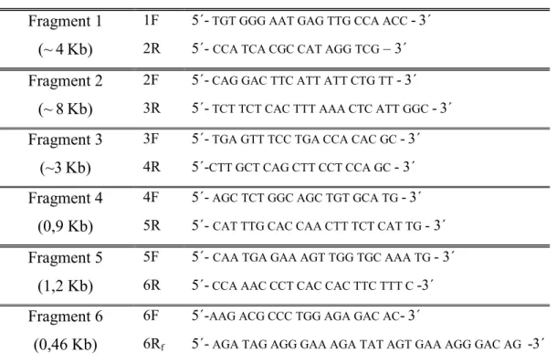
The sequences of the primers used for the amplification of the 6 fragments were reported on table I.
Table 1: Sequences of the primers used for the amplification of Ovis aries PIT1 gene
with the approximate length of each PCR fragment.
PRODUCT SIZE SEQUENCE
Fragment 1 1F 5´- TGT GGG AAT GAG TTG CCA ACC -3´
(~4Kb) 2R 5´- CCA TCA CGC CAT AGG TCG – 3´
Fragment 2 2F 5´- CAG GAC TTC ATT ATT CTG TT -3´
(~8Kb) 3R 5´- TCT TCT CAC TTT AAA CTC ATT GGC -3´
Fragment 3 3F 5´- TGA GTT TCC TGA CCA CAC GC -3´
(~3Kb) 4R 5´-CTT GCT CAG CTT CCT CCA GC - 3´
Fragment 4 4F 5´- AGC TCT GGC AGC TGT GCA TG -3´ (0,9 Kb) 5R 5´- CAT TTG CAC CAA CTT TCT CAT TG - 3´
Fragment 5 5F 5´- CAA TGA GAA AGT TGG TGC AAA TG - 3´
(1,2 Kb) 6R 5´- CCA AAC CCT CAC CAC TTC TTT C -3´
Fragment 6 6F 5´-AAG ACG CCC TGG AGA GAC AC-3´
(0,46 Kb) 6Rf 5´- AGA TAG AGG GAA AGA TAT AGT GAA AGG GAC AG -3´
For amplification of fragments 1, 2 and 3 we used a 50 µl reaction final volume with 200 ng DNA, 16 pmol of each primer and 2,6 U of Taq (Expand Long Template PCR System, Roche). For fragment 1 we used a final concentration of 1,4 mM dNTP and 2,25 mM MgCl2, while for fragment 2 we used 2 mM dNTP and 4 mM MgCl2 and for fragment 3 we used 1,4 mM dNTP and 2,25 mM MgCl2.
For fragment 4, 5 and 6, in a final volume of 25 µl of mixture we used 50 ng DNA, 8 pmol of each primer, 0,2 mM final concentration of each dNTP, 1,5 mM MgCl2 and 1 U of Taq (Pharmacia).
Cloning and Sequencing
The six PCR products were separated on agarose gels and purified by GenElute agarose spin columns (Sigma). For fragments 1, 2 and 3, the ligation was made with pCR-XL-TOPO vector and for fragments 4, 5 and 6 with pCR 2.1-TOPO, according to the suppliers recommendations (TOPO TA Cloning kit; Invitrogen, CA). The selection of colonies was made by blue/white screening in the case of TOPO-TA cloning kit while only recombinant colonies grew in the case of TOPO-XL cloning kit. Plasmid DNA was purified with GenElute Plasmid Miniprep kit (Sigma). Clones were sequenced by T7 sequencing kit, following the supplier’s recommendations (Amersham-Pharmacia), according to the dideoxynucleotide chain termination method. Dried gels were exposed to kodak film (Sigma). In order to comprove the sequencing result, a second set of sequencing was made on both strands in an independent enterprise (MWG-Biotech). Sequences were analysed with the Genetics Computer Group (GCG) program package from Belgian Embonode (BEN).
RESULTS
The PIT1 gene seems to have an essential role in the control of pituitary development and hormone expression. It is an interesting candidate gene for Marker Assisted Selection (MAS). In order to identify natural gene mutations in the coding region in Ovis aries, it was necessary to sequence the intron region immediately adjacent to each exon. For this purpose, we optimized the conditions to amplify six
fragments that were subsequently cloned and sequenced. The scheme of the amplification is present in figure 1, with the approximate length of each fragment and the position of the primers used in each case.
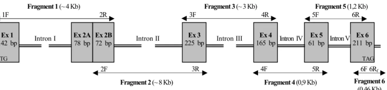
FIGURE 1: Schematic diagram of the Ovis aries PIT1 gene, with respective position of
the PCR primers (arrowheads) used for the amplification of each fragment. Introns are represented as double lines and exons as boxes with the correspondent length. The start point given for exon 1 (142 bp) is the initiation codon and the end of exon 6 (211 bp) is the stop codon.
Figure 2 shows the result of the amplification of the three first fragments. Introns 1, 2 and 3 are very extensive, giving a PCR product of approximately 4, 8 and 3 kb, respectively. Once the initial aim of this work was the partial sequencing of PIT1 gene that could allow the subsequent screening for mutations on the entire exons, introns 1, 2 and 3 were not completely sequence.
Ex 1-2 Ex 2-3 Ex 3-4 Marker 10 Kb 3 Kb 4 Kb 5 Kb 8 Kb 6 Kb 2,5 Kb 2 Kb Fragment 2 (~ 8 Kb) 2F 3R Fragment 4 (0,9 Kb) 4F 5R Fragment 6 (0,46 Kb) 6Rf 6F Fragment 1 (~ 4 Kb) 1F 2R Fragment 3 (~ 3 Kb) 3F 4R Fragment 5 (1,2 Kb) 5F 6R Ex 1 142 bp ATG Ex 2A 78 bp Ex 2B 72 bp Ex 3 225 bp Ex 4 165 bp Ex 5 61 bp Ex 6 211 bp TAG
FIGURE 2: Analysis of PCR purified products on 0,8 % agarose gel, stained with
ethidium bromide. The Marker (Smart Ladder, Eurogentec) shows that fragment 1 (from exon 1 to exon 2) has approximately 4 Kb; fragment 2 (from exon 2 to exon 3) about 8 Kb and fragmant 3 (from exon 3 to exon 4) around 3 Kb.
Figure 3 presents the PCR products of fragments 4, 5 and 6 with the respectively length of 0,9; 1,2 and 0,46 Kb. The length of those fragments allowed the complete sequencing of introns 4 and 5.
FIGURE 3: Analysis of PCR purified products on 2,5 % agarose gel, stained with
ethidium bromide. The Marker (Smart Ladder, Eurogentec) confirms the length of each band. Fragment 4 (from exon 4 to exon 5) has 0,9 Kb; fragment 5 (from exon 5 to exon 6) has 1,2 Kb and fragment 6 has 0,46 Kb.
Ex 4-5 Ex 5-6 Ex 6 Marker 1,0 kb 1,5 kb 0,8 kb 0,6 kb 0,4 kb 0,2 kb
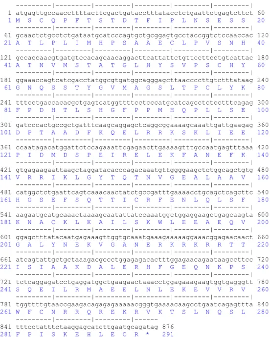
The nucleotide sequence of the coding region (876 bp) of Ovis aries PIT1 gene and the deduced aminoacid sequence are shown on Figure 4.
---|---|---|---|---|---| 1 atgagttgccaaccttttacttcgactgatacctttatacctctgaattctgagtcttct 60 1 M S C Q P F T S T D T F I P L N S E S S 20 ---|---|---|---|---|---| 61 gcaactctgcctctgataatgcatcccagtgctgcggagtgcctaccggtctccaaccac 120 21 A T L P L I M H P S A A E C L P V S N H 40 ---|---|---|---|---|---| 121 gccaccaacgtgatgtccacagcaacaggacttcattattctgttccttcctgtcattac 180 41 A T N V M S T A T G L H Y S V P S C H Y 60 ---|---|---|---|---|---| 181 ggaaaccagtcatcgacctatggcgtgatggcagggagcttaaccccttgtctttataag 240 61 G N Q S S T Y G V M A G S L T P C L Y K 80 ---|---|---|---|---|---| 241 tttcctgaccacacgctgagtcatggttttcctcccatgcatcagcctctcctttcagag 300 81 F P D H T L S H G F P P M H Q P L L S E 100 ---|---|---|---|---|---| 301 gatcccactgccgctgatttcaagcaggagctcaggcggaaaagcaaattgattgaagag 360 101 D P T A A D F K Q E L R R K S K L I E E 120 ---|---|---|---|---|---| 361 ccaatagacatggattctccagaaattcgagaacttgaaaagtttgccaatgagtttaaa 420 121 P I D M D S P E I R E L E K F A N E F K 140 ---|---|---|---|---|---| 421 gtgagaagaattaagctaggatacacccagacaaatgttggggaagctctggcagctgtg 480 141 V R R I K L G Y T Q T N V G E A L A A V 160 ---|---|---|---|---|---| 481 catggctctgaattcagtcaaacaactatctgccgatttgaaaacctgcagctcagcttc 540 161 H G S E F S Q T T I C R F E N L Q L S F 180 ---|---|---|---|---|---| 541 aagaatgcatgcaaactaaaagcaatattatccaaatggctggaggaagctgagcaagta 600 181 K N A C K L K A I L S K W L E E A E Q V 200 ---|---|---|---|---|---| 601 ggagctttatacaatgagaaagttggtgcaaatgaaagaaaaaggaaacggagaacaact 660 201 G A L Y N E K V G A N E R K R K R R T T 220 ---|---|---|---|---|---| 661 atcagtattgctgctaaagacgccctggagagacactttggagaacagaataagccttcc 720 221 I S I A A K D A L E R H F G E Q N K P S 240 ---|---|---|---|---|---| 721 tctcaggagatcctgaggatggctgaagaactaaacctggagaaagaagtggtgagggtt 780 241 S Q E I L R M A E E L N L E K E V V R V 260 ---|---|---|---|---|---| 781 tggttttgtaaccgaagacagagagaaaaacgggtgaaaacaagcctgaatcagagttta 840 261 W F C N R R Q R E K R V K T S L N Q S L 280 ---|---|---|--- 841 tttcctatttctaaggagcatcttgaatgcagatag 876 281 F P I S K E H L E C R * 291
Figure 4: Sequence of the coding region (876 bp) obtained after organization of the
sequenced 5787 bp of the Ovis aries PIT1 gene. In blue there is the predicted translated protein (291 aminoacids).
DISCUSSION
The 5787 bp sequenced in this work include two complete introns (introns 4 and 5) and six exons. This sequence enlarges the information until now published in Ovis aries and will allow the future screening for mutations in exons, using primers located in the intron regions.
The coding region (876 bp) and the predicted aminoacid sequence (291 aminoacids) presented in Figure 4 were compared with bovine and human sequences. We confirmed that PIT1 gene in Ovis aries has a high level of conservation with its human and bovine counterparts showing 91,3% and 98,2% homology at the coding DNA level and 96,2% and 99% at the expected amino acid level, respectively. When we compare the sequence presented here with the mRNA sequence for Ovis aries (Accession U88399), we found 7 bp difference in the coding region (99% homology) and the predicted protein sequence presented in figure 4 differs from the published protein in Ovis aries (Accession P79364) on 3 amino acids (99% of homology).
In Figure 1, the stucture of the gene include exon 2A. This extra exon (78 bp) was first described in humans (Delhase et al., 1995) as being the responsible for an alternative transcript (named PIT2). The genomic sequence revealed that this fragment is also present in sheep, in the same position, showing 88,5 % of homology with the human sequence. With RT-PCR essays from RNA extracted from sheep pituitary we found out that in Ovis aries, as it is described for several other species, there are alternative splicing transcripts for PIT1 gene (data not published) and we are presently investigating the possible implications of those variants.
ACKNOWLEDGMENTS
This work was supported by a Ph.D. grant from the Portuguese Science and Technology Foundation-FCT (BD-1365/2000), by funds from the project PRAXIS XXI 3/3.2/CA/1991/95 and the National Fund for Scientific Research, B-1000 Brussels, Belgium (# 24524.01)
REFERENCES
BODNER,M.,CASTRILLO, J.L.,THEILL, L.E., DEERINCK, T.,ELLISMAN, M.,KARIN,
M.1988. The pituitary-specific transcription factor GHF-1 is a homeobox-containing protein. Cell. 55: 505-518.
BRUNSCH, C., STERNSTEIN, I., REINECKE, P., BIENIEK, J. 2002. Analysis of
associations of PIT1 genotypes with growth, meat quality and carcass composition traits in pigs. J. Appl. Genet.. 43 85-91.
CHEN, R.,INGRAHAM, H., TREACY, M.N., ALBERT, V.R.,WILSON, L.,ROSENFELD,
M.G. 1990. Autoregulation of PIT1 gene expression mediated by two cis-active promoter elements. Nature. 346: 583-586.
COHEN, L.E.,WONDISFORD,F.E., SALVATONI, A.,MAGHNIE, M.,BRUCKER-DAVIS,
F.,WEINTRAUB,B.D.,RADOVICK,S.1995.A “hot spot” in the Pit-1 gene responsible for
combined pituitary hormone deficiency: clinical and molecular correlates. J. Clinical Endocrinology and Metabolism. 8: 679-684.
CUSHMAN, L.J., SHOWALTER, A.D., RHODES, S.J. 2002. Genetic defects in the
development and function of the anterior pituitary gland. Ann. Med.. 34: 179-191.
INGRAHAM, H., CHEN, R., MANGALAM, H.J. ELSHOLTZ, H.P., FLYNN, S.E., LIN,
C.R., SIMMONS, D.M., SWANSON, L., ROSENFELD, M.G. 1988. A tissue-specific
transcription factor containing a homeodomain specifies a pituitary phenotype. Cell. 55: 519-529.
LEFEVRE,C.,IMAGAWA,M.,DANA,S.,GRINDLAY,J.,BODNER,M.,KARIN,M.1987.
Tissue-specific expression of the human growth hormone gene is covered in part by the binding of a specific trans-acting factor. EMBO Journal. 6 : 971-981.
LI, S., CRENSHAW III, E.B, RAWSON, E.J., SIMMONS, D.M., SWANSON, L.W.,
ROSENFELD, M.G. 1990. Dwarf locus mutants lacking three pituitary cell types result
from mutations in the POU-domain gene PIT1. Nature. 347: 528-533.
LIN, C., LIN, S.C., CHANG, C.P., ROSENFELD, M.G. 1992. PIT1 dependent expression of the receptor for growth hormone releasing factor mediates pituitary cell growth. Nature. 360: 765-767.
MARTINEZ-BARBERÁ, J., VILA, V., VALDIVIA, M.M., CASTRILLO, J.L. 1997.
Molecular cloning of gilthead seabream (Sparus aurata) pituitary transcription factor GHF-1/Pit-1. Gene. 185: 87-93.
MCCORMICK, A., BRADY, H., THEILL, L.E., KARIN, M. 1990. Regulation of the
pituitary-specific homeobox gene GHF1 by cell-autonomous and environmental cues. Nature. 345: 829-832.
MONTGOMERY,G.,SISE,J.1990.Extraction of DNA from sheep white blood cells.
New Zealand Journal of Agricultural Research.33:437-441.
NELSON, C., ALBERT, V.R., ELSHOLTZ, H.P.,LU, L.I.W., ROSENFELD, M.G. 1988.
Activation of cell-specific expression of rat growth hormone and prolactin genes by a common transcription factor. Science. 239: 1400-1405.
ONO, M., TAKAYAMA, Y. 1992. Structures of cDNAs encoding chum salmon
pituitary-specific transcription factor, Pit-1/GHF-1. Gene. 116: 275-279.
RENAVILLE,R.,GENGLER,N.,VRECH,E.,PRANDI,A.,MASSART,S.,CORRADINI,C.,
BERTOZZI, C., MORTIAUX, F., BURNY, A., PORTETELLE, D. 1997. Pit-1 gene
polymorphism, milk yield, and conformation traits for Italian Holstein-Friesian bulls. J Dairy Sci. 80: 3431-8.
TATSUMI, K.,NOTOMI, T.,AMINO, N.,MIYAI,K. 1992.Nucleotide sequence of the
complementary DNA for human Pit-1/GHF-1.Biochim. Biophys. Acta.1129:231-234.
THEILL, L.E., CASTRILLO, J.L., WU, D.,KARIN, M.1989. Dissection of functional
domains of the pituitary-specific transcription factor GHF-1. Nature. 342: 945-948.
TUGGLE,C.K.,YU,T.P.,HELM,J.,ROTHSCHILD,M.F.1993. Cloning and restriction
fragment length polymorphism analysis of a cDNA for swine Pit-1, a gene controlling growth hormone expression. Animal Genetics. 24: 17-21.
YAMADA,S.,HATA,J.,YAMASHITA,S.1993.Molecular cloning of fish Pit-1 cDNA
and its functional binding to promoter of gene expressed in the piptuitary. J. Biol. Chem. 268: 24361-24366.
YU,T.P.,TUGGLE,C.K.,SCHMITZ,C.B.,ROTHSCHILD,M.F.1995.Associations of
PIT1 polymorphisms with growth and carcass traits in pigs. J. Animal Sci.. 73(5): 1282-1288.