OATAO is an open access repository that collects the work of Toulouse
researchers and makes it freely available over the web where possible
Any correspondence concerning this service should be sent
to the repository administrator:
[email protected]
This is an author’s version published in: http://oatao.univ-toulouse.fr/21168
To cite this version:
Soumbo, Marvine
and Pugliara, Alessandro
and Monje, Marie-Carmen
and
Roques, Christine
and Despax, Bernard
and Bonafos, Caroline and Carles, Robert
and Mlayah, Adnen and Makasheva, Kremena
Physico-chemical characterization of
the interaction of red fluorescent protein — DsRed with silica layers. (2015) In: 2015
IEEE Nanotechnology Materials and Devices Conference (NMDC), 13 September 2015 -
16 September 2015 (Anchorage, United States).
1 Abstract The Discosoma Recombinant Red Fluorescent
(DsRed) protein is the latest member of the family of fluorescent proteins. It holds great promise for applications in biotechnology and cell biology. However, before being used for rational engineering, knowledge on the underlying mechanisms relating the DsRed structural stability and adsorption properties on solid surfaces is highly demanded. The physico-chemical analysis performed in this study reveals that the interaction of DsRed with SiO2 surfaces does not lead
to protein denaturation. The secondary structure of DsRed is preserved after adsorption and dehydration. The measured contact angles of sessile droplets with different DsRed concentrations determine the interaction as hydrophilic one. The photoluminescence emission of dehydrated DsRed droplets is found to be slightly red-shifted, peaking at 590 nm.
Topic Nano-Biomedicine
Index Terms/Keywords Thin SiO2 layers, Proteins, DsRed
I. INTRODUCTION
RECENT advances in biotechnology offer the possibility to explore chemical, physical and functional properties of proteins, in particular fluorescent proteins, in various applications such as biosensors, bioelectronics, drug delivery systems, etc., [1]. The fluorescent proteins are a family of proteins of 25 30 kDa, mainly applied to study the organization and function of living systems; with the green fluorescent protein (GFP) as most extensively characterized member. The recently cloned from reef coral Discosoma sp. Recombinant Red Fluorescent (DsRed) protein [2] expresses the longest recorded yet excitation and emission maxima at 558 nm and 583 nm, respectively. DsRed holds great promise for biotechnology and cell biology as a spectrally distinct companion or substitute for the GFP.
The current technological achievements lead to exposure
and interaction of proteins with non-biological organic and inorganic solid surfaces. Considering further development our choice is to go with DsRed protein and study its interaction with thin SiO2 layers. Studying thin SiO2 layers is based on their large use in microelectronics as diffusion barrier or thin electrical insulator, in plasmonics as host matrix, etc., [3-6 and the references therein]. Most of the currently applied diagnostic methods are adapted to study the structural stability of proteins in solution. Nevertheless, some advanced diagnostic methods are based on probing proteins after dehydration, for example, with matrix assisted laser desorption/ionization (MALDI) [7] or the Fourier Transform InfraRed (FTIR) spectroscopy [8]. In this work we report results on the physico-chemical characterization of the interaction of DsRed protein with solid silica surfaces for a series of diluted protein solutions.
II. EXPERIMENTAL PART
Thermally grown 100 nm-thick SiO2 layers were used.
Before being exposed to protein deposition, the SiO2/Si
substrates were consecutively cleaned in ethanol and acetone and rinsed in deionized water until attaining zero surface conductivity. DsRed was purchased from Biovision (at least 97% pure, in a freeze dried form). A stock solution of DsRed was made to a concentration of 1 mg/mL in water for injectable preparations (European Pharmacopeae, COOPER) with pH-value of 7.0. The pH-stability of the DsRed stock solution was repetitively controlled during measurements. The assays were performed at room temperature (23°C). The pH-behavior of
DsRed was obtained for the range 3.0 11.0. Sessile droplets of DsRed with very small volume (3.8 ± 0.1 L) were deposited on the surface of SiO2 layers by using a Contact Angle Meter from GBX Scientific Instruments. Optical images of dehydrated
sessile droplets were recorded with a digital microscope Keyence VHX-1000. The droplet diameter was measured on the images. The other parameters of the droplets: thickness, droplet boarder width and height were measured with a 2D-surface profilometer Alpha-Step IQ from KLA-Tencor. Morphological changes of DsRed were observed with a field emission Scanning Electron Microscopy (SEM), ZEISS CrossBeam 1540 XB. The photoluminescence emission of the DsRed was excited with an Ar+ laser operating at 514 nm. The emitted light was dispersed using a Jobin Yvon spectrometer with a 150 grooves/mm grating.
Physico-chemical characterization of the interaction
of red fluorescent protein - DsRed with silica layers
Marvine Soumbo, Alessandro Pugliara, Marie-Carmen Monje, Christine Roques, Bernard Despax,
Caroline Bonafos, Robert Carles, Adnen Mlayah, and Kremena Makasheva, Member, IEEE
III. RESULTS AND DISCUSSION
When a droplet containing given concentration of proteins is brought to a contact with solid surface, the organization and protein behavior rely primarily on the contact angle hysteresis (CAH) at the solid-vapor and liquid-vapor interfaces [9, 10]. Figure 1 shows the DsRed behavior during dehydration for different protein concentrations. The linear decrease over time of the contact angle is common for all the studied DsRed concentrations. The droplet dehydration is more rapid for small concentrations of DsRed (up to 0.1 g/L). This effect is related to the large surface energy variation on the droplet profile close to the triple line (solid-liquid-vapor) and to the protein adsorption mechanisms induced by the liquid convective drive inside the droplet (Marangoni effect) [10].
Fig. 1. Measured contact angles at 23°C as a function of time during sessile droplet dehydration for different DsRed concentrations.
As shown in Table II, all the droplet characteristics are increased when the concentration of DsRed is larger. It means that the DsRed adsorbs on the entire area covered by the droplet with preferential deposition close to the triple line, i.e. to the droplet boarder. The thickness of the adsorbed DsRed layer on solid surfaces can finely be controlled by the protein concentration.
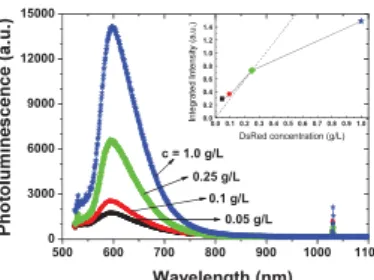
Fig. 2. Photoluminescence spectra of DsRed dehydrated droplets adsorbed on SiO2 surface for different concentrations at pH = 7.0 and 23°C. The inset represents the integrated intensity of the photoluminescence peak as a function of the DsRed concentration.
The obtained photoluminescence spectra are presented in Fig. 2. The photoluminescence emission increases with increasing DsRed concentration. However, it does not scale up linearly most likely due to quenching phenomena (Fig. 2, inset). The photoluminescence emission band is peaking at 590 nm. The slight red-shift with respect to the DsRed emission in solution [11] can be attributed to conformation effects and to interaction of the DsRed with the thin silica layer.
IV. CONCLUSION
Physico-chemical analysis of the interactions of red fluorescent protein, DsRed with thermal SiO2 surfaces shows
that the thickness of the adsorbed DsRed protein layer can finely be tuned by the protein concentration. The measured contact angles of very small sessile droplets containing different concentration of DsRed determine the interaction as hydrophilic one. The DsRed proteins appear stable under pH-variations. The adsorption of DsRed on SiO2 surfaces and the
following dehydration processes do not lead to protein denaturation. The photoluminescence emission of dehydrated DsRed proteins adsorbed on SiO2 layers is found to peak at
590 nm, which is slightly red-shifted compared to the reported value for a solution (583 nm). Potential modification of the DsRed excitation and emission spectra due to the DsRed-SiO2
interactions in solution will be addressed further. REFERENCES
[1] D. M. Chudakov, M. V. Matz, S. Lukyanov, and K. A. Lukyanov, Physiol. Rev., vol. 90, pp. 1103-1163, 2010.
[2] M. V. Matz, et al., Nature Biotechnology, vol. 17, pp. 969973, 1999.
[3] E. A. Irene, in Integrated Cirquits: Chemical and Physical Processing, ACS Symposium Series, vol. 290, pp 3146, 2009.
[4] W. B. Lacy, et al., Anal. Chem., vol. 68, pp. 1003-1011, 1996. [5] R. Carles, C. Farcau, C. Bonafos, G. BenAssayag, M. Bayle, P. Benzo,
J. Groenen, and A. Zwick, ACS NANO, vol. 5, pp. 8774-8782, 2011. [6] A. Pugliara, C. Bonafos, R. Carles, B. Despax,and K. Makasheva,
Material Research Express, vol. 2, pp. 065005, 2015.
[7] A. B. Hughes, Amino Acids, Peptides and Proteins in Organic Chemistry, Weinheim, Germany: Wiley-VCH Verlag & Co. KGaA, 2012.
[8] A. Barth, Biochimica et Biophysica Acta, vol. 1767, pp. 1073-1101, 2007.
[9] Y. L. Chen, C. A. Helm, and J. N. Israelachvili, J. Phys. Chem., vol. 95, pp. 10736-10747, 1991.
[10] J. R. Trantum et al., Langmuir, vol. 29, pp. 6221 6231, 2013.
[11] M. F. Garcia-Parajo, M. Koopman, E. M. H. P. van Dijk, V. Subramaniam, and N. F. van Hulst, PNAS, vol. 98, pp. 1439214397, 2001.
0 5 10 15 0° 10° 20° 30° 40° 50° 60° 70° 80° 90° Control pH = 7.0 DsRed c = 0.05 g/L c = 0.1 g/L c = 0.25 g/L c = 1.0 g/L Time (min) 500 600 700 800 900 1000 1100 0 3000 6000 9000 12000 15000 c = 1.0 g/L 0.25 g/L Wavelength (nm) 0.05 g/L 0.1 g/L 0.0 0.1 0.2 0.3 0.4 0.5 0.6 0.7 0.8 0.9 1.0 0.0 0.2 0.4 0.6 0.8 1.0 1.2 1.4 DsRed concentration (g/L) TABLEII
CHARACTERISTICS OF DSRED SESSILE DROPLETS AT pH=7.0 AND 23°C
DsRed
(g/L) image at 1s Droplet
Contact angle -
Optical image
after dehydration Dehydrated dropleta control pH=7.0 54.3° ± 0.1° n/a n/a 0.05 65.4° ± 0.2° d = 2.8 mm; e = 20 nm; l = 46 m; h = 0.6 m; 0.10 65.5° ± 1.6° d = 2.8 mm; e = 30 nm; l = 67 m; h = 1.4 m; 0.25 ± 1.5° 73.7° d = 2.7 mm; e = 30 nm; l = 84 m; h = 3.0 m; 1.00 ± 2.7° 72.0° d = 2.6 mm; e = 60 nm; l = 200 m; h = 5.2 m. aThe DsRed sessile droplet characteristics after dehydration: d is the droplet diameter; e is the droplet thickness measured inside the droplet, just before the droplet boarder; l is the droplet boarder width; and h is the droplet boarder height. Scale bar on the images equals 500 m.