HAL Id: hal-02475488
https://hal.archives-ouvertes.fr/hal-02475488
Submitted on 12 Feb 2020
HAL is a multi-disciplinary open access
archive for the deposit and dissemination of
sci-entific research documents, whether they are
pub-lished or not. The documents may come from
teaching and research institutions in France or
abroad, or from public or private research centers.
L’archive ouverte pluridisciplinaire HAL, est
destinée au dépôt et à la diffusion de documents
scientifiques de niveau recherche, publiés ou non,
émanant des établissements d’enseignement et de
recherche français ou étrangers, des laboratoires
publics ou privés.
Distributed under a Creative Commons Attribution - ShareAlike| 4.0 International
License
Philippe Silar
To cite this version:
1
Philippe SILAR
3
Podospora anserina
4 Acknowledgments.
Merci à Stéphan d'avoir été patient pendant que je rédigeais ce livre, souvent le matin très tôt ou le weekend ! Thanks to Professor Uwe Braun from the Martin Luther Universität at Halle-Wittenberg and Curator of Halle Herbarium (Germany) for both illustrations of type specimens. Merci à Pierre pour sa relecture !
Legal Notice
This book is under Creative Commons Attribution licence. Attribution-NonCommercial-ShareAlike 4.0 International. To access a copy of the licence, go to the following link https://creativecommons.org/licenses/by-nc-sa/2.0/fr/deed.en or send a mail to Creative Commons, 444 Castro Street, Suite 900, Mountain View, California, 94041, USA.
First edition : february 2020
ISBN : 978-2-9555841-2-5 EAN : 9782955584125
5
SOMMAIRE
Foreword ... I
Podospora anserina: a brief history ... 1
Part 1: Biology of Podospora anserina ... 3
Podospora anserina in the tree of life: classification of the species ... 4
Podospora anserina in its natural biotopes ... 26
Isolation, culture and preservation ... 29
Strains preservation ... 36
Methods for macromolecules extraction and genetic transformation... 37
DNA Extraction ... 37
RNA Extraction ... 39
Protein Extraction ... 40
Protoplasts preparation ... 41
Genetic transformation ... 42
Gene deletion, single nucleotide change and at-will modification of the genome ... 44
Tools for expression analysis... 49
Morphology and cytology ... 50
The mycelium ... 51
The gametes ... 55
The perithecium ... 57
The asci and the ascospores ... 60
The sexual cycle and genetical analysis ... 65
The lifecycle of P. anserina ... 65
Mutant generation ... 69
Genetical analyses: segregation, dominance/recessivity and complementation ... 70
Parasexual cycle ... 74
Developmental genetics: grafting and genetic mosaics ... 74
6
Part 2: Podospora anserina as a model for physiological and molecular analysis... 89
Ascospore ejection and germination ... 91
Mycelium growth and development ... 94
Senescence and the Premature Death Syndrome ... 95
Crippled Growth and other phenotypic instabilities ... 103
Hyphal Interference and Vegetative/heterokaryon Incompatibility ... 111
Appressorium-like structure differentiation ... 119
Sexual reproduction ... 121
Mating and mating types ... 121
Fruiting body differentiation ... 125
Fruiting body production and repartition ... 132
Meiotic Drive Elements: Spore Killers ... 136
Natural products biosynthesis ... 140
Secondary metabolites ... 140 Enzymes ... 142 REFERENCES ... 145 Herbarium Specimen ... 145 Original Articles ... 145 Book Chapters ... 175 Dissertations ... 177 Conference Proceedings ... 181
Miscellaneous documents including edited thesis ... 182
Appendix 1: Original descriptions of Podospora anserina ... 183
I
Foreword
In any good movie, the cast is a large part of its success. Of course, the stars playing the main characters are important. However, a good set of supporting roles is often equally crucial to fully bring to the fore all the qualities of the film. The science of Biology is a little bit like a movie. Indeed, it needs models to decipher the fundamental laws of the living. Some are superstars, such as mouse, drosophila, the bacterium Escherichia coli or the baker’s yeast Saccharomyces cerevisiae and play foremost roles. However, other less-known models are sometime key actors in major discoveries. Examples are numerous: Mendel used peas to decipher laws of heredity and Hämmerling used Acetabularia to show that the cell’s nucleus is the location where genetic information is stored…
For the filamentous fungi, the stars of the cast are Neurospora crassa and Aspergillus nidulans. The former has for example been instrumental in the discovery of the nature of the gene. Indeed, experiments on this model showed that “one gene codes for one enzyme”, a paradigm which still holds for the most part. It is currently used in many labs to study many fundamental processes ranging from the circadian clock to epigenetic gene regulation. The second fungus is used to decipher many cellular phenomena ranging from the cell cycle to the role of the cytoskeleton. It has however been particularly important in the discovery of the parasexual cycle in eukaryotes in the 1950’s. The supporting cast for filamentous fungi is too numerous to cite them all. Cryptococcus neoformans and Magnaporthe grisea are instrumental to study pathogenicity; Coprinopsis cinerea and Sordaria macrospora are used to study reproduction, to cite a few of them. This book will focus on one of the supporting cast, the “friendly mold” Podospora anserina. This fungus is used for over a hundred years in the laboratory to study processes as diverse as senescence, prions and sexual reproduction. Few reviews dealing with its biology are available. However, no large monography exists on this species. This book proposes to fill this void.
1
Podospora anserina: a brief history
Podospora anserina is a filamentous fungus, i.e., a mold, used now for more than a century in
several laboratories to study various biological processes ranging from those typical of fungi such as anastomoses and vegetative incompatibility (= heterokaryon incompatibility), hyphal interference, spore germination, mycelium degeneration and fruiting body formation, to those of general importance for eukaryotes such as sexual reproduction, Meiotic Drive Elements that cheat Mendel’s laws, mitochondrial and peroxisomal physiology and those of general relevance in biology, e.g., senescence, cell differentiation, degeneration and death, respiratory metabolism, or structural and regulatory inheritance caused by prions and prion-like hereditary units.
Early researchers working with P. anserina were mostly concerned with the cytological descriptions of cellular phenomena and structures beyond those used to classify the fungus. Wolf1 in 1912 and Satina in 1916 dealt mostly with the description of the sexual process from the differentiation of the gametes to the production of ascospores, Ames in 1930, 1932 & 1934, Dowding in 1931 and Dodge in 1936 with the breeding system, finally Buller in 1931
with hyphal anastomoses. Page analyzed the complete cycle of P. anserina and related species in 1939. It is however with the work of the French geneticist Georges Rizet (figure 1) that P. anserina was extensively used as a model. Rizet and its students gave the complete explanation of the pseudo-homothallic breeding system and complex nuclear behavior during sexual reproduction of the fungus, enabling them to perform elegant genetic analysis. They describe the vegetative
incompatibility (the “Barrage“phenomenon), the still mysterious Senescence process and the peculiar behavior of the S/s incompatibility system that we now know is due to a prion. They use the fungus to
1
All references are given at the end of the book.
2
study genetic recombination, gene structure, translation and mitochondrial physiology and evolution. Along with Karl Esser, a German mycologist, who visited Rizet’s lab during a postdoc in the 1950’s, they develop all the tools, which are now routinely used, including the growth and germination media as well as the methods for macromolecule extractions and analyses. This culminated by the development of genetic transformation procedures enabling the entry of P. anserina in the era of molecular genetics, by permitting the identification of mutant genes and later on gene deletions and modifications. Finally, it is Rizet’s “scientific progeny” who established in 2008 the complete genomic sequence of the fungus.
Presently, the fungus is still studied in a few labs, mostly in France and Germany as expected from its history. However, many publications also originate from the Netherlands, Mexico, Switzerland, Taiwan… showing an ever increasing popularity! It truly deserves its nickname: the friendly mold…
Figure 2. Podospora anserina, the friendly mold.
3
Part 1: Biology of
4
Podospora anserina in the tree of life: classification of the species
When dealing with living organisms, it is always advisable to know their exact position in the tree of life as this greatly help to outline their main characteristics. P. anserina, also known as the “friendly mold”, is thus a “mold” or a “filamentous fungus” (figure 3). This means that it belongs to the domainEukaryota (figure 4), i.e., the major part of its genetic information is contained inside a nucleus, the
remaining part being located inside the mitochondria. It is related to animals in the sense that like animals it belongs to the Opisthokonta superphylum (figure 4). Species of this superphylum share many characters, like the capacity to use extensively chitin as a coat material, to store carbon as glycogen and to use UGA as a tryptophan codon in the mitochondrial genetic code. Their name stems from the fact that they differentiate flagellated cells with a single posteriorly-orientated flagellum. However, P.
anserina is a terrestrial fungus and, as such, it has altogether lost the capacity to differentiate the
flagellum. Opisthokonta belongs to the subdomain Amorphea, which has been defined by molecular
Figure 3 Podospora anserina. Left at high magnification (bar= 0.3 mm) and grown on toothpicks;
Mycelium and fruiting bodies (the pear-shaped perithecia) are visible. Right: the friendly mold at low magnification (bar= 1 cm) and grown on a minimal medium (M2) Petri plate. The black dots are the perithecia.
5 Amebozoa Metazoa Choanoflagellata Achaeplastida Heterokonta Rhizaria Holozoa Hacrobia Opisthokonta Amorphea Mycetozoa Variosea Lobosa Conosa Tubulinea Discosea Rhodophyta Viridiplantae Glaucophyta Diaphoretickes Pseudomycota Ochrophyta Bygira Alveolata ApicomplexaDinoflagellata
Ciliophora Cercozoa Foraminifera Radiolaria
«
U
ni
kon
ta
»
«
B
ik
on
ta
»
« SAR » Excavata Metamonada Discoba«
Op
im
od
a
»
«
D
ip
ho
d
a
»
Haptista Cryptista Eukaryota Nucleariida Eumycota Holomycota Rozellida Archea EubacteriaFigure 4 P. anserina belongs to the Eumycota, or true fungi. In red, the lineage to which P. anserina
belongs.
phylogeny. Species from Amorphea share little characteristics, hence their name that means “formless”. In Opisthokonta, two phylogenetic lineages are presently recognized: Holozoa that contains the Metazoa or animals, and Holomycota that contains the Eumycota or true fungi, to which P. anserina belongs. Fungi, and hence P. anserina, feeds by osmotrophy, i.e., they degrade their food outside the cells by secreting enzymes and then absorb the released molecules through transporters located in their plasma membrane. Species from the Holozoa lineage, and also those located at the base of the Holomycota, feed by phagotrophy or ingestion, i.e., ingest food as large particles and digest it inside the cells (or digestive
6
Chytridiomycota
Blastocladiomycota
Entomophthoromycota
Kickxellomycotina
Mucoromycotina
Glomeromycota
Dikarya
Neocallimastigomycota
Ascomycota
Basidiomycota
Monoblepharidomycota
Zoopagomycotina
«
Ea
rly
D
iv
e
rgi
n
g
Fu
n
gi
»
Mortierellomycotina
« Olpidium »
land invasion
« chytridiomycetes » « zygomycetes »Entorrhizomycota
Figure 5 Diversity of the Eumycota. In red, the lineage to which P. anserina belongs.
track for animals). Other eukaryotic organisms feed by osmotrophy, the Pseudomycota. They look very much like Eumycota, but have evolved from completely different ancestors (figure 4).
Eumycota is a highly diverse group containing at least 14 phyla (figure 5). Its basal members, the
“chytridiomycetes”, are still aquatic organisms that disperse thanks to a flagellum. However most species are terrestrial and have invaded nearly all biotopes. Some have even returned to an aquatic lifestyle, as whales and dolphins did. These terrestrial or formerly-terrestrial species all share the inability to produce a flagellum. The two major phyla that contain over 90% of the species of Eumycota are the Ascomycota and the Basidiomycota. They are related and able to produce dikaryotic cells (cells with two genetically-different nuclei) during an extended period of their life cycle, hence the name of the lineage that contains them; the Dikarya (figure 5). For most Dikarya species, the major part of their life is completed as a mycelium, a network of interconnected and elongated cells called hyphae. This mycelium is the stage of the life cycle during which the fungus feeds, i.e., the vegetative part of the life cycle. The major difference
7 Orbiliomycetes Lecanoromycetes Eurotiomycetes Geoglossomycetes Laboulbeniomycetes Dothideomycetes Arthoniomycetes Leotiomycetes Sordariomycetes Coniocybomycetes Xylonomycetes
Taphrinomycotina
Taphrinomycetes Saccharomycetes Pneumocystidiomycetes Schizosaccharomycetes Neolectomycetes ArcheorhizomycetesSaccharomycotina
Pezizomycotina
Lichinomycetes Pezizomycetes Leotiomyceta Sordariomyceta Dothideomyceta SaccharomycetaFigure 6 Diversity of the Ascomycota. In red, the lineage to which P. anserina belongs.
between the Basidiomycota and the Ascomycota is the way they differentiate their sexual (meiotic) spores during the reproductive part of their lifecycle. In the Basidiomycota, spores are produced outside the mother cell or basidium that undergoes meiosis; they are called basidiospores. In the Ascomycota, they are formed inside the mother cell or ascus. They are then called ascospores. P. anserina belongs to the phylum of the Ascomycota, also known as the “ascomycetes” in the vernacular language. P. anserina differentiates a typical mycelium during its vegetative phase (figure 3) and produces archetypal asci during sexual reproduction (see those on figure 16).
Ascomycota are further subdivided into three subphyla (figure 6). The basal Taphrinomycotina
8
Pisorisporiales
Koralionastetales
Lulworthlales
Xylariales
Xylariomycetidae
Melanosporales
Coronophorales
Falcocladiales
Torpedosporales
Hypocreales
Microascales
Glomerellales
Pleurotheciales
Conioscyphales
Savoryellales
Hypocreomycetidae
Sordariomycetidae
Meliolales
Coniochaetales
Cordanales
Phyllachorales
Boliniales
Sordariales
Chaetosphaeriales
Trichosphaeriales
Ophiostomatales
Annulatascales
Amplistromatales
Magnaporthales
Togniniales
Calosphaeriales
Jobellisiales
Tirisporellales
Diaporthales
Papulosaceae
Thyridiaceae
Cephalothecaceae
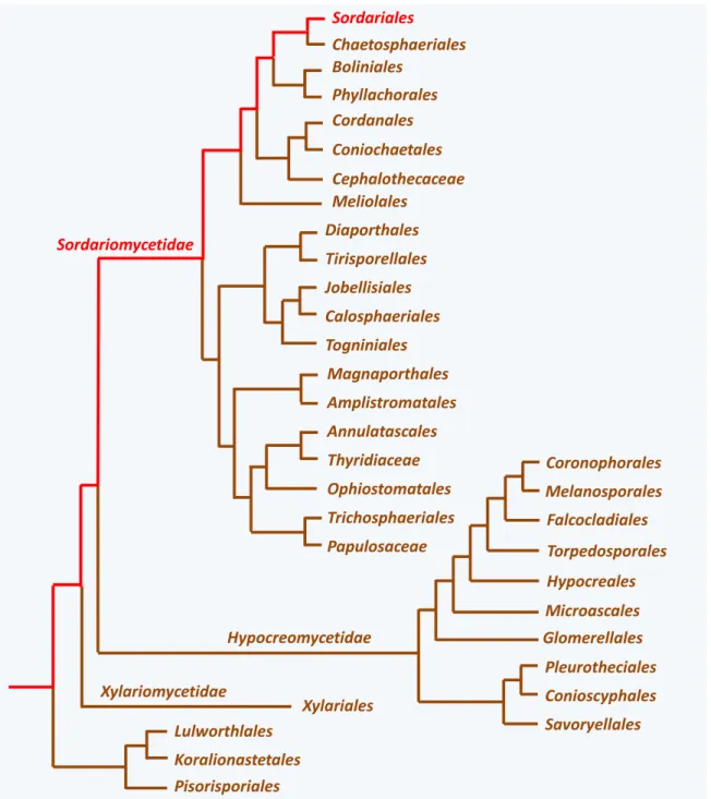
Figure 7 Diversity of the Sordariomycetes. In red, the lineage to which P. anserina belongs.
often live as yeast or as mycelium in which communications between cells remains simple. P. anserina is a member of the third subphylum: Pezizomycotina. The species of Pezizomycotina often have complex life
9
cycles and behaviors, as we shall see for P. anserina. Their mycelium is highly interconnected, firstly by central pores that exist between two consecutive articles (i.e., the elongated cells that made up the hyphae), and secondly by the ability of hyphae to fuse by a process called anastomosis. Different types of hyphae can also be distinguished, primarily by their diameter. Pezizomycotina are often able to differentiate complex multicellular structures, especially during sexual reproduction. For example, P.
anserina differentiate a fruiting body looking like a tiny pear that is called a perithecium (figure 3). Pezizomycotina fungi also disperse through asexual spores, often conidia. P. anserina differentiate
conidia-looking cells that are used as male gamete for fertilization. They are called spermatia. As yet in the laboratory, germination of P. anserina spermatia is achieved with very low efficiency (in the range of one out of 106-107), questioning their role as asexual dispersal unit. However, it cannot be ruled out that the proper conditions for their germination are still unknown.
Differentiating a perithecium during sexual reproduction is a general characteristic of species in the class Sordariomycetes (and also in the related class Laboulbeniomycetes) to which P. anserina belongs (figure 7). More than 10 000 species of Sordariomycetes have been described, but this number is likely largely underestimated. They live as saprobes (i.e., they live freely and feed on dead organic matter) or as parasitic or mutualistic associates of plants and animals. However, none appears to live as lichen or
Chaetomiaceae
Sordariaceae
Lasiosphaeriaceae I
Lasiosphaeriaceae II
Lasiosphaeriaceae III
Lasiosphaeriaceae IV
Figure 8 Diversity of Sordariales. In red, the lineage to which P. anserina belongs. The numbering is
10
mycorrhiza, two lifestyles largely adopted by fungi. P. anserina lives primarily as a saprobe on herbivore dung and less frequently in soil, but it also seems to be able to associate with plants as an endophyte, i.e., it may also live within plants. Molecular phylogeny has recognized three major lineages of
Sordariomycetes, as well as additional as-yet nameless basal groups (figure 7). Most species of Xylariomycetidae produce dark perithecia often embedded collectively within a stroma. Most species of Hypocreomycetidae produce brightly-colored perithecia and most species of Sordariomycetidae, to which P. anserina belongs, produced dark greenish ones. Molecular phylogeny has also helped to refine the
classification of the various Sordariomycetes species into orders (figure 7). The order containing P.
anserina is the Sordariales. Species of this order produce solitary perithecia that most often contain
darkly-pigmented ascospores, as P. anserina does. Sordariales presently contains three families:
Sordariaceae, Chaetomiaceae and Lasiosphaeriaceae. The definition of these families has greatly changed
over time and was previously based on the fine structures of the fruiting bodies as well as the shape and ornamentation of the ascospores. The new data generated by the molecular phylogenies have shown that the Lasiosphaeriaceae is paraphyletic and four distinct phylogenetic groups can be identified. The monophyletic Sordariaceae and Chaetomiaceae are nested within these four lineages (figure 8). We are thus waiting for a complete reclassification of the order. This will entail name changes for three of the
Lasiosphaeriaceae lineages… P. anserina is in the Lasiosphaeriaceae lineage IV that is more closely related
to the Chaetomiaceae. Other well-known species of Sordariales (figure 9) are those of genera Neurospora and Sordaria belonging to the Sordariaceae and of the genus Chaetomium belonging to the
Neurospora crassa
Sordaria macrospora
Chaetomium globosum
Figure 9 Relatives of P. anserina from the order Sordariales. On the left, Neurospora crassa is known to
produce large amounts of orange conidias. On the center, Sordaria macrospora produces typical glabrous perithecium. On the right, Chaetomium globosum differentiates very hairy fruiting bodies.
11
Cercophora
Podospora
Apiosordaria
Triangularia
Zopfiella
Arnium
Figure 10 Main ascospore shapes in the Lasiosphaeriaceae IV. The size of the ascospores can be very
different among the different species. Moreover, some can be decorated with gelatinous appendages not represented here.
Chaetomiaceae. In addition to the famous Neurospora crassa, Neurospora intermedia, Neurospora tetrasperma, Sordaria macrospora and Sordaria fimicola are often used in the laboratories or in
classrooms for genetic studies. Chaetomium species, especially Chaetomium globosum, are well adapted to grow on cellulose and are often responsible for the spoilage of books in humid libraries. Some
Chaetomium species are responsible of very rare but often fatal mycosis in human. To give an insight into
the biodiversity of the Sordariales, species in this order are as genetically diverse as the vertebrates. For example, the genetic divergence between P. anserina and N. crassa is at least as large as that between fishes and humans!
The traditional classification of the Lasiosphaeriaceae is based primarily on the form of the ascospores. Figure 10 depicts the shape of the ascospores of the species presently known in
Lasiosphaeriaceae IV. Alas, this criterion turned out to be highly unreliable to trace the true relationship
between species of Lasiosphaeriaceae. For example, most ascospores of Lasiosphariaceae IV are bicellular with one cell large and melanized and the other smaller and unpigmented. This latter cell may have undergone an apoptosis-like death. Nevertheless, Arnium ascospores are unicellular, showing that having bicellular ascospores is not a character shared by all species. Most genera of this family are thus polyphyletic. For instance, Podospora species are present in Lasiosphariaceae IV, but also in
Lasiosphariaceae I, Lasiosphariaceae II and Lasiosphariaceae III! A paper by Miller & Hundorf in 2005
12 Cladorrhinum (Bahupaathra) Phialophora 50 µm 10 µm 10 µm
Figure 11 Known anamorphs in the Lasiosphaeriaceae IV.
might be a better predictor of the true phylogeny. Unfortunately, too few species have been analyzed with regard to this character to know if it is actually able to predict relationships in the Lasiosphaeriaceae.
In addition to the species differentiating sexual structures (or teleomorph), several species of
Lasisopshaeriaceae clade IV are only known through their asexual forms (or anamorph). These are known
as Cladorrhinum (= Bahupaathra) or Phialophora (figure 11). Finally, it is most likely that many species of
Lasiosphaeriaceae IV are presently unknown and those listed in figure 12 are likely to be only the “tip of
the iceberg”.
To complicate the matter, few studies are devoted to the deciphering of the true phylogeny of the Lasiosphaeriaceae, as this family contains mostly saprobic species with inconspicuous life styles. Note that a fungus responsible for some mycetoma, Madurella mycetomatis, is a close relative, but it is not yet known whether this species belongs to Lasiosphaeriaceae IV or is more likely closely related to the
Chaetomiaceae. Mycetomas caused by M. mycetomatis are rare but very debilitating and among the
most dreadful diseases that one can catch! Presently, molecular phylogeny recognizes three subsets of species in Lasiosphaeriaceae IV (figure 12). In figure 12, the species of each subset are listed (mostly) alphabetically because their actual relationships are as yet unknown.
The two Podospora species most closely related to P. anserina are P. setosa and P.
austroamericana. Both are also coprophilous fungi. P. setosa produces asci with 128 ascospores, P. austroamericana with eight ascospores, while P. anserina asci have four ascospores. P. setosa and P. austroamericana are homothallic, i.e., spores with a single nucleus will give rise to self-fertile thalli. P.
13
Podospora anserina species complex
Podospora austroamericana Podospora brasiliensis Podospora curvula Podospora nannopodelis Podospora platensis Podospora praecox Podospora setosa Podospora tarvisina Podospora unicaudata Apiosordaria backusii Apiosordaria longicaudata Apiosordaria tenuilacunata Apiosordaria tetraspora Apiosordaria verruculosa Apiosordaria yaeyamensis Arnium arizonense Cercophora samala Cercophora squamulosa Cercophora striata Cladorrhinum brunnescens Cladorrhinum microsclerotigenum Cladorrhinum phialophoroides Lacunospora stercoraria Triangularia batistae Triangularia bambusae Zopfiella longicaudata Zopfiella ovina Zopfiella tetraspora Arnium olerum Arnium tomentosum Cercophora coprophila Cercophora grandiuscula Cercophora terricola Cladorrhinum globisporum Cladorrhinum foecundissimum Jugulospora rotula Podospora australis Zopfiella ebriosa Zopfiella leucotricha Apiosordaria hamata Apiosordaria jamaicensis Apiosordaria nigeriensis Apiosordaria sacchari Apiosordaria striatispora Cercophora costaricensis Cladorrhinum australe Cladorrhinum bulbillosum Cladorrhinum flexuosum Cladorrhinum samala Echinopodospora verruculosa Papulaspora equi Podospora fimicola Podospora fimiseda Podospora inflatula Triangularia striatispora Zopfiella macrospora
Figure 12 The species most closely related to P. anserina. Precise phylogeny of these species is still not
known. However, three subsets of species are defined in most analyses. One contains the “Podospora
anserina species complex”. Another contains Podospora fimiseda, the type species of the genus Podospora. The last one includes Podospora australis.
anserina is pseudo-homothallic. Most of its ascospores give rise to self-fertile thalli. They in fact contain
two kinds of sexually compatible nuclei and both are required for starting sexual reproduction. We shall come back to this point in the section dealing with P. anserina reproduction. It is not known whether these two Podospora are the actual closest relatives of P. anserina. Indeed, few molecular data are available for most of the species and the published phylogenies are poorly supported or partial (i.e., they only deal with a few species). Nevertheless, the best candidates appear to date to be Cercophora samala or Zopfiella tetraspora, because these two species have sequences of their Internal Transcribed Spacer (ITS) of the rDNA that are the closest to P. anserina. This is supported by their life style or their morphology (Table 1), the former being coprophilous and the latter producing four-spored asci. Note that
14
Table 1 : some species related to P. anserina
Species habitat/life style fruiting body mating Asci
Apiosordaria backusii soil perithecium homothallic 8-spored
Apiosordaria longicaudata soil perithecium homothallic 4-spored
Apiosordaria tetraspora soil and dung perithecium homothallic ? 4-spored
Apiosordaria verruculosa soil, dung and endophyte perithecium pseudo-homothallic ? 4-spored
Apiosordaria yaeyamensis soil perithecium homothallic ? 8-spored
Arnium arizonense dung perithecium apomictic 4-spored
Cercophora samala dung perithecium heterothallic 8-spored
Cercophora striata decaying stems perithecium homothallic ? 8-spored
Cercophora squamulosa aquatic decaying wood perithecium homothallic ? 8-spored
Cladorrhinum microsclerotigenum endophyte of Musa sp. anamorph unknown NA
Cladorrhinum phialophoroides desert soil anamorph unknown NA
Podospora austro-americana dung and endophyte perithecium homothallic 8-spored
Podospora setosa dung, soil and endophyte perithecium homothallic 128-spored
Triangularia batistae soil and endophyte perithecium homothallic ? 8-spored
Zopfiella longicaudata dung and soil « cleistothecium » homothallic ? 8-spored
Zopfiella tetraspora dung and soil « cleistothecium » homothallic ? 4-spored
known as ”cleistothecia”, although this term is now reserved for species in the Eurotiales); yet an isolate was shown to produce both neck-endowed and neckless fruiting bodies… As seen in Table 1, many other potential applicants are possible. It is striking to see the diversity in the habitat/life style and developmental patterns of these species.
We are now finally reaching the species level in our journey through the classification of P.
anserina. However, recent analyses of several strains of this “species” has reserved some surprises: P. anserina is a morpho-species - meaning that it has been defined by the morphology of its perithecia, asci
and ascospores - that encompasses in fact at least seven species that appear to intercross rarely. All these species present different characteristics, including divergent genome sequences. Before going into the detail of each species, we need to go back to the traditional classification of P. anserina, which has seen battles of experts at to what is the proper name for this fungus!
Indeed, when one is looking in the fungal culture collections for strains of P. anserina, one is surprised to find that in some of them it is labelled as Podospora pauciseta. Moreover, in some early
15
Table 2: The different names of P. anserina
Name Reference
Sphaeria pauciseta (Cesati) Unknown author (1852) Botanische Zeitung 10: 285-288
Malinvernia anserina (Rabhenhorst) Rabenhorst, L. (1857) Hedwigia 1: 116 - pl. 15 fig.4
Sphaeria anserina (Cesati) cited as « in litt. » in Rabenhorst, L. (1857) Hedwigia 1: 116
Sordaria pauciseta (Cesati & De Notaris) Cesati, V & De Notaris, G (1853) Schema di classificazione degli sferiacei italici aschigeri pp 51-53
Sordaria anserina (Rabhenhorst) Winter Winter G. (1873) ) Botanische Zeitung 31: 481-485
Hypocopra anserina (Cesati) cited as « in litt. » in Sacchardo P.A. (1882) A Sylloge fungorum omnium hucusque cognitorum. 1: 238
Hypocopra erecta (Spegazzini) Spegazzini C. (1880) An. Soc. Cient. Argentina 10: 5-33
Podospora anserina (Rabhenhorst) Niessl Niessl G. (1883) Hedwigia 22: 153-156
Sordaria Penicillata (Ellis & Everhart) Ellis, J. B. and B. M. Everhart (1888). The Journal of Mycology 4(8): 73-82.
Pleurage anserina (Rabhenhorst) Kuntz Kuntze, o. (1898). Revisio generum plantarum. 3(2): 504-505.
Sordaria communis var. tetraspora (Spegazzini) Spegazzini, C. (1899). An. Mus. Nac. Hist. Nat. Buenos Aires Ser. 2 6: 289-365.
Podospora pauciseta (Cesati) Traverso Traverso, J. B. (1905) Flora Italica Cryptogama ParsI: Podospora pauciseta. 1(2): 431-432
Bombardia anserina (Rabhenhorst) Migula Migula, W. (1913) Thome's Kryptogamic Flora. 10: 123-129.
Schizothecium anserinum (Rabhenhorst) Bessey Bessey, E. A. (1950). Morphology and Taxonomy of Fungi. pp 264-265
papers, this fungus was called Pleurage anserina. Pleurage anserina is only one of the names that this fungus has been designated and a full list is given in Table 22. This proliferation of names stems from the fact that different authors classified the fungus under different names, for taxonomic purposes. Indeed, the genus is supposed to reflects on the relationships between close species. Depending on the characters used to regroup species, as well as the concept of “close” by the mycologist that has first identified the species, a fungus ends up in an already-known genus or in a new one. The “type” for the new fungus should at the same time be deposited in a herbarium or a culture collection for future analyses. This is called an exsiccata in the case of dried specimen kept in herbaria and it has a voucher for further reference. Then, as knowledge progressed, and as new species are identified, another taxonomist may reexamine the fungus and its name may change because new genera are created to accommodate growing numbers of species (or because this new specialist deemed his/her own set of characters to be important for classification!). Normally, during these transfers between genera the species epithet should
2
Table 2 may be incomplete, as some authors, such as F. Doveri in “Fungi Fimicoli Italici”, list more synonyms, but direct consultation of the cited literature does not permit to conclude that the described species is indeed our friendly mold.
16
42. Sph. pauciseta Ces. mss. Perithecium small, sparse, at first like a wart with few coarse upright hair, then emerging, finally with an ostiole naked and deciduous (?), forming a papilla. Formation of asci in the center, asci taking over the paraphyses; spores uniseriate, oval, simple.
Figure 13 First description of P. anserina as Sphaeria pauciseta in the Botanische Zeitung in 1852 vol. 10 pp 285-288.
not change (although it may be corrected to comply with the Latin terminology). Another source of names is that a species may be identified as new, while it was already known. When this is realized, the two names are synonymized and the first one should be used. All of this participate to a proliferation of names and add to the confusion. Note that this is not restricted to P. anserina, but is common for many fungi, especially when they are molds with tiny fruiting bodies…
So why two species epithets for our friendly mold? Well, the proper name being normally the first one given, it should be P. pauciseta because the fungus appears to have been first formally described in 1852 as a “Sphaeria pauciseta” by an italian botanist called Vincenzo de Cesati (1806-1883). It was deposited in the “Klotsch herbarium mycologicum” under the number n° 1642. However as seen on figure 13, the description given in the Botanische Zeitung of this new species is rather limited (especially, it has no associated iconography) and the description from the herbarium associated with the original exsiccata is identical3. Note that in the description the number of ascospores in asci is not given, nor are the sizes of perithecia, asci and ascospores. There is thus no way to know based on this description whether this fungus is actually P. anserina with its typical four-spored asci. Many species of Sordariales may fit the description of the Botanische Zeitung. There is for example Podospora austroamericana having asci with eight ascospores that may also fit to the description for “Sphaeria pauciseta”. This description was nonetheless validated by Giovanni Battista Traverso (1878-1955), an italian mycologist in 1907 in his “Flora Italica Cryptogama”, this time with an associated drawing (figure 14). The depicted species seems to be indeed our friendly mold, hence, the name validated by the taxonomists, “Podospora
pauciseta (Ces.) Trav.” that is used in some papers and culture collections.
3
Pictures of the original exsiccatas, as well as old publications dealing with the fungus, are appended in the annexes at the end of the book.
17
Figure 14 Illustration of P. pauciseta by Traverso in his “Flora Italica Cryptogama”. Note the similarity of
these drawings with those of Griffiths published six years earlier (figure 17)…
The second historical and formal mention of the fungus appears to be by the German mycologist Gottlob Ludwig Rabenhorst (1806-1881) as “Malinvernia anserina” in the first issue of the journal Hedwigia. The description is also rather scant (figure 15), but is associated with some drawings, most likely the first ones for our friendly mold. In fact, “Malinvernia anserina” is only described in the legend of a figure! The type for this “Malinvernia anserina” was deposited under n° 526 in the “Klotschi herbarium
vivum mycologicum sistens fungorum per totem Germaniam crescentum collectionem perfectuam, ed. II”.
There, the description is more extensive. Many mycologists consider this to be the first accurate description of P. anserina and hence prefer to use “anserina” as the species epithet, especially given the
18
F. 4. Malinvernia Rabenh.Mspt. Sphaeriacearum nov. genus
M. anserina Rabenh. Sphaeria anserina Ces. in Litt.
a. Perithecia at various late stages of maturity
b. Perithecium, isolated, at an even later stage of maturation c. Part of an immature centrum
d.e. Asci at various stages of maturity. length= 45/500 mm. f. Mature ascospores length = 10/500 mm.: width = 5-6/500 mm.
Figure 15 Second description of P. anserina as Malinvernia anserina in the first volume of Hedwigia p 116 Fig. 4 of plate 15.
doubts that shroud the first description as Sphaeria pauciseta and the fact that Cesati also described a “Sphaeria anserina” and a “Hypocopra anserina” in earlier letters (see in litt. in Table 2). Unfortunately, I have not been able to find these letters nor their date of writing, to ascertain whether Cesati was indeed referring to the same species.
All this confusion regarding the proper name was already noted by the American mycologist George Francis Atkinson (1854-1918) in a footnote of a paper published in 1912 by another American mycologist, Frederick Adolph Wolf (1885-1975). Atkinson recommended using the name P. anserina... In fact, we may never know which description for the fungus is the good one, even if we go back to the herbarium specimens. Indeed, at that time pure cultures were rare and descriptions often relied on samples collected from the wild and not on strains isolated in pure cultures. The original specimens for P.
19
P.
t
etra
spo
ra
P.
an
ser
in
a
Figure 16 Podospora tetraspora. This four-spored species may be the one actually described as Malinvernia anserina. It produces slender perithecia with a differently-shaped neck, as well as smaller
ascospores. The bottom pictures are from P. anserina taken at the same magnification for comparison. From left to right, bar= 250 µm, 250 µm & 100 µm.
pauciseta and P. anserina consist in dried dung with potentially more than one fungal species on it!
Accordingly, in his thesis “Nordic Sordariaceae S. Lat.” the Swedish mycologist Nils Lundqvist mentions that he was not able to find P. pauciseta in the “authentic” collections he examined. Intriguingly, it seems to me that the dimensions of the spores given by Rabenhorst, the long appendages on the neck of the fruiting bodies as well as the presence of a bubble in the center of the spores (discernible in figure 15), fit more with P. tetraspora than with P. anserina… This Podospora species looks very much like P. anserina (figure 16). It has similar-sized and -looking perithecia with four-spored asci, but these are smaller than in
P. anserina. This species belongs to Lasiosphaericeae clade I. This casts strong doubts on the original
description of Malinvernia anserina being that of our friendly mold. Along this line, some authors such as Mirza & Cain in their “Revision of the Genus Podospora” in 1969 even state that it is doubtful that P.
pausiceta and P. anserina are the same species! So it is possible that neither “pauciseta” nor “anserina”
should be the proper species epithet…
The quality of the microscope in the mid-19th century likely prevented a more accurate description of our friendly mold by Rabenhorst (the drawings of figure 15 show many inaccuracies in the appendage of the spore and the neck of the perithecium even if the represented species is P. tetraspora
20
Figure 17 Two early drawings of P. anserina. Left (fig. 15-21), by E.C. Hansen in 1876 under the name Sordaria anserina; Right (fig. 4-6) by D. Griffiths in 1901 under the name Pleurage anserina. Compare the
right drawings with those of Traverso (figure 14).
and not P. anserina). Though, improvement in microscope quality rapidly permitted to obtain better drawings for the fungus. Figure 17 gives two of them showing the actual P. anserina. The first one by the Danish mycologist Emil Christian Hansen (1842-1909) in his “Champignons Stercoraires du Danemark” published in 1876 and the second one by the American mycologist David Griffiths (1867-1935) in his “North American Sordariaceae” published in 1901. It is likely that Traverso (figure 14) got some inspiration from the earlier drawings from Griffiths (figure 17)…
At the beginning of the XXth century, confusion was already high regarding P. anserina, when in 1937 a Ukrainian mycologist, M. Milovtzova, described a new species closely related to P. anserina and
21
named it Podospora comata (figure 18). This species was described as having slightly smaller ascospores and perithecia. On the provided drawing, the neck of the perithecium lacks the small brush of hair at the base of the neck that is characteristic of P. anserina. This species was subsequently considered either a true species or only a “minute” form of P. anserina, depending on the mycologist…
Owing to the doubts concerning the original descriptions of P. pauciseta and P. anserina, as well as the uncertain status of P. comata, we reexamined in my lab many strains conserved under these names in culture collections. Thanks to the molecular tools now available, we were able to show that the strains stored in the culture collection under the three names belong in fact to seven bona fide species,
i.e., populations that likely rarely interbreed in the wild, although they can mate with each other in the
laboratory. This means that they accumulated throughout evolutionary times many differences or polymorphisms in their genome. Quantification of the differences between the seven species shows that their genomes differ in average by 1-4% at the nucleotide level, a difference similar to the one between the genomes of human and chimpanzee. Moreover, many genes present in one species, may be missing in the others. We identified for example genes encoding a laccase, a histone or a cytochrome P450 as
Figure 18 Podospora comata, a new species described by M. Milovtzova.
This species is described as having smaller ascospores and perithecia. The drawing on the left of the perithecium does not mention the presence of the small brush of erected hair typical of P. anserina. This species was considered either as a true species or as a minute form of
22
P. anserina P. pauciseta P. comata P. bellae-mahoneyi
P. pseudocomata P. pseudopauciseta
P. pseudoanserina
Figure 19 Perithecium production patterns of the species from the P. anserina species complex are species-specific. The medium is the minimal M2 medium with 4g/L of potato dextrins. On other media,
the strains will exhibit different patterns.
being present in the strain named T and absent in our reference strain named S. Importantly, phenotypic analysis showed that the criteria traditionally used to differentiate P. comata from P. anserina were not valid. For example, strains belonging to the same species could be labelled under different names in the collections and reciprocally strains having different names could belong to the same species! On the contrary, careful analyses showed that the seven species could be differentiated by the way they produced fruiting bodies on several media differing by the carbon source (figure 19). Some also exhibited typical phenomenon not displayed by the others (Table 3). We were able thus to name these seven species and provided new types for them. We kept the three already used species epithets (anserina,
pauciseta and comata) for three species that we formally redefined and proposed four new names
(bellae-mahoneyi, pseudoanserina, pseudocomata and pseudopauciseta). Of course, we chose to give the name P. anserina to the species to which our major working strain belong (strain S or BIG S). Most work on P. anserina has been made with this strain or with strain s (small-s), which fortunately also belongs to the P. anserina species as newly redefined. We designated as belonging to the redefined P. comata, the
23
Table 3: some features of the species from the P. anserina species complex.
Hyphal Interference1 Crippled Growth1 ring of perithecia1 Senescence1
P. anserina efficient yes yes yes
P. pauciseta inefficient no no yes
P. comata inefficient no no yes
P. bellae-mahoneyi inefficient no no yes
P. pseudoanserina inefficient no no yes
P. pseudocomata inefficient no yes yes
P. pseudopauciseta inefficient no no yes
1These phenomena will be described in the chapter “Physiological and molecular analysis: deciphering developmental pathways”.
aforementioned strain called T. This strain has previously been used in few molecular studies under this name. It is the only publicly known cultivated isolate for this species. Fortunately, strain T produce smaller ascospores and perithecia, as did the original isolate described by Milovtzova as a new species. We chose P. pauciseta for a group of three strains, since this will limit name change in the culture collections. These have not been yet used for genetical or biochemical studies, nor have the strains belonging to the four new species. Nonetheless, the seven species share common features. Their ascospores germinate with the same modalities. Their mycelium grows, differentiates aerial hyphae and
Eukaryota………..…….………….Domain Amorphea Opisthokonta Holomycota Eumycota……….……….……..Kingdom Dikarya Ascomycota……….………Phyllum Saccharomyceta Pezizomycotina………..………..…subphyllum Leotiomyceta Sordariomyceta Class……….…………..Sordariomycetes Sordariomycetidae Order……….Sordariales Family ……….Lasiosphaeriaceae IV
Genus………..Podospora anserina species complex
Species………..Podospora anserina Figure 20 Current Identity card of P. anserina. This card has been established thanks to molecular
phylogenetic analyses. Except for potential modifications in the names of the family and genus, pending a revision of the Lasiosphaeriaceae, this identity card should now not change. Prior, P. anserina was included in the now defunct Pyrenomycetes class, Sphaeriales order and Sordariaceae sensus Lato family.
24
accumulates pigment in a similar fashion, even if minor differences between the strains can easily be spotted. They all undergo senescence (Table 3).
P. anserina, as usually known, is thus a species-complex with at least seven members that cannot
be distinguished by simple visual inspection. It is likely that several more species belonging to the complex as yet not isolated exist in the wild. The now newly-redefined P. anserina is only one of the species of the complex. This book will be focused on this particular one with some references to P.
comata. Its complete identity card is given in figure 20. To summarize, P. anserina is a filamentous fungus
from the Pezizomycotina subphylum of the Ascomycota. As such, it has a lifecycle typical of this group of fungi, including the ability to differentiate a multicellular fruiting body. This fruiting body is a perithecium having a greenish color, typical of the Sordariales.
In a turn of fate, as this book was in its final stage of writing, a paper starting to partially revise the phylogeny of Lasiosphaeriaceae was published (in august 2019 by Wang et al. in Studies in Mycology) in which the name of Podospora anserina was changed into “Triangularia anserina (Rabenh.) X. Wei
Wang & Houbraken”, and all the species of the complex had accordingly the name of their genus changed
to Triangularia. A name for the Lasiosphaeriaceae Clade IV was also given: Podosporaceae. While it is most likely that the name Podosporaceae will stick for Lasiosphaeriaceae Clade IV, I think it is highly unlikely that the change to Triangularia will be adopted. Indeed, although it would be the correct way to name the friendly mold, two reasons militate against its usage. Firstly, researcher working on ageing, prions, sexual development, etc., especially those not working on fungi will not understand the need for a change and will thus not use the new name. This is especially true because several hundreds of papers have already been published on the friendly mold with its P. anserina name, and this is not counting the thousands of paper citing studies with this fungus! Changing the name of the fungus will thus only lead to great confusion. Note that very few papers have been published (I am aware of only two) on Podospora
fimiseda the type species of the genus, apart from the purely taxonomic ones. According to me it would
therefore be wise to maintain for all eternity the name of friendly mold as Podospora anserina. This can be done only by changing the type species for Podospora to P. anserina; however this is not an easy task as taxonomist tends to be very very conservative… Note that intense battles other the names of famous fungi (such as Aspergilli) have lately been won by changing the International Code of Botanical Nomenclature. Facilitating the change of genus types for fungi such as Podospora, whose origin is obscure since the herbarium types for the species although available are not useful would be
25
appreciated… Ironically, the taxonomic origin of Podospora fimiseda is as mysterious as the one of P.
anserina, if not more!
A second reason for not adopting the name is that it is most likely that, according to the rule of nomenclature, it will change again shortly! Indeed, the genus adopted for the new name is highly diverse.
P. anserina is in a lineage different from Triangularia bambusae, the type species for Triangularia. Hence,
once additional species are identified of Podosporaceae and their phylogeny sorted out, it is most likely that Triangularia will be split into many genera! Note that as explained above P. anserina has already suffered many battles upon its naming. In the end, it appears that the name Podospora anserina has always prevailed…
Therefore, whatever the fate of the new naming to Triangularia, I have decided to conserve the name under which the friendly mold is known and that is Podospora anserina!!
26
Podospora anserina in its natural biotopes
Many fungal species have evolved through natural selection life strategies that permit them to use dead plant material as carbon and energy sources. These fungi produce and export outside the cells many enzymes that allow them to degrade plant polymers (like cellulose or lignin). Released nutrients are then transported into the fungal cell by very efficient transport systems. Such nutrition strategy is called saprotrophy and the fungi are said to be saprotrophic, saprophytic or saprobic. P. anserina is one such a saprotrophic fungus that has specialized to retrieve its nutrients from materials that have not been completely digested by herbivorous vertebrates, i.e., their dung. About 2000 fungal species are known to inhabit dung including many Podospora species. They are called coprophilous, coprophilic or fimicolous.
Coprophilous fungi fructify sequentially in a fashion that recapitulate what we know about fungal evolution. The first ones to be observed are basal fungi such as species from genera Mucor or
Pilobolus, then basal Ascomycota from the class Pezizomycetes such as species from genera Ascobolus
and Saccobolus. These are followed by species from the class Sordariomycetes including those from genera Sordaria and Podospora that appear just before the final Basidiomycota from genera Coprinopsis,
Coprinus or Cyathus among others. This succession experiences nonetheless many exceptions and it is
not rare for example that fast-growing Coprinus or Coprinopsis appear early and prevent appearance of other fungi. It is likely that all fungi are inoculated in the dung as spores stuck on the plants ingested by the animals. Spores are triggered to germinate while passing through the digestive track. All the coprophilous fungi likely start thus to grow at about the same time. The observed succession is in fact the complex result of interactions between growth speeds of the mycelia and timing of differentiation of the sporophores. Both are modulated upon the ability of each species to use more or less hard to digest plant remnants and their ability to eliminate the bacterial and fungal competitions.
P. anserina is usually one of the last species to fructify. Although very rapid on synthetic medium
or sterile dung (the complete cycle is then completed in one week), when in competition with other microorganisms, P. anserina takes more time to fructify (about two to three weeks). Its growth speed of about 7 mm/day is slower for example than that of Sordaria macrospora which is of 2-3 cm/day.
27
Figure 21 P. anserina perithecia on its natural biotope: dung. The horse dung used here to cultivate the
fungus is composed of partially digested plant debris, which can be further broken down thanks to the numerous enzymes encoded in the genome of P. anserina. Bar=0.5 mm.
However, the genome of P. anserina contains more genes coding for enzymes enabling to cope with lignocellulose and the fungus exhibits hyphal interference towards some fungi, while S. macrospora does not. Hyphal interference is a mechanism whereby hyphae are able to kill hyphae from other species upon contacting them. Although the fruiting bodies of the two fungi appear at roughly the same time on dung, to do so S. macrospora rely mostly on its fast growth and utilize easy to reach cellulose, while P. anserina appears to count on its abilities to extract nutrient from harder to digest plant debris and to kill competition.
Investigation of P. anserina in the wild has presently been made only by visual determination of the perithecia (figure 21), with isolation in pure cultures and molecular determination only in few cases. Therefore, there is no way to know whether the observed specimens belonged actually to P. anserina or
28
to one of the others from the complex. Inventories of fungi growing on dung have shown that these are frequently found on many kinds of dung from birds and mammals originating from all regions of the world. The exact geographic distribution of P. anserina is unknown. We know that the fungus is commonly found in Western Europe during summer, because all strains of P. anserina but one hosted in culture collections come from this region. The only exception originates from Ontario in Canada. The strains from culture collections isolated from other regions of the world often belong to the other species of the complex, tentatively suggesting that there is a geographical structuration of the complex. Each species would have evolved to adapt to the faunas/floras present in each region. This would nicely fit with the fact that they seem to utilize carbons sources differently (see previous chapter, figure 19). P.
anserina would thus be the one adapted to mild climates. However, most P. anserina strains were
isolated from domestic horse and cow dung and it is possible that it cohabits with other species more adapted to dung from other herbivores including birds like geese or smaller mammals like hares, rabbits or even mice.
In addition to being collected from dung, species from the P. anserina complex have also been isolated from a decaying Chinese mat, soil and living plants. Its presence in soil and as a plant endophyte is confirmed by metagenomic data, since its DNA is sequenced along with that of related species when analyzing various plants and soils. At the present time, it is not clear if these alternate ecological niches are part of the normal cycle of the fungus or are occupied accidentally.
Overall, we know little about P. anserina in its natural biotopes, unlike for example Neurospora
spp. for which extensive data regarding strain variations exist. To better understand P. anserina, and the
other species from the complex, we now need to investigate its population structure thanks to molecular technologies. Extensive analyses of many isolates collected around the world will permit to identify more species and to understand if there is a geographical structuration of the populations related to the particular faunas and floras present in the ecosystems. Another needed line of investigation is the catalogue of all the natural biotope the fungus is able to invade.
29
Isolation, culture and preservation
In this section, I will describe how to collect, grow and preserve P. anserina and the other species of the complex, since they all behave similarly. The recipes for the media are the optimal ones and I will not in this section examine how modifications of the media impact on the fungus. This will be dealt in the appropriate sections regarding the modalities of germination growth and reproduction. The toolkit required to isolate and cultivate the fungus is rather simple. In addition to a 10-40 X binocular microscope, it should include tools to manipulate ascospores and mycelia (figure 22). The fungus can be grown at room temperature, but a temperature-controlled chamber should ensure reproducible result. Ideally, it should have also controlled humidity and light. Indeed, optimal growth conditions are 27°C, 70-80% humidity (to prevent desiccation of the Petri plates) and constant illumination or 12 hours alternation of light and dark (to allow for perithecium production). There is no need for a sterilized chamber and all manipulation can be performed on the bench.
If you wish to recover P. anserina strains from nature in order to grow them in the lab, you need to fetch dung from the fields. Horse dung seems rather efficient, but any kind of herbivore dropping
mounted needle
pen point holder and nib pique-huile
alcohol burner
Figure 22 The basic tool kit to work with P. anserina. Pen point
holders are used to slice explants of mycelium from jellified growth plates. The ones with replaceable nibs are ideal. Mounted needles or “pique-huile” are used to collect fruiting bodies and ascospores. These tools can be sterilized with an alcohol burner.
30
fresh dung
humid paper
perithecia visible on the dung
projection plates to collect ascospores
2-3 weeks
Figure 23 Humid chamber and projection plates are ideal to collect new P. anserina strain from the wild.
should do; the fungus has seemingly been first seen on pig and goose dung! Fresh dung is preferable since in old ones it may have already fructified. Incubate the dung in a closed and humid container as to make a humid chamber and let the various fungi grow. Perithecia can be easily spotted on the dung after 10-20 days of incubation and ascospores can be recovered on a projection plate as described in figure 23. Recipe for the projection plate is given in the “projection plate” box. On wild dung, many fungi should grow, some having morphologies quite similar to our friendly mold. I find it easier to recognize the asci of
P. anserina once expelled on the projection plate than the perithecia on dung; those look quite similar to
those of many other species.
Perithecia may be collected with a mounted needle or a “pique-huile” and transported onto a fresh projection plate (see movie n°1). After bursting the perithecium, ascospores may be collected also with a mounted needle and transported onto germination medium. Antibiotics could be added to the germination plates to prevent bacterial contamination. To burst the perithecia, simply squeeze them with a thin tweezer or between two mounted needles or “pique-huiles”. Rosettes of asci are liberated. Individual asci can then be gently probed with a mounted needle as to break them apart. Usually the four ascospores stick together thanks to a small rope connecting them. The trick to collect the ascospores is then to break apart this rope. This demands some skill that one usually masters in two or three sessions of ascospore collection (movie n°2). Individual ascospore stick reversibly to the mounted needle and can be deposited at will on fresh media. Note that the needle must be sterile and this is achieved by flaming