HAL Id: hal-02918230
https://hal.inrae.fr/hal-02918230
Submitted on 20 Aug 2020
HAL is a multi-disciplinary open access
archive for the deposit and dissemination of
sci-entific research documents, whether they are
pub-lished or not. The documents may come from
teaching and research institutions in France or
abroad, or from public or private research centers.
L’archive ouverte pluridisciplinaire HAL, est
destinée au dépôt et à la diffusion de documents
scientifiques de niveau recherche, publiés ou non,
émanant des établissements d’enseignement et de
recherche français ou étrangers, des laboratoires
publics ou privés.
Distributed under a Creative Commons Attribution| 4.0 International License
domestication in parasitic wasps
Fabrice Legeai, Bernardo Santos, Stéphanie Robin, Anthony Bretaudeau,
Rebecca Dikow, Claire Lemaitre, Véronique Jouan, Marc Ravallec,
Jean-Michel Drezen, Denis Tagu, et al.
To cite this version:
Fabrice Legeai, Bernardo Santos, Stéphanie Robin, Anthony Bretaudeau, Rebecca Dikow, et al..
Genomic architecture of endogenous ichnoviruses reveals distinct evolutionary pathways leading to
virus domestication in parasitic wasps.
BMC Biology, BioMed Central, 2020, 18 (1), pp.1-23.
R E S E A R C H A R T I C L E
Open Access
Genomic architecture of endogenous
ichnoviruses reveals distinct evolutionary
pathways leading to virus domestication in
parasitic wasps
Fabrice Legeai
1,2†, Bernardo F. Santos
3†, Stéphanie Robin
1,2†, Anthony Bretaudeau
1,2, Rebecca B. Dikow
3,4,
Claire Lemaitre
2, Véronique Jouan
5, Marc Ravallec
5, Jean-Michel Drezen
6, Denis Tagu
1, Frédéric Baudat
7,
Gabor Gyapay
8, Xin Zhou
9, Shanlin Liu
9,10, Bruce A. Webb
11, Seán G. Brady
3and Anne-Nathalie Volkoff
5*Abstract
Background: Polydnaviruses (PDVs) are mutualistic endogenous viruses inoculated by some lineages of parasitoid wasps into their hosts, where they facilitate successful wasp development. PDVs include the ichnoviruses and bracoviruses that originate from independent viral acquisitions in ichneumonid and braconid wasps respectively. PDV genomes are fully incorporated into the wasp genomes and consist of (1) genes involved in viral particle production, which derive from the viral ancestor and are not encapsidated, and (2) proviral segments harboring virulence genes, which are packaged into the viral particle. To help elucidating the mechanisms that have facilitated viral domestication in ichneumonid wasps, we analyzed the structure of the viral insertions by sequencing the whole genome of two ichnovirus-carrying wasp species, Hyposoter didymator and Campoletis sonorensis.
Results: Assemblies with long scaffold sizes allowed us to unravel the organization of the endogenous ichnovirus and revealed considerable dispersion of the viral loci within the wasp genomes. Proviral segments contained species-specific sets of genes and occupied distinct genomic locations in the two ichneumonid wasps. In contrast, viral machinery genes were organized in clusters showing highly conserved gene content and order, with some loci located in collinear wasp genomic regions. This genomic architecture clearly differs from the organization of PDVs in braconid wasps, in which proviral segments are clustered and viral machinery elements are more dispersed. Conclusions: The contrasting structures of the two types of ichnovirus genomic elements are consistent with their different functions: proviral segments are vehicles for virulence proteins expected to adapt according to different host defense systems, whereas the genes involved in virus particle production in the wasp are likely more stable and may reflect ancestral viral architecture. The distinct genomic architectures seen in ichnoviruses versus
bracoviruses reveal different evolutionary trajectories that have led to virus domestication in the two wasp lineages. Keywords: Endogenous virus architecture, Polydnavirus, Parasitoid wasp, Koinobiont, Campopleginae, IVSPERs
© The Author(s). 2020 Open Access This article is licensed under a Creative Commons Attribution 4.0 International License, which permits use, sharing, adaptation, distribution and reproduction in any medium or format, as long as you give appropriate credit to the original author(s) and the source, provide a link to the Creative Commons licence, and indicate if changes were made. The images or other third party material in this article are included in the article's Creative Commons licence, unless indicated otherwise in a credit line to the material. If material is not included in the article's Creative Commons licence and your intended use is not permitted by statutory regulation or exceeds the permitted use, you will need to obtain permission directly from the copyright holder. To view a copy of this licence, visithttp://creativecommons.org/licenses/by/4.0/. The Creative Commons Public Domain Dedication waiver (http://creativecommons.org/publicdomain/zero/1.0/) applies to the data made available in this article, unless otherwise stated in a credit line to the data.
* Correspondence:anne-nathalie.volkoff@inrae.fr
Fabrice Legeai, Bernardo F. Santos and Stéphanie Robin are Co-first authors.
5DGIMI, INRAE, University of Montpellier, 34095 Montpellier, France
Background
Parasites and their hosts are involved in a continual co-evolutionary arms race, with hosts evolving defense mechanisms and parasites developing strategies to over-come them [1,2]. Identifying the genomic basis of such adaptations is crucial to understand the evolutionary dy-namics of host-parasite interactions [3], with cycles of adaptation and counter-adaptation often resulting in complex biological strategies with far-reaching conse-quences at the genomic level. The use of endogenous vi-ruses by parasitoid wasps provides an example of how complex host-parasite interactions can lead to specific genomic adaptations.
Parasitoid wasps are among the most successful groups of parasitic organisms, potentially comprising several hundred thousand species and playing major ecological roles in terrestrial ecosystems [4]. While the adult wasps are free-living, during their immature stages they develop as parasites of other arthropods, eventually killing their host. Parasitoid wasps have diverse bio-logical strategies, and many groups develop inside a host that remains active after being parasitized (koinobiont endoparasitoids). In order to survive within a developing organism, some lineages of parasitic wasps have evolved strategies to manipulate their host by employing mutual-istic viruses from the Polydnaviridae (PDVs) family. PDV particles are produced exclusively within the calyx re-gion of the ovary during female wasp pupation and adulthood. Particles enclose a packaged genome com-posed of several circular molecules, or “segments,” of double-stranded DNA. Mature virions are secreted into the oviduct and transferred into the host, usually a cater-pillar, during oviposition. Once inside the host, PDVs do not replicate but express genes that induce profound physiological alterations in the parasitized host, such as impairment of the immune response or developmental alterations, which are required for successful develop-ment of the wasp larva [5–9].
Two groups of PDVs have been reported, associated with the hyperdiverse sister wasp families Braconidae (bracoviruses) and Ichneumonidae (ichnoviruses) [10, 11]. In both cases, PDVs persist in all cells of the wasp as integrated sequences (provirus), allowing the vertical transfer of the PDV genetic material [12, 13]. Bracov-iruses and ichnovBracov-iruses differ in their morphology and gene content but share the life cycle described above [14]. Each PDV group derives from the genomic integra-tion of a different virus during the evoluintegra-tion of braconid and ichneumonid lineages [15,16]: the independent ori-gin of these two groups of PDVs illustrates an astonish-ing example of convergent evolution. Bracoviruses, found in wasps from the “microgastroid complex” lineage, result from the integration of a nudivirus gen-ome [17]; about 50,000 species belonging to six braconid
subfamilies are estimated to carry these mutualistic vi-ruses [18,19]. Ichnoviruses descend from the integration of a virus of unknown origin [16] and are found in two distantly related ichneumonid subfamilies, the Campo-pleginae and the Banchinae [20], which together com-prise over 3860 species [21]. Whether ichnoviruses in these two subfamilies result from a single virus integra-tion or two independent events involving related viruses remains unclear [20]. At least one campoplegine, Ven-turia canescens, has lost its PDVs and instead produces virus-like particles devoid of DNA deriving from a third event of virus integration that occurred in this lineage [22].
Following integration, viral sequences retained in the wasp genome underwent rearrangements leading to the present genomic architecture of bracoviruses and ichno-viruses. The integrated PDV genomes include two types of functional components [15, 23, 24]. The first type, hereinafter called“proviral segments,” corresponds to se-quences that serve as templates for the PDV segments packaged within the particles. Proviral segments exhibit direct repeated sequence at their extremities (“direct re-peat junctions,” or DRJs) that allow homologous recom-bination and generation of the circular molecule [25, 26]. The packaged DNA segments of several PDVs have been sequenced, revealing that their content differs be-tween bracoviruses and ichnoviruses (reviewed in [6]). PDV segments encode virulence genes that will be expressed in the parasitoid’s host; no typical virus repli-cation genes have been identified in PDV packaged ge-nomes. Although considered as part of the PDV genome, they probably consist of a mosaic of sequences from various organismic backgrounds (ancestral virus, insect host, and others still unknown) [27]. The second type of PDV endogenous sequences, hereafter “replica-tion genes,” are those involved in the produc“replica-tion of the PDV particles. They are expressed exclusively in the wasp calyx cells during the process of PDV production [16,17,28]), but in contrast with viral segments, are not packaged in the viral particles.
Knowledge on PDV genomic architecture is currently focused on bracoviruses, based on whole genome se-quencing of PDV-carrying braconid wasps [16, 24]. In braconid genomes, most viral segments are located in clusters which may comprise up to 18 segments [15,23, 29] organized in tandem arrays and separated by regions of intersegmental DNA that are not encapsidated. This organization leads to a particular mode of replication of the bracovirus segments in the wasp calyx cells. They are first amplified within replication units encompassing several segments [30–32]. A concatemer is then excised and finally sub-divided in individual segments by hom-ologous recombination between the direct repeats (DRJ) present at each end of the segment. All bracovirus DRJs
contain a conserved tetramer AGCT, shown to be the site where the segment is circularized [26, 30, 33]. On the other hand, the genomic architecture of ichnoviruses is still poorly known: in the absence of PDV-carrying ichneumonid genomes, there is no information on the distribution and organization of the segments within the wasp genome. Note that direct repeats have also been reported in ichnoviruses [25,34], but this finding was re-stricted to a few segments and so far there is no evi-dence of a conserved motif in ichnovirus DRJs, neither if segment excision relies on similar or different mecha-nisms in the two groups of PDV.
The data are slightly more consistent for the two types of PDVs as regards genomic architecture of the replica-tion genes. In braconids, particle producreplica-tion relies on 100 endogenous genes highly similar to nudivirus genes, including a large proportion of the core structural genes seen in nudiviruses [23]. Half of the nudiviral genes identified within the genome of the braconids Cotesia congregata and Microplitis demolitor are located in a “nudiviral cluster;” the other nudiviral genes are dis-persed and isolated in the wasp genome [15, 23, 31]. Genes involved in ichnovirus particle formation, but lacking similarity with known virus genes, have been identified in two ichneumonids belonging to two sub-families, the campoplegine Hyposoter didymator [16,35] and the banchine Glypta fumiferanae [20]. Analysis of bacterial artificial chromosomes (BAC) for these two species revealed that the approximately 40 replication genes that have been identified are organized in three large clusters, named “Ichnovirus Structural Protein En-coding Regions” (IVSPER) [16, 20]. However, the man-ner in which IVSPERs are distributed within the wasp genome is unknown, and other IVSPERs may remain undiscovered.
Elucidating how viral insertions are distributed and or-ganized in the wasp genomes is required both to under-stand the machinery that produces PDVs and to determine the mechanisms that have facilitated the “do-mestication” of viruses by parasitic wasps. While a clear picture of this organization has started to emerge for bracoviruses as shown above, there is still little informa-tion on how PDV sequences are distributed in the gen-ome of ichnovirus-carrying wasps. For example, it is not known whether ichnovirus proviral segments are clus-tered like bracovirus ones, and, if so, whether there are conserved recombination motifs analogous to those in bracoviruses allowing excision/circularization of individ-ual segments. It is also unclear whether ichnovirus repli-cation genes are all clustered in the few loci identified so far, and whether the position and gene composition of IVSPERs are conserved across wasp species within the same clade. Since ichnoviruses and bracoviruses derive from the integration of unrelated viral ancestors,
comparing their genomic characteristics should provide insights regarding the selection forces that have operated on the domestication of the two types of ancestral viruses.
To address these questions, we sequenced the ge-nomes of two ichneumonid wasps from the campople-gine subfamily, Hyposoter didymator and Campoletis sonorensis. Both species are parasitoids of larvae of owlet moths (Lepidoptera, Noctuidae) and are associated with endogenous ichnoviruses. The packaged PDV genomes produced in the two species (H. didymator ichnovirus or “HdIV”, and C. sonorensis ichnovirus or “CsIV”) have been previously sequenced [36, 37], showing they share homologous genes. High-quality genome assemblies were obtained for both species, allowing a clear picture of ichnoviruses genomic architecture and comparisons with that of bracoviruses. The availability of the first ichnovirus-carrying wasp genomes revealed a differing genomic architecture between ichnoviruses and bracov-iruses. Ichnoviral loci include a large number of isolated proviral segments scattered along the genome scaffolds while replication genes are all clustered in half a dozen IVSPERs. The observed differences between ichnovirus and bracovirus gene content and genomic architectures suggest that viral domestication may have followed sub-stantially different evolutionary paths in the two wasp families.
Results
The genomes of the two campoplegine ichneumonid wasps were sequenced using high-throughput Illumina HiSeq technology. The datasets were then assembled using either Supernova v.2.1.1 [38] or Platanus assembler v1.2.1 [39], depending on the species (see “Methods” section). Assembled genomes were then annotated auto-matically using Augustus v3.3 [40] for Campoletis sonor-ensisand BRAKER1 v1.10 [41] for Hyposoter didymator (see “Methods” section). The annotated whole genomes are the first ever produced for ichnovirus-carrying ichneumonids.
Shared features of Hyposoter didymator and Campoletis sonorensis genomes
The draft assembled genome of H. didymator consisted of 199 Mb in 2591 scaffolds ranging in size from 1 kbp to 15.7 Mbp, with a scaffold N50 of 3.999 Mbp and a contig N50 of 151,312 bp (Table 1). The C. sonorensis assembled genome consisted of 259 Mb in 11,756 scaf-folds with sizes ranging from 400 bp to 6.1 Mbp, with an N50 of 725,399 bp and a contig N50 of 315,222 bp (Table 1). For both ichneumonid species, G+C content was similar to most other parasitoid species for which genomes are available (between 33.6 and 39.5%) (Table1).
Transposable elements (TE) represented 15.09% of the H. didymator and 17.38% of the C. sonorensis genomes. The major TE groups (LTR, LINE, SINE retrotranspo-sons, and DNA transposons) contribute to 54% of the total TE coverage in H. didymator and up to 79% in C. sonorensis (Additional file 1: Table S1). The two wasp species differed by the number of class 1 elements (ret-rotransposons), which was higher in C. sonorensis (46% of the TEs) compared to H. didymator genome (24% of the TEs).
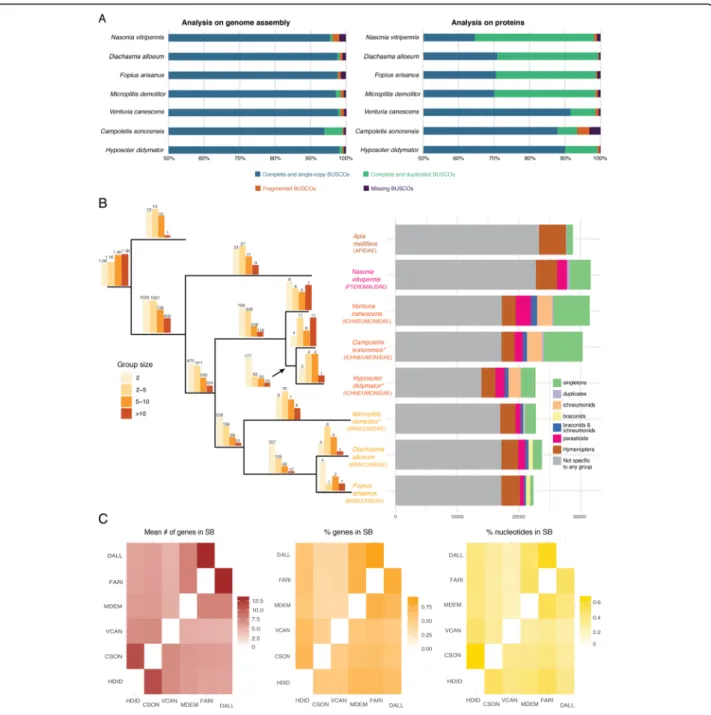
Automatic gene annotation for H. didymator (for which RNA-seq datasets were available) and for C. sonorensis(for which no RNA-seq dataset was available) yielded 18,119 and 21,915 transcripts, respectively (Table 2). These two genome assemblies and annota-tions are available at BIPAA website [46, 47]. Although different software packages were used for gene predic-tion, the two species have similar gene annotation statis-tics, except for transcript size, which was longer in H. didymator, and which also showed a higher predicted in-tron size (Table2). Benchmarking Universal Single-Copy Orthologs (BUSCO) [48] analyses indicated a high level of completeness of the two genome assemblies and an-notations, with 99% of the BUSCO Insecta protein set (1658 proteins) identified as complete sequences (Fig.1a).
Orthologous gene families were identified with Ortho-finder by computing each pair’s similarity among the proteomes of different parasitoid wasps, either harboring polydnaviruses (the braconid Microplitis demolitor) or not (the ichneumonid Venturia canescens, the braconids Fopius arisanusand Diachasma alloeum, and the ptero-malid Nasonia vitripennis). For H. didymator and C. sonorensis, a total of ~ 10,000 orthogroups were
identified (Fig. 1b; Additional file 2: Table S2). The orthogroups included a large majority of the H. didyma-tor (87.1%) and C. sonorensis (71.4%) genes. Among those, only a small portion corresponded to species-specific orthogroups: 11 orthogroups for H. didymator (69 genes) and 32 for C. sonorensis (288 genes). The number of shared orthogroups decreases with the in-creasing evolutionary distance among the other species (higher in the campoplegine ichneumonid Venturia canescens to lower in the dipteran Drosophila
Table 1 Statistics for Hyposoter didymator and Campoletis sonorensis assembled genomes. Summary statistics are compared to other selected parasitoid genomes. Assemblathon2 [42] was used to calculate metrics of genome assemblies
Family Ichneumonid Braconid Pteromalid
Species Hyposoter didymator* Campoletis sonorensis* Venturia canescens§ Microplitis demolitor* Fopius arisanus§ Diachasma alloeum Nasonia vitripennis Number of scaffolds 2591 11,756 62,001 1794 1042 3968 6098 Total length (Mbp) 198.7 258.9 237.8 241.2 153.6 388.8 295.8 Longest scaffold (Mbp) 15.7 6.1 0.85 7.15 5.5 6.61 33.57 Scaffold N50 (Mbp) 4.00 0.73 0.114 1.14 0.98 0.65 0.90 Median scaffold size (nt) 1941 3200 233 2621 12,305 4372 2037 Contig N50 (bp) 151,312 315,222 15,077 14,499 59,408 50,453 18,840 %N 1.35% 0.40% 1.70% 14.65% 8.24% 6.16% 19.33% GC (%) 39.5% 37.2% 39.33% 25.99% 35.42% 36.09% 33.65% Reference / / [22] [31] [43] [44] [45] *Species carrying PDV;§
species that produces virus-like particles (devoid of DNA)
Table 2 Gene annotation statistics for Hyposoter didymator and Campoletis sonorensis assembled genomes. Statistics are given for transcripts, exons, introns, and coding sequences (CDS)
Hyposoter didymator Campoletis sonorensis Transcript number 18,119 21,915
Total transcript size (nt) 95,868,518 68,669,265 Mean transcript size (nt) 5291 3133 Median transcript size (nt) 2428 1934 Total exon number 98,639 93,590 Mean exon number 5.4 4.3 Median exon number 4 3 Total exon size (nt) 28,900,964 27,144,418 Mean exon size (nt) 292 290 Median exon size (nt) 186 192 Total intron size (nt) 66,540,946 41,453,172 Mean intron size (nt) 826 578 Median intron size (nt) 245 296 Total CDS size (nt) 28,900,964 27,144,418 Mean CDS size (nt) 1595 1239 Median CDS size (nt) 1090 806
Fig. 1 Genomic features of Campoletis sonorensis and Hyposoter didymator genomes. a BUSCO analysis of parasitoid wasp genomes (Insecta protein set with 1658 proteins). On the left, results using the genome assemblies; on the right, results using the predicted protein set. b Orthogroups analysis. Left panel: Barplots above each branch of the phylogenic tree indicate the number of orthogroups specific to each species or group of species; the color of the bar indicates the size range of the corresponding orthogroups. Phylogenetic tree was constructed by aligning the complete genomes with Cactus ([49]), converting the resulting HAL alignment to MAF and then to multi fastas with the requirement of full alignment (all taxa present); fasta files were then concatenated into a single matrix (620 kb) and used in a maximum likelihood analysis with RAxML [50] with 1000 fast bootstrap replicates. Asterisks indicate the species carrying polydnaviruses. Right panel: Number of genes for each species that were (i) specific to the species and present either as singletons or duplicates, (ii) present in ichneumonids, (iii) present in braconids, (iv) present in both ichneumonids and braconids, (v) present in all parasitoids, and (vi) present in all hymenoptera. c Heatmaps indicating, for each species pair, the mean number (#) of genes in synteny blocs (SB), the percentage (%) of genes in SBs, and the size of the genome (% nucleotides) in SBs. HDID, Hyposoter didymator (ichneumonid, with PDV); CSON, Campoletis sonorensis (ichneumonid, with PDV); VCAN, Venturia canescens (ichneumonid); MDEM, Microplitis demolitor (braconid, with PDV); FARI, Fopius arisanus (braconid); DALL, Diachasma alloeum (braconid)
melanogaster; Additional file 2: Table S3). Among the orthogroups shared by H. didymator and C. sonorensis genes, 313 were specific to these two ichnovirus-carrying species (Fig.1b; Additional file2: Table S4), representing 875 genes for C. sonorensis and 509 genes for H. didymator.
Global synteny analysis revealed a number of syntenic blocks between the two genomes, enabling evaluation of the genomic reorganization between the two species, even using fragmented assemblies. When comparing the C. sonorensis and H. didymator genomes, the mean number of genes per synteny block obtained was 11.2, one of the highest pairwise values for the evaluated spe-cies (Additional file 3: Table S5), including one other campoplegine species, Venturia canescens (mean num-ber of 7 genes per synteny block). The percentage of re-gions in syntenic blocks shared between C. sonorensis and H. didymator compared to the complete genome size was 67% for H. didymator and 50% for C. sonorensis (Fig. 1c). The percentage of genes that are located in syntenic blocks was 71% for H. didymator and 54% for C. sonorensis (Fig. 1c). The high pairwise values show that global collinearity of H. didymator and C. sonorensis genomes is well conserved, as expected by their close evolutionary relationship.
The two campoplegine genomes include numerous and dispersed ichnovirus loci
To analyze the relationship between the wasps and their endogenous viruses, we identified the location of the viral sequences in those genomes using blast searches with available ichnovirus sequences. In the assembled C. sonorensisgenome, a total of 35 scaffolds, ranging in size from 2.3 kbp to more than 6 Mbp, contained C. sonoren-sisichnovirus (CsIV) sequences (Fig.2A; Additional file4: Table S6). Within these scaffolds, 40 viral loci were iden-tified, corresponding either to viral segments, to clusters of replication genes (IVSPERs), or to isolated replication genes. A total of 31 proviral segments were recognized, with sizes varying from 6.4 to 23.2 kbp (Additional file4: Table S6). They included all segments reported in a pre-vious study [37] except for two (Table 3). Eight previ-ously uncharacterized segments, named CsX1 to CsX8, were additionally identified (Table 3). Two short scaf-folds each contained a repeat element gene (i.e., a mem-ber of a gene family known to be encoded by ichnovirus segments) and were considered as probable additional viral segments (Table 3). Altogether, the C. sonorensis genome contained 33 loci corresponding to CsIV pro-viral segments. Finally, and for the first time in C. sonor-ensis, we identified 48 replication genes located in six different scaffolds, corresponding to five clusters (named Cs_IVSPER-1 to Cs_IVSPER-5) and two isolated genes (named IVSP_U36L and IVSP_U37L). The IVSPERs in
C. sonorensis varied in size from 8.6 to 33.3 kbp and contained from 3 to 19 genes (Additional file 4: Table S6; Additional file5: Table S7).
In the H. didymator assembled genome, a total of 60 proviral loci were identified (Fig. 2A); they were located in 32 scaffolds ranging in size from 1.5 kbp to over 15 Mbp (Additional file4: Table S6). Loci corresponding to all the previously described H. didymator ichnovirus (HdIV) segments [36] were identified in the wasp gen-ome (Table 3). When the first HdIV packaged genome was sequenced [36] some HdIV circular molecules shared part of their sequences (i.e., segments Hd2a and Hd2b, Hd11a and b, Hd17a and b, Hd20a and b, Hd26a and b, Hd31 and Hd34). Mapping on the H. didymator genome revealed that the six segments pairs actually each co-localized at the same proviral locus (Add-itional file 6: Fig. S1). Thanks to the availability of the genome, we found three HdIV segments present in two copies that were not identical but clearly recognizable as duplications (Fig. 2B); two had copies in the same scaf-fold (Hd23.1 and Hd23.2; Hd44.1 and Hd44.2), one in two different scaffolds (Hd45.1 and Hd45.2). Finally, Hd9 was tandemly duplicated at a single locus (Fig.2B). In addition, six previously uncharacterized segments were identified (named Hd46 to Hd51). Altogether, 54 HdIV proviral segments were found, ranging in size from 2.0 to 17.9 kbp (Additional file4: Table S6). Finally, new replication genes were identified in the H. didyma-torgenome. In addition to the three IVSPERs previously described [16], two novel clusters (names Hd_IVSPER-4 and Hd_IVSPER-5) and one isolated gene (IVSP_U37) were identified, making up a total of 54 predicted repli-cation genes present in H. didymator genome. All except one were organized in five IVSPERs, varying in size from 1.6 to 26.6 kbp (Additional file 4: Table S6; Additional file5: Table S7).
Analysis of the genome assemblies revealed a large number of widely dispersed viral loci, separated by wasp sequences with a median size of 115.1 kb for viral frag-ments located on the same scaffold (Additional file 7: Table S8, Fig. S2). To independently confirm dispersion of ichnovirus proviral segment sequences, we carried out fluorescent in situ hybridization in H. didymator, using genomic clones including viral segment loci as probes (Fig.2C). Four probes were used, containing Hd11, Hd6, Hd30, and Hd29 segments, all corresponding to different genomic scaffolds. Each of the probes hybridized with a different chromosome, indicating that HdIV segments are indeed widely dispersed across the H. didymator genome.
To assess whether dispersion of the viral loci could have been mediated during genome evolution by transposable elements, distribution of TEs was investi-gated in the regions surrounding the proviral loci.
The analysis of the families of transposable elements in the regions surrounding H. didymator proviral se-quences (Additional file 8: Table S9) did not reveal any particular enrichment that might suggest a role of TEs in the dispersion of the ichnovirus sequences in the wasp genomes.
Ichnovirus proviral segments harbor direct repeats with variable architecture and multiple putative excision sites
PDV segments are circularized by homologous recombin-ation between direct repeats (DRJs) located at each end of the proviral segment. This mechanism has been verified for bracoviruses, where the DRJs contain a conserved tetramer which is the potential excision site [30]. For
ichnoviruses, although the same mechanism is assumed to take place, based on the analysis of the only two proviral segments for which sequence was available [25,34], it was unknown whether or not ichnovirus DRJs contain a con-served motif, and if so, whether this motif was similar to that described for bracoviruses. To investigate if the exci-sion process for ichnovirus proviral segments could poten-tially rely on mechanisms similar to those described for bracoviruses, we searched for direct repeats at the extrem-ities of the newly identified HdIV and CsIV proviral seg-ments. Flanking direct repeated sequences were found for the large majority of HdIV and CsIV loci. All HdIV seg-ment loci, except for Hd45.1 and Hd45.2, had DRJs, which varied significantly in size, ranging from 69 to 949 bp
(See figure on previous page.)
Fig. 2 Distribution of ichnovirus sequences within Campoletis sonorensis and Hyposoter didymator genomes. A Schematic representation of ichnovirus sequences within wasp genome scaffolds. (a) C. sonorensis scaffolds containing viral loci. C. sonorensis ichnovirus (CsIV) segments are named CsA to CsX8. Segments CsP and CsL, located in short scaffolds, are not shown. IVSPER-1 to IVSPER-5 corresponds to clusters of replication genes; U36L and U37L to isolated replication genes. (b) H. didymator scaffolds containing viral loci. H. didymator ichnovirus (HdIV) segments are named Hd1 to Hd51. Segments Hd45.1, Hd46, and Hd51, located in short scaffolds, are not shown. IVSPER-1 to IVSPER-5 corresponds to clusters of replication genes; the isolated replication gene U37, located in a short scaffold, is not shown. Complete scaffold list available in Additional file
4: Table S5. B Segments duplicated in H. didymator genome. Segments Hd23 (Genbank# KJ586309.1), Hd44 (Genbank# KJ586285.1) and Hd45 (Genbank# KJ586284.1), described as part of the packaged HdIV genome in [36], have two copies (named Hd(n).1 and Hd(n).2) that are either in the same scaffold (Hd23.1 and Hd23.2, Hd44.1, and Hd44.2) but in different insertion sites or in two different scaffold (Hd45.1 and Hd45.2). Nucleotide percentage identity between the two related segment sequences is given on the right part of the figure. By contrast, Hd9 (Genbank# KJ586324.1), initially described as a separate segment, is located in a genomic locus composed of a tandemly duplicated sequence (“copy 1” and “copy 2” in the diagram). C FISH on H. didymator chromosomes using BAC genomic clones containing HdIV segments. Upper panel shows hybridization using the probes containing segments Hd11 (labeled with FITC) and Hd6 (labeled with rhodamine); lower panel the probes containing viral segments Hd30 (labeled with FITC) and Hd29 (labeled with rhodamine). Each of the probes hybridized with a different H. didymator chromosome: Hd11 hybridized with chromosome #12, Hd6 to a medium-sized chromosome (potentially #5), Hd30 with chromosome #2 and Hd29 with chromosome #11
Table 3 Summary of the number of viral loci identified in the genomes of Campoletis sonorensis and Hyposoter didymator, in comparison with data available in NCBI database
Campoletis sonorensis Hyposoter didymator
Number of segments in NCBIa 25 50
Number of segments in genome 31 54
NCBI segments not found in genome 2 (CsA2, CsK) 0
Merged segments (compared to NCBI)b 0 6 (Hd2, Hd11, Hd17, Hd20, Hd26, Hd31–34)
Duplicated segments (compared to NCBI)c 0 3 (Hd23, Hd44, Hd45)
Number of newly identified segments 8 (CsX1 to CsX8) 6 (Hd46 to Hd51) “Isolated” segment genes (short scaffolds) 2 1
Total number of segment loci in genome 33 55
Number of IVSPERs in NCBIa 0 3
Number of IVSPERs in genome 5 5
NCBI IVSPERs not found in genome na 0
Newly identified IVSPERs 5 2
“Isolated” IVSPER genes 2 1
Total number of IVSPER loci in genome 7 6
a
Numbers of segments and IVSPER deposited in NCBI and available before this study
b
H. didymator viral loci corresponding to two segments formerly deposited in NCBI as distincts
c
(Additional file 9: Table S10). Similarly, most CsIV seg-ments (25 of 32) were flanked by DRJs, which ranged in size from 99 bp to as much as 1132 bp (Additional file8: Table S9). The main finding that emerged from the avail-ability of dozens of ichnovirus DRJs is their segment speci-ficity in terms of length and sequence. The number of direct repeats also showed high variability across proviral sequences (Additional file9: Table S10; Additional file10: Fig. S3) even though the majority of the HdIV (28) and CsIV (19) segments contained a single repeated sequence, one copy located on their right and left ends. A few HdIV and CsIV segments also contained internal repeats of the same sequence, potentially allowing the generation of more than one related circular molecule by recombination between different pairs of DRJ copies (nested segments). Other ichnovirus proviral segments (21 HdIV segments, but only one CsIV segment) contained two different re-peated sequences, named DRJ1 and DRJ2, present in two or more copies. Note that the segments initially described as distinct [36] but which were found at the same locus are segments flanked either by a DRJ1 or by a DRJ2 (see Additional file6: Fig. S1). As an example, the proviral seg-ment Hd2 displays two different types of DRJs, which con-sequently allows generation of two segments that share part of their sequence: segment Hd2a, generated through recombination between DRJ1; and segment Hd2b, through recombination between DRJ2 (Additional file 6: Fig. S1). Occurrence of several repeats differing in se-quence and in position suggests that a mixture of overlap-ping and/or nested segments may be generated by homologous recombination in this context.
With the aim of assessing whether a conserved exci-sion site motifs is embedded in ichnovirus DRJ se-quences, as described for bracovirus segments, we used the “regulatory DNA motif identification and analyses” (DMINDA) webserver [51] using all 99 DRJs identified in the two wasp species. From all the motifs found, seven occurred in 70% of all 99 DRJs, and two motifs oc-curred at least once in 98% of the analyzed DRJs (Add-itional file 10: Table S11). However, analysis of the H. didymator genome revealed this motif had the same chance of occurring in the DRJs as in the rest of the wasp genome (Additional file 10: Table S12). Hence, circularization of ichnovirus segments probably does not rely on the presence of a conserved nucleotide motif as in bracoviruses.
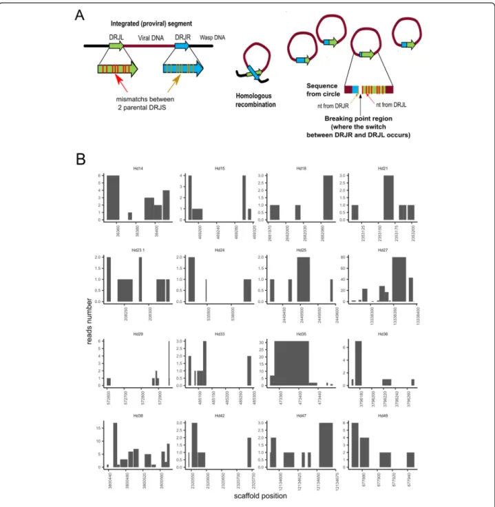
In the absence of a known conserved motif, we searched for potential excision sites allowing to generate ichnovirus circular molecules in H. didymator using an algorithm developed for this purpose, DrjBreakpointFin-der (see“Methods”). The rationale is illustrated in Fig.3a. To identify the excision site, or“breakpoint,” in a given recombined DRJ sequence (i.e., the circular molecule), the latter was compared to sequence of the two copies
present at each end of the segments in their linear inte-grated form. These two copies exhibit several sequence differences (Additional file9: Table S10), allowing identi-fication of the excision site in the corresponding recom-bined DRJ sequence with certain accuracy, depending on the divergence between the parental DRJ copies. Thus, two sets of H. didymator segment sequences (circular DNA molecules extracted from H. didymator ichnovirus particles) were analyzed using DrjBreakpointFinder. Au-tomated analysis of this large set revealed that excision could occur in different sites within a same DRJ, al-though some positions were more frequent than others for a given DRJ (Fig. 3b). This unexpected variability in the excision site position was confirmed for a subset of eight segments by manual comparative analysis of seg-ment isoforms sequenced using Sanger technology (Additional file11).
Proviral sequences serving as templates for ichnovirus packaged genome show features specific to each campoplegine species
To understand evolutionary changes experienced by pro-viral segments in the two different wasp species, we ana-lyzed their characteristics, gene contents, and positions within the two genomes. Altogether, ichnovirus segment loci represent a similar total size for the two species (307.1 kbp for HdIV and 314.1 kbp for CsIV). The total sizes of the endogenous viruses are slightly underesti-mated since two HdIV segments (Hd1 and Hd45.1) and five CsIV segments (CsV, CsX3, CsX4, CsX5, and CsX7) were only partially identified due to genome fragmenta-tion (see Addifragmenta-tional file 4: Table S6 for details). How-ever, general segment features differed between the two wasp species. Prior sequencing of packaged genomes had already highlighted differences between the viral segments in C. sonorensis (CsIV) and H. didymator (HdIV) [36, 37]. CsIV particles enclose only half the number of segments compared to HdIV particles, and CsIV viral segments are on average longer than HdIV segments. Accordingly, the number of proviral loci iden-tified in the H. didymator genome is higher (n = 54) than in the C. sonorensis genome (n = 33), while CsIV proviral segments are on average longer than HdIV ones (Add-itional file12: Fig. S4). Regarding gene number, 111 were predicted for CsIV segment genes whereas a total of 152 genes were predicted in the HdIV segments (Table 4; Additional file 5: Table S7). Within-segment gene com-position varied considerably between the two species. Both encapsidated genomes contain members of the ichnovirus-conserved multimembers families (repeat element genes, vankyrins, vinnexins, cys-motif, and N-genes), but in variable number (Table 4). HdIV contains more viral innexins, whereas CsIV more viral ankyrins and repeat element genes (Table4). In addition, each of
Fig. 3 Segment DRJ variability in terms of excision sites in Hyposoter didymator. a Schematic representation of the homologous recombination between the two DRJs flanking the proviral sequence (left DRJL and right DRJR ends) to produce the circular molecule (segment) containing one recombined DRJ sequence. When excision sites are located at different positions in the DRJ, segments differing in their recombined DRJ sequence are generated. Excision occurs more frequently at some positions, resulting in different relative amounts of each isoform. On the right, rationale of the algorithm developed to identify the“break points.” Mapping of the segment sequence (DRJsegment) with the two parental DRJs, which differ in their sequences (nucleotide (nt) mismatches), allows identification of the regions where the switch from one parental DRJ to the other has occurred (in the diagram, between the first and second mismatch). b Prediction of putative recombination break points in H. didymator DRJs. Each graph corresponds to the left copy of the DRJ for a given segment (indicated below each graph). The X-axis is the position in the scaffold. The Y-axis indicates the number of reads (obtained from sequencing of the packaged circular DNA molecules) confirming that the circle has been recombined between these two positions, based on the observed mismatches at both end of the segment for each read. We observed between 1 and 80 reads per breakpoint region according to the analyzed segment
the genomes enclosed a number of genes (encoding hypothetical proteins; Table 4) specific either to C. sonorensis or to H. didymator. Overall, ichnovirus seg-ments are characterized by gene content that is quite specific to each one of the two wasp species.
The high collinearity in gene order observed between the genomes of H. didymator and C. sonorensis made it possible to assess if the viral segments were located in the same genomic regions in the two species. We com-pared the sequences flanking viral insertions in H. didy-mator with their syntenic genomic regions in C. sonorensis(Additional file12: Fig. S5). For the large ma-jority of syntenic blocks containing an HdIV segment, there was no viral locus in the corresponding C. sonoren-sis block (Additional file12: Fig. S5 a). Two exceptions were found for two segments present in the same H. didymator scaffold (scaffold 351). H. didymator segment Hd18 was flanked by the same wasp genes as C. sonoren-sisIVSPER-5 (Additional file12: Fig. S5 b), and H. didy-mator segment Hd17 was inserted in the same wasp genomic region as C. sonorensis segment CsZ, a region that seemed to have undergone rearrangements like gene duplications (Additional file12: Fig. S5 c). H. didy-mator Hd17 and C. sonorensis CsZ both contain genes from the repeat element genes family, suggesting the two segments may have arisen from the same ancestral sequence, already inserted in this wasp locus, prior to wasp species diversification.
The ichnovirus machinery retained in wasp genomes (IVSPERs) is well conserved
To understand the evolutionary changes experienced by the IVSPER clusters, we analyzed their gene contents, and positions in the wasp genomes. A total of 45 differ-ent predicted IVSPER gene families were iddiffer-entified in the genomes of H. didymator and C. sonorensis (Add-itional file 5: Table S7). The majority (35, or 78%) are shared by both wasp species, with a number of gene
copies within a multigene family that may differ between the two wasp species. Among the 36 different genes/ gene families (corresponding to a total of 48 genes) iden-tified in the C. sonorensis IVSPERs, only one had no homolog in the H. didymator genome. This gene, Gf_ U27L, has similarities (BlastP e-value 1E−31) with an IVSPER gene described in the banchine G. fumiferanae [20]. Among the 44 distinct gene families identified in H. didymatorIVSPERs (corresponding to 54 genes), nine were not detected in the C. sonorensis genome. Of these, three genes (U29, U32, and U33), transcribed in H. didy-mator ovarian tissue based on our transcriptome data (see“Methods” section), had not been previously charac-terized in H. didymator. They were classified as IVSPER genes because of their clustering with other conserved IVSPER genes (within IVSPER-4).
The IVSPERs of the two wasp species show high synteny (Fig. 4A). Regions with conserved gene order are shared, for instance, between Hd_IVSPER-1 and Cs_IVSPER-5, Hd_IVSPER-2 and Cs_IVSPER-2, or Hd_IVSPER-3 and Cs_IVSPER-1. However, there are also rearrangements, inversions, and deletions when the two species are compared. The highest number of rearrangements involves Cs_IVSPER-1 and Cs_ IVSPER-2, with homologs of Cs_IVSPER-1 and Cs_ IVSPER-2 genes dispersed in several different H. didy-mator IVSPERs. Overall, and in contrast with proviral segments, IVSPER are well conserved in terms of gene content and order when comparing the two campoplegine species, suggesting this organization may be required for the IVSPER genes biological function, i.e., to produce the virus particles.
To assess if the IVSPER loci were located in the same genomic regions in H. didymator and C. sonorensis, we compared the genomic regions containing viral inser-tions in H. didymator with their syntenic genomic re-gions in C. sonorensis (Fig. 4B). For three H. didymator loci (Hd_IVSPER-1 and -2, U37), no corresponding C. sonorensis scaffold was found (Fig. 4B, a), making the comparison inconclusive. On the other hand, compari-son was possible for the remaining H. didymator IVSPER loci. For H. didymator IVSPER-3, there was no viral locus in the corresponding C. sonorensis block (Fig. 4B, c). Conversely, 4 and Hd_IVSPER-5, which are both IVSPERs containing a reduced number of genes, are flanked by orthologous wasp genes com-pared to the Cs_IVSPER-4 and U36L loci in C. sonoren-sis genome (Fig. 4B, b and c), indicating that these IVSPER loci have a conserved genomic location in the two genomes. Hence, as observed for viral segments, IVSPERs seem quite mobile in the wasp genomes, except for two small gene clusters that remained in a similar, putatively ancestral, location. The high within-cluster conservation, even between distantly related wasp
Table 4 Comparative segment gene content for the ichnoviruses carried by the campoplegine wasps Hyposoter didymator (HdIV) and Campoletis sonorensis (CsIV)
IV gene family HdIV CsIV Repeat element genes 38 51
Viral innexins 17 6 Viral ankyrins 10 16 Cys-motif proteins 9 13 Polar-residue-rich proteins 5 nd N-genes 3 3 Total 82 89 Hypothetical proteins 70 22 TOTAL 152 111
Fig. 4 Comparative analysis of Campoletis sonorensis and Hyposoter didymator IVSPERs. A Synteny between the IVSPERs identified in H. didymator (Hd) and C. sonorensis (Cs) genomes. B Synteny of H. didymator genomic regions containing IVSPERs compared with C. sonorensis and other parasitoid genomes. (a) Synteny for H. didymator genomic region containing IVSPER-1 and IVSPER-2 (genes from HD016092 to HD016153); no C. sonorensis scaffold corresponded to the H. didymator IVSPER insertion sites. (b) Synteny for H. didymator genomic region containing IVSPER-4 (genes from HD001703 to HD001771); H. didymator IVSPER-4 and C. sonorensis IVSPER-4 are inserted in the same genomic environment. c Synteny for H. didymator genomic region containing IVSPER-3 and IVSPER-5 (genes from HD002066 to HD002111); in the region where H. didymator IVSPER-3 is inserted, there is conservation in gene order compared to C. sonorensis but no viral insertion; conversely, H. didymator IVSPER-5 and C. sonorensis IVSPER-5 are inserted in the same genomic environment. H. didymator genes from HD010503 to HD010526. Hd: Hyposoter didymator; Cs: Campoletis sonorensis; Vc: Venturia canescens (ichneumonid that has lost the ichnovirus [22]); Md: Microplitis demolitor (braconid with a bracovirus); Fa: Fopius arisanus (braconid with virus-like particles). Numbers following the species name correspond to scaffold number for Hd, Cs, and Vc, NCBI project codes for Md and Fa. Triangles within genomic regions correspond to predicted genes; triangles of the same color correspond to orthologs; white triangles are singletons or orphan genes. For better visualization, the name of the gene is indicated only for some viral (in red for segments, in blue for IVSPERs) genes. See Additional file13: Table S13, for H. didymator genes list
species, suggests that they may move as a whole within the wasp genomes by mechanisms still to be elucidated.
Discussion
Dispersal of proviral loci through the wasp genome
The genome assemblies obtained allowed us to perform a comprehensive mapping of the viral inserts into the wasp genome. A major finding is the highly dispersed distribution of the ichnovirus proviral segments. All but two of the 32 CsIV segments loci are located in different scaffolds. Similarly, half of the 54 HdIV viral segments are located in different scaffolds; those located in the same scaffold are usually separated by relatively large, sometimes megabase-long portions of wasp genome. This dispersion was confirmed by FISH experiments for H. didymator, showing that viral loci are distributed across multiple chromosomes. Organization of ichno-virus proviral segments is therefore quite different from that of bracoviruses. Bracovirus segments are generally fewer compared to ichnoviruses, and they are for the most part clustered in a single locus, named the viral macro-locus [15,31,52]. To illustrate this, the Micropli-tis demolitorgenome contains 26 proviral segments dis-tributed in only eight loci, with 14 segments located at the same locus [31]. In contrast, ichnovirus genomes consist of a series of single viral segments scattered throughout the wasp genome. As no enrichment of transposable elements surrounding the ichnovirus seg-ments has been observed, their dispersal in the wasp genome may result from reintegration events, multiple genomic rearrangements events, or yet another still un-known mechanism. In addition, based on the lack of conservation in their genomic position when comparing H. didymatorand C. sonorensis, ichnovirus viral segment diversification and dispersion may result from transpos-ition of individual viral sequence while, for bracoviruses, segment multiplication occurs mainly by duplication of large areas [15].
Conserved IVSPER structure and its significance
As previously described, the proviral segments are the template for the DNA molecules that are packaged and transferred to the parasitized host, whereas replication genes, clustered in IVSPERs, are involved in the produc-tion of the virus particle. Until now, replicaproduc-tion genes were known solely for one campoplegine wasp, H. didy-mator[16], and a banchine species, G. fumiferanae [20]. Our study discovered replication genes in another cam-poplegine wasp genome, C. sonorensis, and revealed a conserved IVSPER architecture when comparing the two campoplegine species. In both H. didymator and C. sonorensisgenomes, the majority of the recognized repli-cation genes are clustered. Indeed, only two isolated rep-lication genes were identified in the C. sonorensis
genome and only one in the H. didymator genome. A large portion of the IVSPER genes are shared between H. didymator and C. sonorensis and arranged in a con-served order. Furthermore, most genes found in campo-plegines are also present in the banchine G. fumiferanae, though the gene order is less conserved in this case [6].
The two ichneumonid subfamilies that harbor PDVs, Banchinae and Campopleginae, do not form a monophy-letic group [53, 54], and ichnoviruses are not reported for other subfamilies in the same lineage [55]. Hence, phylogenetic evidence would suggest separate origins for PDVs in Ichneumonidae. On the other hand, a high pro-portion of IVSPER genes are shared between campople-gine and banchine wasps, including the D5 primase-like and DEDXhelicase-like first described in the banchine G. fumiferanae (corresponding to U37 and U34 respect-ively in H. didymator). This similarity would suggest a common viral ancestor, or related viral ancestors for campoplegine and banchine ichnoviruses. Better under-standing of the evolutionary trajectories of IVSPERs across ichneumonid lineages requires additional sequen-cing of banchine wasp genomes, as well as a thorough screening of species from other subfamilies for the pres-ence of endogenous viruses.
Specific features of bracoviruses and ichnoviruses genome architecture
Our study provides the opportunity to make direct com-parisons of viral composition between ichneumonid and braconid genomes. The genomes of campoplegine wasps associated with ichnoviruses contain numerous dis-persed viral loci consisting of single viral segments and clusters of replication genes. By contrast, genomes of braconid wasps associated with bracoviruses have clus-tered viral segments and more dispersed replication genes. For instance, while proviral segments in M. demo-litorare located in only eight loci, the 76 nudiviral repli-cation genes are dispersed across the wasp genome except for two sets of 12 and 8 genes respectively, sepa-rated by a stretch of 30 kbp, the so-called “nudiviral cluster” [23]. These alternative genomic architectures likely reflect different regulatory mechanisms governing viral replication and particle production in the two PDV taxa.
In both PDV taxa, viral loci are amplified in the calyx cells starting at early pupal stages. However, whereas ichnoviral loci are probably all amplified [16], only the proviral segments and the nudiviral cluster are amplified in braconids [30]. In braconids, viral segments are orga-nized in replication units delimitated by palindromic AT-rich regions (amplification junction sites), an organization that allows simultaneous co-amplification of several segments [30,32]. Amplification junction sites were detected only for the segment DNA, but not at
vicinity of the nudivirus-like cluster [30], which suggests that mechanisms governing viral DNA amplification may differ between the two types of sequences in braco-nids. In contrast, knowledge on the mechanisms govern-ing ichnovirus loci amplification is still lackgovern-ing. Based on their genomic organization, ichnovirus segments are most probably individually amplified, suggesting that segment viral DNA amplification relies on distinct mechanisms in the two PDV taxa. However, more stud-ies are necessary to determine whether or not sequences longer than the proviral ichnovirus segments—presently delimited by DRJs—are amplified, and if so, to analyze the flanking sequences of these ichnoviral “replication units” in order to identify potential amplification junc-tion sites. Similarly, further studies are needed to characterize the limits of the amplified IVSPERs to iden-tify potential amplification sites that could be compared to those of ichnovirus segments.
Different genomic architectures may have conse-quences for the mechanisms regulating the expression levels of replication genes in the calyx cells. In the case of bracoviruses, DNA amplification at the nudiviral clus-ter results in high expression levels of the corresponding genes [56]; however, transcriptional control also relies on the nudiviral RNA polymerase that allows expression of viral genes whatever their location. In ichneumonids, increase in gene copies is one, but also probably not the only, mechanism involved in transcriptional control. In-deed, IVSPER genes differ in their expression level in H. didymator calyx [35], which suggests involvement of other gene-specific mechanisms. Moreover, the cluster-ing of replication genes in ichneumonids may facilitate coordinated regulation of their expression.
Mechanism of excision of proviral loci in ichnoviruses and bracoviruses
Direct repeat junctions (DRJs) have been identified for most of the H. didymator and C. sonorensis proviral seg-ments. DRJs present at the extremities of the integrated form of the viral segment were first described for CsIV [25], and for the bracoviruses associated with Chelonus inanitus [57] and Cotesia congregata [26]. The presence of internal repeats, which allows the generation of mul-tiple circular molecules from the same proviral template in a process termed“segment nesting,” has also been re-ported, mainly for ichnoviruses [25] and very occasion-ally for bracoviruses [15].
Some of the ichnovirus proviral segments identified in this work lacked terminal direct repeats (five CsIV seg-ments of the 32 identified and two HdIV segseg-ments), which suggests they have lost their ability to be excised. For four CsIV segments, this is consistent with the fail-ure to observe their circular form when the packaged genome was sequenced [37]. Pseudo-segments may be
generated from a mutation in the DRJ or from the re-integration of circular forms, thus harboring a single copy of the initial DRJ. The first case has been docu-mented in the braconid Cotesia congregata and the sec-ond in C. sesamiae [15, 58]. In H. didymator and C. sonorensis, we have not detected any repeated sequence at the ends of the aforementioned loci, making the reintegration of circular forms the most plausible hypothesis.
The mechanism and the proteins involved in DNA ex-cision remain to be identified for ichnoviruses. In the case of bracoviruses, there is a first step of amplification of replication units, which contain one or several pro-viral segments delimited by DRJs and surrounded by wasp intervening and flanking sequences [30, 32]. DRJs contain a conserved AGCT tetramer embedded within a larger motif, which corresponds to the site of excision [15,23,59]. The DRJs would act later separating the dif-ferent bracovirus segments in the amplified molecule, thus generating the circular molecules. Bracoviruses have conserved tyrosine recombinase family members of nudiviral origin which are likely involved in regulating this excision step [27].
For ichnoviruses, the present work highlights the vari-ability of DRJs in terms of sequence length and level of homology, and the lack of a detectable motif as found in bracoviruses. Ichnovirus proviral segments are individu-ally amplified and currently there is no data on the limits of the replication units, making it difficult to assess whether DRJs are directly involved in the excision of the segment, or whether there is also a two-step process in-volving additional sequences. Moreover, no virus-derived recombinase has been identified in ichnoviruses so far. It is therefore very unlikely that IV excision relies on a site-specific recombination mechanism as bracov-iruses. The distribution of breakpoints, which spread out over the whole length of the DRJ in provirus circles, sug-gests that a homology-based mechanism is involved. Within the context of circularizing a segment delimited by two direct repeats, mechanisms of homologous re-combination (HR) or single-strand annealing (SSA) can be envisioned, but their outcomes are undistinguishable from each other [60,61]. HR repair is defined by a step of strand invasion catalyzed by the strand-exchange pro-tein Rad51, where one single stranded DNA segment in-vades a double-stranded homologous sequence. HR has the potential of generating products with or without crossover, but only crossovers can generate circles from recombination between direct repeats. The formation of crossover is usually infrequent in somatic cells and would therefore require some specific regulation in order to produce circles with high yield. On the other hand, the mechanism of SSA seems particularly plaus-ible, because this DNA double-strand break repair
mechanism involves annealing of homologous repeat se-quences and produces a deletion rearrangement between the repeats, or it can similarly generate a circle by an-nealing two direct repeats on a linear fragment [62]. However, even low levels of sequence divergence (< 10%) between the repeats have been shown to inhibit strongly HR and SSA [63, 64], making it difficult to ex-plain the efficiency of the process between CsIV and HdIV DRJs, which have on average only 85% identity. One possibility is that the excision process is less sensi-tive to sequence divergence than in previously studied systems: possibly because it takes place on amplified fragments outside the context of the chromosome, or because specific regulations take place in calyx cells. Al-ternatively, it has been proposed that the repair between divergent repeats can be taken on by a composite mech-anism involving early steps of SSA to align the homolo-gous sequences of the DRJs and a bridging mechanism based on annealing between very short homology stretches (< 10 bp), the alternative end joining (ALT-EJ) [62,64]. Newly integrated proviral segments could have initially been excised through the ALT-EJ pathway, which is not expected to favor specific junction sites. From this ancestral situation, the acquisition of direct repeats would have fixed the junctions, allowing for a decrease of non-functional circles due to uncontrolled deletions and maybe a higher efficiency.
Our data also confirms the high level of segment nest-ing in ichnovirus segments, which may harbor multiple DRJs that differ in sequence and number. This complex-ity suggests the capaccomplex-ity to produce, from a single tem-plate, a series of related circular molecules by intrachromosomal homologous recombination. The pos-sibility for this system of generating a large variety of molecules is further accentuated by the finding, using an automated search, of various possible excision and re-combination sites within DRJs, with some sites appear-ing to occur more frequently than others.
Evolutionary implications
Two main conclusions arise from the comparison of the two genomes, providing insights into the evolutionary forces driving ichnovirus domestication in campoplegine wasps. First, the genes potentially involved in ichnovirus particle production (replication genes) are highly con-served in terms of gene content and gene order. In both H. didymatorand C. sonorensis, there are only a few rep-lication gene clusters, and one or two isolated genes. Second, in direct contrast, the viral segments carrying virulence genes are divergent between the two species, despite the existence of common gene families. The two components of the ichnoviral genome also differ in terms of conservation of their genomic localization: pro-viral segments were not flanked by synteny blocks
shared between H. didymator and C. sonorensis, unlike the situation in braconids, where the viral segments re-main in homologous positions [15]. Conversely, two of the five IVSPERs were localized in the same genomic re-gions in H. didymator and C. sonorensis. These two IVSPER harbor related genes, which suggests a shared ancestral origin. These loci may represent ancestral viral insertion sites conserved in both wasp genomes.
PDVs in both braconids and ichneumonids provide a solution to the same adaptive demand: inducing physio-logical changes in the lepidopteran hosts to allow the survival of a koinobiont endoparasitoid. The independ-ent domestication of unrelated viruses in these two line-ages represents a remarkable example of convergent evolution, but the differences in genomic architecture in each virus group suggest that different pathways were followed in these two lineages to achieve these similar solutions. Considering the common life history strategy in both groups, why would the genomic architecture of the virus need to be so different between these groups? While this remains an unresolved question, the answer may be related to the divergent nature of the virus an-cestor, or to pre-existing differences in the genomic en-vironment of ichneumonids and braconids. Previous research has indicated that closely related species tend to share more similar genetic backgrounds, enabling them to use similar pathways to achieve adaptive solu-tions [65]. In contrast, relatively distant relatives may re-quire different biochemical or genetic mechanisms to show the same adaptive functions [66,67].
The conservation of IVSPER genes is consistent with their role in the machinery that allows the wasp to pro-duce virus particles, which presumably prevents rapid change. On the other hand, proviral segments carry viru-lence genes that need to respond to counter-adaptations arising in the immune system of the parasitoid’s host [68]. Since campoplegines are koiniobiont endoparasi-toids that often have a restricted host range [21, 69], proviral sequences are expected to evolve rapidly and in a species-specific manner. Finally, we did identify a shared syntenic block containing a proviral segment in H. didymator and an IVSPER in C. sonorensis. In a sce-nario assuming a common origin for IVSPER and viral segments (i.e., the ancestral virus), this locus may repre-sent an ancestral viral insertion site, which may have contained the complete ancestral virus genome before its separation in two components, the proviral segments, and the replication gene clusters.
Conclusions
We report the whole genome sequencing of two parasit-oid wasps, H. didymator and C. sonorensis, which both harbor integrated ichnoviruses. These annotated full ge-nomes, the first for the family Ichneumonidae, provide a
comprehensive picture of the architecture of ichno-viruses in these wasp genomes (Fig. 5). Our results re-veal a clear duality between the proviral segments and the conserved viral machinery, differences that may be linked to the biological functions of these elements. Pro-viral segments, which are delivered from the parasitoid to its host and harbor virulence genes needed for suc-cessful parasitization, are isolated and scattered across the wasp genomes, in locations that differ between the two wasp species. By contrast, the replication genes re-quired to produce the delivery system (the virus particle) are clustered in the wasp genome, and the gene content and gene order in the clusters (IVSPERs) are highly con-served between the two wasps. While conservation of the viral machinery versus diversification of the viral seg-ments is also observed in bracoviruses, ichnovirus gen-omic organization of each component is in marked contrast to that observed in bracoviruses. This leads to the hypothesis, yet to be validated, that solutions to adaptive demands can arise convergently via different evolutionary pathways. Understanding the origins of the genomic architecture of modern ichnoviruses from the domestication of an ancestral virus will require the iden-tification of the group of viruses to which the ancestrally integrated virus belongs and the sequencing of other ich-neumonid wasps from multiple lineages.
Methods
Target species and insect rearing
Two species from the monophyletic subfamily Campo-pleginae [53, 54] were chosen as target taxa for whole genome sequencing: Hyposoter didymator occurs in all Western Europe and mainly parasitizes Helicoverpa armigera [70] whereas Campoletis sonorensis occurs from North to South America and parasitizes several noctuid species [71].
Specimens of H. didymator were reared on Spodoptera frugiperda larvae as previously described [36]. Male specimens of C. sonorensis wasps were furnished from a colony maintained at the University of Kentucky and reared as described in [72].
C. sonorensis whole genome sequencing, assembly, and automatic annotation
Genomic DNA was extracted from one single adult male using the Qiagen™ MagAttract HMW DNA kit, follow-ing the manufacturer’s guidelines. The resultfollow-ing extrac-tion was quantified using a Qubit™ dsDNA High Sensitivity assay, and DNA fragment size was assessed using an Agilent™ Genomic DNA ScreenTape. Sample li-braries were prepared using 10X Genomics Chromium technology (10X Genomics, Pleasanton, CA), followed by paired-end (150 base pairs) sequencing using one lane
Fig. 5 Steps of virus domestication in ichneumonids. Following integration of an ancestral virus genome in a wasp chromosome, the viral sequences were maintained but underwent significant modifications over evolutionary time. The viral sequences including genes necessary to produce particles (IVSPERs) were conserved through evolution, although they have undergone fragmentation and gene duplications and have lost some genes, including the viral DNA polymerase. The encapsidated sequences (proviral segments) include virulence genes involved in promoting parasitism. Ichnovirus segments likely derive from ancestral viral sequences that have acquired virulence genes from the wasp. Both proviral segments and IVSPERs are amplified in the replicative tissue in a coordinated manner suggesting regulation by a common mechanism and that they may both derive from the ancestral virus. Following amplification, viral segments are excised via homologous recombination or single-strand annealing mechanism (depicted) involving the direct repeated junctions
on an Illumina HiSeqX sequencer at the New York Gen-ome Center (Additional file14: Table S14).
Assembly of the sequenced reads was conducted using Supernova v.2.1.1 [38]. Reads were mapped back to the assembled genome using Long Ranger (https://support.1 0xgenomics.com/genome-exome/software/pipelines/lat-est/what-is-long-ranger) and error correction was per-formed by running Pilon [73]. Note that Supernova recommends 56X total coverage and sequencing deeper than 56X reduces the assembly quality. A full lane of HiSeqX produced several times the sequencing data we needed to reconstruct the genome. Hence, raw reads were divided in four subsets and four separate assem-blies were conducted in parallel, with the best one chosen for downstream analyses.
We ran Kraken 2 [74] on the assembly to check for bacterial contamination. All contigs that were classified as bacteria were removed before proceeding with other analyses. We used RepeatMasker [75] to identify repeat regions using the honeybee, Apis mellifera, as the model species. H. didymator transcripts were aligned to the C. sonorensis genome using BLAT [76]. We created hints files for Augustus from the repeat-masked genome and the BLAT alignments. We also ran Benchmarking Uni-versal Single-Copy Orthologs (BUSCO) version 3.0.2 [48] with the long option both to assess genome com-pleteness and to generate a training set for Augustus. We then ran Augustus v3.3 [40] for gene prediction using the three lines of evidence, the RepeatMasker-generated hints, the BLAT-RepeatMasker-generated hints, and the BUSCO-generated training set.
H. didymator whole genome sequencing, assembly, and automatic annotation
Genomic DNA was extracted from a batch of adult males (n = 30). DNA extractions that passed sample quality tests were then used to construct 3 paired-end (inserts lengths = 250, 500, and 800 bp) and 2 mate pairs (insert length = 2000 and 5000 bp) libraries, and qualified libraries were used for sequencing using Illumina Hiseq 2500 technology (Additional file 14: Table S15) at the BGI. For genome assembly, the raw data was filtered to obtain high-quality reads.
The reads were assembled with Platanus assembler v1.2.1 [39], in 2 steps (contigs assembly and scaffolding), then the scaffold gaps were filled with SOAPdenovo GapCloser 1.12 [77]. Finally, only scaffolds longer than 1000 bp were kept for further analyzes. Assemblathon2 [42] was used to calculate metrics of genome assemblies. For annotation, EST reads from venom [78], as well as reads obtained using GS FLX (Roche/454), Titanium chemistry from total insects [78] and from ovaries [79], were mapped to the genome with GMAP [80], and Illu-mina reads published previously [35], or from the 1KITE
consortium (http://1kite.org/subprojects.html) using STAR [81], and new calyx RNA-seq (SRA accession: PRJNA590863) with TopHat2 v2.1.0 [82]. BRAKER1 v1.10 [41] was used to predict genes in the genome of H. didymator using default settings. Gene annotation was evaluated using BUSCO version 3.0.2 [48] with a refer-ence set of 1658 proteins (conserved in Insecta).
The other parasitoid genomes used in this work for comparison purposes were similarly analyzed (genomes available at NCBI for Microplitis demolitor [31] (PRJNA251518), Fopius arisanus [43] (PRJNA258104), Diachasma alloeum [44] (PRJNA306876), and Nasonia vitripennis [45] (PRJNA13660); Venturia canescens genome [22] available at https://bipaa.genouest.org/sp/ venturia_canescens/).
Manual annotation of the viral loci
Manual annotation of viral regions was performed using the genome annotation editor Apollo browser [83] avail-able on the BIPAA platform (https://bipaa.genouest.org). The encapsidated forms of the HdIV and CsIV genomes were previously sequenced with Sanger technology by isolating DNA from virions [36, 37]. H. didymator IVSPER were also previously sequenced [16]. To identify the viral loci, sequences available at NCBI for campople-gine IV segments and IVSPER sequences from campo-plegine and banchine species were used to search the H. didymator and C. sonorensis genome scaffolds using the Blastn tool implemented in the Apollo interface. To de-termine the limits of the proviral segments, we searched for direct repeats at the ends of the viral loci by aligning the two sequences located at each end using the Blastn suite at NCBI. The start or stop codons of the genes lo-cated at the ends of the IVSPER loci were considered as the borders of the IVSPER.
Transposable element detection
Transposable elements (TEs) were identified in H. didy-matorand C. sonorensis genomes using the REPET pipe-line [84, 85]. The enrichment analysis was performed using LOLA [86] comparing the observed number of each TE family member in a region encompassing IV segments and 10 kbp around, and 1000 random seg-ments of the same length extracted with bedtools shuffle [87].
Orthologous genes and syntenic regions
To identify homology relationships between sequences of H. didymator, C. sonorensis, and other parasitoids with available genomes (one ichneumonid Venturia canescens, three braconids Microplitis demolitor, Fopius arisanus, and Diachasma alloeum, and one pteromalid Nasonia vitripennis), as well as two taxonomically more distant insect sequences (the bee Apis mellifera and the