HAL Id: hal-02966287
https://hal.archives-ouvertes.fr/hal-02966287
Submitted on 4 Dec 2020HAL is a multi-disciplinary open access archive for the deposit and dissemination of sci-entific research documents, whether they are pub-lished or not. The documents may come from teaching and research institutions in France or abroad, or from public or private research centers.
L’archive ouverte pluridisciplinaire HAL, est destinée au dépôt et à la diffusion de documents scientifiques de niveau recherche, publiés ou non, émanant des établissements d’enseignement et de recherche français ou étrangers, des laboratoires publics ou privés.
Technical Note: GATE-RTion: a GATE/Geant4 release
for clinical applications in scanned ion beam therapy
L. Grevillot, D.J. Boersma, H. Fuchs, A. Aitkenhead, A. Elia, M. Bolsa, C.
Winterhalter, M. Vidal, S. Jan, U. Pietrzyk, et al.
To cite this version:
L. Grevillot, D.J. Boersma, H. Fuchs, A. Aitkenhead, A. Elia, et al.. Technical Note: GATE-RTion: a GATE/Geant4 release for clinical applications in scanned ion beam therapy. Med.Phys., 2020, 47 (8), pp.3675-3681. �10.1002/mp.14242�. �hal-02966287�
Technical Note: GATE-RTion: a GATE/Geant4 release for clinical applications in Scanned Ion Beam Therapy. 1
2
Authors: 3
L Grevillot1, D J Boersma1,2, H Fuchs1,3, A Aitkenhead4, A Elia1, M Bolsa1, C Winterhalter5, M Vidal6, S 4
Jan7, U Pietrzyk8, L Maigne9, D Sarrut10 5
6
Institutes: 7
1 MedAustron Ion Therapy Center, Marie Curie-Straße 5, A-2700 Wiener Neustadt 8
2 ACMIT Gmbh, Viktor-Kaplan-Straße 2/1, A-2700 Wiener Neustadt, Austria 9
3 Medical University of Vienna, Austria. Department of Radiation Therapy, Medical University of 10
Vienna/AKH Vienna, Austria 11
4 Division of Cancer Sciences, University of Manchester, Manchester Cancer Research Centre, The 12
Christie NHS Foundation Trust, Manchester, UK 13
5 Division of Cancer Sciences, University of Manchester, The Christie NHS Foundation Trust, 14
Manchester, UK 15
6 Centre Antoine LACASSAGNE, Université Côte d’Azur – Fédération Claude Lalanne, Nice (France) 16
7 UMR BioMaps, CEA, CNRS, Inserm, Université Paris-Saclay, 4 place du Général Leclerc 91401 17
Orsay France 18
8 University of Wuppertal, Germany 19
9 Université Clermont Auvergne, CNRS/IN2P3, Laboratoire de Physique de Clermont, UMR6533, 4 20
avenue Blaise Pascal TSA 60026 CS 60026 63178 Aubière cedex, France 21
10 Université de Lyon, CREATIS, CNRS UMR5220, Inserm U1044, INSA-Lyon, Université Lyon 1, 22 France. 23 24 Abstract: 25 Purpose: 26
GATE-RTion is a validated version of GATE for clinical use in the field of Light Ion Beam Therapy. This
27
paper describes the GATE-RTion project and illustrates its potential through clinical applications developed
28
in three European centers delivering scanned proton and carbon ion treatments.
29
Methods:
30
GATE-RTion is a collaborative framework provided by the OpenGATE collaboration. It contains a validated
31
GATE release based on a specific Geant4 version, a set of tools to integrate GATE into a clinical environment
32
and a network for clinical users.
33
Results:
34
Three applications are presented: Proton radiography applications at the Centre Antoine Lacassagne (Nice,
35
France); Independent dose calculation for proton therapy at the Christie NHS Foundation Trust (Manchester,
36
UK); Independent dose calculation system for protons and carbon ions at the MedAustron Ion Therapy center
37
(Wiener Neustadt, Austria).
38
Conclusions:
39
GATE-RTion builds the bridge between researchers and clinical users from the OpenGATE collaboration in the 40
field of Light Ion Beam Therapy. The applications presented in three European facilities using three completely 41
different machines (three different vendors, cyclotron and synchrotron-based systems, protons and carbon ions) 42
demonstrate the relevance and versatility of this project. 43
44
1. Introduction
45
The OpenGATE collaboration has been created in 2002 with the initial purpose to provide a Geant4-based Monte 46
Carlo (MC) research toolkit for PET and SPECT simulations1. In the very first paper1, the extension of GATE for 47
dosimetry application was discussed, together with the potential of GATE to simulate in-line tomography in 48
hadrontherapy. A few years later, the GATE toolkit was indeed extended to CT and radiotherapy modeling2,3. The 49
developments provided new features, such as the modeling of moving sources and motion, thus allowing for IMRT 50
and arc therapy applications. A carbon ion therapy application combining radiation therapy modeling and emission 51
tomography was presented as a proof of concept of the combined imaging and dosimetric, time-resolved, 52
capabilities of the GATE platform2,4. In this paper, the terminology Light Ion Beam Therapy (LIBT) is used5. Light 53
ions are defined as those nuclei with an atomic number lower or equal to 10, i.e. including all ions from protons to 54
neons6. The terminology Scanned Ion Beam Delivery (SIBD, often called pencil beam scanning)7 is used in 55
contrast with passive beam delivery techniques6. More detailed PET-based dose delivery verification for LIBT 56
were presented elsewhere8,9. The overall capabilities of GATE for radiation therapy and dosimetry applications 57
were reviewed10. In parallel of the GATE developments, the Geant4 MC toolkit was extensively used for passive 58
scattering proton delivery system and TPS evaluation11,12 and the FLUKA MC code was proven to be a useful tool 59
for in-vivo beam delivery and range verification13,14. These pioneer works demonstrated the usefulness of 60
integrating general purpose MC codes into LIBT clinics to support medical physics activities15. Several other 61
general purpose Monte Carlo codes are used in the field of medical physics and LIBT, such as MCNP16, Shield-62
HIT17 and PHITS18. Due to the complexity of Geant4, several Geant4 applications have been developed over the 63
years to simplify the user interactions with Geant4, such as GATE10, GAMOS19, PTSim20 and TOPAS21. To our 64
best knowledge, GATE is historically the first Geant4-based application developed for medical physics purposes 65
and it is currently the reference platform for imaging in nuclear medicine. In addition, following the clinical trend 66
towards SIBD systems , the first modeling of a commercial IBA dedicated nozzle for scanned proton beams was 67
also developed in GATE22,23 and used as Independent Dose Calculation (IDC) system for validating the XiO TPS 68
from Elekta (Stockholm, Sweden)24. These results demonstrated the potential of the GATE platform and generated 69
logically a lot of interest from other LIBT Facilities and TPS vendors. In particular, GATE was used to model 70
complex beam optics variations from the Skandion proton beam lines25, to support the MedAustron facility start-71
up and beam line design for proton and carbon ion beams26–28 and to evaluate the RayStation TPS proton pencil 72
beam algorithm (RaySearch Americas Inc. (NY))29. In addition, extensive validation tests were performed at 73
MedAustron for scanned proton beams 30–32 and carbon ion beams (not yet published). Off-line PET-based 74
treatment monitoring was also considered33. With the increased interest of GATE for clinical purposes, a GATE 75
satellite workshop was organized in March 2017 by David Sarrut, David Boersma and Loïc Grevillot at the 76
Skandion proton therapy center during the Swedish DOTSKAN meeting. The purpose was to define actions to 77
ease the implementation of GATE in clinical centers. The GATE-RTion project has been developed specifically 78
for this reason. It is a project of the OpenGATE collaboration aiming at building the bridge between researchers 79
and clinical users. The GATE-RTion project has been officially approved by the OpenGATE collaboration in May 80
2017 and presented at the first ESTRO physics for health workshop in November 2017 (Glasgow, UK). GATE is 81
free, open-source and benefits of a collaborative development model. Indeed, users do not need C++ to run 82
simulations as GATE can be fully configured and controlled via simple macros. However, experienced users can 83
access the code and, therefore, participate in the development of GATE. In the following sections, the GATE-84
RTion concept is presented together with some validation tests. The result section focuses on clinical applications 85
performed in three different European LIBT facilities. 86
87
2. Materials and Methods
88
a. The GATE-RTion concept
89
The GATE releases follow every new Geant4 release in order to stay compatible with the latest Geant4 versions 90
and to provide additional features specific to GATE. In contrast, the implementation of a general-purpose MC 91
code in clinical centers requires extensive validation before clinical use, which is not compatible with an annual 92
release cycle. To support the implementation of GATE in clinical centers, three key milestones have been 93
identified: 1) Providing a stable and “long-term” GATE release (See Discussion section 4 for more details), called 94
GATE-RTion, having all necessary features for dosimetric applications in LIBT facilities equipped with SIBD 95
systems. 2) Providing a collection of tools to the clinical users for integrating GATE into the clinics. 3) Developing 96
a clinical user network and establish guidelines. GATE-RTionV1.0* based on GATE version 8.1 and 97
Geant4.10.03p03 was released in May 2018. A collection of open source python tools is available in the GateTools 98
repository released since December 2019 and contains in particular the functionalities related to beam modeling, 99
DICOM (image, structure, plan, dose, 3D gamma index computation) management and cluster management. 100
Meanwhile, several clinical centers started collaborating on the use of GATE-RTion, thus fostering the 101
development of clinical applications and guidelines for the use of GATE-RTion in LIBT facilities. 102
103
b. GATE-RTion key features and validation tests
104
An overview of the GATE features are presented in references2,10. The GATE-RTion clinical users share validation
105
tests under the GATE-RTion folder from the GateContrib repository. These validation tests focus on the key
106
features of GATE-RTion necessary for dosimetric applications in LIBT facilities equipped with SIBD systems.
107
Currently two validation tests are available. The first one focuses on the features of the source used for simulating
108
SIBD systems (TPS Pencil Beam Source). The second one focuses on the features allowing scoring energy and
109
dose distributions (Dose Actor†). From a medical physics view point, these validation tests can be considered as 110
acceptance testing. When performing these tests, the user verifies that GATE-RTion delivers scanned beams
111
according to the treatment plan, multiple gantry angles and particle types can be delivered according to the
112
prescription, the nozzle geometry can be explicitly simulated or not, the user can change the beam model and
113
source properties, the dose can be computed in voxelized geometries (such as CT) or non-voxelized phantoms. We
114
rely on Geant4 for the validation of physics processes (Geant4 Medical Physics Benchmarking group‡ and
115
validation testing §). In addition, a list of GATE publications have been presented in Introduction to validate the
116
accuracy of the Geant4 proton physics based on independent data sets and medical physics commissioning data30–
117
*http://www.opengatecollaboration.org/GateRTion
† In GATE terminology, an „Actor“ is a kind of scorer that will output simulation results. ‡ https://twiki.cern.ch/twiki/bin/view/Geant4/G4MSBG
32. Similar work will be performed with carbon ions (and other light ions) in the future. Once accepted at the user
118
facility, the level of accuracy of GATE-RTion must be evaluated as part of the medical commissioning process
119
for each user, as it will depend on user specific input parameters (e.g. the beam model, the CT calibration curves,
120
physics models and settings used, etc.). The definition of tolerances and actions levels used for clinical application
121
must be defined during the medical commissioning process of GATE-RTion for each facility. Of course,
122
establishing GATE-RTion in several clinics will support the users in sharing experience and establishing
123
commissioning guidelines.
124 125
c. Applications of GATE-RTion in clinical centers
126
Usually the first step of the clinical implementation consists in developing and validating a beam model, which is
127
out of scope of this note. Instead, this technical note focuses on the clinical applications performed once the beam
128
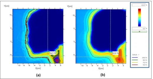
model is available. An overview of the three different LIBT centers and SIBD systems available is provided in
129
Table 1.
130 131
Table 1: SIBD system properties 132
LIBT facility Manufacturer Machine type Ion Species SIBD technique6 Gantry angles Energy range
Centre Antoine Lacassagne (Nice, France) IBA PT (Louvain-la-Neuve, Belgium) Synchro-Cyclotron
(S2C2)34,35 Protons Discrete scanning from 320° to 180° 70-230 MeV
Christie NHS Foundation Trust (Manchester, UK)
Varian (Palo Alto, California,
US) ProBeam (Cylcotron) Protons Discrete scanning from 0° to 360° 70-245 MeV MedAustron
(Wiener Neustadt, Austria)
MedAustron (Synchrotron) MAPTA 7
Protons Quasi-discrete scanning
0° and 90° 60-250 MeV Carbon
ions 0° and 90° 120-400 MeV/n
133
The different centers agreed in using similar physics settings. Physics settings were selected using Geant4
134
recommendations for medical physics applications**, personal communications with Geant4 developers and
135
independent validation tests using GATE. For protons, the physics-builder QGSP_BIC (containing BInary
136
Cascade for nuclear processes) is used31, while for carbon ions the SHIELDING physics-builder (containing the
137
Quantum Molecular Dynamics model for nuclear processes) is used 36,37. For both particle types, the
138
electromagnetic option EMZ (also called electromagnetic option 4, which is the most accurate) is selected30. For
139
more details, the reader is referred to the Physics Reference Manual†† of Geant4. Additional parameters may be
140
set, such as the maximum step size for ions, a range cut and a tracking cut for secondary electrons, positrons and
141
photons, which are usually comprised between 0.1 and 1 mm (compromise between speed and accuracy)38.
142 143
3. Results
144
i. Proton Radiography at Centre Antoine Lacassagne
145
The goal of the project consists in evaluating the potential of proton radiography images for patient positioning
146
using GATE-RTion. An anthropomorphic human head phantom (PBU-50, Supertech, USA) was scanned in a CT.
147
Treatment plans used to generate the proton radiography images of the anthropomorphic phantom were prepared
148
with the RayStation 6.0 TPS, using beam ranges larger than the phantom largest dimensions and a Monte Carlo
149
algorithm. Dose distributions were scored downstream the phantom in a plan perpendicular to the beam direction,
150
resulting in 2D proton radiography images. Proton radiography images were then simulated using GATE-RTion 151
at the same positions. In addition, proton radiography images were acquired using the Lynx 2D scintillator (IBA
152
Dosimetry, Schwarzenbrück, Germany) having a spatial resolution of 0.5 mm. The images were all imported into
153
the MyQA software (IBA Dosimetry, Schwarzenbrück, Germany) for comparisons. Isodoses contours were first
154
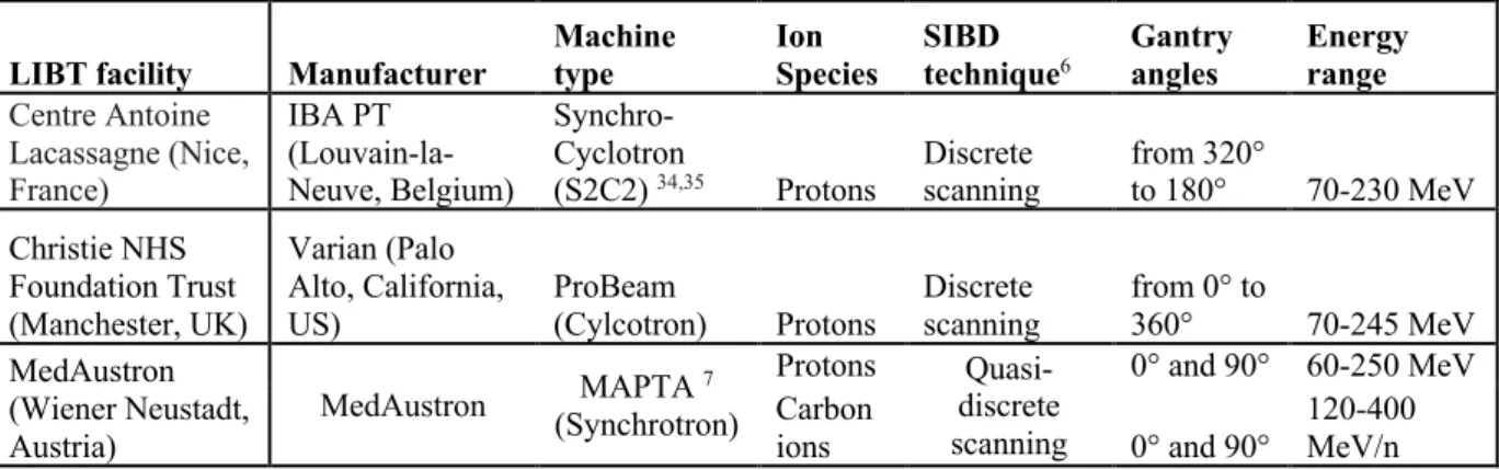
compared qualitatively, and then γ-index analyses were performed. A good qualitative agreement was found for
155
patient contours between Monte Carlo simulations and measurements (Figure 1). Using a γ-index analysis (2%,
156
2mm) between GATE-RTion simulations and TPS, more than 95% of the pixels are passing the test. This study
157
demonstrates the feasibility of using GATE-RTion to predict proton radiography images.
158 159 160 ** https://geant4.web.cern.ch/support/user_documentation †† http://geant4-userdoc.web.cern.ch/geant4-userdoc/UsersGuides/PhysicsReferenceManual/fo/PhysicsReferenceManual.pdf
161
Figure 1: Relative comparison of a GATE dose simulation (a) and a 2D Lynx measurement (b) acquired at the same downstream 162
position for an anthropomorphic phantom. 163
164
ii. Independent Dose Calculation of proton beam therapy plans at The Christie 165
Treatment planning is done using the Varian Eclipse (version 13.7) TPS, with Varian’s
proton-convolution-166
superposition (PCS version 13.7.16) analytical dose calculation algorithm. Prior to treatment, measurement-based
167
Patient Specific Quality Assurance (PSQA) is performed on each plan. In addition, Independent Dose Calculation
168
(IDC) using an in-house system called AutoMC and based on GATE-RTion is performed. AutoMC acts as a
169
wrapper around GATE-RTion to fully automate IDCwith the aim to ease the use and minimize the risk of user
170
error when configuring simulations. It is implemented within an Octave 39 environment, and uses modular
beam-171
models and CT calibrations. An example MC re-calculation in patient CT of the first phase of a 2-phase plan
172
treating a craniospinal axis is presented in Figure 2. The prescription for this phase was 23.4 Gy in 13 fractions,
173
delivered using 5 fields: a pair of left/right fields to the brain, and 3 fields to the spine (superior, mid and
174
inferior). A 5 cm WET range-shifter was used for all 5 fields. The percentage of voxels in the patient having γ ≤ 175
1 was between 92.4% and 95.8% for all fields, and the GATE-RTion simulation was between 1.6% and 2.4% 176
hotter than the TPS in terms of the median dose to the patient. Dose differences occurring outside the patient
177
surface were excluded from the analyses.
178
179
Figure 2: Comparison of TPS (Varian Eclipse) and MC (AutoMC / GATE-RTionV1.0) calculations of a 5-field craniospinal axis 180
pencil beam scanning proton plan, planned at the Christie for delivery on a Varian ProBeam system. Top row: TPS; Middle 181
row: GATE-RTion; Bottom row: Gamma at 3% (local), 3 mm using a 10% lower dose threshold. Voxels in green have γ ≤ 1, 182
while voxels in (red/blue) have γ > 1 and are (hotter/colder) than the TPS respectively. 183
184
iii. Independent Dose Calculation with Scanned Ion Beams at MedAustron
185
Treatment planning is performed using the RayStation version 8B from RaySearch Laboratories (Stockholm, 186
Sweden). For protons, the Monte Carlo algorithm version 4.2 is used. For carbon ions, the pencil beam algorithm 187
version 3.0 and the Local Effect Model (LEM) I40 for Relative Biological Effectiveness (RBE) modeling are used. 188
The measurement-based PSQA process was set-up since the beginning of clinical operation28. It is performed in 189
water only and for a limited number of measurements points28 (using the 3D-block/24 PinPoint ionization 190
chambers type 31015, PTW, Freiburg). In contrary, an IDC has the advantage to evaluate patient treatments in CT 191
geometry and for the entire 3D dose distribution. A key advantage of IDC-based PSQA is to reduce beam time 192
requirements for QA and thus increase the facility treatment capacity. The Independent DosE cAlculation for LIBT 193
(IDEAL) project, including GATE-RTion as dose engine, started in 2017, in a collaboration between the 194
MedAustron ion therapy center, the Medical University of Vienna (MUW) and the Austrian Center for Medical 195
Innovation and Technology (ACMIT). A first prototype has been developed in 2018 in a research network and 196
was transferred into the clinical environment of MedAustron in October 2019. TPS plans are exported to a QA 197
database and IDEAL is run on a cluster of modular capacity (currently featured with 48 cores). A carbon ion 198
treatment recomputed using the IDEAL prototype and including a comparison to the TPS dose distribution is 199
illustrated in Figure 3. This is a curative carbon ion treatment up to 65.6 Gy RBE in 16 fractions of 4.1 Gy RBE 200
(4 fractions per week). The PTV1 is treated with 9 fractions up to 36.9 Gy RBE, using 4 beams with a horizontal 201
beam line and table rotations of 315o, 355o, 320o and 360o. 202
203
204
Figure 3: Comparison of the physical dose distribution for a carbon ion beam having an oblique incidence in the head region 205
of a patient. IDEAL/GATE-RTion dose distribution (Top left) is compared to the TPS (bottom left) in terms of DVH (bottom 206
right) and dose profiles (top right). For DVH and dose profiles, solid lines correspond to IDEAL/GATE-RTion and dotted lines 207
to the TPS. The positions of the two orthogonal dose profiles in the patient are visible in the patient images on the left side 208
(orange and green lines). 209
210
4. Discussion
211
The validated GATE-RTion release 1.0 allows clinical users to build confidence in a specific GATE/Geant4
212
version for clinical applications and share validation results. In parallel, GATE and Geant4 are evolving and may
213
provide new relevant features and improved physics models to the users in the future. For example, with respect
214
to the physics processes, uncertainties of nuclear cross-sections and models are known to be substantial, especially
215
for carbon ions41. This is where most improvements could be achieved in future. In addition, every new GATE
216
release provides new features which may be relevant for certain clinical applications, for example code
217
optimization allowing to perform simulations more efficiently or the scoring of new quantities of clinical interest.
218
Also deep learning methods started to be included during Monte Carlo simulations. In its current state, we believe
219
that the proposed GATE features and Geant4 physics models available in GATE-RTion V1.0 are sufficiently
accurate for most dosimetric applications in LIBT facilities. Every new GATE-RTion release will need to be
221
thoroughly re-validated and re-commissioned by each user before clinical use, which is a major effort. This can
222
only be justified by substantial improvements in the physics models or by the introduction of new features
223
clinically relevant to the users.The release cycle of GATE-RTion is therefore not planned and will depend on user
224
needs and request. However, bug fixes to GATE-RTion can be ported to the current GATE-RTion version via
225
patch mechanism. This presents the advantage of fixing software bugs (if needed), without modifying the
226
underlying Geant4 physics. GATE-RTion specific validation/acceptance could subsequently be re-run, in order to
227
validate that the patch did not affect the rest of GATE-RTion functionalities except fixing the bug. It is therefore
228
important to develop all necessary validation tests with the users, as described in section 2.b. The results provided
229
in Centre Antoine Lacassagne for proton radiography-based patient-positioning are preliminary but very
230
promising. A dedicated application wrapper would certainly help in future to integrate this innovative
GATE-231
RTion-based application into clinical environments. The IDC applications implemented at The Christie (AutoMC)
232
and MedAustron (IDEAL) are serving both the purpose of IDC, with a key difference that MedAustron extends
233
the application to carbon ions. The Christie is using GATE-RTion clinically since the start of the treatment end of
234
2018, while MedAustron is still in development and commissioning phase. The commissioning methodology and
235
dosimetric perfromances of GATE-RTion as implemented at The Christie and MedAustron facilities will be
236
published in order to provide reference commissioning reports to support the clinical community.
237 238 239
5. Conclusions
240
The GATE-RTion projectpaves the way towards the use of the GATE simulation tool in Light Ion Beam Therapy 241
facilities. GATE-RTion version 1.0 was released in May 2018 and the framework includes a validated GATE
242
release based on a specific Geant4 version, a set of tools to integrate GATE into a clinical environment and
243
a network for clinical users. Three completely different machines were modeled (three different vendors, 244
cyclotron and synchrotron-based systems, protons and carbon ions). Applications such as proton radiography and 245
Independent Dose Calculation (IDC) for scanned proton and carbon ion beam therapy were presented. This project 246
builds the bridge between clinical users and researchers using GATE, fostering the transfer of clinically relevant 247
research applications into the end-user’s clinics. While applications at Centre Antoine Lacassagne and 248
MedAustron are still under development, The Christie is running GATE-RTion clinically for IDC since the start 249
of the clinical treatments end of 2018. The results presented within the first two years after the first release of 250
GATE-RTion demonstrate the versatility and relevance of this project. 251
252
Acknowledgments 253
The financial support from ACMIT Gmbh, Medical University of Vienna and MedAustron is gratefully 254
acknowledged. The competence center ACMIT is funded within the scope of the COMET program by Austrian 255
ministries BMVIT and BMWFW, and by the governments of Lower Austria and Tyrol. The competence center 256
program COMET is managed by the Austrian Funding Agency FFG. Part of this work was performed within the 257
framework of the SIRIC LYriCAN Grant INCa-INSERM-DGOS-12563, and the LABEX PRIMES (ANR-11-258
LABX-0063) of Université de Lyon, within the program “Investissements d’Avenir” (ANR-11-IDEX-0007) 259
operated by the ANR. This work was supported by the Science and Technology Facilities Council Advanced 260
Radiotherapy Network [grant number ST/N002423/1]. 261
The authors would like to thank Priv. Doz. Dipl. Ing. Markus Stock for careful review and advices on the 262 manuscript. 263 264 References 265
1. Jan S, Santin G, Strul D, et al. GATE: a simulation toolkit for PET and SPECT. Phys Med Biol. 266
2004;49(19):4543. http://stacks.iop.org/0031-9155/49/i=19/a=007. 267
2. Jan S, Benoit D, Becheva E, et al. GATE V6: a major enhancement of the GATE simulation platform 268
enabling modelling of CT and radiotherapy. Phys Med Biol. 2011;56(4):881-901. doi:10.1088/0031-269
9155/56/4/001 270
3. Grevillot L, Frisson T, Maneval D, Zahra N, Badel J-N, Sarrut D. Simulation of a 6 MV Elekta Precise 271
Linac photon beam using GATE/GEANT4. Phys Med Biol. 2011;56(4):903-918. 272
4. Jan S, Frisson T, Sarrut D. GATE simulation of 12C hadrontherapy treatment combined with a PET 273
imaging system for dose monitoring: A feasibility study. IEEE Trans Nucl Sci. 2013;60(1):423-429. 274
doi:10.1109/TNS.2012.2233496 275
5. Moyers MF, Vatnitsky SM. Practical Implementation of Light Ion BeamTreatments. Medical 276
PhysicsPublishing; 2012. 277
6. ICRU. International Commission on Radiation Units and Measurements report 78: Prescribing and 278
Recording and And Reporting Proton-Beam Therapy: Contents. J ICRU. 2007;7. 279
doi:10.1093/jicru/ndm021 280
7. Grevillot L, Osorio Moreno J, Letellier V, et al. Clinical implementation and commissioning of the 281
MedAustron Particle Therapy Accelerator for non‐isocentric scanned proton beam treatments. Med Phys. 282
2019:1-13. doi:10.1002/mp.13928 283
8. Robert C, Fourrier N, Sarrut D, et al. PET-based dose delivery verification in proton therapy: a GATE 284
based simulation study of five PET system designs in clinical conditions. Phys Med Biol. 285
2013;58(19):6867-6885. http://stacks.iop.org/0031-286
9155/58/i=19/a=6867?key=crossref.5c9d5c8b062bf80543bccc0cd3d280e1. 287
9. Robert C, Dedes G, Battistoni G, et al. Distributions of secondary particles in proton and carbon-ion 288
therapy: a comparison between GATE/Geant4 and FLUKA Monte Carlo codes. Phys Med Biol. 289
2013;58(9):2879-2899. doi:10.1088/0031-9155/58/9/2879 290
10. Sarrut D, Bardiès M, Boussion N, et al. A review of the use and potential of the GATE Monte Carlo 291
simulation code for radiation therapy and dosimetry applications. Med Phys. 2014;41(6):064301. 292
doi:10.1118/1.4871617 293
11. Paganetti H, Jiang H, Lee SY, Kooy HM. Accurate Monte Carlo simulations for nozzle design and 294
commissioning and quality assurance for a proton radiation therapy facility. Med Phys. 2004;31(7):2107-295
2118. 296
12. Paganetti H, Jiang H, Parodi K, Slopsema R, Engelsman M. Clinical implementation of full Monte Carlo 297
dose calculation in proton beam therapy. Phys Med Biol. 2008;53(17):4825-4853. doi:10.1088/0031-298
9155/53/17/023 299
13. Parodi K, Paganetti H, Cascio E, et al. PET/CT imaging for treatment verification after proton therapy: a 300
study with plastic phantoms and metallic implants. Med Phys. 2007;34(2):419-435. 301
14. Parodi K, Ferrari A, Sommerer F, Paganetti H. Clinical CT-based calculations of dose and positron 302
emitter distributions in proton therapy using the FLUKA Monte Carlo code. Phys Med Biol. 303
2007;52(12):3369-3387. doi:10.1088/0031-9155/52/12/004 304
15. Parodi K, Mairani A, Brons S, et al. Monte Carlo simulations to support start-up and treatment planning 305
of scanned proton and carbon ion therapy at a synchrotron-based facility. Phys Med Biol. 2012;57:3759-306
3784. 307
16. Ardenfors O, Dasu A, Kopeć M, Gudowska I. Modelling of a proton spot scanning system using 308
MCNP6. J Phys Conf Ser. 2017;860(1). doi:10.1088/1742-6596/860/1/012025 309
17. Bassler N, Hansen DC, Lühr A, Thomsen B, Petersen JB, Sobolevsky N. SHIELD-HIT12A - A Monte 310
Carlo particle transport program for ion therapy research. J Phys Conf Ser. 2014;489(1):8-13. 311
doi:10.1088/1742-6596/489/1/012004 312
18. Sato T, Niita K, Matsuda N, et al. Overview of the PHITS code and its application to medical physics. 313
Prog Nucl Sci Technol. 2014;4:879-882. doi:10.15669/pnst.4.879 314
19. Goma C, Safai S, Voros S. Reference dosimetry of proton pencil beams based on dose-area product: a 315
proof of concept. Phys Med Biol. 2017;62(12):4991. doi:https://doi.org/10.1088/1361-6560/aa7008 316
20. Akagi T, Aso T, Iwai G, et al. Geant4-based particle therapy simulation framework for verification of 317
dose distributions in proton therapy facilities. Prog Nucl Sci Technol. 2014;4:896-900. 318
21. Perl J, Shin J, Schümann J, Faddegon B, Paganetti H. TOPAS: An innovative proton Monte Carlo 319
platform for research and clinical applications. Med Phys. 2012;39(11):6818-6837. 320
doi:10.1118/1.4758060 321
22. Grevillot L, Bertrand D, Dessy F, Freud N, Sarrut D. A Monte Carlo pencil beam scanning model for 322
proton treatment plan simulation using GATE/GEANT4. Phys Med Biol. 2011;56:5203-5219. 323
23. Grevillot L, Frisson T, Zahra N, et al. Optimization of GATE/Geant4 settings for Proton Pencil Beam 324
Scanning simulations towards TPS Quality Assurance. In: 49th Meeting of the Particle Therapy Co-325
Operative Group (PTCOG). ; 2010. 326
24. Grevillot L, Bertrand D, Dessy F, Freud N, Sarrut D. GATE as a GEANT4-based Monte Carlo platform 327
for the evaluation of proton pencil beam scanning treatment plans. Phys Med Biol. 2012;57:4223-4244. 328
25. Almhagen E, Boersma DJ, Nyström H, Ahnesjö A. A beam model for focused proton pencil beams. 329
Phys Medica. 2018;52:27-32. doi:10.1016/j.ejmp.2018.06.007 330
26. Fuchs H, Grevillot L, Carlino A, et al. Optimizing the MedAustron proton gantry beam delivery: 331
Providing nozzle design recommendations based on Gate/Geant4 Monte Carlo simulation. In: PTCOG 332
55. ; 2016. 333
27. Grevillot L, Stock M, Vatnitsky S. Evaluation of beam delivery and ripple filter design for non-isocentric 334
proton and carbon ion therapy. Phys Med Biol. 2015;60(20):7985-8005. 335
http://www.ncbi.nlm.nih.gov/pubmed/26418366. 336
28. Carlino A. Implementation of advanced methodologies in the commissioning of a Light Ion Beam 337
Therapy facility (PhD thesis, Department of Physics and Chemistry, University of Palermo, Italy). 2017. 338
29. Saini J, Maes D, Egan A, et al. Dosimetric evaluation of a commercial proton spot scanning Monte-Carlo 339
dose algorithm: Comparisons against measurements and simulations. Phys Med Biol. 2017;62(19):7659-340
7681. doi:10.1088/1361-6560/aa82a5 341
30. Fuchs H, Vatnitsky S, Stock M, Georg D, Grevillot L. Evaluation of GATE/Geant4 multiple Coulomb 342
scattering algorithms for a 160 MeV proton beam. Nucl Instruments Methods Phys Res Sect B Beam 343
Interact with Mater Atoms. 2017;410. doi:10.1016/j.nimb.2017.08.006 344
31. Resch AF, Elia A, Fuchs H, et al. Evaluation of electromagnetic and nuclear scattering models in GATE 345
/Geant4 for proton therapy. Med Phys. 2019;46(5):2444-2456. doi:10.1002/mp.13472 346
32. Elia A. Characterization of the GATE Monte Carlo platform for nonisocentric treatments and patient 347
specific treatment plan verification at MedAustron (PhD thesis, INSA Lyon, 2019LYSE002). 2019. 348
33. Meißner H, Fuchs H, Hirtl A, Reschl C, Stock M. Towards offline PET monitoring of proton therapy at 349
MedAustron. Zeitschrift fßr Medizinische Phys. 2019;29(1):59-65. doi:10.1016/j.zemedi.2018.05.003 350
34. Kleeven W, Abs M, Forton E, et al. The IBA Superconducting Synchrocyclotron Project S2C2. Proc 351
Cyclotrons2013. 2013:115-119. 352
https://accelconf.web.cern.ch/AccelConf/CYCLOTRONS2013/papers/mo4pb02.pdf. 353
35. Van de Walle J, Abs M, Conjat M, et al. The S2C2: From Source to Extraction. Proc Cyclotrons 2016. 354
2017;THB01:285-289. http://accelconf.web.cern.ch/AccelConf/cyclotrons2016/papers/thb01.pdf. 355
36. Bolst D, Cirrone GAP, Cuttone G, et al. Validation of Geant4 fragmentation for Heavy Ion Therapy. 356
Nucl Instruments Methods Phys Res Sect A Accel Spectrometers, Detect Assoc Equip. 2017;869:68-75. 357
doi:10.1016/j.nima.2017.06.046 358
37. Böhlen TT, Cerutti F, Dosanjh M, et al. Benchmarking nuclear models of FLUKA and GEANT4 for 359
carbon ion therapy. Phys Med Biol. 2010;55(19):5833-5847. doi:10.1088/0031-9155/55/19/014 360
38. Grevillot L, Frisson T, Zahra N, et al. Optimization of GEANT4 settings for Proton Pencil Beam 361
Scanning simulations using GATE. Nucl Instruments Methods Phys Res Sect B Beam Interact with 362
Mater Atoms. 2010;268(20):3295-3305. 363
39. Eaton JW, Bateman D, Hauberg S WR. GNU Octave version 5.1.0 manual: a high-level interactive 364
language for numerical computations. 2019. https://www.gnu.org/software/octave/doc/v5.1.0/. 365
40. Scholz M, Kellerer AM, Kraft-Weyrather W, Kraft G. Computation of cell survival in heavy ion beams 366
for therapy: The model and its approximation. Radiat Environ Biophys. 1997;36(1):59-66. 367
doi:10.1007/s004110050055 368
41. Dedes G, Parodi K. Monte Carlo Simulations of Particle Interactions with Tissue in Carbon Ion Therapy. 369
Int J Part Ther. 2015;2(3):447-458. doi:10.14338/IJPT-15-00021 370