HAL Id: tel-01249535
https://tel.archives-ouvertes.fr/tel-01249535
Submitted on 4 Jan 2016
HAL is a multi-disciplinary open access archive for the deposit and dissemination of sci-entific research documents, whether they are pub-lished or not. The documents may come from teaching and research institutions in France or abroad, or from public or private research centers.
L’archive ouverte pluridisciplinaire HAL, est destinée au dépôt et à la diffusion de documents scientifiques de niveau recherche, publiés ou non, émanant des établissements d’enseignement et de recherche français ou étrangers, des laboratoires publics ou privés.
establishment of the competence to transdifferentiate in
vivo in C. elegans
Thomas Daniele
To cite this version:
Thomas Daniele. How a differentiated cell can change its identity : study of the role of the LIN-12/Notch pathway in the establishment of the competence to transdifferentiate in vivo in C. elegans. Microbiology and Parasitology. Université de Strasbourg, 2013. English. �NNT : 2013STRAJ038�. �tel-01249535�
ÉCOLE DOCTORALE DES SCIENCES DE LA VIE ET DE LA SANTÉ
ED414
Institut de génétique et de biologie moléculaire et cellulaire
CNRS UMR 7104
THÈSE
présentée par :Thomas DANIELE
soutenue le : 26 septembre 2013pour obtenir le grade de :
Docteur de l’université de Strasbourg
Discipline/ Spécialité
: Sciences de la Vie et de la Santé
How a differentiated cell can change its identity:
Study of the role of the LIN-12/Notch pathway in
the establishment of the competence to
transdifferentiate in vivo in C. elegans
THÈSE dirigée par :
Mme. JARRIAULT Sophie Docteur, Université de Strasbourg
RAPPORTEURS :
Mme. FELIX Marie-Anne Professeur, Ecole Normale Supérieure, PARIS M. POOLE Richard Docteur, University College London, LONDON
AUTRES MEMBRES DU JURY :
Remerciements
Avant toute chose, je souhaite remercier les membres de mon jury, Pr. Marie-Anne Félix, Dr. Richard Poole, Pr. Jean-marc Reichhart et Pr. Olivier Pourquié de me faire l'honneur d'accepter de juger mon travail de thèse.
Il est toujours difficile pour moi de remercier les gens à leur juste valeur, ces quelques lignes ne résumeront jamais toute la gratitude et le respect que j'ai pour vous...
Un énorme merci à Sophie, la chef, qui m'a vu grandir depuis mon master 1. C'est dans ton laboratoire que j'ai fait mes premiers vrais pas de scientifique. Merci pour ta disponibilité, ton écoute et d'avoir toujours essayé de rendre optimiste le pessimiste que je suis. Merci de m'avoir permis de grandir scientifiquement et de m'avoir donné accès à des conditions de recherche exceptionnelles. Je n'oublierais pas que grâce à toi, j'ai fait mes premiers voyages en Europe et dans le monde… Edimbourg, Barcelone et tout seul comme un grand Cold Spring Harbor. Merci pour toutes ces discussions scientifiques et non scientifiques et je n'oublierais jamais les pauses chocolat quand les expériences ne marchaient pas… J'espère avoir été à la hauteur et je souhaite le meilleur à ton laboratoire pour les années à venir.
Merci à toi Nadine, notre maman à tous ! Tu m'auras aidé à grandir ! Merci pour ta disponibilité, ta gentillesse, ton sourire et ton soutien sans failles. Merci pour toutes tes contributions scientifiques et non scientifiques. Merci de m'avoir appris à injecter alors que tu détestes ça. Je garderai également en mémoire, les nombreux gâteaux, tartes et autres tiramisus que tu nous as fait. D'ailleurs tu m'as promis un repas à la maison, j'espère qu'on se fera ça avant que je parte ;-). Je te souhaite que du positif jusqu'à ton futur départ en retraite, et tu peux être sûr que si je suis dans le coin, je viendrais te faire un petit coucou !!
A big shout out to my man Steven (as the americans say ;-)), I have seen your evolution during our four and half year together, you became one the hardest worker I have met in my life. I am sure that you are going to be one leader in the C. elegans community. It was really enriching to work with you, to have all these scientific discussions during our lab meetings and in the lab. Beside this hard worker side, you know as well how to have fun ;-), I remembered our tennis-calendar, dry ice fun and other football games. So, Steve thanks a lot for everything and I wish you the best for your carreer, which will certainly be successfull !!
Merci à Marie-Charlotte, Marie-Boubou, Marie-Chacha ou pour les intimes Maritas (en hommage à ses origines espagnoles). On aura quand même trippé comme des fous ! Scientifiquement, ça n'a pas été facile tous les jours pour nous mais au final on aura quand même su en rire. Reste comme tu es, garde ton âme d'artiste, ça te servira ! Le meilleur pour la suite et surtout accroche-toi !!
Arnaud… il me faudrait une thèse pour parler de toi ! Tu es un des seuls types au monde qui peut me faire rire sans être présent à mes côtés, j'ai juste à penser à ce qu'on a partagé ensemble pour me fendre la poire tout seul… Merci pour tout, scientifiquement tu m'as beaucoup apporté, ton expertise en biologie moléculaire va me servir à vie. Mais c'est surtout humainement que tu m'auras apporté le plus, même quand le moral était au plus bas, tu as toujours su être assez débile pour me faire marrer. Merci pour mon entrainement quotidien de Kung-Fu, nos délires, et surtout pour notre virée au ski, un jour on se refera une petite Velasquez, c'est promis ;-). Bon je m'arrête là sinon j'en ai encore pour des pages. En tout cas au cours de cette thèse le petit collègue tout calme que j'ai connu est devenu un véritable ami plutôt farfelu Lol (comme disent les ados) !! D'ailleurs comme tu le dis si bien, t'es comme un pote mais en mieux ! PS: Tu me dois encore des chewing-gums…
Pour finir avec le laboratoire Jarriault: Merci aux anciens membres du laboratoire, par ordre de départ, Jai Richard, Konstantinos Kagias et Martina Hajduskova, pour tout ce que vous m'avez appris et tous les fous rires que nous avons pu partager. Une pensée aux étudiants que j'ai encadré, mes deux Matthieu, Matthieu Leobold et Matthieu Gendarme, vous m'avez été d'une grande aide, je n'oublierai pas les délires que nous avons eus, mais je dois souligner que je vous ai quand même fait beaucoup travailler, bravo à vous d'avoir tenu le choc à mes côtés.
Passons maintenant à nos formidables voisins, communément appelés les Labouesse:
Commençons par leur chef, Michel… Merci à vous, pour le soutien depuis le master et tous les conseils avisés que vous avez pu me donner en lab meeting. Je dois avouer que c'était toujours un peu stressant de passer devant vous, on savait qu'en fonction des jours ça pouvait être plus où moins difficile. En tout cas, j'ai toujours été admiratif de votre savoir et de votre esprit critique. Ce fut une chance et un honneur de vous côtoyer.
Merci à Christelle, the gossip girl, ça va me manquer de ne pas savoir tout ce qui se passe dans les coulisses de l'institut. Merci aussi pour tous tes conseils, avis, questions et surtout pour ta franchise légendaire, même si des fois ça pique, c'est bon d'avoir en face de soi quelqu'un qui dit ce qu'il pense.
Merci Sophie Q, il faut toujours préciser le Q sinon on ne sait pas de quelle Sophie on parle. Merci pour ton aide, ta disponibilité, ta gentillesse et aussi tes gâteaux fraichement cuit du matin. Ca fait toujours du bien de commencer une journée avec ça dans le ventre.
David, le troll officiel de l'institut, derrière cette carapace, se cache un petit être fragile et plus gentil qu'il ne veut nous le faire croire. On se sera quand même bien marré tous les deux. Merci aussi pour les deux petites virées à moto, dommage que je n'ai pas eu plus de temps pour en faire plus.
Merci à tous les autres membres du laboratoire Labouesse, Gabi, Vincent, Naël, Maaanuuu, Thahn, Agnès, Shashi, Maxime, pour tout ce que vous avez pu m'apporter. C'est toujours un plaisir d'avoir des collègues comme vous !!
Il n'est jamais facile de faire une thèse, mais heureusement bon nombre de personnes vous entourent pour que cela se passe le mieux possible.
Jérôme… C'est comme Arnaud, faudrait une thèse pour parler de toi. T'es juste un tueur, je sais pas combien de délire on a eu depuis le master, ce fut vraiment une chance de te rencontrer. De toute façon on ne se lâche pas, on fait ce qu'on a dit, le premier qui monte son équipe recrute les autres ;-). Je te souhaite de tout cœur le meilleur, pour toi, ta petite femme et la petite Inès.
Adrien, le problème avec toi c'est que tu es tellement discret et secret que je ne pourrais jamais écrire une thèse sur toi (pas comme le vieux Jérôme qui pourrait jamais garder le moindre truc secret…). Tu es un mec en Or, je me fais pas de soucis pour toi pour la suite, t'es fait pour la recherche, tu verras que l'équipe dont tu rêves, tu l'auras j'en suis persuadé ! Bon mise à part ça, on aura aussi déliré comme des foufous, je regrette de ne pas avoir fait tout ce qu'on avait prévu mais on se rattrapera plus tard. Comme pour Jérôme, we stay in touch et dès que ton labo est monté, je t'envoie mon CV.
Merci à la team du RU, Thibaut (Aka Timble), Anne-Sophie, Léa, Stéphanie (avé son accent du sud quand elle revient de vacances), Rose-Marie (La troubadoure), Salim (notre copain issu de l'immigration ;-)), Daaaavid, Sara. Merci pour tous les délires, il est bon de couper notre journée avec de bonnes tranches de rigolade !
Un énorme merci à ma famille (bien trop nombreuse pour être lister ici ;-)), à ma belle-famille (Nathalie, Vevette, Jean-Claude, Amélie, Lionel, Rémy, Renée et Guy) et à mes amis (Julie, Julien, Guillaume, Delacotch, Mimil, Jacques et Josette) d'avoir toujours cru en moi et pour tout le soutien dont vous m'avez fait part.
Maman et Papa, je ne trouverais jamais les mots pour vous remercier pour tout ce que vous avez fait pour moi et tout ce que vous m'avez apporté. Si j'en suis là aujourd'hui c'est grâce à vous. Vous avez toujours été là pour me soutenir quelques soient mes décisions. J'espère vous faire honneur, cette thèse vous est dédiée.
Enfin, merci à toi, Tiphanie, qui me supporte depuis toutes ces années. Je sais que cette thèse n'a pas été simple pour toi aussi. J'espère que des jours meilleurs nous attendent. Je ne suis pas la personne la plus expressive au monde mais tu sais à quel point tu es essentielle à mes yeux, je n'aurais jamais pu tenir si tu n'avais pas été à mes côtés. Merci pour tout !
Résumé en français
L’acquisition d'une identité cellulaire différenciée est souvent considérée comme définitive et figée dans le temps; or un nombre croissant d’études démontre que les cellules différenciées peuvent faire preuve de plasticité sous certaines conditions. Il est par exemple possible de forcer une cellule différenciée à devenir soit une cellule pluripotente, ou directement un autre type cellulaire différencié, via la surexpression de facteurs de transcription clés, on parle alors de reprogrammation cellulaire. Ces phénomènes de plasticité cellulaire peuvent également prendre place naturellement chez certains organismes1. Le fait que l’identité cellulaire ne soit pas figée peut devenir préjudiciable dans le cas de pathologies tel que le cancer où certaines cellules normales changent d’identité pour devenir des cellules tumorales2,3. A l’opposé rendre possible la manipulation aisée de l’identité cellulaire permettrait à long terme de mettre en place de nouveaux protocoles de thérapie cellulaire. Il serait alors possible de remplacer des cellules malades ou endommagées par des cellules saines reprogrammées. Malheureusement, l’application thérapeutique de telles méthodes n’est pas encore à l’ordre du jour, d’une part les protocoles de reprogrammation actuels ne sont efficaces qu’à 0,1 à 1%, d’autre part la stabilité des cellules reprogrammées n’a pas encore été clairement évaluée. Il a été montré que certaines cellules reprogrammées pouvaient perdre leur équilibre et devenir tumorales4. Il est donc indispensable de comprendre les mécanismes sous-tendant la reprogrammation cellulaire afin de pouvoir mieux maitriser la manipulation de l’identité cellulaire.
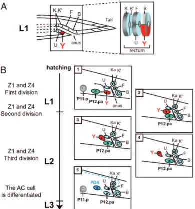
Ainsi, afin de mieux comprendre les phénomènes de reprogrammation cellulaire prenant place naturellement ou lors de cancer, notre laboratoire a établi un modèle unique chez Caenorhabditis elegans (C. elegans) permettant l’étude d’un événement de reprogrammation dans un contexte physiologique à l'échelle de cellules uniques. Nous étudions particulièrement un événement qui prend place au sein du rectum, formé par six cellules. Une de ces cellules, nommée Y, présente un comportement extraordinaire. Y est une cellule rectale épithéliale qui, au cours du développement du ver, va migrer antérieurement puis changer d’identité pour devenir un motoneurone nommé PDA dont les signatures moléculaires et
morphologiques sont totalement distinctes de la cellule Y5. Ce changement direct d’identité cellulaire est défini par le terme de transdifférenciation. Ainsi, cet reprogrammation cellulaire prend place naturellement et de manière stéréotypée: contrairement à la reprogrammation in vitro, cet événement de transdifférenciation à lieu chez 100% des vers sauvages. De façon importante, la possibilité de prédire par avance quelle cellule va changer d'identité nous donne accès aux étapes précoces de ce processus.
Les travaux préliminaires du laboratoire ont montré que la voie de signalisation LIN-12/Notch est le signal le plus précoce nécessaire pour le bon déroulement de la reprogrammation de Y en PDA. De plus, la cellule Y acquière une compétence à changer d'identité et la voie Notch est nécessaire à l’établissement de cette compétence: chez certain mutants pour le gène egl-38 ou mab-9, une cellule Y surnuméraire est formée mais n’est pas capable de se transdifférencier6,7, alors que chez des mutants gain de fonction pour Notch, la cellule Y surnuméraire formée est compétente à se transdifférencier, résultant en la formation de 2 neurones PDA5,8. Mes travaux de thèse ont donc portés sur la compréhension de l’implication de la voie LIN-12/Notch dans cet évènement de reprogrammation cellulaire.
Au cours de ma thèse, nous avons pu mettre en évidence: i) que lors de l’embryogénèse, 2 ligands canoniques (apx-1 et lag-2) semblent agir de façon redondante afin d’activer la voie Notch, et ce dans la cellule Y. ii) l’activation ectopique et contrôlée de la voie Notch est suffisante pour induire la formation d’un second neurone PDA (et ce dans une fenêtre de temps courte autour de la naissance de Y). De façon intéressante, ces résultats nous ont permis d’observer que seul un nombre limité de cellules est alors reprogrammé laissant suggérer l’importance d’un contexte cellulaire permissif. iii) Les facteurs nucléaires que le laboratoire a identifiés comme cruciaux pour l'initiation de cet évènement de transdifférenciation6 sont également importants pour la reprogrammation induite de cette deuxième cellule en neurone PDA formée chez des vers portant une mutation gain de fonction pour Notch. Cela suggère que ces facteurs sont en aval du signal Notch. iv) La suractivation prolongée de la voie Notch dans la cellule Y maintien
l’identité épithéliale de cette dernière, ayant pour conséquence le blocage de la transdifférenciation de Y en PDA.
Ensemble, nos résultats montrent que la voie Notch est nécessaire et suffisante afin d’établir la compétence à transdifférencier et que cela ne peut être réalisé que si la voie Notch est régulée de façon très précise dans la cellule Y.
Plus généralement nos travaux apportent la preuve que la régulation fine et précise des voies de signalisation est indispensable au bon déroulement de la reprogrammation cellulaire.
Bibliographie :
1. Zuryn, S., Daniele, T. & Jarriault, S. Direct cellular reprogramming in Caenorhabditis elegans: facts, models, and promises for regenerative medicine. WIREs Developmental Biology 1, 138-152 (2012).
2. Abollo-Jimenez, F., Jimenez, R. & Cobaleda, C. Physiological cellular reprogramming and cancer. Semin Cancer Biol 20, 98-106 (2010).
3. Hajduskova, M., Ahier, A., Daniele, T. & Jarriault, S. Cell plasticity in Caenorhabditis elegans: From induced to natural cell reprogramming. Genesis (2011).
4. Ben-David, U. & Benvenisty, N. The tumorigenicity of human embryonic and induced pluripotent stem cells. Nat Rev Cancer 11, 268-277 (2011).
5. Jarriault, S., Schwab, Y. & Greenwald, I. A Caenorhabditis elegans model for epithelial-neuronal transdifferentiation. Proc Natl Acad Sci U S A 105, 3790-3795 (2008).
6. Chamberlin, H. M. et al. The PAX gene egl-38 mediates developmental patterning in Caenorhabditis elegans. Development 124, 3919-3928 (1997). 7. Woollard, A. & Hodgkin, J. The caenorhabditis elegans fate-determining gene
mab-9 encodes a T-box protein required to pattern the posterior hindgut. Genes Dev 14, 596-603 (2000).
8. Greenwald, I. S., Sternberg, P. W. & Horvitz, H. R. The lin-12 locus specifies cell fates in Caenorhabditis elegans. Cell 34, 435-444 (1983).
Table of contents
!List of abbreviations ... 14
!List of figures ... 16
!INTRODUCTION ... 18
!I) Cell plasticity ... 19
!I.1) Historical view of development and cell differentiation ... 19!
I.1.1) Epigenesis versus preformationism – The opening debate ... 19!
I.1.2) The cell theory ... 21!
I.1.3) The Waddington’s landscape ... 22!
I.2) When a cell can exhibit plasticity ... 24!
I.2.1) Definition of cell potency ... 24!
I.2.2) Terminology of cell plasticity events ... 25!
I.2.3) Dedifferentiation ... 26!
I.2.3.1) Naturally induced dedifferentiation ... 26!
I.2.3.2) Experimentally induced dedifferentiation ... 28!
I.2.4) Nuclear reprogramming ... 30!
I.2.4.1) Somatic-cell nuclear transfer (SCNT) ... 30!
I.2.4.2) Nuclear reprogramming by cell fusion and cell extract ... 33!
I.2.4.3) Induction of pluripotency by defined factors ... 34!
I.2.5) Transdetermination ... 38!
I.2.6) Transdifferentiation ... 41!
I.2.6.1) Natural transdifferentiation events ... 41!
I.2.6.2) Transdifferentiation during regeneration. ... 44!
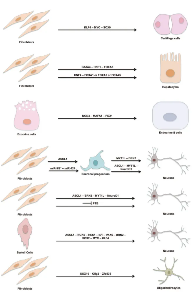
I.2.6.3) Induced transdifferentiation ... 45!
I.2.7) Limitations and future challenges in the cell plasticity field ... 54!
I.2.8) Controversy around cell plasticity ... 56!
I.2.9) Maintenance of cell identity ... 59!
I.2.10) Cell-plasticity based therapeutical approaches ... 62!
I.3) A refreshed view of the Waddington's landscape ... 64!
II) Caenorhabditis elegans as a model to study cell plasticity ... 66
!II.2) C. elegans as a model to study stem cell biology ... 69!
II.2.1) The C. elegans germline, a model to study stem cell-niche interactions ... 70!
II.2.2) The seam cells, a model to study stem cell-like lineages ... 72!
II.3) C. elegans as a model to study cellular reprogramming ... 74!
II.3.1) Induced reprogramming in C. elegans ... 74!
II.3.1.1) Induced reprogramming of C. elegans blastomere by ectopic factors expression .... 74!
II.3.1.2) Induced reprogramming in the germline by factors elimination ... 77!
II.3.1.3) Induced reprogramming of the adult soma by factor elimination ... 80!
II.3.1.4) Chemically induced reprogramming ... 81!
II.3.2) Natural reprogramming in C. elegans ... 82!
II.3.2.1) Cell fate reprogramming during vulva formation ... 82!
II.3.2.2) Transdifferentiation of a rectal epithelial cell into a neuron ... 83!
III) The C. elegans Notch signaling ... 88
!III.1) Mechanistic aspects and components of the C. elegans Notch pathway ... 88!
III.1.1) General mechanisms ... 88!
III.1.2) Core components and mechanistic of the C. elegans Notch signaling ... 91!
III.1.2.1) The C. elegans Notch receptors: LIN-12 and GLP-1 ... 91!
III.1.2.2) The C. elegans Notch ligands ... 92!
III.1.2.3) The C. elegans Notch coligands ... 93!
III.1.2.4) The C. elegans Notch proteases ... 94!
III.1.2.5) The C. elegans Notch nuclear complex ... 95!
III.1.2.6) The C. elegans Notch target genes ... 96!
III.2) Diversity of Notch signaling in C. elegans ... 98!
III.2.1) Lateral specification by the Notch signaling ... 98!
III.2.2) Biased Notch interaction and specification ... 100!
III.2.3) Inductive Notch interactions ... 102!
III.2.3.1) Inductive Notch interactions in the early embryo ... 102!
III.2.3.2) Inductive Notch interaction during gonadal morphogenesis ... 104!
III.2.3.3) Inductive Notch interaction during germ cells proliferation ... 104!
III.3) Notch signaling and cell plasticity ... 107!
III.3.1) Notch signaling and cell plasticity during organ regeneration ... 107!
III.3.2) Notch signaling and cell plasticity in C. elegans ... 109!
I) C. elegans maintenance, strains and alleles ... 111
!II) Plasmid constructions ... 115
!III) Germline transformations and integrations ... 118
!IV) RNAi ... 121
!V) Crossing of lin-12(n950) allele ... 122
!VI) Heat-shock experiments ... 122
!VII) Temperature-shift experiments ... 123
!VIII) LacZ staining ... 123
!IX) Microscopic observations ... 124
!RESULTS ... 125
!I) Scientific context ... 126
!II) Results ... 127
!II.1) Characterization of the lin-12 mutants ... 127!
II.1.1) Phenotype of the lin-12(gf) mutants ... 127!
II.1.2) Conversion of the supernumerary Y into PDA in lin-12(gf) mutant is complete ... 129!
II.2) Only one Notch receptor is involved in Y-to-PDA transdifferentiation .. 132!
II.3) Canonical but not non-canonical ligands are involved in the activation of the LIN-12/Notch pathway in Y. ... 133!
II.3.1) Identification of a sensitized background to study the role of LIN-12/Notch ligands in Y-to-PDA transdifferentiation ... 133!
II.3.2) APX-1 and LAG-2 act redundantly to activates LIN-12 in the Y cell ... 134!
II.3.3) Individual non-canonical ligands are not required for the establishment of the competence to transdifferentiate ... 137!
II.4) LIN-12 is necessary and sufficient around the Y birth to induce the competence to transdifferentiate ... 139!
II.4.1) LIN-12 is necessary and sufficient only around Y birth to induce the competence to transdifferentiate ... 139!
II.4.2) The induced extra PDA is coming from DA9 conversion and suggests a context-dependent action of lin-12 ... 142!
II.5) The supernumerary PDA formed in lin-12(gf) mutants requires the same
principles as the endogenous PDA. ... 143!
II.6) Expression pattern and dynamics of lin-12 and its activating-ligands .. 145!
II.6.1) LIN-12 protein is expressed in Y during a short embryonic time window .. 146!
II.6.2) lin-12 mRNA is expressed in Y and DA9 during a short embryonic time window ... 146!
II.6.3) Expression pattern and dynamics of apx-1 and lag-2 ... 150!
II.7) Maintained LIN-12/Notch activity in Y is deleterious for Y-to-PDA transdifferentiation ... 154!
II.7.1) Maintained LIN-12 activity inhibits Y-to-PDA transdifferentiation ... 154!
II.7.2) Y-to PDA transdifferentiation inhibition by maintained LIN-12/Notch signal is not dependent of an early activation time point ... 156!
II.7.3) Ligands are still available and functional when lin-12 is downregulated .... 158!
II.8) Misregulated LIN-12/Notch signaling inhibits Y-to-PDA transdifferentiation by maintenaining Y epithelial fate and is cell autonomous ... 161!
II.8.1) Y-to PDA transdifferentiation inhibition by maintained LIN-12/Notch pathway is cell-autonomous ... 161!
II.8.2) Prolonged LIN-12/Notch signaling inhibits Y-to-PDA transdifferentiation by maintaining Y fate ... 165!
II.8.2.1) Y expresses epithelial markers when LIN-12/Notch pathway activity is maintained 166! II.8.2.2) Y does not express neuronal markers when Lin-12/Notch pathway activity is maintained ... 167!
II.8.3) The identity of U, B, F, K and K’ cells is not affected by LIN-12/Notch misregulation ... 168!
II.8.4) A threshold of LIN-12/Notch signaling is necessary to be deleterious for Y-to-PDA transdifferentiation ... 169!
DISCUSSION ... 172
!I) Results summary ... 173
!II) Discussion ... 173
!II.1) ABprpppaaaa and ABplpppaaaa have the same developmental potential ... 174!
II.2) Acquisition of the Y fate and the competence to transdifferentiate are
indistinguishable ... 175!
II.3) Transient lin-12 activity is crucial for Y-to-PDA transdifferentiation ... 176!
II.4) Mechanisms of lin-12 downregulation ... 178!
II.5) Establishment of the competence to transdifferentiate is context dependent ... 179!
II.6) Interconnection between LIN-12/Notch signaling and the initiation-promoting factors ... 180!
II.7) LIN-12/Notch signaling and cell plasticity in C.elegans: two opposite roles? ... 182!
II.8) General discussion around the PDA motor neuron ... 182!
III) Future Directions ... 183
!III.1) Re-evaluation of the apx-1 and lag-2 redundancy ... 183!
III.2) Induction of competence in non-competent supernumerary Y cell ... 184!
III.3) What is a permissive context? ... 185!
ANNEXES ... 191
!I) Essay on the induced transdifferentiation ... 192
!II) Participation in the writing of two reviews ... 207
!List of abbreviations
Every genotypes, phenotypes, alleles, and transgenes have been named according to the nomenclature set by the C. elegans community (Horvitz, Brenner, Hodgkin, & Herman, 1979).
5-azaC 5-azacytidine
AC Anchor Cell
BEC Biliary epithelial cell
bFGF Basic fibroblast growth factor BRN2 POU domain 2
Btc Betacellulin
CSL CBF1/Su(H)/LAG-1
DFAT Dedifferentiated fat
DIC Differential interference contrast
DNA Deoxyribonucleic acid
dsRNA Double-stranded RNA DTC Distal tip cell
EGF Epidermal Growth Factor
FOXA3 Forkhead box A3
gf Gain-of-function
GFP Green fluorescent protein
GSC Germline Stem Cell
HAND2 The heart and neural crest derivatives expressed 2 hiPSC Human induced pluripotent stem cell
HNF1A HNF1 homeobox A
HS Heat-shock
iPSC Induced pluripotent stem cell
JNK c-Jun N-Terminal Kinases
kb Kilobase
lf Loss-of-function
MAFA V-maf musculoaponeurotic fibrosarcoma oncogene homologue A
MCS Multiple cloning site
MEF2C Myocyte-specific enahncer factor 2C miRNA MicroRNAs
MYT1L Myelin transcription factor 1-like Ngn3 Neurogenin 3
NICD Notch intracellular domain
nt-ESC Nuclear-transfer-derived embryonic stem cells
PcG Polycomb-group
PDX1 pancreatic and duodenal homeobox 1 PEC Pigmented epithelial cell
PTB RNA-binding polypyrimidine tract-binding
RNA Ribonucleic acid
RNAi RNA interference
RPE Retinal pigmented epithelium
SCNT Somatic-cell nuclear transfer TBX5 T-box5
VPC Vulval precursor cells
VU Ventral uterine precursor cell
List of figures
Figure
Page
Figure 1 The Hartsoeker's homunculus 20
Figure 2 The Waddington's epigenetic landscape 23
Figure 3 Regeneration of the Zebrafish fin through dedifferentiation of osteoblasts cells 25
Figure 4 Examples of cell dedifferentiation 29
Figure 5 Somatic-cell nuclear transfer 31
Figure 6 Nuclear reprogramming by cell fusion 33
Figure 7 Nuclear reprogramming by cell extract 34
Figure 8 Nuclear reprogramming by over-expression of defined factors 34 Figure 9 Phases of the nuclear reprogramming by over-expression of defined factors 37
Figure 10 Drosophila imaginal disc fate map 38
Figure 11 Transdetermination of imaginal discs 40
Figure 12 Example of transdifferentiation in Drosophila 42 Figure 13 Summary of the described induced transdifferentiation events 52-53
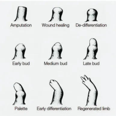
Figure 14 Regeneration of the urodele limb 59
Figure 15 A refreshed view of the Waddington's landscape 65
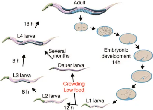
Figure 16 The C. elegans life cycle 67
Figure 17 The cell lineage of the pharynx 68
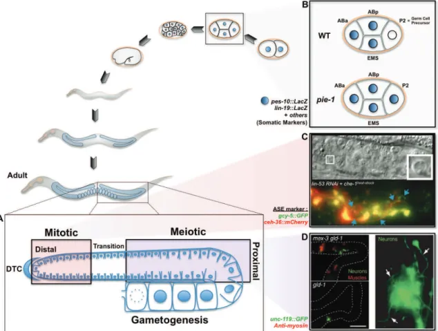
Figure 18 The C. elegans germline 70
Figure 19 The C. elegans seam cell division pattern 73 Figure 20 C. elegans blastomere conversion by ectopic factors expression 76
Figure 21 C. elegans germ cell conversion 79
Figure 22 C. elegans chemical reprogramming 82
Figure 23 The Y-to-PDA transdifferentiation 84
Figure 24 Detailled steps of the Y-to-PDA transdifferentiation 87
Figure 25 Key Events of the Notch Pathway 89-90
Figure 26 Organization of the LIN-12 and GLP-1 Notch receptors 91 Figure 27 The C. elegans Notch Ligands and Coligands 93
Figure 28 Structure of lag-1 bound to DNA 95
Figure 29 Domain organisation of the SEL-8 protein 96 Figure 30 Validated C. elegans Notch target genes. 97 Figure 31 AC/VU decision in wild-type and genetic mosaics 99 Figure 32 Feedback mechanism during the AC/VU decision 100 Figure 33 Model of biased Notch signaling during VPC specification 101 Figure 34 Inductive Notch signaling during early embrogenesis 103
Figure 35 Inductive Notch signaling during germ cell proliferation 106 Figure 36 Description of the lin-12(gf) phenotype 128 Figure 37 Fate of PDA and DA9 in lin-12 gain and loss-of-function mutants 131 Figure 38 Scoring of loss and gain-of-function mutant for glp-1 133 Figure 39 Identification of a sensitized background for the analysis of LIN-12/Notch components 135 Figure 40 APX-1 and LAG-2 act redundantly in Y-to-PDA transdifferentiation 136 Figure 41 Individual non-canonical ligands are not required for the establishment of the competence to transdifferentiate 138 Figure 42 lin-12 is sufficient to promote transdifferentiation 141 Figure 43 Extra PDA formed after ectopic activation of the LIN-12/Notch pathway around Y birth is coming from DA9 143 Figure 44 Supernumerary PDA formed in lin-12(gf) mutant required the same factors as the endogenous PDA 145 Figure 45 Expression pattern and dynamics of LIN-12 protein 148 Figure 46 Expression pattern and dynamics of lin-12 mRNA 149 Figure 47 Expression pattern of lin-12 in L1 larva 150 Figure 48 Expression pattern and dynamics of apx-1 152 Figure 49 Expression pattern and dynamics of lag-2 153 Figure 50 Maintained LIN-12/Notch pathway activation is deleterious for Y-to-PDA transdifferentiation 156 Figure 51 Y-to-PDA transdifferentiation inhibition by maintained LIN-12/Notch pathway is independent of an early activation 157 Figure 52 Ligand are still available when lin-12 is downregulated: Validation of the approach 160 Figure 53 Ligand are still available when lin-12 is downregulated 161 Figure 54 Activation of the LIN-12/Notch pathway in U, F, K and K’ has no effect on Y-to-PDA transdifferentiation 162 Figure 55 Surprising expression of egl-20::ICLGFP in all the rectal cells including Y 163
Figure 56 Mosaic analysis of ICLGFP expression 164
Figure 57 Verification of ICL functionality 165
Figure 58 Blocked Y cell expresses epithelial markers 166 Figure 59 Blocked Y cell does not express neuronal markers 167 Figure 60 Identity of B, F, U, K and K’ is not affected by LIN-12/Notch overactivation 168 Figure 61 High dose of LIN-12/Notch signaling is deleterious of Y-to-PDA transdifferentiation 170 Figure 62
Experimental design to identify the transcriptome of the wild-type Y cell and the blocked Y cell when LIN-12/Notch is misregulated
188
I) Cell plasticity
The term cell plasticity can be generally defined by the ability of a cell to change its identity. In this part of the introduction, general principles encompassed by the term "cell plasticity" will be discussed. However, before entering in the depths of cell plasticity, the historical perspectives of developmental biology will be presented to show that old concepts postulated with rudimentary tools are not that far from the actual known biological reality. Early scientists were able to postulate important concepts, which are still relevant today and others that turns to be less fixed than expected
I.1) Historical view of development and cell differentiation
I.1.1) Epigenesis versus preformationism – The opening debateEpigenesis and preformation were two embryonic and developmental theories in competition during the past centuries. The epigenesis theory considers that the final form of an organism is built gradually. During development, the embryo gets more and more specified and acquires its final form over time. In contrary, the preformationist theory considers that an organism is already formed at its origin and just grows during development, in other words any adult being comes from the growth and consolidation of its miniature form.
The epigenesis theory arises with Generation of Animals (Aristotle translated By Peck, 1979) of Aristotle (384-322 B.C.) in which he describes the development of several organisms. By looking carefully at chicken development, he could notice that the early egg was not prepatterned, the egg does not contain a preformed little chick. Instead, he noted that the egg gradually acquires its form and specific features as a beating heart or developing eyes. He postulated that the starting material is certainly available at the beginning but the final form is obtained through embryonic development. At this period Aristotle thought that the epigenetic
elements affecting development are contained in the soul of the developing organism.
This concept of epigenesis was well accepted, even by the church, but new observations and interpretations of development made during the 17th and 18th century went for the preformationist theory. Jan Swammerdam (1637-1680) studied in detail the development of insects and amphibians and could observe that the same organism was preserved during development and was just growing packed inside different structure (as egg, larva or pupa). These findings were reinforced by the development of microscopy and the observations of spermatozoids by Nicolaas Hartsoeker (1656-1725). In is Essai de Dioptrique (Hartsoeker, 1694), Hartsoeker claimed that he could observe a microscopic men inside the sperm cell. He called this little human trapped in a spermatozoid: homunculus (Figure 1).
Due to their scientific contribution and reputation Swammerdam and Hartsoeker (with many others) imposed the preformation theory, in which development is just a matter of growth and any living being is pre-determined. This pre-deterministic view of development stand for a long time and fueled the debate between epigenesist and preformationist, but in 1759, because of the evolution of
optics and microscopes, work performed by Kaspar Friedrich Wolff (1733-1794) allowed the complete reinterpretation of the observations made by previous researchers. Wolff studied meticulously the development of plants and animals and proposed that group of cells initially not specialized differentiate during development to give rise to tissues, organs and complete systems and qualified this phenomenon "morphogenesis" (Maienschein, 2012).
I.1.2) The cell theory
The debate between preformationism and epigenesis ended with the emergence of the cell theory. During the 19th century, work from Matthias Schleiden (1804-1881), Theodore Schwann (1810-1882) and later Rudolf Virchow (1821-1902) showed that cells are the bases of all the elements constituting any living being. They went further by postulating that living cells can only come from other pre-existing cells. Many questions arise from these findings and especially how a single cell (the egg or the sperm) can give the information of all the traits found in an adult being? Or how the features found in an organism can be inherited through the egg or the sperm cell?
Observations of fertilization partially answered this last question, and showed the nuclei of the egg and the sperm cells have to fuse in order to let development to take place. From these observations scientists proposed that the nucleus was the vehicle of inheritance. In 1882, Walter Flemming (1843-1905) described mitosis and showed that in the nucleus, structures called chromosomes have to split equally between two sister cells during cell division. For the first time the physical nature of inheritance was described. It is only in 1910 that experimental embryology and genetics meet to prove that chromosomes and specific regions of chromosome support the inheritance information. Thomas Hunt Morgan (1866-1945) found some male mutant Drosophila having white-eyed among population of red-eyed wild-types flies. Morgan could show that this eye color trait is linked to the sexual chromosome giving for the first time the evidence that chromosomes carry the hereditary information (Heard, 2013).
Based on the available evidence and knowledge, scientists had different point of view on the behavior of the inheritable material during cell differentiation. Because cells become more and more specialized, some scientists thought that specialization was linked to a progressive loss of genetic material and only relevant material was kept in the cells (except for the germline where it had to be preserved). On the contrary, new generations of geneticists thought that genes and chromosomes were the same in every cell but their activity were different.
The ultimate demonstration that the genetic material was conserved all along cell differentiation was brought by experiments performed by Briggs (1911-1983) and King (1921-2000) (Briggs & King, 1952) in 1952 and ten years later by John Gurdon (Gurdon, 1962). By transferring purified nucleus from a somatic cell into an enucleated egg they could reconstitute fully a viable tadpole, showing that indeed the entire genetic material was kept through cellular differentiation and by consequence development is the result of gene expression changes.
I.1.3) The Waddington’s landscape
The biggest remaining question was to understand how the genetic information could be differentially used during cell differentiation. One brilliant paleontologist, zoologist, geneticist, embryologist and philosopher, Conrad H. Waddington (1905-1975) postulated several theories to try to bridge genetics and embryology. From his observations and personal experience, Waddington supposed that a single gene can have multiple effects on different organ and multiple genes can affect a single organ (Waddington, 2012). He later defined epigenetics as "the branch of biology which studies the causal interactions between genes and their products which bring the phenotype into being" and formulated his famous epigenetic landscape theory (Waddington, 1957) (Figure 2, upper part). A schematic representation of his hypothesis shows a ball (representing a cell) rolling down a landscape made of valleys. Development is represented by the progression of the ball down the landscape. During its descent, the ball encounters several roads representing the different cell fates that a cell could acquire; once the ball has committed to a road, its destiny will be chosen and fixed for the rest of its life. Waddington imagines that differential gene expression is the foundation of the
topology of the landscape (Figure 2, lower part). At that time, Waddington was already arguing that mutation could change the topology of the landscape and by consequence change the road taken by the cell.
This unidirectional view of development (from a undifferentiated to differentiated cell) was really innovative for the era and has become the prevalent view since then.
This view of cell differentiation considers that commitment of a cell into a certain lineage and the acquisition of the final identity is a blocked phenomenon. Once the cell has reached its final destination, nothing can perturb this state, no cell fate switch or reversion could take place.
In the following parts we are going to discuss how this view of development and cell differentiation has changed. It is now well accepted that the acquisition of a
Figure 2 – The Waddington's epigenetic landscape. In the upper part the ball
represent a cell that will roll down the landscape. Each valley represents a possible cell fate. The lower part represents how the topology of the landscape is made because of genes represented by the black boxes.
cell fate is not a fixed situation and that a cell can change its identity under certain circumstances.
I.2) When a cell can exhibit plasticity
I.2.1) Definition of cell potencyCell potency is defined by the ability of a cell to differentiate into an other cell type, if we look back at the Waddington landscape (Figure 2), we can remark that according to this unidirectional view of development, cells possess a large potential of differentiation at the beginning its life. Then throughout the development this potency becomes more and more restricted.
According to their differentiation potential, cells can be group in different categories:
Totipotent cells: These cells are the most undifferentiated and potent cells
found in early development. They can give rise to all the embryonic and extra embryonic tissues (Smith, 2006). (Example: mammalian embryonic cells up to the 8-cell stage embryo, plant meristem 8-cells).
Pluripotent cells: can give rise to cells belonging to the three embryonic
germ layers (endoderm, mesoderm, ectoderm) (De Miguel, Fuentes-Julian, & Alcaina, 2010; Ratajczak et al., 2012) (Example: embryonic stem cells)
Multipotent cells: can give rise to several types of cells constituting one or
more tissues (Example: Mesenchymal stem cells (Augello, Kurth, & De Bari, 2010))
Oligopotent cells: can give rise to two or more cell types within a specific
tissue. (Example haematopoietic stem cells (Metcalf, 2007) or oligopotent stem cells in mammalian eyes (Majo, Rochat, Nicolas, Jaoude, & Barrandon, 2008)
Unipotent cells: Can form a specific lineage and differentiate into one cell
type only (example muscle stem cell (Bentzinger, Wang, von Maltzahn, & Rudnicki, 2013))
Differentiated cells: specialized cell performing a particular task in a organ or
During development, the first cells from the early embryo are totipotent. Throughout the cell divisions, this totipotency is gradually lost and the differentiation potential of the cell is reduced until the cells are differentiated and fulfill a specific function inside an organ or a tissue. Cell differentiation was seen as the last possible state for a cell. Once the differentiation program of cell is launched, we cannot go back to a primitive state or acquire another fate.
I.2.2) Terminology of cell plasticity events
Even though evidence have been obtained by the scientific community that the fate of a cell can be modified, it is only since 2006 with the work of the Yamanaka's lab (Takahashi & Yamanaka, 2006) (detailed given in the following parts) that the fact that a cell can change its identity has been widely accepted. Since this date, the cell plasticity field is booming and many examples of cell fate manipulations have been reported.
Denomination and terminology problems are often encountered when a field is in rapid expansion. In order to be clear among them, scientists have to use the same terms and denomination to describe the same phenomena. For example, the epigenetic field had to set a nomenclature for histone denomination in order to simplify their description (Talbert et al., 2012). The same problem is encountered in the cell plasticity field. Unique cell plasticity events are sometimes defined by different terms. So far, no one really tried to unify the different terminologies used to describe cell plasticity phenomena. In order to be clear for the rest of the manuscript here are the definitions that I will use for the notions detailed in the following parts. The proposed definitions are the one currently used by the scientists of the cell plasticity field . Nevertheless it is possible that in a near future, theses definitions will be modified.
Dedifferentiation: is defined by the reversion of a terminally differentiated cell
into a cell that does not exhibit any specialised or differentiated characters anymore. This encompasses what has been called by some retro-differentiation, the reversion of a terminally differentiated cell into a less differentiated (progenitor) stage within its
own lineage. Note that dedifferentiation is not automatically coupled to reversion to a more potent state.
Nuclear reprogramming: is defined by the reversal of the differentiation
state of a mature cell to a pluripotent state (Hochedlinger & Jaenisch, 2006).
Transdetermination: is defined by the switch from a committed state to
another (Manohar & Lagasse, 2009).
Transdifferentiation: is defined as the stable switch from a differentiated cell
type to another (Eguchi, 1995).
I.2.3) Dedifferentiation
I.2.3.1) Naturally induced dedifferentiation
Dedifferentiation has been reported to take place in vivo under specific circumstances. Some organisms have the capacity to regenerate some organs after amputation or injury and dedifferentiation processes take place to reform the missing organ.
Zebrafish can regenerate some organs such as the heart, spinal cord, retina or the fins (Poss, 2002). After amputation of up to 20% of the heart ventricle, regeneration can take place through dedifferentiation of cardiomyocytes (Jopling et al., 2010; Kikuchi et al., 2010). Cardiomyocytes present an important contractile sarcomeric apparatus. During regeneration, dedifferentiation of cardiomyocytes lead to the disassembly of the sarcomeres and decrease in the expression of sarcomeric genes. During this dedifferentiation process, cardiomyocytes separate and prior to proliferation start to express positive cell cycle regulators. After proliferation, this new pool of cells differentiates again into cardiomyocytes to reform the missing part of the heart.
Similar results have been reported during Zebrafish fin regeneration. After amputation of the fin, a zone of undifferentiated proliferating cells (called blastema) forms and gives rise again to the amputated part. Using careful cell tracking, it has been shown that after amputation, osteoblast cells dedifferentiate and become
proliferative in the blastema. The newly produced cells will then differentiate again into osteoblast to form the bones present in the fin (Knopf et al., 2011) (Figure 3).
In mammals very few examples of clear natural cell dedifferentiation have been reported. In the brain, Schwann cells are glial cells wrapping around neuronal axon and form a myelin sheath. During development, Schwann cell precursors are made from the neural crest cells. These precursors differentiate into proliferative immature Schwann cells and finally differentiate into mature Schwann cells able to wrap around axons. During injury, when Schwann cells lose the contact with the axons, they dedifferentiate by expressing immature Schwann cell genes, proliferate and differentiate again to form new mature Schwann cell (Mirsky et al., 2008; Woodhoo et al., 2009).
Surprisingly, only few well characterized cases of natural dedifferentiation have been reported. This absence of knowledge is due in particular to a lack of tools allowing proper cell tracking during development. Unfortunately, in the cell plasticity field this lack of tool was the source of several controversies that will be detailed later.
Figure 3 – Regeneration of the Zebrafish fin through dedifferentiation of
osteoblasts cells. After amputation, osteoblasts dedifferentiate and proliferate in a blastema. After proliferation, the new cells differentiate again into osteoblasts to reconstruct the fin (Knopf et al., 2011).
I.2.3.2) Experimentally induced dedifferentiation
To date, only few examples of experimentally induced dedifferentiation have been reported. Some of them were found in vitro, while only one clear example has been discovered in vivo.
In some organisms such as the newt, myotubes can dedifferentiate and proliferate whereas mammalian myotubes do not have this feature. This lack of plasticity could be due to several possibilities: mammalian myotubes lack specific genes allowing dedifferentiation or irreversibly express genes that lock the myotube fate. It can be possible as well that extrinsic factors present during newt myotube dedifferentiation are not available in mammals. To test these hypothesis scientists have applied extracts from newt regenerating limbs on C2C12 mammalian myotubes. They could observe that expression of myotube specific genes as MyoD, myogenin or troponin T was reduced and that cell can reenter the cell cycle suggesting a dedifferentiation followed by proliferation (McGann, Odelberg, & Keating, 2001). These data suggested that newt-specific extrinsic factors help to allow myotube dedifferentiation and could explain why this cell plasticity difference exists between newt and mammalian myotubes.
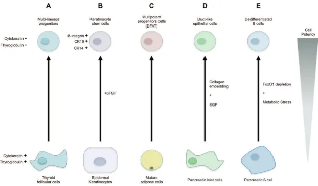
Purified human thyroid cells have been put in culture with a serum-free medium. A lot of the cultured cells died but a small proportion survived and proliferate. These proliferating cells show reduced expression in thyroid specific genes such as thyroglobulin or cytokeratine-18. If these cells are kept in these culture conditions, they do not express any mature thyroid specific markers. Surprisingly, these newly obtained undifferentiated cells were able to differentiate into thyroid cells and into neuronal or adipogenic lineages indicating that the starting cells dedifferentiate into multipotent cell (Suzuki et al., 2011) (Figure 4.A).
Human epidermal keratinocytes grown in culture with basic fibroblast growth factor (bFGF) and no extra intervention can dedifferentiate by re-expressing native keratinocyte stem cell markers such as ß-integrin, CK19 or CK14 (Sun et al., 2011) (Figure 4.B).
It has been reported that upon simple specific culture strategy, which allow adipocyte observation and prevent cell to fuse, that mature adipocyte (thought to be
non-proliferative) can dedifferentiate into cells called dedifferentiated fat (DFAT). These DFAT cells become multipotent and present adipogenic, osteogenic, chondrogenic and myogenic potential (Shen, Sugawara, Yamashita, Ogura, & Sato, 2011) (Figure 4.C).
Several cases of pancreatic cells dedifferentiation have been reported. In a first study, made in vitro, human islet cells embedded in collagen and in contact with epidermal growth factor have been shown to dedifferentiate into proliferative duct-like epithelial cells (Hanley, Assouline-Thomas, Makhlin, & Rosenberg, 2011) (Figure 4.D). In a second study, made in vivo, it has been reported that adult murine ß cells can modify their differentiation state under physiological stress conditions. Maintenance of the ß cell identity relies on the specific transcription factor FoxO1. Ablation of this gene in adult murine ß cell has no effect in normal condition. However under stress condition, the pancreas present a dramatic loss of ß cells. It was though that this loss is the result of increase cell death in insulin producing cells but instead ß cells seem to dedifferentiate into multipotent endocrine progenitors. This dedifferentiation could be an advantage allowing the ß cells to survive in case of strong metabolic stress (Talchai, Xuan, Lin, Sussel, & Accili, 2012) (Figure 4.E).
Figure 4 – Examples of cell dedifferentiation adapted from (Eguizabal, Montserrat, Veiga, &
All these examples point the fact that differentiated cells can revert into a less specialized state under specific conditions. Moreover, dedifferentiation is not limited to a specific embryonic origin (i.e. Pancreatic cells originate from the endoderm whereas keratinocytes originate from the ectoderm), suggesting that any cell type could increase its potency under specific circumstances.
I.2.4) Nuclear reprogramming
As mentioned previously, nuclear reprogramming was used to describe the switch from a differentiated cell state to a pluripotent state. In other word, by nuclear reprogramming, a fully specialized cell fulfilling its role in a tissue can be reprogrammed into a non-specialized cell able to give rise to cells from the three germ layers. Different methods can be used to completely revert the differentiation state of cell. Nuclear reprogramming can be done by somatic-cell nuclear transfer (SCNT), by cell fusion, by using cell extracts or by over-expression of specific factors.
I.2.4.1) Somatic-cell nuclear transfer (SCNT)
During the 1950's the key question in developmental biology was whether the genome of cells was irreversibly modified during cell differentiation. To answer this question Briggs & King transferred the nucleus from a blastula into an enucleated frog egg and could obtain living tadpoles (Briggs & King, 1952). These experiments showed for the first time that the genetic information is not modified during cell differentiation. However, this view was challenged by Briggs and King themselves, when they tried to repeat this experiment with a nucleus coming from a late developmental stage. Indeed, using these new nuclei, they could not be able to obtain living tadpoles. They conclude that irreversible nuclear changes could take place when cells differentiate. Later, John Gurdon decided to do the same experiment in another species of frog. Somatic nuclei from post-blastula embryo and intestinal nuclei were transferred into enucleated eggs. In both cases, living and fertile frogs have been obtained bringing the proof that genetic information is not lost during cell differentiation. These results highlighted an other important concept: based on its experiments John Gurdon proposed that the fate of a differentiated nucleus can be reverted to a pluripotent one, which can drive the development and
formation of a complete new living organism. Indeed in the Gurdon's experiments, intestinal cells were extracted from a functional organ. The nucleus of these cells is "programmed" to execute the function of the cell. When this nucleus is transferred into an enucleated egg, its program and function are erased by intrinsic determinants of the egg. This newly reprogrammed nucleus is now totipotent, and can be committed into any cell differentiation program. At the time, the scientific community remained skeptical and thought that during the nuclear transfer procedure, Gurdon took the nucleus from a contaminating undifferentiated cell, which may exist in the post-blastula embryo or in the intestine (Figure 5).
Although, John Gurdon showed that nuclei from skeletal muscle, skin, lung or kidney transferred into an egg could be reprogrammed (Gurdon, Brennan, Fairman, & Mohun, 1984; Laskey & Gurdon, 1970), it is only when the first mammal, Dolly the sheep, has been obtained by SCNT that the hypothesis that differentiated nucleus can be reprogrammed into a pluripotent state begun to be widely accepted (Wilmut, Schnieke, McWhir, Kind, & Campbell, 1997)]. Because all the nuclear reprogramming experiments performed so far have a success rate close to 1%, some people were still arguing that reprogramming is only due to the use of contaminating undifferentiated nuclei. The laboratory of Rudolf Jaenisch brought the ultimate evidence that this argument was wrong. They could show that reprogramming of highly specialized cells such as mouse olfactory neurons or mouse T and B lymphocyte can be performed showing that any differentiated nucleus can be reprogrammed into a pluripotent state (Eggan et al., 2004; Hochedlinger & Jaenisch, 2002). Indeed the viable mouse obtained by SCNT of T or B-lymphocytes carry the same genomic rearrangement than the donor cells.
Figure 5 – Somatic-cell nuclear transfer: A single nucleus from a somatic cell is injected into an
enucleated egg. The developing embryo can give rise to a fertile adult, a fetus or can be used to derive embryonic stem cell line (Gurdon & Melton, 2008).
The success rate of nuclear reprogramming has been shown to be dependent on the differentiation state of the donor cell. The more differentiated the cell is, the less reprogramming is efficient. The nucleus of differentiated cells acquires through development an epigenetic restriction. For example these restrictions can be brought by DNA methylation, histone modification, histone variants and non-coding RNAs (Pasque, Jullien, Miyamoto, Halley-Stott, & Gurdon, 2011). All these restrictions have to be suppressed at the same time to allow reprogramming explaining why this phenomenon hardly takes place.
Because nuclei from differentiated cells are more resistant to reprogramming, scientist passes through the formation of nuclear-transfer-derived embryonic stem cells (nt-ESCs). Rarely, transferring a nucleus (from any species) from a differentiated cell leads to the formation of a living blastula. From those embryos, nt-ESCs can be derived and used as a donor cell to perform new nuclear transfer into new enucleated egg or oocyte. When nt-ESCs are used more than 20% of the newly transferred nuclei are reprogrammed. Because of this technique nt-ESCs lines have been derived from cows, dogs, and non-human primates.
A very recent study showed that SCNT could work as well with human tissue. The recipient cell used in human SNCT is an oocyte blocked in metaphase II of the meiosis. During the SCNT experiment, the penetration of the needle used to transfer the somatic nucleus acts as a sign of fertilization for the oocyte, this manipulation triggers the completion of the meiosis in the oocyte and inhibits the reprogramming of the transferred nucleus. Caffeine has been applied to metaphase II oocytes to prevent premature completion of meiosis. Because of this molecule, the transferred nucleus can be reprogrammed; the embryo can develop until the blastocyst stage (around 100 cells) and can be used to derive embryonic stem cell lines (Tachibana et al., 2013).
Note: This paper is under investigation because of potential mislabeling and figure duplication. The Cell press group is studying if these errors can completely change the conclusion of the paper and in the worst-case lead to a retraction.
I.2.4.2) Nuclear reprogramming by cell fusion and cell extract
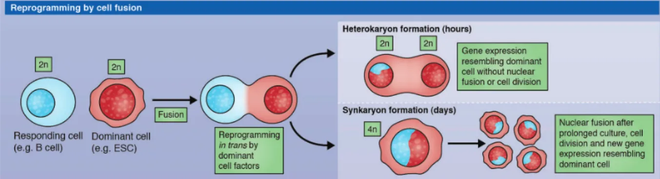
Cells can be fused leading to the formation of a non-proliferative tetraploid cell called heterokaryon. During cell fusion the less differentiated cell is the dominant cell and impose its fate to the partner cell (Yamanaka & Blau, 2010). Reprogramming of differentiated cells such as mouse T lymphocytes can be done by fusion with an embryonic stem cell (Tada, Takahama, Abe, Nakatsuji, & Tada, 2001). The resulting heterokaryon expresses pluripotency genes after a few hours and when injected into mouse blastocyst can give rise to chimeric embryos. Similar results have been obtained with human cells (Cowan, 2005). Unfortunately the use of these heterokaryons is really limited. On one hand their non-proliferative status is not suitable to derive embryonic stem cell line, on the other hand their tetraploid status could lead to aberrant gene reactivation from one nucleus leading for example to loss of pluripotency (Figure 6).
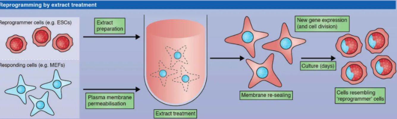
Nuclear reprogramming has been shown to be also possible after exposure to protein extracts from pluripotent cells (Taranger et al., 2005). Cultured epidermal cells (293T cells) have been permeabilized, exposed to embryonic stem cell or carcinoma cell extracts and re-sealed. After culture, the cells in contact with the extract represses 293T specific gene and begin to express genes normally up-regulated in the cells used for the extract. The reprogrammed cells can be differentiated into neurogenic, adipogenic, osteogenic, and endothelial lineages. The efficiency of reprogramming with this method is really low. Moreover we don't know if the resulting cells are fully pluripotent; their potency have not been tested with stringent test (i.e. teratoma or chimera formation) (Figure 7).
Figure 6 – Nuclear reprogramming by cell fusion. A somatic cell can be reprogrammed by fusion
with a dominant cell (e.g. an embryonic stem cell). The resulting cell can be a tetraploid heterokaryon or a synkaryon if the cell is kept under culture (Halley-Stott, 2013).
I.2.4.3) Induction of pluripotency by defined factors
In 2006, the lab of Shinya Yamanaka sat off of a revolution in the stem cell and cell plasticity field. They could show that mouse, then human embryonic fibroblasts can be reprogrammed into pluripotent stem cell by over-expression of specific factors (Takahashi & Yamanaka, 2006; Takahashi et al., 2007). A screen was designed to find the factors able to induce pluripotency in somatic cell. They developed a modified mouse fibroblast cell line in which a drug resistance gene can be express under an embryonic cell specific promoter (Fbxo1). They over-expressed 24 pluripotency associated factors in this cell line and could obtain drug resistant colonies with the morphology of embryonic stem cells. With consecutive series of elimination of individual factors they could define the minimal cocktail able to induce pluripotency in fibroblasts, which is composed by Klf4, Sox2, c-Myc and Oct-4 (Figure 8). This newly pluripotent cells are called induced pluripotent stem cells (iPSC), express pluripotent stem cell markers such as SSEA-1 and Nanog and can generate teratomas when injected in immune-deficient mice.
Figure 7 – Nuclear reprogramming by cell extract treatment. Somatic cell can be reprogrammed
after permeabilization and cultured with embryonic stem cell extract. The reprogrammed cell takes the identity of the cell used to make the extract (Halley-Stott, 2013).
Figure 8 – Nuclear reprogramming by over-expression of defined factors. Over-expression of Klf4,
However these first generations of iPSCs exhibit incomplete DNA demethylation of the Oct4 promoter compare to embryonic stem cells and were not able to generate chimeras when injected into blastocysts showing their plausible incomplete reprogramming. Later, improvements of this reprogramming technique, based on a more stringent method to select the reprogrammed cells, have been done resulting in iPSCs molecularly and functionally close to embryonic stem cells. These "new generation" of iPSCs were able to give rise to viable chimera attesting that these cells are fully pluripotent (Maherali et al., 2007; Okita, Ichisaka, & Yamanaka, 2007; Wernig et al., 2007). iPSCs have been derived from many species such as mouse, human, rats (Li et al., 2009), pig (Esteban et al., 2009), and rhesus monkey (Liu et al., 2008) showing that induction of pluripotency requires elements which have been conserved through evolution.
Since 2006, the number of publication related to iPSCs completely exploded (Scott, McCormick, Derouen, & Owen-Smith, 2011). An intensive race between labs was launched after the publication of the first iPSCs paper. Scientists tried to reprogram other cell type than fibroblast and could show that, for example, pancreatic ß cells (Stadtfeld, Brennand, & Hochedlinger, 2008), keratinocytes (Aasen et al., 2008) or hepatic and stomach cells can be reprogrammed (Aoi et al., 2008), indicating that reprogramming is not dependent on the starting cell fate. The first iPSCs were obtained using over-expression, after genomic integration, of the four reprogramming factors; people have tried to find alternative methods to reprogram cells and not modify the genome of the reprogrammed cells. Some non-integrative methods using adenoviruses (Stadtfeld, 2008) or plasmids (Okita, 2008) have been used. Some DNA free reprogramming methods have been developed using either recombinant protein (Zhou et al., 2009) or synthetic modified RNAs (Warren et al., 2010).
In parallel of this reprogramming improvement race, stem cell scientists tried to understand the mechanisms underlying induced pluripotency. The first stricking element is that efficiency rate of reprogramming with defined factors oscillate in average around 0,02% for mouse and human fibroblasts (Brambrink et al., 2008; Stadtfeld, 2008; Wernig et al., 2008). In addition to this, reprogramming is slow; it takes around 2 weeks to see the first reprogrammed clone appearing. From these