Carbon nanotubes: an insight into the mechanisms
of their potential genotoxicity
Damiën van Berloa, Martin Cliftb, Catrin Albrechta, Roel Schinsa a
IUF-Leibniz Research Institute for Environmental Medicine, Dusseldorf, Germany
b
Adolphe Merkle Institute and Fribourg Center for Nanomaterials, University of Fribourg, Marly, Switzerland Summary
After the health catastrophe resulting from the widespread use of asbestos which was once hailed as a new miracle material, the increasing use of carbon nanotubes (CNTs) has spawned major concern due to their similarities in terms of size, shape and poor solubility. Assessment of genotoxicity has shown that CNTs can damage DNA in vitro and in vivo. The genotoxic potential of different CNT samples varies considerably, however, with negative find-ings reported in a number of studies, probably due to the enormous heterogeneity of CNTs. The observed spectrum of genotoxic effects shows similarities with those reported for asbestos fibres. Mutagenicity has been found in vivo but in bacterial assays both asbestos and CNTs have mostly tested negative. An overview of key experimental observa-tions on CNT-induced genotoxicity is presented in the first half of this review.
In the second part, the potential mechanisms of CNT-eli-cited genotoxicity are discussed. Whereas CNTs possess intrinsic ROS-scavenging properties they are capable of
List of abbreviations:
Aprt Adenine phosphoribosyltransferase BER Base excision repair
CNTs Carbon nanotubes DSB Double strand breaks fpg Formamidopyrimidine glycosylase
hpgrt Hypoxanthine-guanine phosphoribosyltransferase iNOS Inducible nitric oxide synthase
MDA Malondialdehyde
MW-CNTs Multi-walled carbon nanotubes NER Nucleotide excision repair OGG1 8-oxoguanine-DNA glycosylase 8-OHdG 8-hydroxydeoxyguanosine PARP Poly(ADP-ribose) polymerase
PL-PEG Phospholipids with a polyethylene glycol moiety RNS Reactive nitrogen species
ROS Reactive oxygen species SOD Superoxide dismutase SSB Single strand breaks
SW-CNTs Single-walled carbon nanotubes TBARS Thiobarbituric acid reactive substances
TUNEL Terminal deoxynucleotidyl transferase dUTP nick end labeling
generating intracellular ROS upon interaction with cellular components, and can cause antioxidant depletion. These ef-fects have been attributed to their Fenton-reactive metals content. In addition, CNTs can impair the functionality of the mitotic apparatus. A noteworthy feature is that frus-trated phagocytosis, which is involved in asbestos-induced pathology, has been observed for specific CNTs as well. The involvement of other mechanisms generally implicated in particle toxicity, such as phagocyte activation and impairment of DNA repair, is largely unknown at present and needs further investigation.
Key words: carbon nanotubes; asbestos; genotoxicity; mutagenicity; reactive oxygen species
Introduction Carbon nanotubes (CNTs)
In recent decades, considerable interest has focused on the development of novel nanomaterials that can be useful for a plethora of medical, industrial and consumer applications. Due to their physico-chemical, electrical, mechanical and thermal properties, carbon nanotubes (CNTs) can enhance a variety of consumer or industrial products, ranging from use in electronics or protective clothing to biomedical ap-plications such as drug delivery or genetic modification of crops. CNTs feature a very large surface per weight ra-tio, which is useful in catalytic processes, a tensile strength that is many times higher than that of steel and excel-lent conduction of heat and electricity. They also possess good thermal stability and are resistant to many chemic-als. However, due to their very small diameter inhalation of such materials is possible and they are thus capable of reaching the most distal areas of the pulmonary system, i.e. the alveolar sacs [1].
CNTs possess a robust graphene structure which is bioper-sistent. They are produced in several forms, the main dis-tinction being the number of carbon atom layers; CNTs with one layer of carbon atoms are referred to as single-walled CNTs (SW-CNTs), while those with multiple layers are denominated multi-walled CNTs (MW-CNTs). Both of these forms can become aerosolised in occupational
set-tings [2–5]. The diameter of SW-CNTs is generally 0.4 to 3 nm [6], but they readily form bundles held together by van der Waals forces called nano-ropes, which are commonly 20–30 nm in diameter [7] up to 500 nm [6]. MW-CNTs have a diameter that is more variable, ranging from roughly 5–100 nm, while MW-CNT nano-ropes can be 3μm in dia-meter [6]. To further improve their properties, MW-CNTs can be functionalised, which involves the addition of mo-lecules to their carbon-based structure. CNTs can be as long as 18 cm [8], providing an incredible aspect ratio (i.e. the ratio of length to width). Clearly, nanotubes represent a highly heterogeneous hazard, with marked differences in size, rigidity, single- vs. multi-walled structures and pos-sible functionalisation of their surface.
Due to their small size (providing a substantial atom-to-surface ratio), low biodegradability and fibrous structure (defined as an aspect ratio of at least 3:1), the important question arises whether the known severe health issues as-sociated with exposure to another durable fibrous material (i.e. asbestos) might be relevant for CNT exposure. Asbestos is a natural fibrous material that was once hailed as a new miracle material that was durable, easy to process and possessing remarkable flame-repellent properties. However, when these fibres become fractured, resulting in dimensions sufficiently small to be inhalable, they can cause severe pulmonary pathology, characterised by chron-ic inflammation, fibrosis and cancer – specifchron-ically meso-thelioma.
Carcinogenesis is a multi-step process involving initial in-sult to the genome, compromised cellular defence mech-anisms and proliferation. A first hint for the carcinogenic potential of CNTs was published in 2008. In heterozygous p53+/- mice, featuring reduced function of the crucial tu-mour suppressor protein p53, intraperitoneal injection of CNTs was reported to elicit mesothelioma formation [9]. However, the results from this study have been disputed since the investigators administered a very large dose (3 mg per mouse) of CNT aggregates that were several hundreds of μm in diameter. Fibres of these dimensions are not capable of reaching the alveoli upon inhalation and thus will not be taken up by macrophages, which is known to be of critical importance for asbestos-induced pathogenes-is [10]. Moreover, the model used has not been validated to demonstrate applicability for fibre-induced mesotheli-oma [10]. In a follow-up to this study the authors addressed the first point of criticism related to dosimetry. Dose-de-pendent induction of mesothelioma was observed in the same p53+/-mouse strain upon intraperitoneal injection of MW-CNT at much lower doses of 3, 30 and 300 μg per mouse. At the two highest doses, most mice (17 and 19 out of 20) developed mesothelioma within a year [11]. In rats, with an in vivo model widely accepted for the eval-uation of fibre pathogenesis and mesothelioma formation, increased development of mesothelioma has been observed after intraperitoneal injection of 1 or 10 mg MW-CNT. Ef-fects were stronger for thin and rigid MW-CNTs compared to samples with a larger diameter [12]. However, another study involving intraperitoneal injection in rats has repor-ted contrasting results. The effects of MW-CNT injection were evaluated after 2 years and compared to the positive control crocidolite asbestos. Even at a dose tenfold
high-er than that used for the asbestos sample, enhanced occur-rence of mesothelioma was only found for asbestos [13]. The authors discuss the question whether this lack of effect might be due to the relatively short length of the CNTs used (<1 μm on average) and the absence of an inflammatory response, as well as the antioxidant capacity of the CNT sample. Due to contrasting observations on CNT carcino-genesis, this issue is at present unresolved.
Genotoxicity and mutations
Genotoxicity can be defined as damage to the genetic ma-terial encrypted within our DNA. When DNA integrity is compromised by a damaging agent, a number of scenari-os are pscenari-ossible. These depend upon the type of damage, its extent and persistence [14]. Well-described forms of DNA damage include strand breakage, oxidative DNA damage and formation of bulky adducts. There are many agents with DNA-damaging potential. These include reactive oxy-gen and nitrooxy-gen species (ROS and RNS), aromatic com-pounds (e.g. benzo(a)pyrene), and also some types of particles and fibres (e.g. quartz, diesel engine exhaust particles, asbestos) (e.g. [15]). For particles (including fibres) specifically, genotoxicity has multiple mechanistic components. Primary genotoxicity is induced in the ab-sence of an inflammatory response, while secondary gen-otoxicity is driven by the activation of inflammatory cells such as neutrophils and macrophages which can generate substantial amounts of reactive species in the so-called ox-idative burst [15]. In addition, genotoxicity of particles can be direct and indirect. Direct genotoxicity can result from physical interactions of the particulate material with the genomic DNA. Indirect genotoxicity can result from in-creased ROS generation upon interaction with other cellu-lar components (e.g. mitochondria, cell membrane) or can arise when particles elicit depletion of intracellular anti-oxidants. In this scenario ROS that are generated during physiological cellular processes (e.g. cellular respiration) can accumulate and damage the DNA.
Eukaryotic cells possess extensive defensive mechanisms to avert and counter damage to the genome. For instance, their antioxidant array repels the adverse action of penet-rating or intracellular generated oxidants. Important anti-oxidants include glutathione, thioredoxin and superoxide dismutase (SOD). DNA repair enzymes restore damaged DNA to its original form, mainly via base excision repair (BER) and nucleotide excision repair (NER), while mis-match repair and direct repair via DNA alkytransferases are minor mechanisms. When the protective arsenal fails, a cell can undergo controlled cell death to prevent the fixation of mutations, which contributes to carcinogenesis. Altern-atively, in seriously damaged cells necrotic cell death can occur, which is an uncontrolled process. When these mech-anisms fail, fixation of the DNA mutation is a fact and the alteration is replicated upon cellular mitosis.
Various types of DNA mutations are known. Mutations of key loci within the genetic code are the molecular hallmark of cancer. These key genes can be divided into proto-onco-genes that stimulate cellular growth and proliferation (e.g. K-ras), and tumour suppressor genes that inhibit prolifer-ation or are involved in DNA repair (e.g. p53). In addi-tion to their dreaded role in carcinogenesis, DNA mutaaddi-tions
can cause various pathologies and can modulate susceptib-ility to disease. Mutations can involve relatively small se-quences, like single genes, or can occur on a larger scale. Examples of small-scale mutations are point mutations (i.e. transitions or transversions), in which one nucleotide is replaced by another; insertions, which add nucleotides to the genetic code; and deletions, which remove nucleotides from DNA.
Mutations can also affect chromosomal structures. Examples of clastogenic effects, affecting part(s) of a chro-mosome, are: amplifications, which augment the number of copies of chromosomal regions; chrosomal region de-letions, which leads to the opposite and thus loss of in-formation; chromosomal translocations, whereby sections of non-homologous chromosomes are interchanged; inter-stitial deletions, in which parts of a chromosome are re-moved, potentially creating fusion proteins from two pre-viously unconnected sequences; chromosomal inversions, involving the rearrangement of segments within the same chromosome; and loss of heterozygosity, the loss of one allele of a gene for which the other allele was previously rendered inactive.
When chromosome segregation between daughter cells is disturbed during their mitosis, aneugenic effects can occur, resulting in alteration of the chromosome number. Aneuploidy can result from weakening or deactivation of mitotic checkpoints, attachment of the kinetochore (which bind chromosome centromeres to the microtubuli of the mitotic spindle to allow for segregation) to both mitotic spindles and formation of additional spindles or the loss of one.
Aim and contents
Although the (nano-) toxicological community has directed much attention towards possible adverse effects of CNTs, many questions are still unanswered due to the short time span available for toxicological assessment of this recently
emerging potential hazard. The present review sets out to discuss whether CNTs may elicit genotoxicity, with a spe-cial focus on the underlying mechanisms. Assessment of genotoxicity is considered a useful tool for understanding of the potential carcinogenic risk of particles [16]. In section II, findings on CNT-induced genotoxicity in ex-perimental in vivo and in vitro systems are presented. Available data are summarised in tables 1 and 2 for SW-CNTs and MW-SW-CNTs respectively. A discussion on the similarity to the known genotoxic and carcinogenic hazard asbestos and an overview of studies comparatively evalu-ating the genotoxicity of CNTs and asbestos will be found in section III. Subsequently, in section IV, hypothetical mechanisms of CNT-associated genotoxicity are discussed. The proposed mechanisms are visualised in figure 1. Investigations on CNT genotoxicity DNA strand breakage
Breakage of the DNA backbone can occur due to the direct action of reactive species such as hydroxyl radicals (HO•) [17]. However, strand breaks can also be elicited by adduct formation and subsequent DNA repair enzymes (e.g. en-donucleases) that transiently cleave the DNA. In addition, DNA strand breakage is one of the features of apoptosis [18].
Both single and double DNA strand breakage can occur. Whereas single strand breaks (SSB) are generally repar-able, double strand breaks (DSB) are not. SSB (and alkali labile sites which are converted into SSB) are usually measured by the comet assay in its alkaline variant. Higher levels of SSB increase the ability of DNA to migrate out of nucleoids of lysed cells during electrophoresis. The pH-neutral version of the comet assay detects DSB. This type of DNA strand breakage can also be assessed by measuring H2AX phosphorylation, which results in the formation of
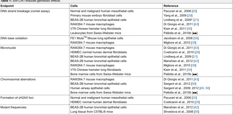
Table 1: SW-CNT-induced genotoxic effects.
Endpoint Cells Reference
DNA strand breakage (comet assay) Normal and malignant human mesothelial cells Primary mouse embryo fibroblast cells BEAS-2B human bronchial epithelial cells RAW264.7 mouse macrophages V79 Chinese hamster lung fibroblasts Leukocytes from Swiss-Webster mice
Pacurari et al., 2008 [20] Yang et al., 2009 [29] Lindberg et al., 2009* [21] Di Giorgio et al., 2011 [43] Kisin et al., 2011 [30] Patlolla et al., 2010b [■■] DNA base oxidation FE1 MutaTMMouse lung epithelial cells
RAW264.7 mouse macrophages
Jacobsen et al., 2008 [38] Migliore et al., 2010 [39]
Micronuclei RAW264.7 mouse macrophages
HDMEC normal human dermal fibroblasts BEAS-2B human bronchial epithelial cells BEAS-2B human bronchial epithelial cells RAW264.7 mouse macrophages V79 Chinese hamster lung fibroblasts Bone marrow cells from Swiss-Webster mice
Di Giorgio et al., 2011 [43] Cveticanin et al., 2010 [28] Lindberg et al., 2009 [21] Manshian et al., 2012 [42] Migliore et al., 2010 [39] Kisin et al., 2011 [30] Patlolla et al., 2010b [■■]
Chromosomal aberrations RAW264.7 mouse macrophages
BEAS-2B human bronchial epithelial cells Human airway epithelial cells
Bone marrow cells from Swiss-Webster mice
Di Giorgio et al., 2011 [43] Sargent et al., 2012 [50] Sargent et al., 2009; 2012 [49, 50] Patlolla et al., 2010b [■■] Formation of γH2AX foci Normal and malignant human mesothelial cells
HDMEC normal human dermal fibroblasts
Pacurari et al., 2008 [20] Cveticanin et al., 2010 [28] Mutant frequencies BEAS-2B human bronchial epithelial cells
Lung tissue from C57BL/6 mice
Manshian et al., 2012 [42] Shvedova et al., 2008 [55] * The SW-CNT sample in this study had a purity of about 50%, the remaining fraction contained other nanotubes.
γ-H2AX foci that are considered a marker for DSB. Activ-ation of the poly(ADP-ribose) polymerase (PARP) enzyme is considered another indicator of strand breakage because of its role in DNA repair [19].
Effects have been evaluated in several cell types:
Mesothelial cells: importantly, Pacurari et al. explored the effects of SW-CNTs in normal and malignant mesothelial cells, the target cell for mesothelioma development. They
Figure 1
Proposed mechanisms of CNT genotoxicity. The presence of structural defects and transition metals has been implicated in their genotoxic potential. CNTs can induce ROS formation by interaction with cellular components, such as mitochondria and the cell membrane, and are capable of compromising intracellular antioxidant (AOX) defences. Contamination with transition metals may also contribute to oxidative DNA damage induction. In addition, CNT have been reported to disturb the mitotic apparatus, which could explain aneugenic effects as observed in several studies. Whether CNT are capable of inhibiting DNA repair is currently unknown. Secondary genotoxic effects may result from ROS that are generated from recruited phagocytes during CNT-induced inflammation and/or activation of these cells. Further research is needed to unravel the relevance of each of the above postulated mechanisms that should take into account realistic exposure scenarios.
found enhanced DNA strand breakage in both cell types, assessed by comet assay and by measurement of histone H2AX phosphorylation and activation of poly(ADP-ribose) polymerase 1 (PARP-1) [20].
Lung epithelial cells: a CNT sample consisting of SW-and MW-CNTs elicited significant, dose-dependent DNA strand breakage measured by the comet assay in BEAS-2B human bronchial epithelial cells. Effects were observed at all concentrations (1–100 μg/cm2) and time points (24h, 48h and 72h) tested. Graphite nanofibres (which are of sim-ilar length but are roughly 100–200 times as thick) showed far lower genotoxic potency in the same assay [21]. In A549 lung epithelial type II cells (human carcinoma cell line), MW-CNT treatment elicited enhanced DNA strand breakage determined by the comet assay [22]. Enhanced DNA strand breakage was induced by MW-CNTs in A549 cells [23–25], especially by nanofibres that were relatively thick and long [23]. SW-CNTs elicited smaller effects [23]. Fibroblasts: in normal human dermal fibroblasts, both nor-mal and functionalised MW-CNTs elicited enhanced DNA strand breakage [26, 27]. The occurrence of H2AX foci was induced in human fibroblasts by normal SW-CNTs as well as amide-functionalised SW-CNTs [28]. Comparative evaluation of several particle types (including CNT, car-bon black, zinc oxide [ZnO] and silica [SiO2]) in primary mouse embryo fibroblasts, showed that CNTs were the most potent inducers of DNA strand breakage [29]. In V79 lung fibroblasts, SW-CNT treatment elicited enhanced DNA strand breakage [30].
Endothelial cells: treatment of human umbilical vein en-dothelial cells with MW-CNTs resulted in enhanced form-ation of γH2AX foci [31].
Mouse embryonic stem cells: enhanced expresson of the double strand break repair protein Rad51 and increased H2AX phosphorylation were found after treatment with MW-CNTs [32].
In vivo: In bronchoalveolar lavage cells obtained from apolipoprotein E deficient mice exposed to SW-CNTs via
Table 2: MW-CNT-induced genotoxic effects.
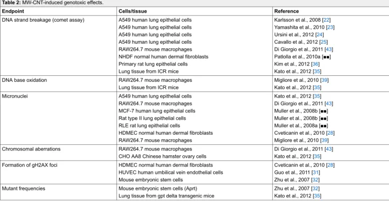
Endpoint Cells/tissue Reference
DNA strand breakage (comet assay) A549 human lung epithelial cells A549 human lung epithelial cells A549 human lung epithelial cells A549 human lung epithelial cells RAW264.7 mouse macrophages NHDF normal human dermal fibroblasts Primary rat lung epithelial cells Lung tissue from ICR mice
Karlsson et al., 2008 [22] Yamashita et al., 2010 [23] Ursini et al., 2012 [24] Cavallo et al., 2012 [25] Di Giorgio et al., 2011 [43] Patlolla et al., 2010a [■■] Kim et al., 2012 [36] Kato et al., 2012 [35]
DNA base oxidation RAW264.7 mouse macrophages
Lung tissue from ICR mice
Migliore et al., 2010 [39] Kato et al., 2012 [35]
Micronuclei A549 human lung epithelial cells
RAW264.7 mouse macrophages MCF-7 human lung epithelial cells Rat type II lung epithelial cells RLE rat lung epithelial cells
HDMEC normal human dermal fibroblasts RAW264.7 mouse macrophages
Kato et al., 2012 [35] Di Giorgio et al., 2011 [43] Muller et al., 2008b [■■] Muller et al., 2008b [■■] Muller et al., 2008a [■■] Cveticanin et al., 2010 [28] Migliore et al., 2010 [39]
Chromosomal aberrations RAW264.7 mouse macrophages
CHO AA8 Chinese hamster ovary cells
Di Giorgio et al., 2011 [43] Kato et al., 2012 [35] Formation of gH2AX foci HDMEC normal human dermal fibroblasts
HUVEC human umbilical vein endothelial cells Mouse embryonic stem cells
Cveticanin et al., 2010 [28] Guo et al., 2011 [31] Zhu et al., 2007 [32] Mutant frequencies Mouse embryonic stem cells (Aprt)
Lung tissue from gpt delta transgenic mice
Zhu et al., 2007 [32] Kato et al., 2012 [35]
intratracheal instillation, a statistically significant induction of DNA strand breakage was detected compared to the ef-fect in control mice [33]. In a more recent study, geno-toxic effects of functionalised and non-functionalised MW-CNTs were evaluated in murine bone-marrow cells after repeated intraperitoneal injection. Significant induction of DNA strand breakage was observed in leukocytes. Also, the purified functionalised MW-CNTs demonstrated stronger genotoxic potential than non-functionalised MW-CNTs [34].
In lung tissue obtained from mice exposed to a single dose of MW-CNTs (0.05 or 0.2 mg per animal), dose-dependent enhanced DNA strand breakage was measured by comet assay [35]. Significant induction of DNA strand break-age was also observed in primary lung cells isolated from rats which were exposed to aerosolized MW-CNTs (0.94 mg/m3) for 5 days, 6 h per day. Cells were analysed using the comet assay either directly after exposure ended or one month post-exposure [36].
Oxidative DNA damage
The induction of oxidative stress is considered a main drive behind particle exposure-associated genotoxicity (e.g. [37]) The prime example of an oxidative DNA lesion is 8-hydroxydeoxyguanosine (8-OHdG), which can for in-stance be detected using high performance liquid chroma-tography, gas chromatography coupled mass spectrometry. A more recent alternative to these highly sensitive meth-ods is the comet assay plus additional treatment with the enzyme formamidopyrimidine glycosylase (fpg), which re-cognises 8-OHdG and subsequently incises the DNA back-bone.
In vitro: SW-CNT treatment induced oxidative DNA dam-age in the FE1-MutaTMMouse lung epithelial cell line, but no direct strand breakage was detected [38]. In SW-CNT-and MW-CNT-treated RAW 246.7 cells, enhanced oxida-tion of purines and pyrimidines was observed by endonuc-lease III- or fpg-modified comet assay. However, whereas purines were found to be oxidised at treatment concentra-tions as low as 1 μg/ml, pyrimidine oxidation was meas-ured only after treatment with 100 μg/ml SW-CNTs [39]. No oxidative DNA damage, assessed using the fpg-modi-fied comet assay, was detected in A549 cells after treatment with either pristine [24, 25] or OH-functionalised MW-CNT [24].
In vivo: in rats, oral administration of SW-CNTs (0.064 and 0.64 mg/kg b.w.) has been reported to lead to augmented 8-OHdG formation in the lung and liver [40]. Enhanced formation of 8-OHdG and heptanone etheno-deoxyribo-nucleosides, indicative of oxidative DNA damage, was found in lungs of MW-CNT-exposed ICR mice [35]. Clastogenic and aneugenic effects
Clastogenic effects arise when part(s) of a chromosome is/ are deleted, added or rearranged. Commonly, chromosomal damage is assessed using the chromosomal aberration as-say or by the micronucleus test, which respectively evalu-ate structural chromosomal alterations using a microscope and the formation of micronuclei, which can be formed when a part of a chromosome is broken off. Both methods are also used for evaluation of aneugenic effects which
res-ult from insres-ults inducing numerical modification of mosomes. Aneuploidy can be measured using the chro-mosomal aberration test by the numerical assessment of stained chromosomes. In the micronucleus test it can be identified by fluorescent in situ hybridisation with probes targeting the chromosomal centromeres. The latter method allows discrimination of micronuclei containing a complete chromosome (i.e. containing a centromere) or merely chro-mosomal fragments (i.e. without centromere).
In a recent study, induction of genotoxicity by MW-CNTs and chrysotile asbestos was comparatively evaluated using Chinese hamster lung cells. Both samples were internalised upon treatment. Similar genotoxic effects and potential were found; both samples elicited enhanced numbers of bi-and multi-nucleated cells bi-and aneuploidy. However, only the asbestos fibres induced marginal micronucleus forma-tion [41]. In BEAS-2B cells a mixture of SW-CNTs and MW-CNTs induced MN formation at concentrations of 10 μg/cm2and over, with 100 μg/cm2representing the highest concentration tested [21]. Another study reported a signi-ficant dose- and time-dependent induction of micronuclei in BEAS-2B cells for three SW-CNT samples with dif-fering aspect ratios, in the absence of cytotoxicity [42]. A significant induction of micronuclei by MW-CNTs was observed in A549 cells, while enhanced sister chromatid exchanges were found in CHO AA8 cells [35]. Enhanced micronucleus formation was also observed in RAW 246.7 murine macrophages treated with SW-CNTs and MW-CNTs, even at low doses (0.1 and 1 μg/ml) [39]. In RAW 264.7 murine macrophages, enhanced chromosomal aber-rations were detected upon treatment with SW-CNTs and MW-CNTs [43]. Using a human fibroblast cell line as well as human lymphocytes, enhanced micronucleus formation was observed after treatment with MW-CNTs, SW-CNTs and amide-functionalised SW-CNTs [28]. In another study, enhanced induction of micronucleus formation was also observed in lung fibroblasts after SW-CNT treatment [30]. However, baytubes (agglomerates of MW-CNTs) did not elicit chromosomal aberrations at concentrations of 2.5–10 μg/ml in the same V79 cell line, in presence or absence of S9 mix [44]. No chromosomal damage, measured by the sister chromatid exchange and micronucleus tests, was found in human lymphocytes [45].
Exposing MW-CNTs to high temperatures (600 or 2400 °C) to modify their structure (annealing of defects) and metal content, was shown to reduce their ability to elicit micronucleus formation in rat lung epithelial cells. However, grinding of heated samples restores their geno-toxic potential, which suggests that this potential is driven by their structural defects [46].
In vivo, a significant, dose-dependent increase of micro-nucleus formation was observed in lung epithelial cells ob-tained from rats after 3 days of intratracheal instillation of 0.5 or 2 mg MW-CNTs [47]. After intraperitoneal injec-tion, enhanced induction of structural chromosomal aber-rations and micronucleus formation was detected in leuko-cytes obtained from Swiss-Webster mice. Stronger effects were found for functionalised MW-CNTs [34].
The ability of CNT to trigger aneugenic effects has been explored in several studies: both centromerepositive and -negative micronuclei were induced in lung epithelial cells
treated with MW-CNTs, indicating both clastogenic and aneugenic effects [47]. These observations have resulted in the hypothesis that CNTs are capable of disturbing mitot-ic spindle formation [48]. Similar findings were obtained using RAW 264.7 murine macrophages by both SW-CNTs and MW-CNTs; enhanced breakage, decondensation and numerical alteration of chromosomes were reported [43]. Sargent et al. found numerical chromosome changes, frag-mented centrosomes, multiple mitotic spindle poles and anaphase bridges in SW-CNT-treated human airway epi-thelial cells [49]. Moreover, they showed the presence of SW-CNTs within the cell nuclei, interacting with the centrosomes and mitotic spindle using confocal micro-scopy [49]. To investigate potential effects in an exposure scenario estimated to be realistic for CNTs, BEAS-2B and primary human lung epithelial cells were treated with low concentrations of SW-CNT equivalent to a 20-week in-halation exposure at the OSHA (Occupational Safety and Health Administration) Permissible Exposure Limit of 5 mg/m3. Doses of 0.024 μg/cm2and over (24–72 h treat-ment) elicited chromosome fragmentation, mitotic spindle disruption and aneuploidy. At the lower treatment concen-trations these findings were in the absence of cytotoxicity [50]. The potential of CNTs to associate with the cellular mitotic machinery is hypothesised to depend on their sim-ilarity to cellular microtubuli; they are robust and possess similar dimensions, especially when they form nano-ropes [7].
Gene mutations
Mutations can be detected using bacterial systems (e.g., Salmonella typhimurium in the Ames test), in cell lines or in tissues from animals. In particular, a number of genetic-ally modified in vitro and in vivo models have been deve-loped to facilitate detections of mutations (e.g., Big Blue® rats or FE1-MutaTMMouse cells).
Bacterial assays: In Ames tests using five different bacteri-al strains, no mutagenicity was found in the presence or ab-sence of metabolic activators after treatment with two MW-CNT samples [51] or with SW-CNT [52]. In another study, two MW-CNT samples with high (diameter 10–15 nm, length 10 μm) and low aspect ratio (diameter 10–15 nm, length 150 nm) did not induce mutagenicity in the Ames test [53]. Similarly, using three different bacterial strains, MW-CNTs did not show mutagenicity, either using the CNT sample alone or with the additional presence of the metabolic activator S9 [54]. At concentrations up to 5,000 μg/plate, no mutagenicity was detected in the Ames test following treatment with baytubes either with or without S9 mix [44].
In vitro (mammalian cells): Treatment of mouse embryonic stem cells with MW-CNTs has been reported to result in a 2-fold induction of the adenine phosphoribosyltransferase (aprt) mutation frequency compared to non-treated cells [32]. Hprt mutations were observed in SW-CNT-treated BEAS-2B cells, at doses of 25 ug/ml or higher and only for the sample with medium aspect ratio (length of 1–3 μm) [42]. In contrast, in Chinese hamster lung cells, no hypoxanthine-guanine phosphoribosyltransferase (hpgrt) mutagenicity was detected after MW-CNT treatment [41]. Nor was enhancement of chromosome aberrations detected
in Chinese hamster ovary cells (CHO-k1) treated with two MW-CNT samples featuring a high and low aspect ratio [53]. After long-term treatment (24 days) of FE1-MutaTMMouse cells with SW-CNTs, no increased mutant frequency was measured in the CLL gene, contrast-ing with earlier observations uscontrast-ing carbon black and diesel engine exhaust particles [38].
In vivo: Also, a 4-day (5 h/day) inhalation exposure to 5mg/m3SW-CNTs resulted in enhanced pulmonary muta-tion of the proto-oncogene K-ras in C57BL/6 mice [55]. No induction of micronucleus formation compared to con-trol animals was detected in bone marrow cells from ICR mice exposed to MW-CNT (two samples, high and low as-pect ratio) by intraperitoneal injection [53]. In the lungs of MW-CNT-exposed gpt delta transgenic mice, enhanced gpt mutation frequencies were found [35]. After oral exposure of rats to SW-CNTs or MW-CNTs, no enhanced urinary mutagenicity was detected using the Ames test [45].
Comparison to asbestos
When attempting to perform risk assessment for the po-tential genotoxic hazard posed by CNTs, available data on asbestos, a known genotoxic, mutagenic and carcinogenic fibre, might prove to be particularly useful. Exposure to asbestos fibers can lead to the development of malignant tumours of the lung and the mesothelium of the pleura, peritoneum and pericardium (mesothelioma). Properties in-volved in their bioactivity and toxicity are their dimen-sions, aspect ratio, surface reactivity, crystallinity, chemical composition and transition metal content [6].
Six different silicates are classified as asbestos: the serpent-ine mserpent-ineral chrysotile and the amphibole mserpent-inerals actin-olite, amosite, anthophyllite, crocidolite and tremolite. All of them are characterised by a fibrous structure, and all are known carcinogens for humans. The serpentine fibre chrysotile has the smallest diameter, ranging from 25 to 100 nm, while the other (amphibole) fibres possess a dia-meter that ranges from 100 to 200 nm [56]. The existing fibre paradigm considering the pathogenic potential of high aspect ratio particles, such as asbestos, has identified thin, long and biopersistent as the most toxic. It has been sug-gested that this paradigm is applicable to carbon nanotubes as well (e.g. [57]): asbestos shares a number of properties with CNTs; their dimensions and shape are comparable, es-pecially considering the fact that CNTs tend to form nanor-ope bundles, increasing their diameter to values similar to asbestos. Also, like asbestos, CNTs are considered to be bi-opersistent. Lower clearance from and thus higher reten-tion in the pleura has been shown for long CNTs compared to shorter fibres, in mice instilled with CNTs directly into the pleura [58]. CNTs are highly durable and poorly sol-uble [59], and have been observed to frustrate macrophage clearance [60].
The genotoxic potency of asbestos is well known, although the mechanisms involved are still incompletely understood. In asbestos-exposed workers significantly increased oxid-ised pyrimidines and chromosomal aberrations were detec-ted compared with non-exposed controls [61]. Augmen-ted formation of 8-nitroguanine, a mutagenic DNA lesion associated with chronic inflammation, has been measured
in asbestos-exposed mice [62]. In rats, induction of DNA strand breaks, mutations and 8-OHdG has been observed [63,64]. Enhanced mutation frequencies have also been re-ported in transgenic mice [65].
In vitro a range of genotoxic effects have been observed, including oxidative DNA damage [66, 67], DNA strand breakage [68–73], clastogenic effects [74–76], aneugenic effects [76,77] and mutations [78,79].
As has been observed in several experiments involving as-sessment of CNT mutagenicity in bacterial systems (sum-marised earlier), many negative results have been reported for asbestos using these systems (e.g. [64,67,78,80]). This is in stark contrast to observations described following ex-perimentation with other tests. Vital differences between the cell membrane of mammalian cells and bacteria, result-ing in different uptake mechanisms, have been suggested as crucially limiting the applicability of bacterial assays for toxicological assessment of nanomaterials and nano-particles (e.g., [80,81]). In particular the use of bacteria-based tests for nanoparticle-associated mutagenicity is dis-puted and further study is necessary.
Some genotoxicity studies have included both asbestos and CNTs for comparative evaluation. In Chinese hamster lung (CHL/IU) cells treated with CNTs or asbestos, very similar findings on genotoxicity were reported. Both types of fibre elicited enhanced formation of polyploidy and an increased number of multi-nucleated cells while structural chromo-some aberrations and hprt mutagenicity were not induced. The one exception to these highly similar responses was the slight induction of micronuclei that was observed only in asbestos-treated cells [41]. In V79 Chinese hamster lung fibroblasts, both crocidolite asbestos and SW-CNTs elicited DNA strand breakage and micronucleus induction [30]. Taken together, the observed similarity between asbestos and CNTs in terms of physicochemical properties and their responses in in vitro genotoxicity tests, warrants mechan-istic investigation into CNT-elicited genotoxicity. The fol-lowing section will provide an overview and discussion of current knowledge on potentially involved mechanisms, in-cluding a comparison with asbestos.
Potential mechanisms of CNT-associated genotoxicity
Intrinsic ROS generation
Generation of ROS has been considered as a crucial driving mechanism of toxic effects attributed to particles and fibres including asbestos (e.g. [15, 16, 82, 83]). Many types of particle possess intrinsic ROS-generating potential, which has been linked to a number of characteristics such as trans-ition metal content (e.g., [82]), surface area (e.g. [84]), carbon-centered radicals (e.g. [85]) and the presence of polycyclic aromatic hydrocarbons and/or quinones (e.g. [86]). Mechanisms of intrinsic particulate ROS generation include Fenton chemistry (linked to the presence of trans-ition metals) [87] and quinone cycling of organics [88]. ROS are generally extremely short-lived, but some species (such as hydrogen peroxide [H2O2] and superoxide [O2•-]) have been found to be capable of negotiating cellular mem-branes [89] and after extracellular production can induce
effects within the cell. The most reactive of all is the hy-droxyl radical (•OH), that almost instantly reacts with the first molecule it encounters after it is generated. Stable species such as H2O2can enter cells, potentially reaching the genomic material within the nucleus, where they might form •OH radicals via Fenton chemical processes. CNT properties potentially involved in their ROS-generat-ing potency include metals, which can be present as con-tamination/impurities or deliberately added as catalysts, as an example of CNT functionalisation. A broad range of metal impurities, both qualitative and quantitative, have been measured in CNT samples [90].
In addition, they possess a large surface area per unit of mass and might contain carbon-centred radicals due to their graphene structure, which can be introduced via function-alisation to improve reactivity. However, findings on ROS generation show that in acellular environments both MW-CNTs [91,92] and SW-CNTs [93] exert quenching activity. In fact they have been proposed as novel antioxidants [94]. Interestingly, the toxic potential of CNTs has been linked to the presence of structural defects, which are reported to be a possible driving force of their scavenging activity [95]. Thus, intrinsic ROS generating properties could be inversely related to genotoxic effects, a concept that is presently not understood.
Although there are clear similarities between asbestos fibres and CNTs, considering their dimensions, biopersist-ence and aspect ratio, there appears to be a marked differ-ence considering their ROS-generating potential. Asbestos fibres have been reported to induce ROS generation (e.g., [64]) and intrinsic ROS-generating potential is considered to be implicated in their genotoxicity [83].
ROS generation on cellular activation
The interaction of CNTs with cellular components or con-stituents, such as cell membranes or mitochondria, can res-ult in the formation of radicals, as has been described for other particles. For instance, the interaction with the bi-lip-id membrane can give rise to lipbi-lip-id peroxbi-lip-idation products, which are known to be capable of eliciting intracellular ox-idative stress.
MW-CNTs have shown very high ROS-generating poten-tial in Jurkat lymphocytic cells and A549 lung epithelial cells [96]. In MH-S murine macrophages enhanced ROS formation measured by DCF-DA fluorescence was only in-duced by the MW-CNT sample with the smaller diameter of two samples tested (diameter 9.4 and 70 nm, length <5 μm for both) [92]. Another study adopted a more extensive approach in characterising ROS formed by CNTs in vitro, employing mesothelial cells. In suspensions of these cells treated with SW-CNTs, enhanced formation of •OH radic-als was measured using electron paramagnetic resonance which was inhibited by addition of the antioxidant catalase or the metal chelator deferoxamine. In addition, the authors showed that the formation of O2•-and H2O2, measured by specific dyes, was increased in CNT-treated cells; these ef-fects could be abrogated by pre-treatment with superox-ide dismutase (SOD) or catalase [20]. The effectiveness of deferoxamine in reducing •OH formation shows the in-volvement of transition metals. Moreover, the induction of DNA strand breaks by SW-CNT treatment was
dimin-ished by co-incubation with catalase, SOD or deferoxam-ine, causally linking DNA damage to ROS formation. Guo et al. confirmed these findings in HUVEC cells; MW-CNT treatment elicited enhanced ROS generation, measured by DCFH-DA assay [31]. When cells were previously treated with the glutathione precursor N-acetyl cysteine, ROS gen-eration as well as the formation of gamma-H2AX foci was markedly reduced, showing the involvement of ROS in in-duction of DSB by MW-CNTs [31]. The ability of SW-CNTs to elicit intracellular ROS formation has been attrib-uted in another study to iron traces, since the acid-treated sample did not elicit ROS formation in contrast to the un-treated sample [97].
A similar mode of action has been described for asbestos; ROS formation is considered to be heavily involved in its ability to damage DNA, while the presence of iron is known to be involved in its ability to generate ROS as well [98,99].
a. Interaction with mitochondria: the importance of mi-tochondrial interactions in particle-induced adverse effects has been shown for ultrafine environmental particulate matter and asbestos [86,100]. Also, inhibition of the mito-chondrial respiratory chain abolished quartz-induced oxid-ative DNA damage in epithelial cells, suggesting that ox-idative DNA damage in quartz-exposed cells results from mitochondria-derived ROS [101]. A likely candidate for this is the relatively stable oxidant H2O2, formed by spon-taneous or enzyme-mediated dismutation of superoxide an-ions leaking from the respiratory chain. A similar mechan-ism of genotoxicity may also occur for asbestos fibres for which oxidative stress responses were found to depend on the mitochondrial respiratory chain [100,102].
Several findings report mitochondrial localisation, damage and ROS generation in response to CNT exposure. SW-CNTs functionalised with phospholipids with a polyethyl-ene glycol moiety (PL-PEG), a modification used for medi-cinal applications, were shown to localise predominantly in the mitochondria of tumour and healthy cells [103]. Mito-chondrial localisation of SW-CNTs was also reported in a second study; this translocation induced collapse of mito-chondrial membrane potential, which leads to exaggerated ROS production [104]. In rat lung epithelial cells, MW-CNT treatment induced collapse of mitochondrial mem-brane integrity, mitochondrial apoptotic factor and cyto-chrome C release into the cytosol, while it reduced cellular ATP content [105]. Treatment of the NR8383 and A549 cell lines, representing alveolar macrophages and type II pneumocytes respectively, with SW-CNTs and MW-CNTs resulted in a dose- and time-dependent decrease in mi-tochondrial membrane potential. In contrast, acid-treated SW-CNTs for which residual metal traces were eliminated showed no effect [97].
Contradicting these observations, similar superoxide form-ation was observed in the mitochondria of SW-CNT-treated HeLa cells and controls, measured by MitoSOXTMRed [106]. In lung epithelial cells, ROS formation was induced by SW-CNT treatment. Application of the mitochondrial inhibitor rotenone did not modify this effect, showing that other sources were involved [107]. In A549 cells ROS formation induced by MW-CNTs was found to be
inde-pendent of mitochondrial activity measured by MTT assay [108].
b. Lipid peroxidation: when cellular membranes which consist of a lipid bilayer suffer an oxidative insult, lipid peroxidation can occur. This is a chain reaction in which mutagenic compounds such as malondialdehyde (MDA) and 4-hydroxynenal can be formed [109].
Lipid peroxidation has been observed in vitro and in vivo after CNT exposure. Increased thiobarbituric acid reactive substances (TBARS), which are low-molecular-weight products formed during the decomposition of lipid perox-idation products, were detected in HEK293 kidney cells upon treatment with MW-CNTs [110]. Guo et al. found en-hanced MDA formation in MW-CNT-treated HUVEC cells [31]. MW-CNT treatment of A549 human alveolar epitheli-al cells resulted in enhanced MDA production [108]. MDA levels were significantly increased in rat blood after intraperitoneal injection of DNA functionalised SW-CNTs [111]. After MW-CNT exposure via intratracheal instilla-tion, enhanced MDA levels were measured in the blood [112] and bronchoalveolar lavage fluid [113] of rats. High-er MDA levels wHigh-ere also measured in the blood of mice ex-posed for 3 months to SW-CNTs intravenously [114]. In summary, SW-CNTs and MW-CNTs can induce the in-tracellular formation of ROS and this has been linked to their genotoxic potential. Intracellular ROS generation has been attributed in a number of studies to the presence of transition metals in the CNT sample. Offering a further mechanistic explanation for the induction of intracellular ROS, CNTs were shown to be able to translocate to the mi-tochondria and impact on their function, thereby eliciting exaggerated ROS formation. Once more, their transition metal content was found to be associated with perturbation of mitochondrial function. It should be noted however, that several studies report negative findings concerning CNT-induced effects on the mitochondria, and that this might depend on the specific CNT sample or the cell type used. Another mechanism that might explain intracellular ROS generation is lipid peroxidation, which has been observed in vitro as well as in murine models.
Depletion of antioxidants
Intracellular antioxidant defences can be enzymatic, such as SOD and catalase, and non-enzymatic, like glutathione and thioredoxin. When they are compromised, the potential of subsequently generated free radicals to cause oxidative insult to the genome is increased [115]. A number of find-ings have described the potential of CNTs to dent the ex-tensive cellular antioxidant armour, described below per cell type/experimental system.
Lung epithelial cells: A dose- and time-dependent deple-tion of intracellular antioxidants was observed in rat lung epithelial cells treated with MW-CNTs at concentrations of 0.5 up to 10 μg/ml [116]. Catalase and glutathione activity were found to be decreased in MW-CNT-treated A549 hu-man type II epithelial cells [108]. SW-CNT treatment was also found to elicit reduced levels of glutathione as well as SOD-1 and -2 in rat lung epithelial cells [107].
Macrophages: After treatment of RAW 246.7 macrophages with two SW-CNT samples with a high (26 wt.%) and a low (2 wt.%) iron content, a significant reduction in
in-tracellular glutathione was detected for the former sample only. Moreover, catalase partially inhibited the effect of the iron-rich CNTs, confirming the role of iron in the modu-lation of the intracellular redox state [117]. Another study reported enhanced glutathione depletion in MH-S murine macrophages after treatment with an MW-CNT sample with a small diameter (9.4 nm), while a sample with a dia-meter of 70 nm tested negative in this assay [92].
Other cell types: Treatment with two MW-CNT samples elicited glutathione depletion in HEK human kidney cells [118]. SW-CNTs caused antioxidant depletion in human epidermal keratinocytes [119]. MW-CNT treatment modi-fied glutathione peroxidase levels and enzyme activity of SOD in HUVEC cells [31]. Treatment of human embryonic kidney cells (HEK293) with MW-CNTs resulted in de-creased intracellular glutathione levels [110].
In vivo: In Balb/c mice, seven-day inhalation exposure to SW-CNTs and MW-CNTs elicited significant decreases in the pulmonary levels of glutathione, SOD and catalase [120]. Glutathione depletion was also found in the lungs of SW-CNT-exposed C57Bl/6 mice [121]. In mice receiving a diet deficient in vitamin E, SW-CNT treatment elicited enhanced depletion of pulmonary antioxidants compared to control mice put on the same diet [122]. The induction of antioxidant depletion by CNT exposure may extend to ex-trapulmonary organs, as was shown by the lower levels of glutathione and SOD, the reduced activity of catalase and the lower total antioxidant response in the serum of rats in-tratracheally instilled with MW-CNTs [112]. Another clue to potential extrapulmonary effects was delivered by the finding that the antioxidant capacity in rat blood, measured as hydrogen donating capacity, was sharply decreased after intraperitoneal administration of functionalised SW-CNTs [111]. When mice were exposed for 3 months to SW-CNTs administered intravenously, decreasing glutathione concen-trations were observed in their blood [114].
Summarising the above, antioxidant depletion has been ob-served after CNT exposure in several in vitro and in vivo systems. One study showed that the iron content of SW-CNTs, known to be responsible for oxidant generation via Haber-Weiss chemistry, plays a role in this adverse effect. Whether this finding can be extrapolated to other cell sys-tems remains to be established, as does its relevance for the in vivo situation. Noteworthy are the findings reported in two studies concerning antioxidant depletion in blood after exposure via intratracheal instillation and intraperi-toneal injection to MW-CNTs and SW-CNTs respectively. Bystander effects
Bystander effects occur when a response (either positive or negative) to, for instance, a drug or environmental factor is passed on to another cell via the intercellular milieu or gap junctional communication. These effects are well described in relation to radiation exposure, have been observed in re-sponse to aging and cancer [123] and have also been dis-cussed in relation to particles. Bhabra et al. [124] found that 29.5 nm chromium-cobalt nanoparticles caused damage to mitochondria in a placental cell line used as a surrogate cel-lular barrier. This resulted in indirect, ATP-induced DNA damage in the fibroblast cell line grown below the BeWo barrier cells, despite the absence of physical contact with
the CoCr nanoparticles [124]. In a more recent study, CoCr nanoparticles induced DSB and chromosomal aberrations (predominantly tetraploidy) in human fibroblasts across the BeWo cellular barrier, while ceramic nanoparticles caused SSB in the same system [125]. Intercellular genotoxicity is also considered as a potential pathogenic mechanism for asbestos [126], but so far no investigations of potential bystander effects of CNTs have been published.
Inhibition of DNA repair
Inhibition of DNA repair has been described as a main mechanism for the carcinogenicity of certain metals (e.g. arsenic, lead and nickel) in humans [127]. It is possible that CNTs affect DNA repair, thus perhaps leading to (facilita-tion) of genotoxic processes. Activation of p53, higher ex-pression of 8-oxoguanine-DNA glycosylase (OGG1) and Rad 51 were detected in mouse embryonic cells treated with MW-CNTs, indicative of modulation of DNA repair. One of the tumour suppressing functions of p53 is the ac-tivation of DNA repair proteins, while OGG1 is involved in the base excision repair of oxidative DNA lesions such as 8-OhdG, and Rad51 represents an essential protein in DNA repair by homologous recombination [32]. In renal epithelial cells, SW-CNT treatment induced the protein ex-pression of p53 and p21 [128]. Similarly, activation of p53 and p21 was observed in response to MW-CNTs in lung epithelial cells [105]. Expression of p53 was also upregu-lated in the lungs of mice instilled with SW-CNTs up to 28 days post-exposure [129]. SW-CNTs were found to activate cleaved PARP, known to facilitate DNA repair, on its ac-tivation by DNA strand breaks, in human mesothelial cells [20]. Protein expression of the DNA mismatch repair pro-tein Msh2 was modified in MW-CNT-treated U937 human monoblastic leukaemia cells [130]. In J774.1 murine mac-rophages, PARP was not activated by MW-CNTs [131]. Although the implications of the observed mRNA expres-sion, protein expression and/or protein activity changes in these studies for CNT genotoxicity remain to be elucidated, they can be considered as potential markers of DNA dam-age. A potential adverse effect of CNTs on DNA repair re-mains to be demonstrated. At present one can only spec-ulate that the expression changes and activation observed in various in vitro models could be due to a feedback loop which is activated in response to disturbed DNA repair. For asbestos, a recent study has evaluated its effect on PARP1 expression and activity. While elevated PARP1 ex-pression was detected in asbestos-exposed subjects and mesothelioma patients, its activity was relatively low in pleural biopsies and in mesothelioma biopsies. This finding led to the hypothesis that asbestos inhibits PARP1 activity, contributing to mesothelioma development [132].
Activation of inflammatory cells
To eliminate pathogens, professional phagocytes such as neutrophils and macrophages have the capability to release large amounts of toxic reactive species in the respiratory or oxidative burst. Depending on their type and dose, in-haled particles can also induce the phagocytic oxidative burst. For fibres specifically, frustrated phagocytosis can occur when their length is larger than 20 μm and the fibres are rigid, resulting in prolonged ROS generation by
pha-gocytes. This has been shown for asbestos (e.g. [133]) and may also be applicable to CNTs. In addition to a direct DNA-damaging effect, some ROS, such as hypochlorous acid (HOCl) and nitric oxide (NO•), potently inhibit DNA repair [134–136]. HOCl is formed by the conversion of H2O2, catalysed by neutrophilic myeloperoxidase. Pulmon-ary inflammation (featuring the influx of inflammatory cells such as neutrophils and macrophages in the acute phase) in response to CNT exposure has been observed in rats (e.g. [137]) and mice (e.g. [129]). Currently, however, data are lacking on the potential of CNTs to stimulate the phagocytic oxidative burst, which is the driving factor for secondary genotoxicity. To the best of our knowledge no observations on extracellular ROS generation after CNT treatment have been reported in the literature. Enhanced in-tracellular ROS generation has been observed in innate im-mune cells (e.g. [43,117,121,138,139], but it still needs to be established whether this represents the oxidative burst or the upregulation of cellular signalling, for which small amounts of ROS are needed.
Also, reactive nitrogen species (RNS) could be involved in CNT-induced pathogenic effects. The main source of RNS in the lung is the pulmonary macrophage, of which most reside within the alveoli. For asbestos, the involve-ment of nitric oxide in its toxicity has been shown despite some conflicting data [140]. Formation of peroxynitrite (ONOO-), a reactive product formed on reaction of O2•-and nitric oxide (NO•), has been reported to be the mechan-ism of DNA damage caused by some nanoparticles (TiO2 and C60 fullerenes) [141]. Upon pharyngeal aspiration of MW-CNT, expression of the inducible nitric oxide synthase (iNOS) gene was affected in mice at both time points as-sessed, i.e. 7 days and 56 days post-exposure [142]. However, treatment of rat alveolar macrophage cell line NR8383 with SW-CNTs and two MW-CNT samples did not result in enhanced NO• release [97]. In RAW 246.7 cells treated with SW-CNT, production of NO• was also not induced [121]. The same group subsequently compared ef-fects of an iron-rich and iron-stripped SW-CNT sample in the same cell line and found no increased intracellular pro-duction of NO• [117]. A more recent study using peritoneal macrophages treated with SW-CNTs confirmed these find-ings [143].
Inflammatory effects of CNTs do of course drive secondary genotoxicity and are logically expected to contribute to this mode of action. However, findings on the induction of pro-inflammatory signalling and release of pro-pro-inflammatory factors by CNTs are not within the scope of the current manuscript.
Summing up of conclusions
The hypothesised mechanisms of CNT-induced genotox-icity are shown in figure 1. SW-CNTs and MW-CNTs have displayed genotoxic potential in a number of in vivo and in vitro systems. In vitro, oxidative DNA damage, DNA strand breakage (single and double), clastogenic and an-eugenic effects were observed, while positive findings in (bacterial) gene mutation tests were not reported. Periton-eal exposure in rodents was found to induce various mark-ers of genotoxicity in bone marrow cells and leukocytes
[34]. A few other studies report pulmonary genotoxicity upon inhalation or instillation exposure [33, 35, 40, 47,
55]. With SW-CNT, in vitro genotoxicity has been also ob-served in mesothelial cells [20]. However, interpretation of these data is difficult at present; CNT translocation has been observed in rodents, but it is unclear whether they translocate to the pleura on deposition in the human lung and to what extent [60]. Moreover, relevant human expos-ure levels cannot at present be accurately predicted. There is thus a pressing need for estimation of the (biologically effective) dose affecting the lung. When this is established, in vitro studies may be designed using CNT concentra-tion ranges that mimic realistically achievable target doses in vivo. Genotoxic effects observed in vitro at very high treatment concentrations may not be applicable to the in vivo situation. An indication of the potential genotoxicity of SW-CNT in a realistic exposure scenario is provided by a recent study in which BEAS-2B and primary human lung epithelial cells were treated by a dose roughly equivalent to 20-week inhalation exposure of a worker. In this study, genotoxic effects were observed at the estimated realistic dose which translated to treatment with 0.024 μg/cm2 SW-CNTs in vitro [50].
As discussed in this review, there are several similarities to asbestos genotoxicity studies; for many of the endpoints similar findings have been reported for these mineral fibres as well, including their propensity to test negatively in bacterial mutagenicity assays. For particles in general, the relevance of bacterial test methods has been questioned [81]. Of specific interest is the fact that, exactly like as-bestos [144, 145], CNTs have been reported to interact with mitotic spindle components, leading to aneuploidy [7,
49,50]. This has been hypothesised as being due to their similarity to spindle components, i.e. tubulin fibres [7]. The “frustrated phagocytosis” phenomenon, characterised by impaired clearance and exaggerated ROS production, is considered highly important for asbestos-induced patho-genesis [146]. Long and rigid CNTs have been described as mimicking the notorious asbestos fibres in this regard; frus-trated phagocytosis was observed for rigid CNT samples longer than 20μm [60].
For both CNTs and asbestos, the presence of transition metals has been shown to be involved in adverse effects. However, unlike the propensity of asbestos fibres to gener-ate ROS intrinsically, CNTs exhibit an antioxidant potential in an acellular environment [91–94]. Moreover, the pres-ence of structural defects, considered a driving force for their scavenging activity, has been linked to their toxicity [95]. However, CNT have been shown to be capable of causing ROS generation within cells, probably by physical interaction with and/or activation of cellular constituents. These findings match observations in asbestos-exposed test systems. Although translocation to mitochondria and dis-turbance of mitochondrial function have been observed in CNT-treated cells [103–105], inhibition experiments with rotenone have shown that other cellular sources of ROS are also involved [107]. In both in vivo and in vitro ex-periments, lipid peroxidation has been observed, represent-ing a possible cause of oxidative DNA damage (e.g. [31,
108,114,115]). It is also possible that a compromised in-tracellular antioxidant status, shown in several studies upon
CNT exposure (e.g. [92,108,118]) contributes to oxidat-ive stress and oxidatoxidat-ive DNA damage within CNT-treated cells. In vivo, antioxidant depletion has been observed as well [111,114,120–122].
The potential role of further mechanisms shown to be rel-evant for particle-elicited genotoxicity, such as bystander effects and modulation of DNA repair, remains to be demonstrated. Also, the relevance of secondary genotox-icity arising through phagocyte recruitment and/or activ-ation is largely unknown. Using macrophages, enhanced ROS or RNS production was not measured after CNT treat-ment [97, 117, 143]. However, neutrophils represent the main source of ROS within the inflamed lung, and experi-ments using these phagocytes are required to answer ques-tions regarding their contribution.
The hypothesised mechanisms of CNT-induced genotox-icity are shown in figure 1:
It is important to stress that both in vitro and in vivo negat-ive results were obtained for various endpoints. The likely explanation for this is the extreme heterogeneity of CNTs: marked differences exist in length, rigidity, structure (i.e. multi-walled vs. single-walled) and in addition, a plethora of modifications have been implemented to achieve func-tionalisation (improvement of properties for various applic-ations), with many more under development.
Several factors limit the unifying interpretation of genotox-ic studies with CNT. As mentioned before, a great variety of CNT types can be (and are being) generated, possess-ing markedly different physico-chemical properties. Chart-ing those of their characteristics that are relevant for toxico-logical hazard and risk assessment should be a major focus of future research. Currently available genotoxicity stud-ies have employed different dispersion protocols; therefore careful characterisation of their physico-chemical proper-ties in treatment medium is a pre-requisite for meaningful research [147]. Moreover, CNTs, like other types of nano-materials, feature various physico-chemical properties dis-tinct from soluble chemicals, which can interfere with as-say reagents or measuring equipment in toxicity asas-says designed to evaluate toxic properties of chemical com-pounds [81,148].
In conclusion, the currently available literature indicates that exposure to carbon nanotubes could represent a gen-otoxic hazard. However, the inconsistent findings reported among different studies provide clear evidence that this hazard may be highly variable in dependence of their het-erogeneous physicochemical properties. However, as the toxic responses to CNT may also depend on the choice of dispersing agents and/or culture medium composition, no solid statement can be made at present regarding the actu-al hazard. The chactu-allenge for future research will be to ad-dress genotoxic effects of CNT in controlled experiment-al settings and selection of an appropriate dose range on the basis of available human exposure data. Such testing should also take into account an extensive characterisation of the CNT under the applied assay conditions, and ideally, aim for the identification of underlying mechanism of ac-tions.
Funding / potential competing interests: No financial support
and no other potential conflict of interest relevant to this article were reported.
Correspondence: Roel Schins, MD, IUF Leibniz-Institut für
umwelt-medizinische Forschung GmbH, Auf'm Hennekamp 50, DE-40225 Düsseldorf, Postfach 10 30 45, Germany,
roel.schins[at]uni-duesseldorf.de References
1 Gehr P, Muhlfeld C, Rothen-Rutishauser B, Blank F, editors. Particle-lung interactions, second edition. New York: Informa Healthcare USA, Inc.; 2009.
2 Maynard AD, Baron PA, Foley M, Shvedova AA, Kisin ER, Castranova V. Exposure to carbon nanotube material: aerosol release during the handling of unrefined single-walled carbon nanotube material. J Toxic-ol Environ Health A. 2004;67(1):87–107.
3 Aitken RJ, Chaudhry MQ, Boxall AB, Hull M. Manufacture and use of nanomaterials: current status in the UK and global trends. Occup Med (Lond). 2006;56(5):300–6.
4 Han JH, Lee EJ, Lee JH, So KP, Lee YH, Bae GN, et al. Monitoring multiwalled carbon nanotube exposure in carbon nanotube research fa-cility. Inhal Toxicol. 2008;20(8):741–9.
5 Yeganeh B, Kull CM, Hull MS, Marr LC. Characterization of airborne particles during production of carbonaceous nanomaterials. Environ Sci Technol. 2008;42(12):4600–6.
6 Sanchez VC, Pietruska JR, Miselis NR, Hurt RH, Kane AB. Bioper-sistence and potential adverse health impacts of fibrous nanomaterials: what have we learned from asbestos? Wiley Interdiscip Rev Nanomed Nanobiotechnol. 2009;1(5):511–29.
7 Sargent LM, Reynolds SH, Castranova V. Potential pulmonary effects of engineered carbon nanotubes: in vitro genotoxic effects. Nanotoxic-ology. 2010;4:396–408.
8 Wang X, Li Q, Xie J, Jin Z, Wang J, Li Y, Jiang K, Fan S. Fabrication of ultralong and electrically uniform single-walled carbon nanotubes on clean substrates. Nano Lett. 2009;9(9):3137–41.
9 Takagi A, Hirose A, Nishimura T, Fukumori N, Ogata A, Ohashi N, et al. Induction of mesothelioma in p53+/- mouse by intraperitoneal application of multi-wall carbon nanotube. J Toxicol Sci. 2008;33(1):105–16.
10 Donaldson K, Stone V, Seaton A, Tran L, Aitken R, Poland C. Re: In-duction of mesothelioma in p53+/- mouse by intraperitoneal application of multi-wall carbon nanotube. J Toxicol Sci. 2008;33(3):385; author reply 386–8.
11 Takagi A, Hirose A, Futakuchi M, Tsuda H, Kanno J. Dose-dependent mesothelioma induction by intraperitoneal administration of multi-wall carbon nanotubes in p53 heterozygous mice. Cancer Sci. 2012;103(8):1440–4.
12 Nagai H, Okazaki Y, Chew SH, Misawa N, Yamashita Y, Akatsuka S, et al. Diameter and rigidity of multiwalled carbon nanotubes are critical factors in mesothelial injury and carcinogenesis. Proc Natl Acad Sci U S A. 2011;108(49):E1330–8.
13 Muller J, Delos M, Panin N, Rabolli V, Huaux F, Lison D. Absence of carcinogenic response to multiwall carbon nanotubes in a 2-year bioas-say in the peritoneal cavity of the rat. Toxicol Sci. 2009;110(2):442–8. 14 Schins RP, Hei TK. Genotoxic Effects of Particles. In: Particle
toxic-ology (Eds: Donaldson K, Borm P). Boca Raton, CRC Press/Taylor & Francis Group; 2006:285–98.
15 Schins RP, Knaapen AM. Genotoxicity of poorly soluble particles. Inhal Toxicol. 2007;19(Suppl 1):189–98.
16 Greim H, Borm P, Schins R, Donaldson K, Driscoll K, Hartwig A, et al. Toxicity of fibers and particles. Report of the workshop held in Munich, Germany, 26–27 October 2000. Inhal Toxicol. 2001;13(9):737–54. 17 Eastman A, Barry MA. The origins of DNA breaks: a consequence
of DNA damage, DNA repair, or apoptosis? Cancer Invest. 1992;10(3):229–40.