Publisher’s version / Version de l'éditeur:
Journal of Glaciology, 18, 79, pp. 315-323, 1977
READ THESE TERMS AND CONDITIONS CAREFULLY BEFORE USING THIS WEBSITE.
https://nrc-publications.canada.ca/eng/copyright
Vous avez des questions? Nous pouvons vous aider. Pour communiquer directement avec un auteur, consultez la première page de la revue dans laquelle son article a été publié afin de trouver ses coordonnées. Si vous n’arrivez pas à les repérer, communiquez avec nous à [email protected].
Questions? Contact the NRC Publications Archive team at
[email protected]. If you wish to email the authors directly, please see the first page of the publication for their contact information.
NRC Publications Archive
Archives des publications du CNRC
This publication could be one of several versions: author’s original, accepted manuscript or the publisher’s version. / La version de cette publication peut être l’une des suivantes : la version prépublication de l’auteur, la version acceptée du manuscrit ou la version de l’éditeur.
Access and use of this website and the material on it are subject to the Terms and Conditions set forth at
Technique for studying structure of sea ice
Sinha, N. K.
https://publications-cnrc.canada.ca/fra/droits
L’accès à ce site Web et l’utilisation de son contenu sont assujettis aux conditions présentées dans le site LISEZ CES CONDITIONS ATTENTIVEMENT AVANT D’UTILISER CE SITE WEB.
NRC Publications Record / Notice d'Archives des publications de CNRC:
https://nrc-publications.canada.ca/eng/view/object/?id=bc14d4b2-ae25-4554-afe7-f357aa7abbe5 https://publications-cnrc.canada.ca/fra/voir/objet/?id=bc14d4b2-ae25-4554-afe7-f357aa7abbe5t
r
National Research
Conseil national
1
9
Council Canada
de recherches Canada
0 . 7 4 9
c
a
TECHNIQUE FOR STUDYING STRUCTURE
OF SEA ICE
by
N.K.
Sinha &BUILDING RESEARCH
-
LIBRARY
-
n
Reprinted from Journal of Glaciology Vol. 18, No. 79, 1977 p. 315-
323DBR Paper No. 749
Division of Building Research
This publication is being d i s t r i b u t e d by the Division of Building R e s e a r c h of the National R e s e a r c h Council of Canada. It should not be reproduced in whole o r in p a r t without p e r m i s s i o n of the original publisher. The Di- vision would be glad to b e of a s s i s t a n c e in obtaining s u c h p e r m i s s i o n .
Publications of the Division m a y b e obtained by m a i l - ing the a p p r o p r i a t e r e m i t t a n c e ( a Bank, E x p r e s s , o r P o s t Office Money O r d e r , o r a cheque, m a d e payable to the R e c e i v e r G e n e r a l of Cgnada, c r e d i t NRC) to the National R e s e a r c h Council of Canada, Ottawa. K1A OR6. S t a m p s a r e not acceptable.
A l i s t of allpublications of the Division i s available and m a y be obtained f r o m the Publications Section. Division of Building R e s e a r c h , National R e s e a r c h Council of Canada, Ottawa. KIA OR 6.
Reprinted from Journal of Glaciology VoL. 18,
No.
79 J o u m l of Glaciologv, Vol. 18, No. pg. 1977I N S T R U M E N T S A N D M E T H O D S
T E C H N I Q U E F O R S T U D Y I N G S T R U C T U R E O F S E A I C E
By N. K. SINHA
(Division of Building Research, National Research Council of Canada, Ottawa, Ontario K I A oR6, Canada)
ABSTRACT. A microtoming and replicating technique has been developed for examining the micro- structure of sea ice optically and by scanning electron microscope. This dual observation method is useful for studying the grain and sub-grain structure of sea ice, the nature of the brine pockets, and the precipitation pattern of the salt crystals at low temperatures.
RESUME. Une technique pour 1'8ude de la structure de la glace d'eau de mer. Une technique de microtomie et de rtplique a ttC dtveloppte afin d'examiner la microstructure de la glace d'eau de mer optiquement et avec un microscope A balayage Clectronique. Cette double mtthode d'observation est utile pour Ctudier la structure granulaire et subgranulaire de la glace d'eau de mer, la nature des inclusions de saurnure et le mode de prCcipitation des cristaux de sel 21 basses temptratures.
ZUSAMMENFASSUNG. Eine Methode rum Studium der Struktur uon Meereis. Zur Untersuchung der Mickro- struktur des Meereises auf optischem Wege und durch Abtasten mit dem Elektronenmikroskop wurde eine Diinnschliff- und Nachbildungstechnik entwickelt. Diese doppelte Beobachtungsmethode kann zum Studium der Korn- und Molekularstruktur des Meereises, der Natur der Salzwassertaschen und des Ablage- rungsmusters der Salzkristalle bei tiefen Temperaturen herangezogen werden.
I . INTRODUCTION
Recent interest in off-shore drilling for oil and natural gas in the Arctic has increased the need for solutions to engineering problems associated with sea ice. Knowledge of the structure of sea ice is essential for an understanding of its mechanical behaviour, classification, structure and texture.
This paper briefly describes a microtoming and replicating technique that permits examination of the substructure of sea ice with greater clarity than has yet been achieved. Successful observations have been made, both optically and electron-optically, of the shape and size of brine pockets and the pattern of precipitation of the enclosed salts.
2. SPECIMEN AND STORAGE
First-year sea-ice cores were obtained from Strathcona Sound, N.W.T., Canada, during November, February and June. They were packed in cylindrical polyethylene bags, trans- ported in a n insulated box filled with dry ice and stored in special refrigerators a t -40 to -45OC. Salinity measurements were takenfin the field and again in the laboratory to make sure that no brine drainage occurred in the intervening period. Structural analyses reported in this writing were made within two or three weeks of removal of the specimens from the sea. Laboratory examination was carried out at various temperatures, but this presentation will be limited mainly to findings at -30°C, which is below the eutectic point of the predominant salts in the brine pockets.
3. SPECIMEN PREPARATION AND OBSERVATION TECHNIQUES
The specimens were thermally stabilized a t -30°C before any examination commenced. Thick sections ( 5 mm) were cut both vertically and horizontally from the cores with a band saw. Each was mounted on a clear glass plate by freezing a few drops of water at the edge, making sure that no water entered the space between the glass and the specimen. The exposed surface of the section was then microtomed to a mirror finish in three stages. First,
3 1 ~ J O U R N A L O F G L A C I O L O G Y
500 pm was removed from the surface, taking only 10 pm at a time. This was followed by removal of the next 200 pm in 5 pm layers, cleaning the microtome blade with a soft tissue paper after every pass. Final finish was given by taking another 50 pm from the surface in I to 2 pm layers, ensuring that the blade was clean before each pass. The quality of the surface
was examined visually, using reflected light from a distant source. The specimen was then removed from the glass plate by careful cutting of the bonding ice at the edge with a sharp razor blade, remounted on another clean glass plate with the prepared surface facing the glass, and microtomed until the second surface had a mirror finish. The sections were micro- tomed to a thickness of about 0.5 mm for photographing with polarized light transmitted through the sections.
Freshly prepared sections were kept in a transparent box containing crushed ice to minimize the rate of sublimation and were examined under a microscope with both polarized and unpolarized, filtered monochromatic light. Areas of interest were selected for making replicas to be later examined with a scanning electron microscope. The replicas were made by pouring a 5% solution of Formvar (polyvinyl formar) in ethylene dichloride on the mirror- finish ice surface and allowing the plastic film to dry. The technique of e x a m i ~ i n g the replica with the scanning electron microscope, essentially followed in this study, has been described by Kuroiwa (1969). Microtoming the surface before replicating, however, influences the quality of the replica, particularly that of sea ice at low temperature. The first replica of sea ice almost always contains debris from the microtoming procedure. I n the present instance, a second replica of the same area of the specimen was found to be relatively free of debris. Thus the surface was further cleaned for subsequent observations by additional applications of this replicating technique.
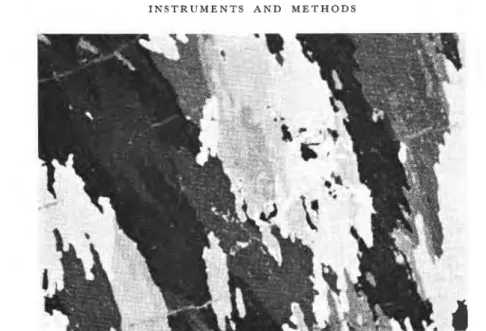
Sea ice normally has a characteristic columnar-grained structure established a few centimetres beneath the surface. The crystallographic c-axes of the columnar grains usually tend to be in the horizontal plane, with random direction in that plane (Pounder, 1965; Weeks and Assur, 1967). Peyton (1966) pointed out that the structure of columnar ice 50 cm or more beneath the surface is characterized by a horizontal c-axis and strongly preferred c-axis orientation in that plane. Peyton called this "bottom" ice.
A typical photomicrograph of a thin horizontal section of columnar-grained ice under cross-polarized light (Fig. I ) exhibits highly irregular, interlocking grain boundaries and internal substructure. Individual grains can be identified by the areas having the same tonal quality; brine pockets can be seen as minute white specks. This photograph clearly demons- trates the advantages and disadvantages of using polarized light and a black-and-white photograph, although this is commonly practised by investigators in this field. Grains having their c-axes parallel or perpendicular to the polarizer appear to be black when the analyser is at a right angle to the polarizer and the brine pockets become visible owing to the scattering of the incident polarized light. I n contrast, grains having their c-axis 45' to the polarizer appear to be bright, thereby eliminating the contrast in tonal quality brought about by the brine pockets. A black-and-white photograph was found to be enriched when supported by a
1
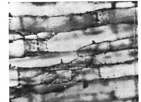
corresponding photograph (Fig. 2) using the brine pockets as scattering sources. Brine pockets, present at the grain boundaries and inside the grains, gave the characteristic pattern 1 of sea ice.I t may be noted in Figures I and 2 that the inclusions appear as individual pockets and that there is no indication of the platy substructure so clearly visible in thin-section photo-
!
graphs provided by previous workers (for example, Tabata and Ono, 1957; Weeks and Assur, 1967; Weeks and Hamilton, 1962). One explanation that can readily be given is that the grain and sub-grain boundaries melt and become impregnated with liquid brine in thinI N S T R U M E N T S A N D M E T H O D S 3'7
Fig. I . Photograph of a horizontal thin section of columnar-grained sea ice at -3ooC, under polarized light.
sections prepared at elevated temperatures or by the hot plate technique commonly used. Little attention has been paid in the past to the details of preparation and examination of thin sections, but it was observed during this investigation that even the choice of the source of light influenced the nature of the thin sections. The absorption of infra-red radiation from the light source was found to introduce undesirable morphological changes in the thin sections. Green (546 nm filter) monochromatic light, near which the normal eye is most sensitive and the absorption coefficient of ice is negligible, was therefore used.
A microscopic view (Fig. 3) of a horizontal thin section reveals details of distribution of brine pockets inside a grain of sea ice. Brine pockets are present not only in arrays parallel to
318 J O U R N A L O F G L A C I O L O G Y
the basal plane of the grain but also along the c-axis. This distribution seems to outline the original boundaries of the dendritic structure formed at the ice-water interface during freezing. The entrapment process of brine in the sea ice is usually explained by the constitu- tional supercooling at the ice-water interface, causing a planar interface to become unstable and change to a highly cellular or dendritic interface (Harrison and Tiller, 1963; Lofgren and Weeks, 1967).
Vertical sections were also prepared to permit examination of the distribution of the brine pockets along the length of the grains. Comparison of the two sections revealed that the majority of brine pockets were quite irregular, although cylindrical and ellipsoidal-tabular shapes were also present.
According to phase relations of sea-water (Assur, I g58), precipitation of salts depends upon the temperature of the ice. Although significant discrepancies of the phase diagram at low temperatures have been discussed by Weeks (1967), they are not important, at least not down to about -30°C. Precipitation of the salts from sea-ice brine at various temperatures was deduced from changes in brine composition and stability ranges of individual salts in the corresponding pure-salt-water systems. A survey of the literature, however, indicates that no one has yet observed under controlled conditions the location and morphology of the precipitated salt crystals in brine pockets.
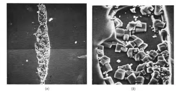
Figures 4 and 5 exemplify irregular and cylindrical cavities, respectively, in a vertical section containing salt crystals and probably air inclusions or the remaining brine. The shallow depth of field of the optical microscope and the presence of the pockets below the ice surface were, however, responsible for lack of clarity of the shape of crystals in these cases. This apparent difficulty was removed by the great depth of field of the scanning electron microscope, which clearly showed details of a cylindrical cavity (Fig. 6). As the photograph is of a replica of a horizontal section, the brine pocket appears as solid matter extending from the surface. Salt crystals appear to adhere to the replica. Vertical sections of a typical pocket
Fig. 3. Photomicrograph of a horizontal section of sea ice at -30°C exhibiting the distribution of brine pockets along the boundaries of the dendrites formed at the ice-water interface during freezing (mgniJication: 30 X ).
I N S T R U M E N T S A N D M E T H O D S
3'9
Fig. p. Photomicrograph of irregular brine pockets and the salt crystals inside the cavities (magniJcation: IOO X ).
320 J O U R N A L O F G L A C I O L O G Y
! / I !b:
Fig. 6. a. Scanning electron micrograph of a replica of a horizontal section of columnar-gained &styear sea ice from 30 cm
below the sugace. (Temperature at time of replicating = -30°C; salinity First replica, magnijcation: 40 X .)
b. Enlarged view of a section of brine pocket situated on the left side of Figure Ga~(magn$cation:, 450 x ).
( a : jb)
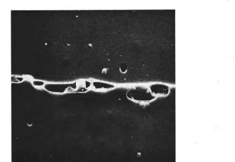
Fig. 7. a. Scanning electron micrograph of a vertical section of a brine pocket at -30°C, salinity 4%,. (Second replica,
magniJcation: go x .) b. Vertical section of a brine pocket at -30°C (magniJcation: 500 x ).
(Fig.
7 )
show the random distribution of the crystals. Individual crystals have not yet been positively identified, although the majority of the salt crystals in Figures 6 and 7 appear to be, as expected, NaCl.nH,O.The majority of the crystals were loosely packed in the cavities and could be removed by making successive replicas. The salt crystals in the exposed cavities could also be removed by washing the microtomed section with kerosene oil before replicating. This also facilitated replicating the pockets for examination of their actual shape (Fig. 8).
I N S T R U M E N T S A N D M E T H O D S
321
Fig. 8. Scanning electron micrograph of a replica of vertical section of brine pockets at -30°C. Microtomed ice surface was washed with kerosene before replicating (rnagniJication: 500 x ).
The random distribution of the precipitated salts in the pockets apparently does not support the theory postulated by Assur ( I 958) and later revived by Peyton ( I 966) of various models of salt reinforcement at low temperatures. The reinforcing structure was assumed as a hollow cylinder of mixed salt and ice filled with the remaining brine. I t is difficult, however, to comment on this subject at the present time, but it should be possible to pursue further this aspect of the strength of sea ice.
Figure g illustrates the replica of brine pockets at - lo°C. These are quite different from
those of Figure 6. The liquid brine in the pockets at - 10°C resulted in the smooth walls of the
replicas. Only sodium sulphate (Na2S04. IoH,O) crystals are expected to be present in the pockets in noticeable quantities at this temperature. Calcium carbonate (CaCO,
.
6H,O), which begins to precipitate at -2.2OC, is present in trace quantities. I t is conceivable that the salt crystals are pushed either to the side (Fig. ga) or near the bottom (Fig. gb) of the cavities by the replicating solution. This indicates that the salt crystals remain in the liquid brine, in which case no reinforcement due to Na2S04. 1oH,0 is possible, contradicting Peyton (1966).Replicating is a useful method of extracting crystals from brine pockets and could be used for further analysis of the precipitation of various salts as a function of temperature, positively identifying the crystals and thereby providing a procedure for examining the phase diagram (Assur, I 958).
Significant advancement has been made in the last twenty years with the analysis of strength and structurally sensitive properties of sea ice in relation to its substructure, which is controlled by growth parameters (Weeks, 1967; Weeks and Assur, 1967) Little attention, however, has been paid to the analysis of the sub-grain boundary, the section of ice that acts
as the ice-to-ice bond between The Formvar solution could, under suitable condi-
tions, be used for etching ice and replicating the etched ice surface. The technique, now under investigation at DBRINRC, could be applied to the analysis of sub-grain boundaries; it could also be applied in developing a better understanding of the region of the ice near the ice-water interface.
322
J O U R N A L O F G L A C I O L O G Y( (1 ) ( / I ;
Fig. 9. a. Replica of a brine pocket at - ro0Cshowing the smooth walls and salts crystals on one side. b. Replica showing the
salt cvstals near the bottom of the brine pocket at - ro°C.
The microtoming and replicating technique has been found suitable for examining the undisturbed microstructure of sea ice. I t is conceivable that further application may lead to better understanding of growth morphology at the ice-water interface during freezing, brine drainage through the sub-grain and grain boundaries, and the dependence of rheology and strength behaviour in relation to the microstructure of sea ice.
Thin sections of sea ice produced by microtoming have been examined under cross- polarized monochromatic light and their grain and sub-grain structure studied. Brine pockets were clearly visible by photographing the scattering characteristics of the thin sections. Optical microscopy of the prepared surfaces revealed that the distribution of brine pockets is related to the boundaries of dendrites formed at the ice-water interface during freezing. Details of brine pockets and the precipitation pattern of the enclosed salts could be observed by replicating the microtomed surface and examining the replica with a scanning electron microscope. Most brine pockets were irregular and the salt crystals loosely packed in the cavities in a random manner.
The success of this dual observation technique lies in the clarity with which the micro- structure of sea ice can be examined.
ACKNOWLEDGEMENTS
The author wishes to thank R. Frederking for valuable suggestions and E. Quinn for his technical assistance. H e would also like to convey his appreciation to the Department of Public Works and the Ministry of Transport, Government of Canada, for arrangements at Strathcona Sound, N.W.T.
This paper is a contribution from the Division of Building Research, National Research Council of Canada, and is published with the approval of the Director of the Division.
I N S T R U M E N T S A N D M E T H O D S 323
R E F E R E N C E S
Assur, A. 1958. Composition orsea icr and ik tcnsilr s~rength. (In Arctic sea ice. Washington, D.C., p. 106-38.
([U.S.] National .4cademy of Sciences-Yational Rrsearch Council Publication 598.))
Harrison, J . D.. and Tiller. W . .4. rgfi3. Conaollrd freezing of water. (In Kingery, W. D., ed. Ice and snow;
propdies: jwct\nrr, lrnd applicntions: proceeding< of ( I mnfirence held at the Massachusetts Institute of Technolgo, February
r r - 1 6 , 1.96~. Cambridge, Mas?., >I,I.T. I'reqs, p. zr5-25.)
Kuroiwa, D. 1969. Surface topography or etrhrd i r e crystals observed by a scanning electron microscope.
J o m a i ' of Glncio10,q. Vol. 8, No. 51, p. 47543.
1-nfgren, G., and IVeeb, 12.'. F. 1967. Effect of growth parameters on substructure spacing in NaCl ice crystals.
U.S. Cold R<gionr R~sparch and engineer in^ J~horabory. Research Report 195.
Peytnn, El. R . 1*6. Sea k c sfreriq[lr. Collrge, Alaska, Cmphysical Institute, University of Alaska. (UAG R-182.)
Poundcr, F,. R. 1965. TIM physic3 of k c . OuTord, erc.. Pergamon Press. (The Commonwealth and International Library. Geophysics Division.)
Tabata, T., and Ono, N. 1957. Kaihy6 no k6z6 tsuite [On the structure of sea ice]. Tezon-kagaku: Low Temperature
Science, Ser. A, [No.] 16, p. 197-210.
Weeks, W. F. 1967. Understanding the variations of the physical properties of sea ice. U.S. Cold Regiom Research
and Engineering Laboratory. Speczal Report I I 2.
Weeks, W. F., and Assur, A. 1967. The mechanical properties of sea ice. U.S. Cold Regions Research and Engineering Laboratory. Cold regzons science and engineering. Hanover, N.H., Pt. 11, Sect. CQ.
Weeks, W. F., and Hamilton, W. L. 1962. Petrographic characteristics of young sea ice, Point Barrow, Alaska.


![[PDF] Cours Android développement d'applications avancées PDF - Free PDF Download](data:image/gif;base64,R0lGODlhAQABAIAAAP///wAAACH5BAEAAAAALAAAAAABAAEAAAICRAEAOw==)