HAL Id: hal-00854474
https://hal.archives-ouvertes.fr/hal-00854474
Submitted on 27 Aug 2013HAL is a multi-disciplinary open access archive for the deposit and dissemination of sci-entific research documents, whether they are pub-lished or not. The documents may come from teaching and research institutions in France or abroad, or from public or private research centers.
L’archive ouverte pluridisciplinaire HAL, est destinée au dépôt et à la diffusion de documents scientifiques de niveau recherche, publiés ou non, émanant des établissements d’enseignement et de recherche français ou étrangers, des laboratoires publics ou privés.
Calcium and α-Tocopherol Suppress Cured Meat
Promotion of Chemically-Induced Colon Carcinogenesis
in Rats and Reduce Associated Biomarkers in Human
Volunteers
Fabrice H.F. Pierre, Océane Martin, Raphaëlle Santarelli, Sylviane Taché,
Nathalie Naud, Françoise Guéraud, Marc Audebert, Jacques Dupuy, Nathalie
Meunier, Didier Attaix, et al.
To cite this version:
Fabrice H.F. Pierre, Océane Martin, Raphaëlle Santarelli, Sylviane Taché, Nathalie Naud, et al.. Cal-cium and α-Tocopherol Suppress Cured Meat Promotion of Chemically-Induced Colon Carcinogenesis in Rats and Reduce Associated Biomarkers in Human Volunteers. The American Journal of Clinical Nutrition, 2013, in press. �hal-00854474�
Calcium and α-Tocopherol Suppress Cured Meat Promotion of
Chemically-Induced Colon Carcinogenesis in Rats and Reduce
Associated Biomarkers in Human Volunteers
Fabrice H.F. Pierre1,7, Océane C.B. Martin1, Raphaelle L. Santarelli1,2, Sylviane Taché1,
Nathalie Naud1, Françoise Guéraud1, Marc Audebert1, Jacques Dupuy1, Nathalie
Meunier3,4, Didier Attaix3, Jean-Luc Vendeuvre2, Sidney S. Mirvish5, Gunter C.G. Kuhnle6,
Noel Cano3,4, Denis E. Corpet1
1 Université de Toulouse, INRA, UMR1331 Xénobiotiques, BP-87614, 23 Ch. des Capelles, F-31076 Toulouse, France
2 IFIP-Institut du Porc, 149 rue de Bercy, F-75595 Paris Cedex 12, France
3 INRA, UMR 1019, UNH, CRNH Auvergne, F-63000 Clermont-Ferrrand, France 4 CHU Clermont-Ferrand, Service de Nutrition, F-63000 Clermont-Ferrrand, France
5 Eppley Institute for Research in Cancer, University of Nebraska Medical Center, Omaha, Nebraska, 68198, USA
6 Department of Food & Nutritional Sciences, University of Reading, Reading RG6 6AP, United Kingdom
7 To whom correspondence should be addressed to Fabrice H.F. Pierre; Phone: +33 561 193 289; Fax : +33 561 491 263; E-mail: f.pierre@envt.fr
Running title: Cured Meat Promotion of Colon Carcinogenesis
Key words: cured meat, colorectal cancer, prevention, calcium, tocopherol, preneoplastic lesions, fat oxidation, cytotoxicity, N-nitroso compounds.
Financial Support: This work was supported by ANR-French Agency (ANR-PNRA Heme Cancer project), by the COST B35 (European Cooperation in Science and Technology) and by NIH Grant R01-CA-1434600 from the United States National Cancer Institute. Abbreviation: ACF, aberrant crypt foci; DCNO, dark cooked meat with nitrite, oxidized; DHN-MA, 1,4-Dihydroxynonane mercapturic acid; DMEM, Dulbecco-modified essential medium; MDF, mucin depleted foci; TBARS, Thiobarbituric acid reactive substances. Names for PubMed indexing: Pierre, Martin, Santarelli, Taché, Naud, Guéraud, Audebert, Dupuy, Meunier, Attaix, Vendeuvre, Mirvish, Kuhnle, Cano, Corpet
Abstract
Background: Processed meat intake is associated with increased colorectal cancer risk. We have shown that cured meat promotes carcinogen-induced preneoplastic lesions and increases specific biomarkers in the colon of rats.
Objectives: To investigate whether cured meat modulates biomarkers of cancer risk in human volunteers, and whether specific agents can suppress cured meat induced preneoplastic lesions in rats, and associated biomarkers in rats and in humans.
Design: Six additives (calcium carbonate, inulin, rutin, carnosol, α-tocopherol and trisodium pyrophosphate) were added to cured meat given to groups of rats for fourteen days, then fecal biomarkers were measured. Based on these results, calcium and tocopherol were kept for further experiments: cured meat, with or without calcium or tocopherol, was given to dimethylhydrazine-initiated rats (47% meat diet for 100 d), and to human volunteers in a cross-over study (180 g/d for 4 d). Rat colons were scored for mucin depleted foci, putative pre-cancer lesions. Biomarkers of nitrosation, lipoperoxidation, and cytotoxicity were measured in rats and volunteers’ urine and feces.
Results: Cured meat increased nitroso-compounds and lipoperoxidation in humans stools (both P<0.05). Calcium normalized both biomarkers in rats and humans’ feces, while tocopherol only decreased nitro-compounds in rats and lipoperoxidation in volunteers’ feces (all p<0.05). Lastly, calcium and tocopherol reduced the number of mucin depleted foci per colon in rats compared with non supplemented cured meat (P=0.01).
Conclusion: The data suggest that addition of calcium carbonate to the diet or of α-tocopherol to cured meat may reduce colorectal cancer risk associated with cured meat intake.
Introduction
Colorectal cancer is the third most common type of cancer in the USA (1). Epidemiological studies suggest that there is an association between colorectal cancer and intake of meat (2), and that risk associated with cured meat is higher than with fresh red meat (3). The World Cancer Research Fund panel stated that "the evidence that red meat and processed meat are a cause of colorectal cancer is convincing" and recommended to “limit intake of red meat and avoid processed meat”. However, if these recommendations were adhered to, iron and zinc supply would decrease, with possible impact on women and elderly people (4). Experimental studies shows that a cooked ham and an experimental cured meat promote colon carcinogenesis in rats (5, 6).
Four major hypotheses have been proposed to explain the effect of processed meat on colorectal cancer: (i) fat present in meat promotes carcinogenesis by raising intestinal bile acids; (ii) cooking meat at high temperature forms carcinogenic heterocyclic amines; (iii) endogenous nitrosation yields nitroso compounds that can be carcinogenic; (iv) heme iron in red meat promotes carcinogenesis because it increases cell proliferation in mucosa, through lipid oxidation and cytotoxicity of fecal water. Nitrosation and heme iron hypotheses have received most experimental support (3): Eating fresh red meat or cured meat increases nitroso compounds concentration in human stools and in mice and rats’ feces, and heme is responsible for this effect (6-10). Fecal nitroso compounds concentration is associated with the NO-specific DNA adduct O6-carboxymethylguanine in the human colon (11), and with mucin depleted foci (MDF,
a putative pre-cancer lesion) promotion in carcinogen-initiated rats given cured meat (6). Aberrant crypt foci (ACF, another putative pre-cancer lesion), are induced in mice by gavages with nitroso compound extracted from hot dogs (12). On the other hand, dietary beef, hemoglobin, and chlorine hemin promote dose-dependently formation of ACF and MDF in rats, and raise biomarkers of intestinal fat oxidation and cytotoxicity of fecal water, suggesting that heme iron is a cancer promoter and that specific fecal biomarkers are associated with cancer promotion (13, 14).
Specific additives can suppress the promoting effect of heme: A calcium-rich diet abolishes most effects of dietary heme, including beef meat-induced promotion of MDF (13-15). These data suggest that calcium could reduce colorectal cancer risk in meat eaters and support the concept that toxicity associated with excess intake of a specific food component, heme iron, may be prevented by another nutrient, calcium (13). Besides, antioxidant molecules added into meat can also suppress heme-induced promotion of carcinogenesis (14).
In this study, we first investigated in rats the effect of six additives on fecal biomarkers induced by cured meat eating. Two additives were chosen for inclusion in a carcinogenesis study in the chemically induced rat model of colon carcinogenesis. These two compounds were also evaluated in healthy volunteers to test their ability to counteract the modulation of fecal and urinary biomarkers by cured meat.
SUBJECTS AND METHODS
Animal studiesTwo sequential studies were performed in rats. A 14-day study was conducted to test the effect of six agents added to diet on fecal and urinary biomarkers in rats. Subsequently, a 100-day carcinogenesis study was conducted to test protection afforded by two agents selected out of the six ones.
Fourteen-day study: animals, design and diets.
Female Fischer 344 rats (n=35) were purchased at five weeks of age from Charles Rivers (St. Germain l’Arbresle, France). The study was conducted in an accredited animal colony by approved staff, and animal care was in accordance with the guidelines of the European Council on Animals used in Experimental Studies. Rats were housed individually in metabolic cages. They were kept at 22°C and a 12 h-12 h light-dark cycle, and were allowed free access to standard AIN76 semi-purified diet and tap water (16). After two days of acclimatization, rats were randomly allocated to seven groups of five rats. Each group was given an experimental cured meat for 14 days, added with one of six potential protective agents, or none (control). Body weights were monitored on days 0, 7 and 14, food and water intakes on days 6-7 and 12-13. Feces were collected during days 13 and 14 and were frozen at -20°C. Urines were collected on day 13 and processed immediately.
Diet was made by the IFIP workshop (Paris, France) by mixing 55 g (dry weight) of an experimental cured meat (IFIP) with 45 g of a modified AIN76-base powder made by UPAE (INRA, Jouy, France) containing (g/100g total diet): sucrose, 23.8; corn starch, 6; cellulose, 5; safflower oil, 5; AIN76 calcium-free AIN76 mineral mix, 3.5; AIN76 vitamin mix, 1.0; methionine, 0.3; calcium phosphate, 0.2; choline bitartrate, 0.2. Diet contained 40% protein, 15% fat and 0.27% calcium. Meat was not freeze-dried but given moist, to avoid fat oxidation (5, 17). The experimental cured meat, similar to air-exposed picnic ham and called DCNO (for Dark Cooked meat with Nitrite, Oxidized), was chosen because it promotes carcinogenesis in rats (6). Dark red Supraspinatus pig muscle (15-17 mg heme /100g) (18) was cured with 2 g/100g salt containing 0.6 g/100g sodium nitrite, and 0.36 g sodium erythorbate. It was then heated inside a vacuum-sealed plastic bag immersed in a 70°C water-bath for 1 h. This “cooking” denatures myoglobin and frees heme iron from protein (19). Cured meat was then exposed to air at 4°C in the dark for five days before being given to the rats. Several antioxidant agents and calcium salts can reduce heme-induced fat peroxidation in the gut, and heme-induced carcinogenesis promotion in rats (13, 14). These agents (rutin, carnosol, α-tocopherol and calcium carbonate) and two additives already in use in cured meat (a fat substitute: inulin; a pH corrector: trisodium pyrophosphate) were tested in DCNO at dose already known to counteract heme toxicity. Three of them were incorporated inside meat during curing process: rutin (0.1% of total diet; Sigma Aldrich, St.Quentin, France), carnosol (0.07%; extracted from rosemary, Naturex, Avignon, France), or α-tocopherol (0.05%; Sigma Aldrich). Two others were added to diet powder: calcium carbonate (CaCO3, 1.5g/100g diet equivalent to
150µmol/g; Sigma) or inulin (4.5%; Orafti HP, Azelis, Oreye, France). A last treatment was to buffer meat pH with trisodium pyrophosphate (Na3PO4, 0.84%; La Bovida, le Subdray, France). Each diet was storedunder vacuum at –20°C, and was dispensed daily at 5:00 p.m.
One-hundred-day study: animals, design, diets
Rats (n=36), same kind as above, were housed in pairs in stainless steel wire-bottomed cages, in the same animal colony as above. After acclimatization they received a single i.p. injection of 1,2-dimethylhydrazine (Sigma; 180 mg/kg) in NaCl (9g/l water). Seven days later, the rats were randomly allocated to three groups (n=16, 10, 10) and fed the experimental diets daily for 98-99 days before CO2 euthanasia. Colons were removed,
washed with cold Ringer solution, opened, coded, and fixed flat between two sheets of filter paper in 10% buffered formalin (Sigma) before ACF and MDF scoring. Body weight was monitored every week during the four first weeks, then every two weeks. Food and water intakes were measured at day 15, 59 and 94. Feces were collected on days 85-95 and kept at -20°C. Each rat was placed in a metabolic cage and urine was collected on days 70-74 and kept at -20°C.
Diet was obtained from the same providers as above. Control diet composition was (g/100g): moist cured meat DCNO, 47 (dry weight), sucrose, 28.7; casein, 5; safflower oil, 5; corn starch, 4.8; cellulose, 4.8; AIN76 calcium-free mineral mix, 3.35; AIN76 vitamin mix, 0.95; methionin, 0.3; calcium phosphate, 0.21; choline bitartrate, 0.17. Two potential protective agents were tested: calcium carbonate was added to the control diet (1.5g/100 diet, at the expense of casein). A-tocopherol was added to DCNO during the curing process (0.05% of diet). Each diet was storedat –20°C under vacuum, and was dispensed daily at 5:00 p.m.
Human study: volunteers, cross-over design and diets
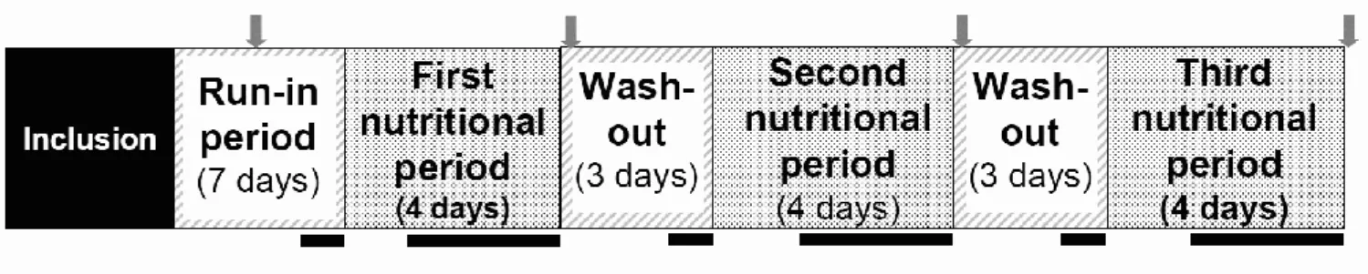
This study was performed in the Nutritional Investigation Unit of the Human Nutrition Research Center of Auvergne (Clermont-Ferrand, France). A single-blind randomized, controlled cross-over trial was conducted in human volunteers satisfying the following criteria: men aged 40 to 75 y, body mass index from 20 to 30, no history or clinical symptom of colonic disease, no history of family colon cancer, normal standard blood tests (blood cell counts, renal and liver function, C-reactive protein, serum glucose and lipids), alcohol and tobacco consumption respectively less than 30 g and five cigarettes per day, and no detectable microscopic bleeding in the stools. The volunteers have been recruited from the Nutritional Investigation Unit data file, which is declared to the National Commission on Informatics and Liberties. All the participants have gave freely their written informed consent before their selection in the study and after information on the objectives, benefits, risks, as well as the nature of the study products. Volunteers with a calcium dietary intake ≥ 1500 mg/day and those treated by vitamins A, C or E or calcium supplements were excluded. After phone contact and first selection, volunteers had a medical visit, a dietetic evaluation and biological measurements for inclusion criteria checking. Finally, 18 volunteers were included after giving their written informed consent.
As shown in study design (Figure 1), during the one-week run-in period adaptation volunteers were asked to eat a diet without beef or pork meat and low in antioxidant products (the no-meat control period). Volunteers were then randomly submitted to three alternated four-day intervention periods: experimental cured meat (DCNO), DCNO and calcium capsule (DCNO+CaCO3) and tocopherol-enriched DCNO
(DCNO+tocopherol), in a random order (doses are given below). Intervention periods were separated from each other by a wash-out period of at least 3 days (same diet as run-in period). Urine and stool samples were collected during the last three days of each intervention period, and at the end of each washout period. Each subject came to the Nutrition Investigation Unit four times in addition to the screening visit. Before the first
intervention period (visit 1), volunteers were given cured meat needed for the whole study (three packages of ham, one per period). Volunteers were asked to keep the ham frozen at home and to open packages in their fridge at 4°C four days before consumption, to match previous rat study design, and to mimic the oxidation that occurs usually when ham is not vacuum-packed. Placebo or calcium capsules for the first intervention period were also distributed at visit 1, depending on randomization. During visits 2, 3 and 4, each volunteer brought back ham packaging, and collected frozen urine and stools. At visits 2 and 3, volunteers were given capsules corresponding to the intervention periods 2 and 3. The study was approved by a written decision of the Person Protection Committee (French CPP Sud-Est VI, 08/09/2009) and authorization of French Ministry of Health (Afssaps, 19/05/2009) and was registered on the registry of the website http://clinicaltrials.gov/ with the number NCT00994526.
Volunteers were given the same experimental cooked ham previously tested in rats, DCNO, cured by Fleury-Michon Charcuterie® (Pouzauges, France). Volunteers were asked to eat 180 g/day of DCNO (four 45 g-slices) during each four-day intervention periods. One calcium carbonate capsule (500 mg calcium/capsule, Montalembert pharmacy, Clermont-Ferrand, France) was consumed twice a day during the four-day DCNO+CaCO3 period (1 g calcium/day). Placebo capsules taken during the two other
intervention periods contained 500 mg of crystalline cellulose. Alpha-tocopherol was incorporated during meat curing (0.05% w/w) to provide meat for the DCNO+tocopherol intervention period. Compliance to diet and supplements was assessed after collection of food and drug packages at the end of intervention periods.
Analytic techniques
Analysis of heme and TBARS in fecal water, DHN-MA in urine
Fecal values were measured in fecal water because, according to bile acids studies, the soluble fraction of colonic contents interacts more strongly with the mucosa than the insoluble fraction (20). Rats: fecal pellets were collected under each cage for 24 h. One mL of sterilized water was added to 0.3 g dried rat feces. Samples were then incubated at 37˚C for one hour, stirring thoroughly every 20 min. After centrifugation at 20,000 g for 15 min, fecal water (supernatant) was collected and kept at –20˚C until use. Volunteers: to 1 g of human stool 5 mL of Dulbecco-modified essential medium (DMEM) were added before stirring vigorously during 30 s. Mix was then submitted to centrifugation and fecal water harvested and kept as described above. Heme was measured by fluorescence in fecal water according to Sesink et al. as already described (21, 22). Thiobarbituric acid reactive substances (TBARS) were measured in fecal water according to Ohkawa et al., exactly as previously described (23). 1,4-Dihydroxynonane mercapturic acid (DHN-MA) is the main urinary metabolite of 4-hydroxynonenal, a major toxic end product of endogenous fat peroxidation. DHN-MA assay was done by competitive enzyme immunoassay as previously described, using DHN-MA-linked acetylcholinesterase enzyme (24). Each urine sample was assayed in duplicate.
Fecal water cytotoxicity
Fecal water cytotoxicity was quantified on three cell lines as previously described (13). The Apc mutation is detected in the majority of MDF in rats and of human colorectal cancers. Apc mutated cells resist to cytotoxic aldehydes found in the gut of meat-fed rats: this resistance leads to the selection of premalignant cells, and would explain cancer promotion by red meat (25). To investigate whether the same mechanism could explain the promotion of carcinogenesis by cured meat, cytotoxicity of fecal water was
quantified on three cell lines: (i) a cancerous mouse colonic epithelial cell line, CMT93 (European Collection of Animal Cultures); (ii) a colon epithelial cell lines derived from C57BL/6J mice (Apc +/+) and (iii) from Min mice Apc +/- (26). Use of this triple cellular model including wild-type cells (Apc +/+), preneoplastic cells (Apc Min/+) and cancerous cells (CMT93), could contribute to our understanding of the effects of digestive content on early steps of colon carcinogenesis.
CMT 93 cells were seeded in 96-well plates at 37°C, 1.6 x 10⁴ cells per well in 200 L of
DMEM. At confluence, cells were treated for 24 h with fecal water sample diluted at 10 % (v/v) in the culture medium. Cells were then washed with phosphate-buffered saline (PBS). Apc +/+ and Apc Min/+ cells have a temperature-sensitive mutation of the simian virus 40 large tumor antigen gene (tsA58), under the control of interferon . These cells are ‘immortalized’, as they express active SV40 at the permissive temperature (33 °C). Cells were cultured at permissive temperature of 33 °C in DMEM supplemented with 10 % (v/v) fetal calf sera, 1 % (v/v) penicillin/streptomycin, and 10 U/ml interferon until subconfluence. The studies were performed at non-permissive temperature of 37 °C, and without interferon , to inhibit the SV40 transgene and limit proliferation. Apc +/+ and Apc Min/+ cells were seeded into a 96-well culture plates at the seeding density of 104 cells in DMEM culture medium. Cells were grown at 33 °C with interferon for 72 h
until subconfluence. They were then transferred at 37°c without interferon for 24 h. Cytotoxicity of fecal water was quantified by the 3-(4,5-dimethyldiazol-2-yl)-2,5 diphenyl tetrazolium bromide test (0.45 mg/ml in PBS). The reaction product was solubilized in 100µl lysis buffer (SDS 10%, NaOH 0.01M) before color of reaction product was quantified using a plate reader at 570 nm and 690 nm. Fecal water genotoxicity was also assessed in the present study by measuring H2AX induction on
Apc +/+ and Apc Min/+ cell lines (See Supplemental Material).
Apparent total N-nitroso compound analysis in fecal samples from volunteers and from rats
Apparent Total N-Nitroso Compound (ATNC) were analyzed in fecal water of rats by SSM (University of Nebraska, USA) using a published method (27). Briefly, 425 μL of fecal water was mixed with 25 μL of 2 N HCl and 50 μL of a freshly prepared saturated solution of sulfamic acid in water to destroy any nitrite present. After storage for 15 min at room temperature and <4 h in ice, 100 or 200 μL samples of the mixture were injected into the reaction vessel. ATNCs were decomposed to NO by an HCl/HBr/HOAc/EtOAc mixture refluxing at <0.1 mm Hg and 28 °C. NO was swept by an argon stream through four wash bottles containing NaOH and Na2SO4 at room
temperature and two empty wash bottles kept at −30 °C to remove water vapor and acids and was then determined in a Thermal Energy Analyzer (Advanced Chromatographic Systems, Charleston, SC).
ATNC were analyzed in fecal water from volunteers by GCGK (University of Reading, UK) with an Ecomedics CLD 88 Exhalyzer (Ecomedics, Duernten, Switzerland) using a modification of a published method (28). Briefly, 100 µl of fecal water were incubated with 500 µl of a 5 % (wt/vol) sulfamic acid solution, to remove nitrite and samples were injected into a purge vessel kept at 60 °C and filled with a standard tri-iodide reagent (38 mg I2 was added to a solution of 108 mg KI in 1 ml water. To this mixture, 13.5 ml glacial acetic acid was added) to determine total ATNC.
The reported values are the concentrations (in µmol/L), measured in 100 µL sample.
Rats were euthanized by CO2 asphyxiation in a random order at day 98-99 of experimental diet. Fixed colons were scored for ACF by Bird’s procedure (29): after methylene blue staining, numbers of ACF per colon, and of crypts per ACF, were counted under light microscope at x 40 magnification in duplicate by two independent readers, blinded for the origin of the colon.
Colons were then stained with the high-iron diamine alcian blue procedure. Two blinded investigators evaluated the number of MDF per colon and the number of crypts per MDF. MDF scoring criteria were focus containing at least two crypts with no or very little apparent mucin (30, 31).
Statistical Analysis:
Data were analyzed using Systat 10 software for Windows andreported as means ± SD (except in Figure 2). Biochemical values were considered firstly using one-way ANOVA. If a significant difference was found between all groups (P < 0.05), comparison of each experimental group with the control group was made using Dunnett's test. ACF and MDF scoring was done in duplicate, these variables were thus tested first using 2-way ANOVA (groups and readers). The (group x reader) interaction was never significant,and when total ANOVA was significant (p<0.05), pairwise differences between groups were analyzed using Fishers’s least-significant-difference test. Difference of fecal water cytotoxicity between Apc +/+ and Apc Min/+ cell lines was tested with Students-t-test. Human volunteers data were analyzed with the Wilcoxon’s signed rank test, each volunteer being its own control. Bonferronni’s correction for three comparisons (i.e., DCNO vs no-meat control period, DCNO+CaCO3 vs. DCNO, and DCNO+tocopherol vs.
Results
Fourteen-day animal study
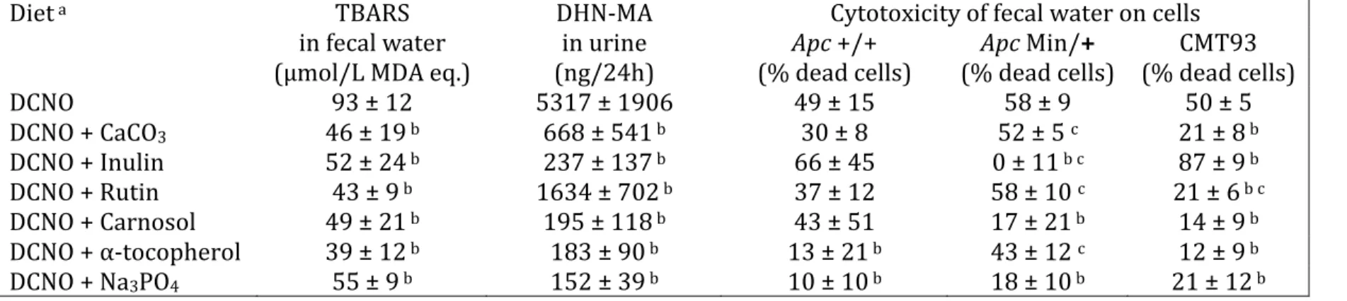
The mean body weight of rats was 145±11 g on day 14. Rats given rutin- and tocopherol-supplemented DCNO cured meat gained more weight than DCNO-fed control rats (p<0.05, data not shown), but dietary and water intakes were similar in all groups (10±2 and 25±6 g/day, respectively). All the tested additives decreased fecal water oxidation (TBARS) and urinary DHN-MA concentration compared with DCNO control (p<0.05, Table 1). Fecal water from rats given DCNO showed similar cytotoxicity on the three tested cell lines, but the addition of CaCO3, rutin or α-tocopherol to the diet
resulted in a survival advantage of wild Apc +/+ cells compared to mutated Apc Min/+ cells. All tested additives, except inulin, decreased fecal water cytotoxicity against CMT 93 cells compared with unchanged DCNO. Fecal water from rats given DCNO plus inulin was highly cytotoxic to Apc +/+ and CMT93 cells, but not at all to mutated Apc Min/+ cells (Table 1).
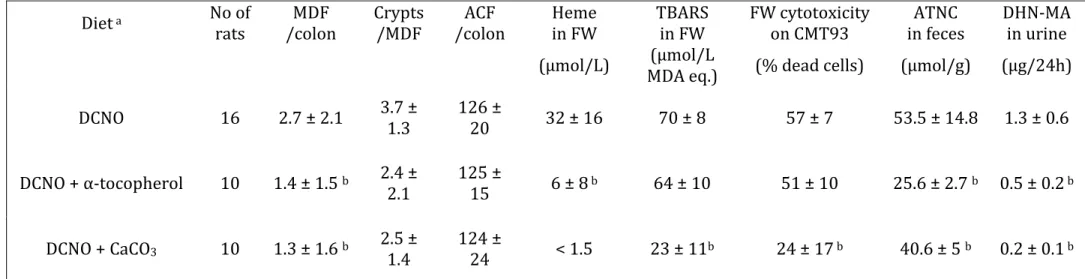
One hundred-day animal study
The final body weight of rats was 217±1 g, similar in all three groups, and the food intake was not significantly different. Rats given DCNO+CaCO3 or DCNO+tocopherol had
less MDF per colon than control rats given DCNO only (Table 2, both P=0.01). In contrast, the two tested additives did not reduce the number of ACF/colon, and the size of MDF (crypts/MDF, Table 2) was not significantly reduced. The addition of CaCO3 to
the DCNO diet decreased the amount of all tested biomarkers previously associated with carcinogenesis (Heme, TBARS and cytotoxicity in fecal water, ATNC in feces and urinary DHN-MA, all P<0.05, Table 2). In contrast, the addition of α-tocopherol to cured meat decreased only concentration of heme in fecal water and DHN-MA in urine (p<0.05, Table 2). Fecal ATNC concentration was significantly decreased by the two tested additives (Table 2).
Human study
General observations
The nutritional intervention was scheduled with 18 healthy volunteers, but one of them lately declared he was also participating to another trial. This volunteer was thus excluded, and 17 persons completed the study and were analyzed. They were aged 56.0 ± 9.5 y and their body mass index was 24.9 ± 2.3 kg/m2. Blood cell counts were normal
in all subjects. Serum creatinine was 79.8 ± 8.8 µmol/L, glucose 5.4 ± 0.5 mmol/L, cholesterol 5.2 ± 0.5 mmol/L, triglycerides 0.9 ± 0.3 mmol/L, serum alanine amino transferase 24.6 ± 10.6 IU/L, gamma glutamyl transferase 29.1 ± 20.9 IU/L, prothrombine time 100 ± 9 % and C-reactive protein 1.2 mg/L [0.7-8.5]. The assessment of compliance showed that the diet guidelines were strictly followed, but one day, one volunteer ate three slices of ham instead of four. All calcium and placebo supplements were taken without any detected fault.
TBARS, ATNC, cytotoxicity and urinary DHN-MA:
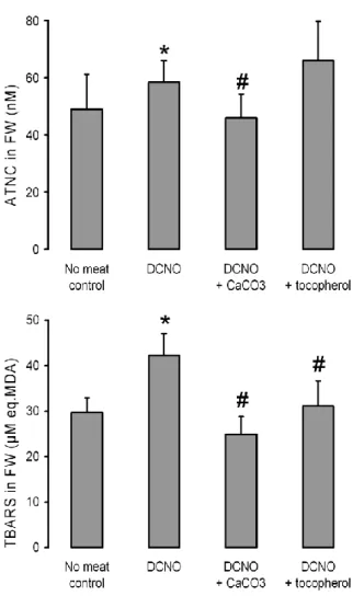
Biomarker measurements showed significant increase in ATNC and TBARS concentrations in fecal water of human volunteers fed 180 g cured meat (DCNO) for four days compared with control periods (Figure 2). The addition of calcium carbonate to the cured meat diet significantly decreased fecal ATNC and TBARS. Addition of tocopherol into cured meat had no effect on fecal ATNC concentration, but decreased
fecal water TBARS. Urinary DHN-MA and fecal water cytotoxicity were not changed by the dietary changes in volunteers (data not shown).
Discussion
The present study is the first one to show that the same cured meat that increased carcinogenesis in rats also increased promotion-associated fecal biomarkers in rats and in human volunteers. It also shows that this increase, and the promotion of carcinogenesis in rats, can be suppressed by dietary calcium or α-tocopherol.
Promotion of colon carcinogenesis was shown with a surrogate endpoint biomarker, mucin depleted foci (MDF). MDF, formed by dysplastic crypts devoid of mucin, have been identified in the colon of humans at high risk for colon cancer (32). Like tumors, MDF harbor mutations in genes affecting colon carcinogenesis (Apc and K-ras) and show Wnt signaling activation (33), a dramatic reduction of MUC2 expression (34), and a strong activation of the inflammatory process (35), all features suggesting that MDF are precancerous. Rodents studies suggest that MDF are better predictors of colorectal cancer than ACF are, this is why we focused on MDF data (30).
Promotion of colon carcinogenesis by fresh moist cured meat (DCNO) in rats is associated with increased fecal nitroso-compounds (ATNC) concentration, and with increased fecal biomarkers of fat peroxidation (TBARS) (6). Hence, we chose to use DNCO to test whether a cured meat could increase those early biomarkers in human volunteers and to identify prevention strategies (6). The results of our cross-over study show that feeding DCNO for four days was enough to increase ATNC and TBARS concentration in volunteers’ stools, compared with the control period. A similar study in volunteers by Joosen et al. showed that the ingestion of 400 g/d processed meat for 14 days increases fecal ATNC from 3.5 nmol/g in controls given a vegetarian diet to 181 nmol/g (9). This increase in fecal ATNC is not associated with increased genotoxicity. In contrast, the cured-meat intervention decreases fecal water-induced DNA strand breaks compared with the vegetarian diet (9). Fecal water genotoxicity was assessed in the present study by measuring H2AX induction on Apc +/+ and Apc Min/+ cell lines: compared with control group or period, the cured-meat diet tended to reduce H2AX induction by fecal water from rats and from volunteers (not significant, Supplemental
Figure S1 and S2). This is in striking contrast with Hebels et al.’s observation that a beef
meat diet intervention did not change fecal ANTC but increase fecal water genotoxicity in volunteers (36), likely because beef meat contains more heme iron than cured pork meat. Intake of cured meat can thus modulate biomarkers associated with promotion of colon carcinogenesis in rats (TBARs, cytotoxicity but not genotoxicity), which gives experimental support to the epidemiology-based conclusion that processed meat could be a cause of colorectal cancer (37, 38).
Since nitrite and heme seem necessary to promote carcinogenesis in rats (6), the reduction of their concentration in meat could reduce toxicity of cured meat. This strategy is not easy to implement, as it would increase microbiological risks and reduce the sensory qualities of cured meat. This is why we looked for additives that could reduce the toxicity. This study shows that dietary calcium carbonate inhibited cured meat promotion of colon carcinogenesis in rats. MDF number reduction was associated with normalization of fecal TBARS and ATNC in rats (Table 2), and parallel normalization was seen in volunteers’ stools (Figure 2). A high-calcium diet consistently blocked the effects of red meat and dietary heme iron on the gut mucosa, including proliferation, carcinogenesis promotion and associated biomarkers (13-15). This can explain why beef meat and bacon do not promote rodent carcinogenesis when added into a high-calcium diet (39).
Present data suggest that cured meat effect could be neutralized by adding calcium to the meat or by consuming a calcium-rich food in the same meal. Indeed, calcium carbonate supplements reduce the risk of recurrent colorectal adenomas in volunteers (40): chemoprevention by calcium might be due in part to the binding of dietary heme iron. This beneficial effect of calcium has thus a drawback: it would increase the risk of iron deficiency.
This study also shows that addition of α-tocopherol into cured meat inhibited the promotion of colon carcinogenesis in rats. MDF number reduction by α-tocopherol was associated with normalization of urinary DHN-MA in rats fed cured meat (Table 2). We proposed that promotion by heme iron would be due to fat oxidation end-products (13, 14, 22): the Apc mutation renders cells resistant to 4-hydroxy-2-nonenal, an end-product of heme-induced fat oxidation (25), which we measured by its urinary metabolite, DHN-MA. Selection of Apc mutated cells by cytotoxic peroxides would thus explain heme-induced promotion of colon carcinogenesis (41). Here, in the 14 d study, reduction in fecal TBARS by α-tocopherol, calcium carbonate and rutin (Table 1) was associated with reduced cytotoxicity against non mutated Apc +/+ cells, which might explain
the reduced promotion. However, this reduction in fecal TBARS and cytotoxicity was not seen in the 100 d study (Table 2), which casts doubt that cytotoxic peroxides and selection of Apc mutated cells, would explain promotion by cured meat. On the other hand, protection by α-tocopherol was associated with reduced fecal ATNC in rats (Table 2). It supports the hypothesis that N-nitroso compounds are the major pro-cancer molecules from cured meat, a hypothesis supported by Mirvish’s studies in rodents (8, 12, 27, 42, 43), Bingham’s studies in volunteers (7, 10, 28, 44), and a previous carcinogenesis study from this team (6). A-tocopherol addition to cured meat halved fecal ATNC in rats (table 2), but did not reduce significantly fecal ATNC in volunteers. It suggests that vitamin E would be not sufficient to reduce cured meat toxicity in humans. Our previous carcinogenesis studies suggest that fat peroxides like 4-hydroxynonenal would explain promotion by fresh red meat (13, 14, 22, 24), while this study and a previous one (6) suggest that nitrosation and ATNC would explain promotion by cured meat.
Recommendations to avoid processed meat intake may reduce colorectal cancer burden (37, 38). However, people of lower social status whose processed meat intake is high are not receptive to such nutritional messages and are less likely than affluent people to change risky behaviors (45, 46). Resulting consequences on the quality and length of life are dramatic, e.g., disability-free life expectancy is 70 years in affluent British neighborhoods but declines to 53 years in deprived ones (47), and colorectal cancer incidence varies between socio-economic classes and therefore contributes to health inequalities (48). Simply conveying information on risks and benefits has almost no impact on food choices in less educated people, and thus tend to enlarge the gap. Changing the food, e.g., by adding protective additives to cured meat, might prevent cancer promotion by meat, and could be an answer to health inequalities.
In conclusion, consumption of cured meat for a few days increased biomarkers associated with heme-induced promotion of colon carcinogenesis. Dietary calcium and α-tocopherol counteracted this promoting effect in carcinogen-injected rats. This protection was associated with normalization of fecal biomarkers in rats and in humans. We previously proposed the concept that a nutrient (calcium carbonate) can inhibit the promoting effect of another nutrient (heme iron) (13). We demonstrate here that this
concept stands true in humans at the biomarker level. This paper also suggests that curing process might be changed to reduce cancer promoting properties of cured meat. This could lead to protective strategies to decrease colorectal cancer burden in those that are most exposed, by changing the food, not the consumer.
ACKNOWLEDGMENTS
We thank all of the volunteers, who generously gave their time for this research. We
thank Xavier Blanc (UPAE, INRA) and the IFIP workshop staff for the preparation of experimental diets, Raymond Gazel and Florence Blas Y Estrada for the care of the animals and Mohammed M. Anwar, (Eppley Institute) for performing most of the ATNC analysis.
The authors’ responsibilities were as follows—FHFP and DEC: study design, coordination of the study, data analysis, manuscript writing, and primary responsibility for final content; OCBM, and RLS: animal care, sample analysis, data analysis, manuscript writing; ST, NN: animal care, sample analysis, data analysis; FG: sample analysis for DHN-MA; NM: volunteers management and human samples collection; MA, JD: genotoxicity analysis of fecal water; DA, and NC: coordination of the human volunteers study, manuscript writing; SSM, and GCGK: sample analysis for ATNC; JLV: study design and food processing. All authors except JLV (dead) read and approved the final manuscript.
DISCLOSURE OF POTENTIAL CONFLICTS OF INTEREST
Raphaelle L. Santarelli and J.L. Vendeuvre were paid by the French Pork Institute. This article was written “in memoriam” of J.L. Vendeuvre. JLV was an IFIP employee who contributed to study design and meat processing. RLS was a doctoral student whose PhD was paid by IFIP: she did the rats’ study and contributed to drafting the manuscript. The other
References
1. Siegel R, Naishadham D, Jemal A. Cancer statistics, 2012. CA Cancer J Clin. 2012;62:10-29.
2. Chan DS, Lau R, Aune D, Vieira R, Greenwood DC, Kampman E, Norat T. Red and processed meat and colorectal cancer incidence: meta-analysis of prospective studies. PLoS One 2011;6:e20456.
3. Bastide NM, Pierre FH, Corpet DE. Heme iron from meat and risk of colorectal cancer: a meta-analysis and a review of the mechanisms involved. Cancer Prev Res (Phila) 2011;4:177-84.
4. Geissler C, Singh M. Iron, meat and health. Nutrients 2011;3:283-316.
5. Pierre FH, Santarelli RL, Allam O, Tache S, Naud N, Gueraud F, Corpet DE. Freeze-dried ham promotes azoxymethane-induced mucin-depleted foci and aberrant crypt foci in rat colon. Nutr. Cancer 2010;62:567-573.
6. Santarelli RL, Vendeuvre JL, Naud N, Taché S, Guéraud F, Viau M, Genot C, Corpet DE, Pierre FH. Meat processing and colon carcinogenesis: cooked, nitrite-treated, and oxidized high-heme cured meat promotes mucin-depleted foci in rats. Cancer Prev Res (Phila) 2010;3:852-64.
7. Lunn JC, Kuhnle G, Mai V, Frankenfeld C, Shuker DE, Glen RC, Goodman JM, Pollock JR, Bingham SA. The effect of haem in red and processed meat on the endogenous formation of N-nitroso compounds in the upper gastrointestinal tract. Carcinogenesis 2007;28:685-690.
8. Mirvish SS, Haorah J, Zhou L, Hartman M, Morris CR, Clapper ML. N-nitroso compounds in the gastrointestinal tract of rats and in the feces of mice with induced colitis or fed hot dogs or beef. Carcinogenesis 2003;24:595-603.
9. Joosen AM, Kuhnle GG, Aspinall SM, Barrow TM, Lecommandeur E, Azqueta A, Collins AR, Bingham SA. Effect of processed and red meat on endogenous nitrosation and DNA damage. Carcinogenesis 2009;30:1402-7.
10. Cross AJ, Pollock JRA, Bingham SA. Haem, not protein or inorganic iron, is responsible for endogenous intestinal n-nitrosation arising from red meat. Cancer Research 2003;63:2358-2360.
11. Lewin MH, Bailey N, Bandaletova T, Bowman R, Cross AJ, Pollock J, Shuker DE, Bingham SA. Red meat enhances the colonic formation of the DNA adduct O6-carboxymethyl guanine: implications for colorectal cancer risk. Cancer Res 2006;66:1859-65.
12. Davis ME, Lisowyj MP, Zhou L, Wisecarver JL, Gulizia JM, Shostrom VK, Naud N, Corpet DE, Mirvish SS. Induction of colonic aberrant crypts in mice by feeding apparent N-nitroso compounds derived from hot dogs. Nutr Cancer 2012;64:342-9.
13. Pierre F, Santarelli R, Tache S, Gueraud F, Corpet DE. Beef meat promotion of dimethylhydrazine-induced colorectal carcinogenesis biomarkers is suppressed by dietary calcium. Br J Nutr 2008;99:1000-6.
14. Pierre F, Tache S, Petit CR, Van der Meer R, Corpet DE. Meat and cancer: haemoglobin and haemin in a low-calcium diet promote colorectal carcinogenesis at the aberrant crypt stage in rats. Carcinogenesis 2003;24:1683-90.
15. Sesink ALA, Termont DSML, Kleibeuker JH, VanDerMeer R. Red meat and colon cancer: dietary haem-induced colonic cytotoxicity and epithelial hyperproliferation are inhibited by calcium. Carcinogenesis 2001;22:1653-1659.
16. American IoN. Report of the American Institute of Nutrition Ad Hoc Committee on standards for nutritional studies. J Nutr 1977;107:1340-1348.
17. Gasc N, Tache S, Rathahao E, Bertrand-Michel J, Roques V, Gueraud F. 4-hydroxynonenal in foodstuffs: heme concentration, fatty acid composition and freeze-drying are determining factors. Redox Rep 2007;12:40-4.
18. Estevez M, Morcuende D, Cava R. Oxidative and colours changes in meat from three lines of free-range reared Ibarian pigs slaughtered at 90kg live weight and from industrial pig during refrigerated storage. Meat Science 2003;65:1139-1146.
19. Pegg RB, Shahidi F. Nitrite curing of meat: the N-nitrosamine problem and nitrite alternatives. The color of meat. Food & Nutrition PressInc., Trumbull, Connecticus 06611 USA 2000:23-66.
20. Lapre JA, Vandermeer R. Diet-Induced Increase of Colonic Bile Acids Stimulates Lytic Activity of Fecal Water and Proliferation of Colonic Cells. Carcinogenesis 1992;13:41-44.
21. Sesink ALA, Termont DSML, Kleibeuker JH, Vandermeer R. Red meat and colon cancer: the cytotoxic and hyperproliferative effects of dietary heme. Cancer Research 1999;59:5704-5709.
22. Pierre F, Freeman A, Tache S, Van der Meer R, Corpet DE. Beef meat and blood sausage promote the formation of azoxymethane-induced mucin-depleted foci and aberrant crypt foci in rat colons. Journal of Nutrition 2004;134:2711-2716. 23. Ohkawa H, Ohishi N, Yagi K. Assay for lipid peroxides in animal tissues by
thiobarbituric acid reaction. Analytical Biochemistry, 1979:351-358.
24. Pierre F, Peiro G, Taché S, Cross AJ, Bingham SA, Gasc N, Gottardi G, Corpet DE, Guéraud F. New marker of colon cancer risk associated with heme intake: 1,4-dihydroxynonane mercapturic Acid. Cancer Epidemiol Biomarkers Prev 2006;15:2274-9.
25. Pierre F, Tache S, Gueraud F, Rerole AL, Jourdan ML, Petit C. Apc mutation induces resistance of colonic cells to lipoperoxide-triggered apoptosis induced by faecal water from haem-fed rats. Carcinogenesis 2007;28:321-7.
26. Forest V, Clement M, Pierre F, Meflah K, Menanteau J. Butyrate restores motile function and actin cytoskeletal network integrity in apc mutated mouse colon epithelial cells. Nutrition and Cancer 2003;45:84-92.
27. Mirvish SS, Davis ME, Lisowyj MP, Gaikwad NW. Effect of feeding nitrite, ascorbate, hemin, and omeprazole on excretion of fecal total apparent N-nitroso compounds in mice. Chem Res Toxicol 2008;21:2344-51.
28. Kuhnle GG, Bingham SA. Dietary meat, endogenous nitrosation and colorectal cancer. Biochem Soc Trans 2007;35:1355-7.
29. Bird RP. Observation and quantification of aberrant crypts in murine colon treated with a colon carcinogen: preliminary findings. Cancer Lett 1987;37:147-151.
30. Caderni G, Femia AP, Giannini A, Favuzza A, Luceri C, Salvadori M, Dolara P. Identification of mucin-depleted foci in the unsectioned colon of azoxymethane-treated rats: correlation with carcinogenesis. Cancer Research 2003;63:2388-2392.
31. Femia AP, Bendinelli B, Giannini A, Salvadori M, Pinzani P, Dolara P, Caderni G. Mucin-depleted foci have beta-catenin gene mutations, altered expression of its protein, and are dose- and time-dependent in the colon of 1,2-dimethylhydrazine-treated rats. Int J Cancer 2005;116:9-15.
32. Femia AP, Giannini A, Fazi M, Tarquini E, Salvadori M, Roncucci L, Tonelli F, Dolara P, Caderni G. Identification of mucin depleted foci in the human colon. Cancer Prev Res (Phila) 2008;1:562-7.
33. Femia AP, Dolara P, Giannini A, Salvadori M, Biggeri A, Caderni G. Frequent mutation of Apc gene in rat colon tumors and mucin-depleted foci, preneoplastic lesions in experimental colon carcinogenesis. Cancer Res 2007;67:445-9.
34. Femia AP, Tarquini E, Salvadori M, Ferri S, Giannini A, Dolara P, Caderni G. K-ras mutations and mucin profile in preneoplastic lesions and colon tumors induced in rats by 1,2-dimethylhydrazine. Int J Cancer 2008;122:117-23.
35. Femia AP, Dolara P, Luceri C, Salvadori M, Caderni G. Mucin-depleted foci show strong activation of inflammatory markers in 1,2-dimethylhydrazine-induced carcinogenesis and are promoted by the inflammatory agent sodium dextran sulfate. Int J Cancer 2009;125:541-7.
36. Hebels DG, Sveje KM, de Kok MC, van Herwijnen MH, Kuhnle GG, Engels LG, Vleugels-Simon CB, Mares WG, Pierik M, Masclee AA, et al. Red meat intake-induced increases in fecal water genotoxicity correlate with pro-carcinogenic gene expression changes in the human colon. Food Chem Toxicol 2011;50:95-103.
37. WCRF. Food, nutrition, physical activity, and the prevention of cancer: a global perspective. WCRF and American Institute for Cancer Research, Washington DC 2007:1-537.
38. WCRF. WCRF/AICR Systematic Literature Review Continuous Update Project Report. The Associations between Food, Nutrition and Physical Activity and the Risk of Colorectal Cancer. WCRF and American Institute for Cancer Research, Washington DC 2011:1-855.
39. Parnaud G, Pignatelli B, Peiffer G, Tache S, Corpet DE. Endogenous N-nitroso compounds, and their precursors, present in bacon, do not initiate or promote aberrant crypt foci in the colon of rats. Nutrition and Cancer 2000;38:74-80. 40. Baron JA, Beach M, Mandel JS, van Stolk RU, Haile RW, Sandler RS, Rothstein R,
Summers RW, Snover DC, Beck GJ, et al. Calcium supplements for the prevention of colorectal adenomas. New England Journal of Medicine 1999;340:101-107. 41. Corpet DE, Tache S, Peiffer G. Colon tumor promotion: is it a selection process?
effects of cholate, phytate, and food restriction in rats on proliferation and apoptosis in normal and aberrant crypts. Cancer Letters 1997;114:135-138. 42. Mirvish SS, Haorah J, Zhou L, Clapper ML, Harrison KL, Povey AC. Total N-nitroso
compounds and their precursors in hot dogs and in the gastrointestinal tract and feces of rats and mice: possible etiologic agents for colon cancer. Journal of Nutrition 2002;132:3526S-3529S.
43. Zhou L, Haorah J, Perini F, Carmella SG, Shibamoto T, Mirvish SS. Partial purification from hot dogs of N-nitroso compound precursors and their mutagenicity after nitrosation. J Agric Food Chem 2006;54:5679-87.
44. Bingham SA, Hughes R, Cross AJ. Effect of white versus red meat on endogenous N-nitrosation in the human colon and further evidence of a dose response. J. Nutr 2002;132:3522S-3525S.
45. Aston LM, Smith JN, Powles JW. Meat intake in Britain in relation to other dietary components and to demographic and risk factor variables: analyses based on the National Diet and Nutrition Survey of 2000/2001. J Hum Nutr Diet 2012;26:96-106.
46. Darmon N, Drewnowski A. Does social class predict diet quality? Am J Clin Nutr 2008;87:1107-17.
47. Smith MP, Olatunde O, White C. Inequalities in disability-free life expectancy by area deprivation: England, 2001-04 and 2005-08. Health Stat Q 2010:36-57. 48. Weiderpass E, Pukkala E. Time trends in socioeconomic differences in incidence
rates of cancers of gastro-intestinal tract in Finland. BMC Gastroenterol 2006;6:41.
Table 1: Effect of agents added to cured meat given to rats on lipoperoxidation markers in feces and urine, and on cytotoxicity of fecal
water
Diet a TBARS DHN-MA Cytotoxicity of fecal water on cells
in fecal water in urine Apc +/+ Apc Min/+ CMT93 (µmol/L MDA eq.) (ng/24h) (% dead cells) (% dead cells) (% dead cells) DCNO 93 ± 12 5317 ± 1906 49 ± 15 58 ± 9 50 ± 5 DCNO + CaCO3 46 ± 19 b 668 ± 541 b 30 ± 8 52 ± 5 c 21 ± 8 b DCNO + Inulin 52 ± 24 b 237 ± 137 b 66 ± 45 0 ± 11 b c 87 ± 9 b DCNO + Rutin 43 ± 9 b 1634 ± 702 b 37 ± 12 58 ± 10 c 21 ± 6 b c DCNO + Carnosol 49 ± 21 b 195 ± 118 b 43 ± 51 17 ± 21 b 14 ± 9 b DCNO + α-tocopherol 39 ± 12 b 183 ± 90 b 13 ± 21 b 43 ± 12 c 12 ± 9 b DCNO + Na3PO4 55 ± 9 b 152 ± 39 b 10 ± 10 b 18 ± 10 b 21 ± 12 b
a DCNO, an experimental cured pork meat Dark Cooked with Nitrite, and Oxidized. Concentration of each additive is given in the Material
and Methods section. Data are means ± SD (N=5)
b significantly different from DCNO (column’s stats, Dunnett’s t test, p<0.05)
Table 2: Effect of dietary α-tocopherol and calcium carbonate on colon carcinogenesis biomarkers (MDF and ACF) and on fecal and urinary biomarkers associated with meat-induced promotion, in rats previously injected with dimethylhydrazine and fed cured meat (DCNO) for 98-99 days
Diet a No of
rats /colon MDF Crypts /MDF /colon ACF in FW Heme TBARS in FW FW cytotoxicity on CMT93 in feces ATNC DHN-MA in urine (µmol/L) MDA eq.) (µmol/L (% dead cells) (µmol/g) (µg/24h) DCNO 16 2.7 ± 2.1 3.7 ± 1.3 126 ± 20 32 ± 16 70 ± 8 57 ± 7 53.5 ± 14.8 1.3 ± 0.6 DCNO + α-tocopherol 10 1.4 ± 1.5 b 2.4 ±
2.1 125 ± 15 6 ± 8 b 64 ± 10 51 ± 10 25.6 ± 2.7 b 0.5 ± 0.2 b DCNO + CaCO3 10 1.3 ± 1.6 b 2.5 ± 1.4 124 ± 24 < 1.5 23 ± 11b 24 ± 17 b 40.6 ± 5 b 0.2 ± 0.1 b a Diet contained 47% DCNO, a dark cooked meat, treated with nitrite and oxidized by air. Tocopherol (0.05%) was added into DCNO
(second row). Calcium carbonate (150µmol/g) was added to the diet (third row). Detailed composition: see Material and Methods. Data are means ± SD (n=16 for DCNO and 10 for DCNO + α-tocopherol and DCNO + CaCO3)
Figure 1: Clinical trial flow chart. The gray arrow symbol corresponds to the medical
Figure 2: Effect of cured meat diets on nitrosated compounds (ATNC, top panel) and fat
peroxides (TBARS, bottom panel) formation, in the fecal water (FW) of human volunteers at day 4 of each nutritional period
Design, diet composition, and analytical methods are given in the Subjects and Methods section. Dietary periods were run in a random order for each volunteer, and separated by wash-out periods.
Control: No- red meat period; DCNO: Four-day period where each volunteer was given 180 g/d cured meat (Dark Cooked meat with Nitrite, Oxidized). DCNO+CaCO3 :
calcium carbonate capsules (1 g/d calcium) and 180 g/d cured meat; DCNO+tocopherol : α-tocopherol-enriched cured meat (168 g/d, 0.05% tocopherol)
Values are means ± SEM, n=17.
*:significantly different from no-meat control period (Wilcoxon’s test, p < 0.017). #:significantly different from DCNO period (Wilcoxon’s test, p < 0.017).
Supplemental material
Calcium and α-Tocopherol Suppress Cured Meat Promotion of
Chemically-Induced Colon Carcinogenesis in Rats and Reduce
Associated Biomarkers in Human Volunteers
Fabrice H.F. Pierre1,7, Océane C.B. Martin1, Raphaelle L. Santarelli1,2, Sylviane Taché1,
Nathalie Naud1, Françoise Guéraud1, Marc Audebert1, Jacques Dupuy1, Nathalie
Meunier3,4, Didier Attaix3, Jean-Luc Vendeuvre2, Sidney S. Mirvish5, Gunter C.G. Kuhnle6,
Noel Cano3,4, Denis E. Corpet1
SUBJECTS AND METHODS
Genotoxicity assays. Fecal water genotoxicity was assessed by measuring H2AX
induction on Apc +/+ and Apc Min/+ cell lines after 24 hours treatment by in cell western (1, 2). Cells were seeded into 96-well culture plates at a seeding density of 5*103 cells per well in DMEM supplemented with 10% fetal calf sera, 1%
penicillin/streptomycin, and 10 U/mL interferon γ at permissive temperature of 33°C. After 72h, cells were transferred at 37°C without interferon γ for 24h. Cells were then treated with fecal water (pool of fecal water sample diluted at 10% or 20% (v/v) in the culture medium (10% rats, 20% humans) within a 96-well cell culture plate. Culture medium without interferon γ and without fetal calf sera was used for untreated control wells, etoposide was used as a positive control.
Results
In comparison with DMEM, fecal waters from rats and human volunteers slightly but significantly induced -H2AX. However, as shown on figures S1 and S2, no difference was seen between experimental groups (rats), nor between nutritional periods (human volunteers). Furthermore, H2AX induction was slightly (but non significantly) reduced in humans given cured meat (DCNO) compared with the no-meat period (Run-In).
References
1- Graillot V, Takakura N, Hegarat LL, Fessard V, Audebert M, & Cravedi JP. Genotoxicity of pesticide mixtures present in the diet of the French population. Environ Mol Mutagen 2012;53:173-184.
2- Audebert M, Dolo L, Perdu E, Cravedi JP, & Zalko D. Use of the γH2AX assay for assessing the genotoxicity of bisphenol A and bisphenol F in human cell lines. Arch
0 0,5 1 1,5 2 2,5 3 3,5
DMEM ETO DCNO DCNO +
CaCO3 DCNO + α-tocopherol
H2
A
X
in
d
u
ct
io
n
Apc +/+ Apc Min/+Supplemental Figure S1: Genotoxicity of fecal water from rats given experimental diets with cured meat.
DMEM, DMEM without fecal water ; ETO : DMEM + etoposide ; DCNO : fecal water (FW) of DCNO fed rats at 1/10 in DMEM ; DCNO + CaCO3 : fecal water (FW) of DCNO + CaCO3 fed rats at 1/10 in DMEM, DCNO + a-tocopherol : fecal water (FW) of DCNO + tocopherol fed rats at 1/10 in DMEM. n=4, data are means +/- SEM
Supplemental Figure S2: Genotoxicity of fecal water from human volunteers given cured meat
DMEM, DMEM without fecal water ; ETO : DMEM + etoposide ; Control : fecal water (FW) of period without meat at 1/5 in DMEM DCNO : fecal water (FW) of DCNO period at 1/5 in DMEM ; DCNO + CaCO3 : fecal water (FW) of DCNO + CaCO3 period at 1/5 in DMEM, DCNO + a-tocopherol : fecal water (FW) of DCNO + tocopherol period at 1/5 in DMEM. n=4, data are means +/- SEM