HAL Id: hal-03065813
https://hal.archives-ouvertes.fr/hal-03065813
Preprint submitted on 22 Dec 2020
HAL is a multi-disciplinary open access
archive for the deposit and dissemination of sci-entific research documents, whether they are pub-lished or not. The documents may come from teaching and research institutions in France or abroad, or from public or private research centers.
L’archive ouverte pluridisciplinaire HAL, est destinée au dépôt et à la diffusion de documents scientifiques de niveau recherche, publiés ou non, émanant des établissements d’enseignement et de recherche français ou étrangers, des laboratoires publics ou privés.
stimulations through functional Near Infrared
Spectroscopy
Coralie Debracque, Thibaud Gruber, Romain Lacoste, Didier Grandjean,
Adrien Meguerditchian
To cite this version:
Coralie Debracque, Thibaud Gruber, Romain Lacoste, Didier Grandjean, Adrien Meguerditchian. Brain activation lateralization in monkeys ( Papio Anubis ) following asymmetric motor and auditory stimulations through functional Near Infrared Spectroscopy. 2020. �hal-03065813�
Brain activation lateralization in monkeys (Papio Anubis) following asymmetric motor
1
and auditory stimulations through functional Near Infrared Spectroscopy
2 3 4
Debracque, C.1±, Gruber, T.1±*, Lacoste, R.2, Grandjean, D.1§, & Meguerditchian, A.2,3§* 5
6 7
1 Neuroscience of Emotion and Affective Dynamics Lab, Faculty of Psychology and 8
Educational Sciences and Swiss Center for Affective Sciences, University of Geneva, Geneva,
9
Switzerland;
10
2 Station de Primatologie UPS846- CNRS, Rousset-sur-Arc, France; 11
3 Laboratoire de Psychologie Cognitive UMR7290, CNRS, Univ Aix-Marseille, Marseille, 12
France;
13 14
± joint first authors 15
§joint senior authors
16 17
*Correspondence to: thibaud.gruber@unige.ch (Gruber, T.);
18
and adrien.meguerditchian@univ-amu.fr (Meguerditchian, A.)
19
[ORCID (T. Gruber): https://orcid.org/0000-0002-6766-3947]
20
[ORCID (A. Meguerditchian): https://orcid.org/0000-0003-3754-6747]
Abstract
22
Hemispheric asymmetries have long been seen as characterizing the human brain; yet, an
23
increasing number of reports suggest the presence of such brain asymmetries in our closest
24
primate relatives. However, most available data in non-human primates have so far been
25
acquired as part of neurostructural approaches such as MRI, while comparative data in humans
26
are often dynamically acquired as part of neurofunctional studies. In the present exploratory
27
study in baboons (Papio Anubis), we tested whether brain lateralization could be recorded
non-28
invasively using a functional Near-Infrared Spectroscopy (fNIRS) device in two contexts:
29
motor and auditory passive stimulations. Under light propofol anaesthesia monitoring, three
30
adult female baboons were exposed to a series of (1) left- versus right-arm passive movement
31
stimulations; and (2) left- versus right-ear versus stereo auditory stimulations while recording
32
fNIRS signals in the related brain areas (i.e., motor central sulcus and superior temporal
33
cortices respectively). For the motor condition our results show that left-arm versus right-arm
34
stimulations induced typical contralateral difference in hemispheric activation asymmetries in
35
the three subjects for all three channels. For the auditory condition, we also revealed typical
36
human-like patterns of hemispheric asymmetries in one subject for all three channels, namely
37
(1) typical contralateral differences in hemispheric asymmetry between left-ear versus
right-38
ear stimulations, and (2) a rightward asymmetry for stereo stimulations. Overall, our findings
39
support the use of fNIRS to investigate brain processing in non-human primates from a
40
functional perspective, opening the way for the development of non-invasive procedures in
41
non-human primate brain research.
42 43
Keywords
44
fNIRS, hemispheric lateralization, primate, motor perception, auditory perception
Introduction
46 47
Lateralization is often presented as a key characteristic of the human brain, which separates it
48
from other animal brains (1, 2); yet, an increasing number of studies, particularly in non-human
49
primates (from here onward, primates), dispute this claim in a broad array of topics ranging
50
from object manipulation, gestural communication to producing or listening to species-specific
51
vocalizations (3-8). For instance, several primate studies present behavioral evidence of manual
52
lateralization (4, 9), which have been associated with contralateral hemispheric correlates at
53
the neurostructural level (5, 6). Other examples show orofacial asymmetries during vocal
54
production, as evidenced by more pronounced grimaces on the left side of the mouth, which is
55
suggestive of right hemisphere dominance in monkeys and great apes (7, 8), as has been
56
documented in humans (10). In addition, comparative structural neuroimaging has shown that
57
particular areas known to be leftwardly asymmetric in humans, such as the Planum Temporale
58
in the temporal cortex, presented also leftward asymmetry in both monkeys and great apes
(11-59
14), although the bias at the individual level seems more pronounced in humans (15, 16).
60 61
At the neural functional level using functional Magnetic Resonance Imaging (fMRI) or
62
Positron Emission Tomography (PET) scan, most available studies in primates focused on
63
lateralization of perception of synthesized sinusoidal or more complex vocal signals and
64
reported inconsistent results. For instance, in rhesus macaques (Macaca mulatta), the
65
processing of species-specific and/or heterospecific calls as well as non-vocal sounds, elicited
66
various patterns of lateralized activations within the Superior Temporal Gyrus (STG) such as
67
in the left lateral parabelt, either toward the right hemisphere or the left depending on the study
68
(17-20). In chimpanzees (Pan troglodytes), a similar PET study reported a rightward activation
69
within STG for processing conspecific calls (21). In general, such a variability of direction of
70
hemispheric lateralization for processing calls appears similar to hemispheric lateralization
71
variability described in humans for language processing depending of the type of auditory
72
information and of language functions that are processed (22-24).
73 74
Compared to the leftward bias suggested for language, research investigating emotion
75
perception in primates has strengthened the idea of a right bias in lateralization specific to
76
emotion processing (3). For example, Parr and Hopkins (25) found that right ear temperature
77
increased in captive chimpanzees when they were watching emotional videos, consistent with
78
a greater right hemisphere involvement (25). The rightward hemisphere bias documented in
chimpanzees is also found in other primate species such as olive baboons (Papio anubis) during
80
natural interactions, as evidenced by studies investigating the perception of visual emotional
81
stimuli (26-29). Yet, while the right hemisphere has understandably received much focused,
82
the left hemisphere is also involved for emotion processing. For example, Schirmer and Kotz
83
have suggested that the left hemisphere is particularly involved in the processing of short
84
segmental information during emotional prosody decoding (24). Whether this functional
85
differentiation, essential for speech perception in humans (30), is also present in non-humans
86
is unclear. Baboons appear in this respect a particularly interesting animal model to study for
87
lateralization, with several recent studies underlying the similarities in manual and brain
88
asymmetries with humans (5, 14, 31). Furthermore, the baboon brain is on average twice as
89
large as the macaque brain (32), which may facilitate the specific investigation of sensory
90
regions. Finally, this species has all the primary cortical structures found in humans (33).
91 92
However, a major drawback in current studies lies in the complexity with which brain
93
asymmetry can be investigated comparatively in primates. Here, we used functional
Near-94
Infrared Spectroscopy (fNIRS) to test whether the blood oxygen level dependent (BOLD)
95
response in baboon brains differed accordingly between the two hemispheres following left-
96
versus right-asymmetric auditory and motor stimulations. fNIRS is a non-invasive optical
97
imaging technique that has been developed to investigate brain processes in potentially at-risk
98
populations such as human premature newborns, but which is now widely used with adult
99
human participants. fNIRS is a relatively young imaging technique, with around two decades
100
of use for functional research (34). Considering its portability and its lessened sensitivity to
101
motion artefacts (35) compared to other non-invasive techniques, it might be an excellent
102
methodology to study brain activations in primates under more ecologically relevant testing
103
conditions, for example with a wireless and wearable device. As a first step, the present study
104
tested fNIRS in baboons immobilized under light anesthesia monitoring. In relation with each
105
of the stimulation types, we targeted relevant corresponding brain regions of interest – the
106
motor cortex within the central sulcus and the auditory cortex regions in the temporal lobe
107
respectively - by positioning the two sets of fNIRS channels in both hemispheres (one by
108
hemisphere for a given region). We predicted that, if fNIRS was suitable to record brain signal
109
in baboons, it would reflect contralateral hemispheric asymmetries in signals for each
110
stimulation type within their corresponding brain region of interest, namely the motor cortex,
111
associated with right- versus left-arm movements, and the temporal cortex, associated with the
112
right- versus left- versus stereo ear auditory presentations. Our latter prediction was modulated
by the knowledge that auditory regions are less lateralized, with about fifty percent of fibers
114
projecting in the bilateral regions (36, 37), compared to cortical motor regions.
115 116
Material & Methods
117 118
Subjects
119
We tested three healthy female baboons (Talma, Rubis and Chet, mean age = 14.6 years, SD ±
120
3.5 years). The subjects had normal hearing abilities and did not present a neurological
121
impairment. All animal procedures were approved by the “C2EA -71 Ethical Committee of
122
neurosciences” (INT Marseille) under the application number
APAFIS#13553-123
201802151547729 v4, and were conducted at the Station de Primatologie CNRS (UPS 846,
124
Rousset-Sur-Arc, France) within the agreement number C130877 for conducting experiments
125
on vertebrate animals. All methods were performed in accordance with the relevant French
126
law, CNRS guidelines and the European Union regulations (Directive 2010/63/EU). All
127
monkeys were born in captivity from 1 (F1) or 2 generations (F2), and are housed in social
128
groups at the Station de Primatologie in which they have free access to both outdoor and indoor
129
areas. All enclosures are enriched by wooden and metallic climbing structures as well as
130
substrate on the group to favour foraging behaviours. Water is available ad libitum and monkey
131
pellets, seeds, fresh fruits and vegetables were given every day.
132
Subject’s hand preference in communicative gesture and bi-manual task
133
The impacts of subject’s handedness on cerebral lateralization of language, motor and visual
134
functions are well known in human neuroscience (38). For that purpose, we report here the
135
hand preference of each baboon during visual communicative gesturing (CG - slapping one
136
hand repetitively on the ground in the direction of a conspecific to threaten it) and during a
bi-137
manual tube task (BM - holding a PVC tube with one hand while removing the food inside the
138
tube with the fingers of the other hand). In both contexts, Talma was left-handed (CG: n=27,
139
HI=-0.56, z-score=-2.89; BM: n=31, HI=-0.42, z-score=-2.33) whereas Rubis showed a
140
preference toward the right hand (CG: n=16, HI=0.25, score = 1; BM: n=79, HI= 1,
z-141
score=8.88). Conversely, Chet was left-handed in communicative gesture (n=25, HI = -0.44,
142
z-score = -2.2) but right-handed in the bi-manual tube task (n=11, HI = 0.45, z=score = 1.51).
143 144
Recordings
We selected one of the most wearable, wireless and light fNIRS devices available on the market
146
(Portalite, Artinis Medical Systems B.V., Elst, The Netherlands) to measure the brain
147
activations in baboons during the motor and auditory stimulations. The data were obtained at
148
50 Hz using six channels (three by hemisphere), three inter-distance probes (3 – 3.5 – 4 cm)
149
and two wavelengths (760 and 850 nm). To localize our regions of interests (ROIs), the motor
150
and auditory cortices, the fNIRS probes were placed using T1 MRI scanner images previously
151
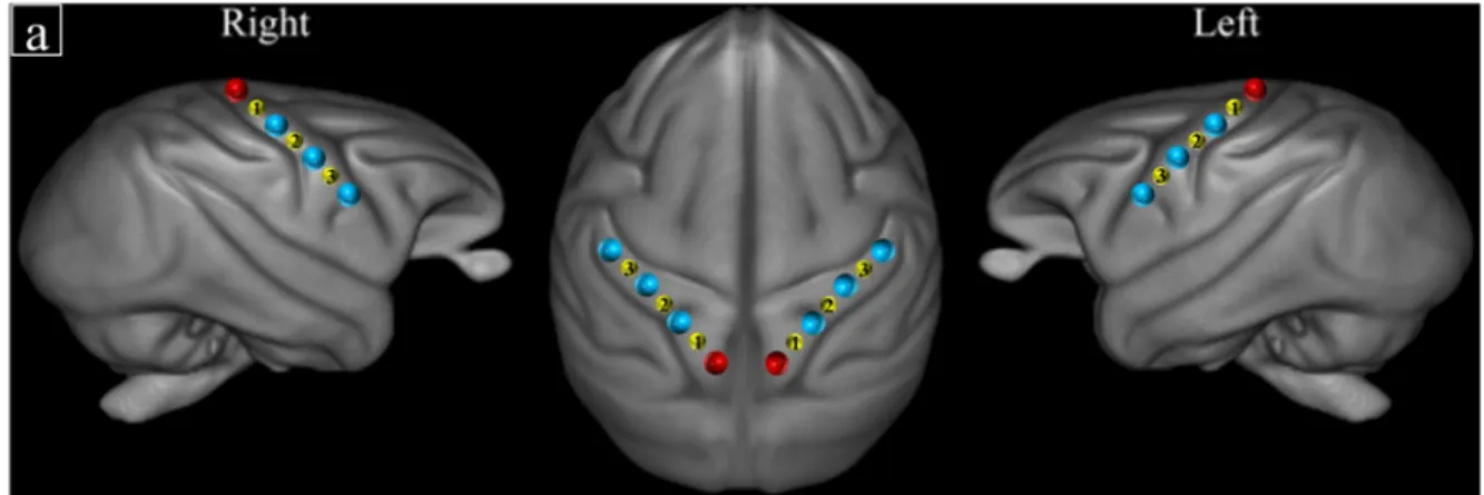
acquired by the LPC group on baboons (see Figure 1).
152 153
Each fNIRS session was planned during a routine health inspection undergone by the baboons
154
at the Station de Primatologie. As part of the health check, subjects were isolated from their
155
social group and anesthetized with an intramuscular injection of ketamine (5 mg/kg - Ketamine
156
1000®) and medetomidine (50µg/kg - Domitor®). Then Sevoflurane (Sevotek®) at 3 to 5%
157
and atipamezole (250 µg/kg - Antisedan®) were administered before recordings. The area of
158
interest on the scalp was shaved. Each baboon was placed in ventral decubitus position on the
159
table and the head of the individual was maintained using foam positioners, cushions and
160
Velcro strips to remain straight and to reduce potential motion occurrences. Vital functions
161
were monitored (SpO2, Respiratory rate, ECG, EtCO2, T°) and a drip of NaCl was put in place
162
during the entire anaesthesia. Just before recording brain activations, sevoflurane inhalation
163
was stopped and the focal subject was further sedated with a minimal amount of intravenous
164
injection of Propofol (Propovet®) with a bolus of around 2mg/kg every 10 to 15 minutes or by
165
infusion rate of 0.1 – 0.4 mg/kg/min. After the recovery period, baboons were put back in their
166
social group at the Station de Primatologie and monitored by the veterinary staff.
167 168
170
Figure 1: Schematic representation of fNIRS channel locations on ROIs according to T1 MRI
171
template from 89 baboons (39) for (a) the motor and (b) the auditory stimulations. Red and
172
blue dots indicate receivers and transmitters’ positions respectively. Yellow dots indicate the
173 channel numbers. 174 175 Motor stimulations 176
The motor stimulations consisted of 20 successive extensions of the same arm, alternatively
177
right and left repeated three times according to the same set plan (L-R-R-L-L-R) for all
178
baboons, resulting in a total of 120 arm movements. One experimenter on each side of the
179
baboon extended slowly their respective arm while stimulating the interior side of the hand
180
(gentle rhythmic tapping) with their fingers throughout the duration of the extension (about 5s)
181
upon a brief vocal command triggered by another experimenter. Between each block, there was
182 a 10s lag. 183 184 Auditory stimulations 185
The auditory stimuli consisted of 20s-long series of agonistic vocalizations of baboons and of
186
chimpanzees recorded in social settings (in captivity in an outside enclosure for baboons; and
187
in the wild for chimpanzees). Equivalent white noise stimuli matched for the energy dynamics
188
(i.e. the sound envelopes) were produced and used for comparison to control for the sound
189
energy dynamic differences. In the present study and analysis, we only examine the effect of
190
the lateralization of auditory stimulations (i.e., left ear versus right ear versus stereo) as a whole
191
on hemispheric asymmetry and thus do not distinguish between auditory signal types or species
192
(e.g. white noise and vocalizations). The auditory stimuli were broadcast pseudo-randomly,
193
alternating voiced and white noise stimuli and separated by 15s silences, either binaurally
194
(stereo), only on the left side, or only on the ride side. Due to signal artefacts and anaesthesia
195
shortfalls, the number of stimuli between the three baboons differs slightly. For Talma, the total
sequence consisted of 37 stimuli; for Rubis, the total sequence consisted of 47 stimuli; and for
197
Chet, the total sequence consisted of 25 stimuli.
198 199
fNIRS signal
200
We performed the first level analysis with MatLab 2018b (Mathwortks, Natick, MA) using the
201
SPM_fNIRS toolbox (40, https://www.nitrc.org/projects/spm_fnirs/) and homemade scripts.
202
Hemoglobin conversion and temporal preprocessing of O2Hb and HHb were made using the 203
following procedure:
204
1. Hemoglobin concentration changes were calculated with the modified Beer-Lambert
205
law (41);
206
2. Motion artifacts were removed manually in each individual and each channel for the
207
auditory stimulations. Thus, 10 seconds in total (1.3%) were removed from the O2Hb 208
and HHb signals of Rubis and 35 seconds (4.8%) for Talma and Chet fNIRS data;
209
3. A low-pass filter based on the hemodynamic response function (HRF) (42) was applied
210
to reduce physiological confounds.
211
4. A baseline correction was used for both the motor and auditory stimulations by
212
subtracting respectively (i) the average of 10 seconds intervals preceding each block;
213
(ii) the average of the 15 seconds of silence preceding each sound.
214
According to the temporal properties of the BOLD responses for each baboon, the O2Hb 215
concentration was averaged for Talma in a window of 4 to 12 s post stimulus onset for each
216
trial; and for Rubis and Chet in a window of 2 to 8 s post stimulus onset in order to select the
217
range of maximum concentration changes (µM). The difference of concentration range is
218
explained by the presence of some tachycardiac episodes for both Rubis and Chet during the
219
experiment, involving an HRF almost twice as fast as the one found for Talma.
220 221
AQ score calculation
222
Asymmetry Quotients (AQ) were derived for each subject and each experimental condition
223
(i.e: stimulation of the right arm and of the left arm for the motor experiment; right, left and
224
stereo audio stimulation for the auditory blocks) by first calculating the difference between the
225
right hemisphere (RH) and the left hemisphere (LH) values, to which we subsequently
226
subtracted the same difference during the preceding baseline block for the same subject to
227
normalize across trials. In particular, for motor stimuli, the baseline represented the 10s block
228
without motor activity immediately before a passive stimulation block of the right or left arm.
229
For auditory stimuli, the baseline was calculated on the 15s silence block that immediately
preceded the auditory stimuli. In this analysis, all auditory stimuli (baboon and chimpanzee
231
calls, and corresponding white noises) were analysed together. All calculated AQs were then
232
normalized using the scale function of R studio (R studio (2015) Inc:, Boston, MA, url:
233
http://www.rstudio.com/). For this analysis, we excluded one block ‘chimpanzee white noise
234
audio stereo’ (2.7% of O2Hb signal) for Rubis, and two blocks ‘chimpanzee white noise audio 235
stereo’ and ‘baboon white noise audio stereo’ (8.3%) for Talma as the recorded data revealed
236
themselves artefactual beyond repair. Positive AQ values indicate a rightward asymmetry and
237
negative values indicate a leftward asymmetry. Finally, using the aov function of R studio, we
238
performed one-way ANOVAs with pairwise comparisons on individual baboons by comparing
239
the AQ of all trials in the different stimulation conditions (right versus left motor stimulation;
240
right versus left versus stereo auditory stimulation) enabling to generalize the data of each
241 individual. 242 243 Results 244 245 Motor stimulations 246
One-way Anova analyses revealed significant differences between the left and right arm
247
stimulations across the three channels and baboons. Hence, for Rubis and Chet, comparisons
248
between left and right arms stimulations were all significant at p < .001 (Rubis: Ch1: F1,118 = 249
52.63; Ch2: F1,118 = 50.63; and Ch3: F1,118 = 42.35; for Chet: Ch1: F1,118 = 30.16; Ch2: F1,118 = 250
28.21; and Ch3: F1,118 = 24.77). Regarding Talma, significant differences were found at p <.05 251
in channel 1 (F1,118 = 3.821) and channel 3 (F1,118 = 6.521). The pairwise comparison in channel 252
2 (F1,118 = 14.71) was significant at p < .001. 253
Overall, the difference of AQ between left- versus right-arm stimulations were consistently
254
contralateral across the three subjects for all three channels: activation asymmetries were more
255
leftward for right-arm stimulations than for left arm stimulations and, were more rightward for
256
left-arm stimulations than for right arm stimulations (Figure 2; see Table 1 in supplementary
257
material for the mean AQ values).
258 259
260
Figure 2: Normalized averaged AQ (and corresponding SE) in the motor cortex following
261
motor stimulations in the three adult female baboons (see Figure 1 for localization of the
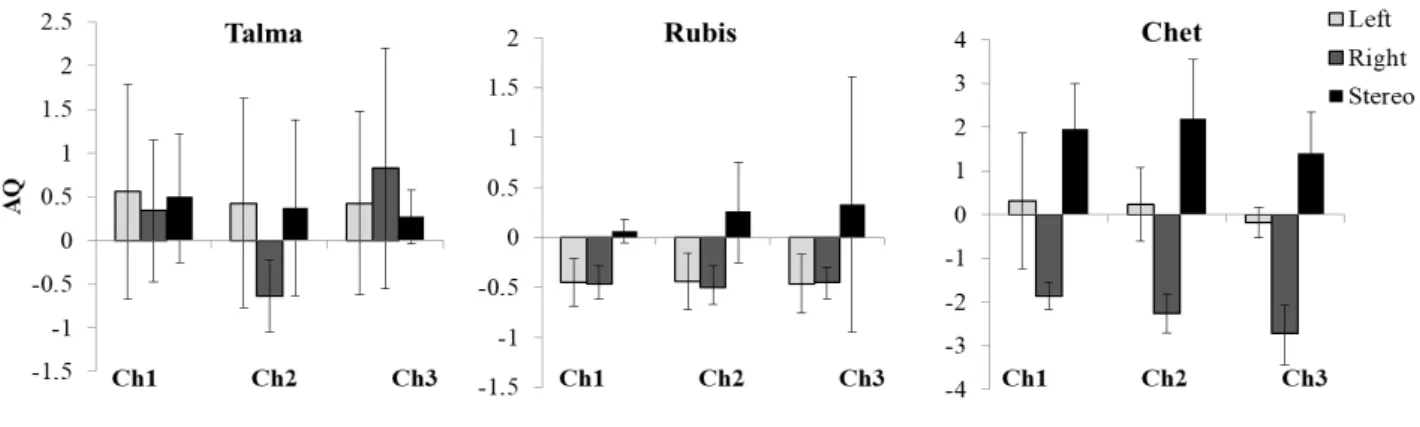
262 channels). 263 264 Auditory stimulations 265
We only found significant overall differences between, right, left and stereo ear stimulations
266
(p <.05) for subject Chet (Figure 3) for all channels (Ch1: F2,6 = 7.073; Ch2: F2,6 = 6.473; and 267
Ch3: F2,6 = 4.289). Pairwise comparison for right versus left ear stimulations were significant 268
(p <.05) in Ch1 (F1,6 = 5.216) and Ch2 (F1,6 = 5.043). Furthermore, significant differences 269
between right and stereo ear stimulations appeared across all channels (Ch1: F1,6 = 22.55; Ch2: 270
F1,6 = 16.56, p <.001; Ch3: F1,6 =15.95, p <.05). Note that the comparison left versus stereo did 271
not reach significance for any channels (Ch1: F1,6 = 1.827; Ch2: F1,6 = 1.825; Ch3: F1,6 =0.989, 272
all p >.05).
273
Hence, for Chet, there was a larger bias toward the left hemisphere with right ear stimulation
274
compared to stereo (for all our channels) and left ear stimulation (for channels 1 and 2 only;
275
Figure 3). No difference was recorded as significant for the two other baboons (see Table 2 in
276
supplementary material for the mean AQ values).
277 278
279 280
Figure 3: Normalized averaged AQ (and corresponding SE) above the temporal cortex
281
following auditory stimulations in three adult female baboons (see Figure 1 for localization of
282 the channels). 283 284 285 Discussion 286 287
The results of the present study clearly demonstrate that non-invasive fNIRS is a valid imaging
288
technique to investigate functional lateralization paradigms in a nonhuman primate species.
289 290
Our most potent results were found with the motor stimulation where we observed a strong
291
contralateral hemispheric asymmetry of the fNIRS signals in the motor cortex across baboons.
292
Right arm movements elicited greater leftward asymmetry than left arm movements and vice
293
versa in each of the three baboons for all three fNIRS channels. Results were clear-cut for
294
Rubis and Chet, though interestingly opposed, with Rubis having a strong leftward asymmetry
295
as a result of her right arm being stimulated, and Chet showing a strong rightward asymmetry
296
for her left arm. Results for Talma were somewhat similar to Rubis’ since right arm movements
297
elicited more leftward asymmetry than the left arm in channels 1 and 3. Results in channel 2
298
were most in line with our original prediction, namely a clear mirror pattern of contralateral
299
asymmetries between the two arms: the right arm movements elicited leftward asymmetry and
300
the left arm, a rightward asymmetry. Our results are consistent with previous studies in
301
primates: for arm/hand movements, 90% of the corticospinal pathway project to the
302
contralateral spinal cord (43-47). Hence, our study replicates these findings, with brain signals
303
differences detected by non-invasive fNIRS. Despite the robust consistency of findings across
304
subjects concerning the direction of the effect between the left and the right arms, the reasons
305
for inter-individual variabilities as well as the lack of mirror pattern of results between the two
arms (channel 2 of Talma excepted) remains unclear. In particular, potential involuntary
307
differences in arms’ stimulation degree between the two experimenters involved in each of the
308
subject’s arms manipulations, as well the handedness of each individual baboon may have had
309
an impact on our results.
310
311
Our results were also consistent with predicted asymmetries regarding auditory stimulations
312
for one subject. Contralateral differences of asymmetry were found for Chet in all three
313
channels, with the stimulation of both ears and left ear eliciting overall more rightward
314
asymmetries than right ear stimulations. Nevertheless, for Talma and Rubis, the direction and
315
degree of asymmetry varied irrelevantly of whether the sound was presented to the right or left
316
ear, namely toward the left temporal areas for Rubis and toward the right temporal areas for
317
Talma. These mixed results related to auditory stimulation might be interpreted with respect to
318
some characteristics of the hemispheric organization of the brain. It is well-known that at least
319
one third of the auditory fibres from the olivary complex project to ipsilateral brain regions
320
inducing less lateralization compared to motor brain regions. Furthermore, it has been shown
321
that receptive fields in some regions sensitive to somatosensory input from the auditory cortex
322
are 50% contralateral and 50% bilateral (48, 49); and that temporal regions such as the belt,
323
parabelt and STS receive strong ipsilateral connections in rhesus macaques (50, 51), suggesting
324
overall a less marked lateralization for auditory processing compared to motor regions.
325
Interestingly, the subject’s handedness in communicative gesture could also explain these
326
mixed results. In fact, our left-handed subject Talma, showed a clear right hemisphere bias for
327
most stimuli (to the exception of the right ear stimulation in channel 2); whereas Rubis,
right-328
handed in communicative gesture, showed a stronger bias toward the left hemisphere for the
329
sounds broadcast in right and left ears. These preliminary findings may thus highlight the
330
impact of hand preference in communicative contexts on contralateral brain organization in
331
baboons during auditory processing but would need further investigations in a larger cohort of
332
subjects.
333
Overall, given the lack of statistical power related to low sample size, we cannot draw any
334
conclusion regarding the direction of hemispheric lateralizations at a population-level for
335
sounds processing in baboons, or their relation to hand preference for communicative
336
gesturing. Nevertheless, some of our findings remain consistent with the literature on human
337
auditory pathways: for example, Kaiser and collaborators found that stimuli presented in stereo
338
activated more the right hemisphere compared to lateralized sounds showing a left hemisphere
339
bias (52). These results suggest that stereo sounds involve additional processing steps resulting
in stronger and more rightward brain activations (53). This pattern of rightward asymmetry for
341
stereo and left sounds processing in the baboon “Chet” is also somewhat consistent with
342
previous rightward asymmetries reported in rhesus monkeys (17) and in chimpanzees (21) for
343
processing conspecific calls. Hence, our data suggest that a phylogenetic functional approach
344
to vocal perception appears possible with fNIRS.
345 346
In conclusion, our study shows that fNIRS is a valid methodology to access brain signals in
347
primates non-invasively. In particular, we have replicated findings in the literature about brain
348
contralateral hemispheric activation in two different modalities showing that fNIRS is able to
349
capture such functional differences even in a context in which baboons were anesthetized.
350
However, we have also uncovered large variation between individuals. This may be due to
351
interindividual differences leading to the inability to precisely record in the same spot for all
352
baboons. Indeed, while we based our placing of optodes on our subjects based on an averaged
353
structural MRI pattern to which all tested individuals contributed, we cannot exclude small
354
variation across cortices. In the future, fNIRS should thus be coupled with structural imaging
355
techniques such as MRI that allow a precise positioning of the optodes for each individual. Yet,
356
the need to couple fNIRS with existing techniques does not deny a more widespread use of
357
fNIRS in the future. To the contrary, we believe that our study opens new avenues for brain
358
investigation in nonhuman primates using fNIRS for two main reasons. First, fNIRS has been
359
used in a multitude of contexts when other brain imaging techniques could not be used, for
360
example in the field with greater ecological conditions (54). While our data have been recorded
361
in anesthetized baboons, a logical next step is to train and habituate baboons to accept wearing
362
a fNIRS device. Our experimental paradigms could then be extended in awake monkeys with
363
more sophisticated design involving behavioural contingencies related to different kinds of
364
stimulation. Second, our study stresses that fNIRS could in the future become a valuable
365
method to explore brain activations in lateral regions in a non-invasive way in nonhuman
366
animals without attempting the physical integrity of the subjects, which would ultimately make
367
investigation of brain mechanisms in animal much more accessible and flexible.
368 369 370
Acknowledgements
371
CD and TG were supported by the Swiss National Science Foundation (grants
372
P1GEP1_181492 to CD and CR13I1_162720 / 1 to DG-TG). AM has received funding from
373
the European Research Council under the European Union's Horizon 2020 research and
innovation program grant agreement No 716931 - GESTIMAGE - ERC-2016-STG. We thank
375
the Société Académique de Genève for their financial support allowing purchasing the fNIRS
376
equipment. We thank the vet Pascaline Boitelle for monitoring heath and anaesthesia of
377
baboons during experiment and the animal care staff as well as Jeanne Caron-Guyon, Lola
378
Rivoal, Théophane Piette and Jérémy Roche for assistance during the recordings.
379 380 381
References
382
1. Eichert N, et al. (2019) What is special about the human arcuate fasciculus?
383
Lateralization, projections, and expansion. Cortex 118:107-115.
384
2. Crow TJ ed (2004) The speciation of modern Homo saiens (Oxford University Press,
385
Oxford).
386
3. Lindell AK (2013) Continuities in emotion lateralization in human and non-human
387
primates. Frontiers in Human Neuroscience 7:464.
388
4. Meguerditchian A, Vauclair J, & Hopkins WD (2013) On the origins of human
389
handedness and language : A comparative review of hand preferences for bimanual
390
coordinated actions and gestural communication in nonhuman primates. Dev
391
Psychobiol 55:637–650.
392
5. Margiotoudi K, et al. (2019) Handedness in monkeys reflects hemispheric
393
specialization within the central sulcus. An in vivo MRI study in right- versus
left-394
handed baboons (Papio anubis). Cortex 118:203-211.
395
6. Meguerditchian A, Gardner MJ, Schapiro SJ, & Hopkins WD (2012) The sound of
396
one hand clapping : handedness and perisylvian neural correlates of a communicative
397
gesture in chimpanzees. Proceeding of the Royal Society Biology 279:1959-1966.
398
7. Fernández-Carriba S, Loeches Á, Morcillo A, & Hopkins WD (2002) Asymmetry in
399
facial expression of emotions by chimpanzees. Neuropsychologia 40:1523–1533.
400
8. Hook-Costigan MA & Rogers LJ (1998) Lateralized use of the mouth in production
401
of vocalizations by marmosets. Neuropsychologia 36:1265–1273.
402
9. Fitch WT & Braccini SN (2013) Primate laterality and the biology and evolution of
403
human handedness: A review and synthesis. Annals of the New York Academy of
404
Sciences 1288:70–85.
405
10. Moreno C, Borod J, Welkowitz J, & Alpert M (1990) Lateralization for the expression
406
and perception of facial emotion as a function of age. Neuropsychologia 28:199-209.
407
11. Hopkins WD, Marino L, Rilling JK, & MacGregor LA (1998) Planum temporale
408
asymmetries in great apes as revealed by magnetic resonance imaging (MRI).
409
Neuroreport 9:2913–2918.
410
12. Gannon PJ, Holloway RL, Broadfield DC, & Braun AR (1998) Asymmetry of
411
chimpanzee planum temporale: humanlike pattern of Wernicke's brain language area
412
homolog. Science 279:220-222.
413
13. Pilcher DL, Hammock EAD, & Hopkins WD (2001) Cerebral volumetric
414
asymmetries in non-human primates: A magnetic resonance imaging study. Laterality
415
6(2):165-179.
416
14. Marie D, et al. (2017) Left Brain Asymmetry of the Planum Temporale in a
417
Nonhominid Primate: Redefining the Origin of Brain Specialization for Language.
418
Cerebral Cortex 28(5):1808-1815.
15. Yeni-Komshian GH & Benson DA (1976) Anatomical study of cerebral asymmetry in
420
the temporal lobe of humans, chimpanzees, and rhesus monkeys. Science 192:387–
421
389.
422
16. Rilling JK (2014) Comparative primate neuroimaging: insights into human brain
423
evolution. Trends in Cognitive Sciences 18:46-55.
424
17. Poremba A, et al. (2004) Species-specific calls evoke asymmetric activity in the
425
monkey’s temporal poles. Nature 427: 448–451.
426
18. Gil-Da-Costa R, et al. (2006) Species-specific calls activate homologs of Broca's and
427
Wernicke's areas in the macaque. Nature Neuroscience 9(8):1064-1070.
428
19. Joly O, Ramus F, Pressnitzer D, Vanduffel W, & Orban GA (2012) Interhemispheric
429
differences in auditory processing revealed by fMRI in awake rhesus monkeys.
430
(Translated from eng) Cereb Cortex 22(4):838-853 (in eng).
431
20. Petkov CI, et al. (2008) A voice region in the monkey brain. Nature Neuroscience
432
11:367-374.
433
21. Taglialatela JP, Russell JL, Schaeffer JA, & Hopkins WD (2009) Visualizing vocal
434
perception in the chimpanzee brain. Cerebral Cortex 19:1151–1157.
435
22. Zatorre RJ & Belin P (2001) Spectral and temporal processing in human auditory
436
cortex. Cerebral Cortex 11(10):946-953.
437
23. Belin P, Zatorre RJ, Lafaille P, Ahad P, & Pike B (2000) Voice-selective areas in
438
human auditory cortex. Nature 403(6767):309-312.
439
24. Schirmer A & Kotz SA (2006) Beyond the right hemisphere: Brain mechanisms
440
mediating vocal emotional processing. Trends in Cognitive Sciences 10:24-30.
441
25. Parr LA & Hopkins WD (2000) Brain temperature asymmetries and emotional
442
perception in chimpanzees, Pan troglodytes. Physiology & Behavior 71(3-4):363-371.
443
26. Casperd JM & Dunbar RIM (1996) Asymmetries in the visual processing of
444
emotional cues during agonistic interactions by gelada baboons. Behavioural
445
Processes 37(1):57-65.
446
27. Morris RD & Hopkins WD (1993) Perception of human chimeric faces by
447
chimpanzees — Evidence for a right-hemisphere advantage. Brain and Cognition
448
21(1):111-122.
449
28. Baraud I, Buytet B, Bec P, & Blois-Heulin C (2009) Social laterality and
450
‘transversality’ in two species of mangabeys: Influence of rank and implication for
451
hemispheric specialization. Behavioural Brain Research 198(2):449-458.
452
29. Wallez C & Vauclair J (2011) Right hemisphere dominance for emotion processing in
453
baboons. Brain and Cognition 75(2):164-169.
454
30. Grandjean D (2020) Brain Networks of Emotional Prosody Processing. Emotion
455
Review 0(0):1754073919898522.
456
31. Meguerditchian A & Vauclair J (2006) Baboons communicate with their right hand.
457
Behavioral Brain Research 171:170-174.
458
32. Leigh SR (2004) Brain growth, life history, and cognition in primate and human
459
evolution. . Am J Primatol 62:139–164.
460
33. Kochunov PV, et al. (2010) Mapping primary gyrogenesis during fetal development
461
in primate brains: High-resolution in utero structural MRI of fetal brain development
462
in pregnant baboons. Front Neurosci 4:1–11.
463
34. Boas DA, Elwell CE, Ferrari M, & Taga G (2014) Twenty years of functional
near-464
infrared spectroscopy: Introduction for the special issue. Neuroimage 85:1-5.
465
35. Ballardin JB, et al. (2017) Imaging brain function with Functionnal Near-Infrared
466
Spectroscopy in unconstrained environments. Front Hum Neurosci 11:258.
36. Robinson CJ & Burton H (1980) Organization of somatosensory receptive fields in
468
cortical areas 7b, retroinsula, postauditory and granular insula of M. fascicularis. J
469
Comp Neurol 192(1):69-92.
470
37. Smiley JF & Falchier A (2009) Multisensory connections of monkey auditory
471
cerebral cortex. Hearing Research 258(1):37-46.
472
38. Willems RM, der Haegen LV, Fisher SE, & Francks C (2014) On the other hand:
473
including left-handers in cognitive neuroscience and neurogenetics. Nature Reviews
474
Neuroscience 15(3):193-201.
475
39. Love SA, et al. (2016) The average baboon brain: MRI templates and tissue
476
probability maps from 89 individuals. NeuroImage 132:526–533.
477
40. Tak S, Uga M, Flandin G, Dan I, & Penny WD (2016) Sensor space group analysis
478
for fNIRS data. Journal of Neuroscience Methods 264:103–112.
479
41. Delpy DT, et al. (1988) Estimation of optical pathlength through tissue from direct
480
time of flight measurement. Phys Med Biol 33:1433-1442.
481
42. Friston KJ, et al. (2000) To smooth or not to smooth? Bias and efficiency in fMRI
482
time-series analysis. NeuroImage 12(2):196–208.
483
43. Dum R. P. & Strick PL (1996) Spinal cord terminations of the medial wall motor
484
Areas in macaque monkeys. The Journal of Neuroscience 16:6513–6525.
485
44. Brösamle C & Schwab ME (1997) Cells of origin, course, and termination patterns of
486
the ventral, uncrossed component of the mature rat corticospinal tract. The Journal of
487
Comparative Neurology 386:293–303.
488
45. Lacroix S, et al. (2004) Bilateral corticospinal projections arise from each motor
489
cortex in the macaque monkey: a quantitative study. The Journal of Comparative
490
Neurology 473:147–161.
491
46. Rosenzweig ES, et al. (2009) Extensive spinal decussation and bilateral termination
492
of cervical corticospinal projections in rhesus monkeys. The Journal of Comparative
493
Neurology 513:151–163.
494
47. Heming EA, Cross KP, Takei T, Cook DJ, & Scott SH (2019) Independent
495
representations of ipsilateral and contralateral limbs in primary motor cortex. eLIFE.
496
48. Robinson CJ & Burton H (1980) Somatotopographic organization in the second
497
somatosensory area of M. fascicularis. J. Comp. Neurol. 192:43–68.
498
49. Smiley J & Falchier A (2009) Multisensory connections of monkey auditory cerebral
499
cortex. Hearing research 258.
500
50. Cipolloni PB & Pandya DN (1989) Connectional analysis of the ipsilateral and
501
contralateral afferent neurons of the superior temporal region in the rhesus monkey.
502
The Journal of Comparative Neurology 281(4):567–585.
503
51. Hackett TA, Stepniewska I, & Kaas JH (1998) Subdivisions of auditory cortex and
504
ipsilateral cortical connections of the parabelt auditory cortex in macaque monkeys.
505
The Journal of Comparative Neurology 394(4):475–495.
506
52. Kaiser J, Lutzenberger W, Preissl H, Ackermann H, & Birbaumer N (2000)
Right-507
Hemisphere Dominance for the Processing of Sound-Source Lateralization. J.
508
Neurosci. 20(17):6631-6639.
509
53. Jäncke L, Wüstenberg T, Schulze K, & Heinze HJ (2002) Asymmetric hemodynamic
510
responses of the human auditory cortex to monaural and binaural stimulation. Hearing
511
Research 170(1):166–178.
512
54. Piper SK, et al. (2014) A wearable multi-channel fNIRS systemfor brain imaging in
513
freely moving subjects. Neuroimage 85:64–71.
514 515