HAL Id: hal-03137373
https://hal.archives-ouvertes.fr/hal-03137373
Submitted on 10 Feb 2021
HAL is a multi-disciplinary open access
archive for the deposit and dissemination of
sci-entific research documents, whether they are
pub-lished or not. The documents may come from
teaching and research institutions in France or
abroad, or from public or private research centers.
L’archive ouverte pluridisciplinaire HAL, est
destinée au dépôt et à la diffusion de documents
scientifiques de niveau recherche, publiés ou non,
émanant des établissements d’enseignement et de
recherche français ou étrangers, des laboratoires
publics ou privés.
emergence and spread of plant, animal and human
diseases. A chronicle of epidemics foretold in South of
France
Didier Fontenille, Astrid Cruaud, Laurence Vial, Claire Garros
To cite this version:
Didier Fontenille, Astrid Cruaud, Laurence Vial, Claire Garros. Understanding the role of arthropod
vectors in the emergence and spread of plant, animal and human diseases. A chronicle of epidemics
foretold in South of France. Comptes Rendus Biologies, Elsevier Masson, 2020, 343 (3), pp.311-344.
�10.5802/crbiol.34�. �hal-03137373�
Comptes Rendus
Biologies
Fontenille Didier, Cruaud Astrid, Vial Laurence and Garros Claire
Understanding the role of arthropod vectors in the emergence and
spread of plant, animal and human diseases. A chronicle of epidemics
foretold in South of France
Volume 343, issue 3 (2020), p. 311-344. <https://doi.org/10.5802/crbiol.34>
© Académie des sciences, Paris and the authors, 2020.
Some rights reserved.
This article is licensed under the
Creative Commons Attribution 4.0 International License.
http://creativecommons.org/licenses/by/4.0/
LesComptes Rendus. Biologies sont membres du Centre Mersenne pour l’édition scientifique ouverte
2020, 343, n 3, p. 311-344
https://doi.org/10.5802/crbiol.34
Articles / Reviews / Articles / Revues
Understanding the role of arthropod vectors in the
emergence and spread of plant, animal and human
diseases. A chronicle of epidemics foretold in South
of France
Comprendre le rôle des arthropodes vecteurs dans l’émergence et
la propagation des maladies infectieuses végétales, animales et
humaines. Chronique d’épidémies annoncées dans le sud de la
France
Fontenille Didier
∗, a, Cruaud Astrid
b, Vial Laurence
c , dand Garros Claire
c , daMIVEGEC unit, Université de Montpellier, Institut de Recherche pour le
Développement (IRD), CNRS, BP 64501, 34394 Montpellier, France
bCBGP, INRAE, CIRAD, IRD, Montpellier SupAgro, Université de Montpellier,
Montpellier, France
cASTRE unit, Université de Montpellier, CIRAD, INRAE, Montpellier, France dCirad, UMR ASTRE, 34398 Montpellier, France
E-mails: didier.fontenille@ird.fr (F. Didier), astrid.cruaud@inrae.fr (C. Astrid),
laurence.vial@cirad.fr (V. Laurence), claire.garros@cirad.fr (G. Claire)
Abstract. Southern France, like the rest of the world, is facing the emergence of diseases affecting
plants, animals and humans, of which causative agents (viruses, parasites, bacteria) are transmitted by arthropod vectors. Global changes are accelerating the emergence and spread of these diseases. After presenting some examples related to vectors of yellow fever and dengue viruses (Aedes aegypti and
Ae. albopictus), Crimean-Congo hemorrhagic fever (Hyalomma marginatum), Bluetongue (Culicoides
sp.), and the phytopathogen Xylella fastidiosa (Hemiptera spp.), we will discuss what are the intrinsic and extrinsic factors that make an arthropod a vector in a given place and at a given time. We also propose some thoughts regarding these emergences, possible scenarios for their evolution and some recommendations for the future.
Résumé. Le sud de la France, comme le reste du monde, est confronté à l’émergence de maladies
affectant les plantes, les animaux et les populations humaines, dont les agents étiologiques (virus, parasites, bactéries) sont transmis par des arthropodes vecteurs. Les changements globaux auxquels nous faisons face accélèrent l’apparition et la diffusion de ces pathologies. Après avoir pris quelques exemples concernant les vecteurs des virus de la fièvre jaune et de la dengue (Aedes aegypti et Ae.
albopictus), de la fièvre de Crimée-Congo (Hyalomma marginatum), de la fièvre catarrhale ovine
(Culicoides sp.) et de la bactérie phytopathogène Xylella fastidiosa (Hemiptera spp.), nous verrons quels sont les facteurs intrinsèques et extrinsèques qui font qu’un arthropode devient un vecteur en un lieu et à un moment donné. Nous proposons des pistes de réflexion sur ces émergences et le possible devenir des maladies transmises par arthropodes. Nous concluons par quelques recommandations pour mieux anticiper les émergences.
Keywords. Vector, Mosquito, Tick, Midges, Xylella, Emergence, Risk.
Mots-clés. Vecteur, Moustique, Tique, Moucherons, Xylella, Émergence, Risque. Manuscript received and accepted 8th December 2020.
Are the tiger mosquito (Aedes albopictus), the gi-ant tick (Hyalomma marginatum), the biting midge
Culicoides imicola and the meadow spittlebug (Phi-laenus spumarius), all still unknown in Southern
France a few years or decades ago, our new plagues of Egypt?
These four species of arthropods are related to vector-borne diseases (VBDs). The superlatives sometimes associated with these new scourges con-cerning the health of humans, animals and plants, reflect the concerns they generate in the general population, among livestock breeders, farmers and among decision-makers.
Enemies needing surveillance or a fight, against which we seem to be quite helpless, these vectors used as examples give us the opportunity to ask the following questions: how did we get there? What has led these arthropods and pathogens to become a threat?
Beyond the stereotypes on very real phenomena with serious consequences such as climate change, environmental modifications, changes in agricul-tural practices, urbanization, intensification of world trade, it is often the conjunction of a set of events, most of the time with a low probability of occurrence, which leads to the emergence of the transmission of a pathogen by a vector: insect, mite, mollusc, nema-tode (see definition below). In other words, the worst is never sure, but it is the worst that holds our atten-tion as humans, when we are directly concerned.
For humans only, the World Health Organization (WHO) estimates that vector-borne diseases (such as malaria, dengue, Chagas disease, leishmaniosis, hu-man African trypanosomiasis) account for more than
17% of infectious diseases and are responsible for more than 700,000 deaths per year worldwide. Eco-nomic costs of human VBDs (surveillance, medical and non-medical costs, vector control, productivity losses) account for billions of euros annually.
Moreover, the economic consequences of some of these vector-borne diseases on agriculture and livestock production are colossal. They have key direct macro-economic impacts: morbidity of live-stock, economic losses such as reduced produc-tion and farm income, ban on livestock and se-men trade, imposed quarantine, costs of biological tests, surveillance and control measures. For exam-ple, in Germany and in the Netherlands [1, 2], the impact of Bluetongue on cattle was estimated up-wards of hundreds of million Euros. In the field of plant health, Citrus greening (Huanglongbing dis-ease, HLB) caused by Candidatus Liberibacter spp. transmitted by psyllids, is the most destructive citrus pathosystem worldwide with an estimated cost for Florida of $8.92 billion in revenue and $4.62 billion in gross domestic product between the 2006/2007 and 2010/2011 crop productions [3]. Sharka, caused by
Plum pox virus transmitted by Aphididae, is the most
devastating disease of stone fruit trees. It generates annual losses accounting for hundreds of millions of euros [4].
In addition, there are also indirect socio-economic impacts that concern any VBDs affecting plants ani-mals or humans: inability of breeders, farmers, work-ers to achieve daily demands, disruption of mar-ket chains, and concerns of consumers and citi-zens regarding health threats and environmental is-sues (concerns of contracting disease, change in
consumption habits, public authorities questioned on use of insecticides, vaccines, massive culling, environmental and sanitary side effects), costs of surveillance and tests for the (early) detection of the disease to avoid large outbreaks etc.
Risk assessment, prevention and control of these emerging diseases or their invasive vectors are major health, ecological, social and economic challenges. The most problematic recent examples in metro-politan France concern dengue fever, Zika disease, chikungunya, West Nile fever, arboviruses transmit-ted by sandflies, Lyme disease, as far as humans are concerned; Bluetongue, Crimean-Congo hemor-rhagic fever although it is a zoonosis, African swine fever, anaplasmosis, for livestock; numerous viral (e.g. Sharka, tomato leaf curl New Delhi virus), phy-toplasma (e.g. flavescence dorée) and bacterial (e.g.
Xylella fastidiosa, Citrus greening) diseases for wild
and cultivated plant species. In the absence of ef-fective control measures (curative treatments, bio-cides, vaccines), prevention, reduction, and risk mit-igation requires the identification of colonization routes, the early detection of introduction, and an ef-ficient control of vectors. Currently, vector control is mostly based on the use of polluting chemical insec-ticides against which arthropods develop resistance. A dramatic change in approaches and practices is needed and requested by society that wants health-ier environment and better health. This is an
objec-tive of many French Regions, of the European Union with the Green deal, and of the United Nations in a “One Health” strategy integrating sustainable devel-opment goals.
However, years of research and development ef-forts have not been able to entirely solve the prob-lems and there is a perpetual arms race between humans, vectors and pathogens. New tools are being developed (not all of them are consensual), or are yet to be invented: diagnostics, treatments, green chemistry, plant-based insecticides, traps, competi-tors, parasites, predacompeti-tors, sterile insect techniques, transgenesis, transmission-blocking vaccines, in-sect densoviruses, Wolbachia endosymbionts, mi-crobiota, repellents, but also education, behavioral approaches, participatory sciences and risk model-ing. An effort is also needed to better understand processes of emergence for better prediction and prevention.
We will take some historical and contemporary ex-amples, particularly from the south of France (the Mediterranean coast from the Spanish to the Ital-ian borders, covering two regions: Occitanie and Provence-Alpes-Côte-d’Azur), regarding human, an-imal and plant health to illustrate how and why an arthropod or a pathogen emerges from anonymity and becomes a public enemy. From these models, we will see the conditions necessary for these emer-gences to occur, and speculate on the future.
Definition of a vector
A vector is any organism that actively transmits an infectious agent from one host to another (either animal or plant).
The concept of active transmission requires that the vector, through its behaviour, allows the transmission of an infectious agent by taking it from one host and transmitting it to another host. The infectious agent may or may not multiply in the vector. This definition therefore excludes most intermediate hosts that passively release infectious agents into the environment.
1. Spreading of primate Flaviviruses by Aedes
aegypti and Aedes albopictus mosquitoes
Yellow fever (YF) was among the most devastating mosquito borne diseases, in Africa, in the Ameri-cas and even in some European ports, during cen-turies. Its story sheds light on the mechanisms of the emergence, establishment and spread of a zoono-sis from African forests to American cities.Unfortu-nately, what happened five centuries ago can happen again, as the following examples show.
Natural history of yellow fever in Africa has been deciphered decades ago [5]. The YF virus circulates in African forests from monkey to monkey, trans-mitted by forest mosquitoes belonging to the genus
Aedes (Ae. africanus, Ae. luteocephalus, Ae. furcifer, Ae. simpsoni s.l., Ae. opok, etc.), with incursions
Ae. aegypti are established, and able to replicate and
transmit YF virus [6]. The virus, once in humans, is introduced through the movement of humans into African cities, where it finds urban Ae. aegypti pop-ulations able to generate outbreaks.
This human-adapted yellow fever virus and urban Ae. aegypti, were introduced into tropical America, via infected people and/or Ae. aegypti transported on ships, during the triangular slave trade, which started in the 16th century [7, 8]. Upon arrival to the Central and South American rainforests, the virus found both susceptible New World mon-keys and new competent endemic mosquitoes from two genera, Sabethes sp. and especially Haemagogus sp. Yellow fever virus thus had two events of cross species transmission: African monkeys to can monkeys, African Aedes mosquitoes to Ameri-can mosquitoes. New cycles of jungle yellow fever have thus developed, involving South American pri-mates and tree-dwelling Haemagogus mosquitoes. Phylogenetically, the genus Haemagogus seems to be quite close to Aedes [9], which may explain the ability of Haemagogus species to often transmit the same viruses: yellow fever, dengue [10], Zika [11], and chikungunya [12], all introduced into South America. At the same time, Ae. aegypti adapted to American cities, and even non-tropical locations were subject to yellow fever epidemics, such as the well-known Philadelphia epidemic in 1793 (5000 deaths).
This combination of both a sylvan and an urban cycle for yellow fever in the Americas regularly fu-elled epidemics that greatly affected the history of colonization of the New World [13]. These cycles have spread throughout South and Central America, where they are still active, as shown by the 2016–2018 yellow fever epidemic in Brazil, with more than 700 deaths [14].
Interestingly, endemic yellow fever has never been reported in Asia, Madagascar or other Indian Ocean islands, for reasons that are still poorly understood. Primates (monkeys and humans) from these regions are susceptible, and Ae. aegypti and other experimen-tally competent Aedes mosquitoes are present there [15–17]. Importantly, Haemagogus sp. and Sabethes sp. are absent from Africa, the Indian Ocean and Asia.
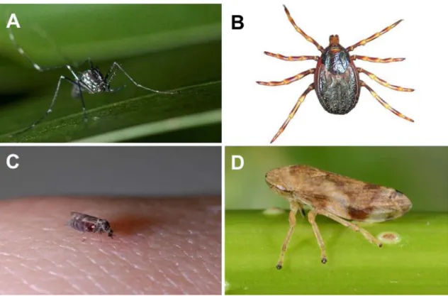
Aedes albopictus (Figure 1), a mosquito
phyloge-netically closed to the YF mosquito, Ae. aegypti, has recently become a real nuisance and increasingly a public health problem in the world, including south
of France. Although it has been present for more than 100 years in the French department of La Re-union Island in the Indian Ocean, it only arrived in continental France in 2004 [18]. Aedes albopictus is now established in 58 departments of metropol-itan France (data French ministry of Health, Santé
publique France).
In Southern France, all the 13 departments of the Occitanie Region are now colonized by Ae.
al-bopictus and 10 departments have more than 40%
of their population in contact with Ae. albopictus. In this région the Ministry of Health reported 124 imported cases of Chikungunya, dengue or Zika (8 CHIK, 114 DEN, 2 ZIKA) in 2019 and “only” 79 dengue imported cases in 2020 (a year with much less travel). In 2020, for metropolitan France, the Ministry of Health reported 545, 5 and 1 imported cases of dengue, chikungunya and Zika respec-tively, and 11 dengue autochthonous cases, all in southern France. Indigenous cases have been ob-served almost every year for the last decade, and this trend will increase. (data Santé Publique France: https://www.santepubliquefrance.fr/maladies- et-traumatismes/maladies-a-transmission- vectorielle/chikungunya/articles/donnees-en- france-metropolitaine/chikungunya-dengue-et- zika-donnees-de-la-surveillance-renforcee-en-france-metropolitaine-en-2020).
Aedes albopictus was really neglected until the
1980’s, when it was gradually discovered on all con-tinents outside Asia and caused a chikungunya pan-demic [19–22]. Aedes albopictus takes blood from many vertebrate species [23], its life expectancy is about one month (which is quite long for a mosquito) [24], and it can transmit more than 40 viruses naturally or experimentally [22]. Aedes
al-bopictus therefore perfectly fulfils the necessary
criteria to be an excellent vector of virus to humans.
Aedes albopictus is considered to be native to
Southeast Asia [19]. This assumption is based on the fact that this mosquito is present everywhere in the forested areas of this region and that many species close to the Albopictus subgroup (members of the Scutellaris group) are present in Southeast Asia. However, the notoriety of Ae. albopictus, com-pared to its relatives, comes from the fact that it has moved out of its area of origin, becoming world-wide in 50 years, adapting perfectly to urbanization, temperate climates, and international transport, as
Figure 1. Arthropod vectors of human, animal and plant diseases. A. Aedes (Stegomyia) albopictus,
female ©Nil Rahola, IRD. B. Hyalomma marginatum ©F Stachurski, CIRAD. C. Culicoides nubeculosus, female ©JB Ferré, EID méditerranée. D. Philaenus spumarius ©Jean-Yves Rasplus, INRAE.
well as being involved in several dengue and chikun-gunya epidemics [25].
In tropical Southeast Asia, the five morphologi-cally closely related species of the Albopictus sub-group live in forests (Aedes novalbopictus, Aedes
pa-triciae, Aedes seatoi, Aedes subalbopictus and Aedes pseudalbopictus) and lay their eggs in tree holes and
bamboo stumps [26,27]. These species are likely to be vectors of arboviruses to vertebrates from which they take blood. These forest-confined viruses are largely unknown, but may emerge through increased con-tact with humans or domestic animals. They could then be transmitted by vectors established in villages and cities, such as Ae. aegypti and Ae. albopictus, in a pattern similar to the emergence of the yellow fever virus in Africa from forests to villages and towns [5].
Based on current knowledge, it seems that Aedes
albopictus is the species that has been the most
suc-cessful in adapting to modern human settings among the Albopictus subgroup. In some Asian countries, such as Laos, Ae. albopictus is reported from deep
natural forest [28], rubber forest and secondary for-est [29], as well as in towns and villages, while in other countries, like in Malaysia, Ae. albopictus is rare in forests [30]. In Yunnan Province, China, if
Ae. albopictus is sometimes more abundant than Ae. pseudalbopictus in bamboo forests, it is often
ab-sent from deep forests [27].
Mogi et al. [27], suggest that the spread of Ae.
al-bopictus from its original tropical forest region was
possible following evolution from an ancestral wild species, due to adaptation to man-made habitats and then migration with humans to temperate climate re-gions, where Ae. albopictus developed a winter di-apause. In most introduced localities, it probably encountered only limited competition from native mosquitoes and when it did, Ae. albopictus proved to be a robust competitor, e.g., with Ae. aegypti [31]. It is likely that the ancestral wild species was already a vector of forest vertebrate arboviruses, for exam-ple, dengue-like flaviviruses, or chikungunya-like al-phaviruses, which are monkey viruses [32].
In both its native range Asia and recently invaded South America [33] and Africa (like in Gabon, Paupy,
pers. com.), Ae. albopictus has retained its ancestral
capacity to colonize forest environments, however most of the time on the ecotone edges, laying eggs in natural pools of water and taking blood from non-domestic vertebrates. It is logical to think that under these conditions, Ae. albopictus populations would take blood from vertebrates carrying as yet unknown viruses, such as from monkeys, terrestrial mammals, birds or reptiles, and then be excellent bridge vec-tors from animals to humans allowing the emergence of forest viruses. We can speculate that this is most likely to occur in Asia. In comparison, in Africa, Ae.
aegypti has already brought many, perhaps most,
hu-man adapted viruses from the forest to huhu-man habi-tats, such as yellow fever, dengue, chikungunya, and Zika viruses [6].
From regions recently colonized by Ae.
albopic-tus, “modern” populations, highly adapted to the
ur-ban environment and excellent vectors, spread to all continents. It is likely that invasive populations re-invaded regions where ancestral populations already existed, such as in Asia and the Indian Ocean. These ancestral populations then found themselves in un-favourable competition and modern Ae. albopictus replaced them [34].
In the coming years, geographical distribution of
Ae. albopictus will likely increase, especially in
tem-perate regions and its control by insecticides will be-come more difficult due to the emergence of resis-tance. Likewise, Ae. albopictus-vectored epidemics are almost certain to increase. In addition to dengue, chikungunya and Zika, it could be responsible for yel-low fever epidemics, or of currently unknown viruses transmitted by forest edge Aedes mosquitoes of South American, South East Asia or Central Africa. A ma-jor unknown for Ae. albopictus’ impact in temper-ate regions is whether arbovirus replication at lower temperatures is selected to be rapid enough to reach saliva before the female dies.
2. Crimean-Congo Hemorrhagic Fever
emer-gence in Southern Europe and Hyalomma
ticks: having the right name doesn’t make
the vector
Crimean-Congo Hemorrhagic Fever (CCHF) is a zoonotic disease caused by a RNA virus (Nairovirus,
Bunyavidae), which results in an acute and po-tentially fatal infection in humans while it remains asymptomatic in animals. The Crimean-Congo Hem-orrhagic Fever virus (CCHFv) is transmitted in the wild through a “tick-non human vertebrate–tick” syl-vatic cycle. Human infections occur punctually via a tick bite or from contact with contaminated body fluids of infected livestock or human patients. Only a few human infections lead to typical hemorrhagic symptoms, and are reported as CCHF cases, the tip of the iceberg. Ticks, especially those belonging to the
Hyalomma genus, are considered as biological
vec-tors but also reservoirs for CCHFv, as they are able to maintain the virus for several months or years indi-vidually or among tick populations [35]. Although the viremia in vertebrate hosts is very short, some hosts, such as lagomorphs, hedgehog, cattle, small rumi-nants, deer and wild boar in Europe, are able to be-come a source of infection for tick vectors [36]. CCHF is widespread throughout Eastern Europe, Africa, Asia and Middle East, and is emerging in West-ern Europe and the Mediterranean Basin [37, 38]. First attempts to correlate CCHF human cases to environmental conditions worldwide with focus on Turkey, showed that the disease likely emerged under warmer temperatures and sufficient number of days >5 °C in April, which probably increased develop-ment rate of thermophilous Hyalomma tick vectors and contributed to early activation of adult stages in spring [39, 40]. However, such modelling approaches suggested much more complex multi-factorial situa-tions than only changing climate.
The issue of the suitable conditions for CCHF emergence in humans is particularly crucial in Southern France although the disease has never been reported there. Indeed, the presence of one of the main vectors of CCHFv, Hyalomma
margina-tum (Figure 1), has been confirmed recently while
it was considered absent in the past, except in Cor-sica [41, 42]. Its northern extension into mainland France may have resulted from natural preexistent tick introduction events through host movements, especially bird migrations, and successful establish-ment of the tick due to global increase of temper-ature and drying of habitats under current climate changes [43]. Hyalomma marginatum is continuing its spread throughout Southern France with speci-mens recorded in localities where it was absent two years earlier. In addition, several hotspots with more
or less abundant tick populations have now been identified in seven departments of Occitanie and Provence-Alpes-Côte d’Azur regions in mainland France (Vial, non-published data). Bio-ecological investigations are being conducted to better under-stand environmental conditions constraining the establishment of such an “invasive” tick, which is as-sumed to adapt to a relatively large panel of temper-ature and humidity compared to other Hyalomma species [44]. In addition to the evidence for tick vec-tor extension, CCHFv is suspected to already cir-culate in Southern France since specific antibodies against CCHFv have been detected in Corsica from 13% and 2–3% of cattle and small ruminants, re-spectively, with up to 80% of seropositive animals in some infected farms [45]. No data are yet available for mainland France but analyses are in progress. In Spain, scientists have first detected CCHFv in
Hyalomma ticks in 2010 [46] and autochthonous
hu-man cases have occurred punctually since 2016 [47]. A similar evolution in France could be expected. However, the situation is a bit different. Indeed, al-though H. marginatum is historically largely dis-tributed in the Iberian Peninsula, CCHF has emerged in humans through the biting of another “unusual” tick, Hyalomma lusitanicum, which is present locally in Iberian forested oak areas and had never been re-ported before as an efficient CCHFv vector [48]. This specific case proved that H. marginatum could be present, abundant and infected with CCHFv without becoming necessarily an efficient vector. Past CCHF outbreaks from various ecosystems among the world can inform on common patterns or rather local specificities that result in efficient vectorial transmis-sion of CCHFv and disease emergence in humans.
CCHF is occurring punctually but regularly in the Balkans as well as in South and West Africa, suggest-ing an endemic epidemiological situation. People are used to engaging in outdoor activities with high ex-posure to infected ticks and livestock. Human cases occur in synchrony with seasonal tick vector dynam-ics and with an increase in virus transmission in the sylvatic cycle. In these endemic regions, H.
margina-tum for Balkans and H. rufipes (formerly known as a
subspecies of H. marginatum) for Africa have been identified as the main CCHFv tick vectors, at least in humans. As viremia is short and transitory in an-imal hosts, their likelihood for infecting tick vectors remains low and other transmission modes among
ticks without vertebrate hosts are necessary [36]. Moreover, it has been estimated that transovarial transmission of CCHFv from infected females to lar-val ticks does not occur with sufficient frequency to allow maintaining the virus in the absence of am-plification of infection in vertebrate hosts [49]. In this peculiar context, it was shown in Africa the in-tercession of another “partner of choice”, the tick
H. truncatum that was confirmed to be able to
am-plify CCHFv among ticks through co-feeding trans-mission on non-viremic animals [50, 51]. As another consequence of low transovarial transmission rate in such “two-hosts” Hyalomma ticks, the infection of immature ticks on small vertebrate hosts constitutes a much more important amplifying mechanism for circulation of the virus than infection of adult ticks on large ungulates [51]. Among the likely hosts for im-mature stages of H. marginatum and H. rufipes, birds are unable to replicate CCHFv while hares or hedge-hogs are considered good amplifiers but remain very scarce in Sahelo-Sudanian environments. However, red-beaked hornbills that are widely distributed in Senegal and frequently infested by H. rufipes imma-tures, were experimentally infected with CCHFv and succeeded to transmit the virus to tick larvae during blood feeding [52]. In South Africa, ostriches which are often infested by immature and adult Hyalomma ticks were also demonstrated as good CCHFv am-plifiers and thus potential sources for infecting large amounts of tick vectors [53]. The intervention of such “helper” hosts could partially explain the emergence of CCHF in Spain through the biting of H.
icum instead of H. marginatum. Hyalomma lusitan-icum parasitizes quasi exclusively hares as
imma-tures and red deer as adults, which are both known as very good CCHFv amplifiers. A serological study in the red deer recorded a prevalence twice as high as in domestic ungulates from Corsica [54]. Conversely, H.
marginatum infests lagomorphs but also many birds
at immature stages, and its adult stages prefer do-mestic ungulates in particular horses that do not de-velop sufficient viremia to infect ticks. In France and Spain where hares are rare and horses abundant, this may contribute to maintain a low rate of infection in
H. marginatum through what is known in
epidemiol-ogy as “dilution effect” [55].
The epidemiological situation of CCHF in the for-mer Soviet Union and Turkey is also informative as CCHFv is also transmitted by H. marginatum but
the disease shows a very different profile. It emerged in the form of massive outbreaks, in 1944, affecting about 200 Soviet soldiers in the Crimean Peninsula and then followed by smaller recurrences [56], and since 2002 in Turkey with an annual disease inci-dence reaching a plateau of 700 to 1300 cases/year until recently [57]. In both regions, agriculture lands were abandoned during wartime, terrorist activities, or due to strict prohibition of hunting in some ar-eas, and resulted in an increase of wild fauna and tick populations that favors the amplification of CCHFv natural transmission. Then, militaries or new set-tlers conducting farming and cattle breeding activ-ities were massively introduced in those regions al-lowing CCHFv transmission to naïve susceptible hu-mans, through tick bites but also via direct contact with animal infected fluids and nosocomial contami-nations between humans. In such regions, apart from suitable conditions increasing the probability of in-fection for tick vectors, changes in human exposure also contributed to the emergence of CCHF cases. Al-though H. marginatum is described as particularly “aggressive” among Hyalomma ticks with a strong hunting behavior against its vertebrate hosts [44] and a relatively high affinity for humans at adult stage, we assume that its biting remains rare and needs spe-cial conditions to be boosted. Indeed, this tick is large and is thus frequently detected and removed before attaching, as observed during tick sampling in the field. In addition, in France, tick abundances remain lower than in southern countries where H.
margina-tum has been well established for a long time, and
this may much more contribute to a relatively low rate of tick-human meeting.
3. Unexpected vectors, new virus: how
Culi-coides biting midge species threaten the
livestock production worldwide
Culicoides imicola is a biting midge species from the
Ceratopogonidae family, responsible for the trans-mission of viruses of veterinary importance such as the Bluetongue virus in the Afrotropical and Mediter-ranean regions. Bluetongue is a disease of which eti-ological agent is a virus of the Orbivirus genus trans-mitted by species of the Culicoides genus (Figure 1) to domestic and wild ruminants. Culicoides imicola
is emblematic for two reasons: the debate on its inva-sive status and pattern and the role of human activi-ties; and the importance that a supposed invasive ex-otic species can take at the expense of native species. Bluetongue disease is an emerging disease in Europe, associated with two distinct mechanisms: (i) the set-tlement of perennial populations of an Afrotropi-cal vector species in the Mediterranean region, and (ii) the transmission of exotic serotypes by competent autochthonous Palearctic species. It is undoubtedly one of the rare cases of emergence of a vector-borne disease for which the link with the global rise of tem-peratures is also strongly suspected [58, 59].
As of 1998, Southern Europe underwent a new se-ries of emerging cases of Bluetongue disease involv-ing different serotypes (1, 2, 4, 8, 9, 16) mainly in areas where C. imicola was considered to be absent [60]. The first outbreaks were observed on several Greek islands before they rapidly and progressively reached all the regions of the Mediterranean basin. In 1999, outbreaks were reported in North Africa, then be-tween the years 2000 and 2004, in Spain, Continen-tal Greece, Sardinia and in France (Corsica) [60]. Fol-lowing these outbreaks, given the demonstrated vec-tor role of C. imicola in the Afrotropical region, en-tomological surveys were led in the regions with out-breaks to highlight the presence of this species. These surveys allowed to quickly draw a map of the distri-bution of C. imicola in the Mediterranean basin, but it appeared to be absent in the north of Greece and in Bulgaria [60]. At the same time, C. imicola was col-lected in Italy (mainland, Sicilia and Sardinia). Ento-mological surveys indicated a northern limit at 44° N (at the north of Toscana), with a relatively rare pres-ence beyond this limit [61]. In 2002, the prespres-ence of C. imicola was confirmed in Corsica [62] where populations turned out to be widespread and very abundant (with more than 10,000 insects collected per night) [63]. More entomological surveys followed, which confirmed the presence of the species in the Balearic Islands [64], in the north of Spain and in Catalonia [65]. On the French mainland, the species was collected for the first time in 2003 in the Var de-partment in very low abundances compared to Cor-sica [63]. After this introduction and up to this day, the entomological surveillance implemented on the Mediterranean coast shows a presence of C. imicola in a limited area [63], (Var and Alpes-Maritimes de-partments). A second introduction was reported in
2008 in the Pyrénées-Orientales department where entomological surveys showed a limited distribu-tion and low abundance, as was the case in the Var. Since 2005, the Var population is the northernmost population of the species. No individual has been collected in the Pyrénées-Orientales since 2012, and this population is considered as extinct [66]. A pos-itive capture in Switzerland is considered by experts as a roaming individual or a trap artifact (the trap was previously used in Spain).
For a long time, the hypothesis of a recent col-onization of Mediterranean territories by C. imicola was the dogma, the observed outbreaks being ex-plained by a dispersal of infected females. This re-cent invasive status being based on the sole basis of historical observations of its presence in the Mediter-ranean basin, large sampling efforts were deployed in all the region to find the species. In retrospect, en-tomological investigations prior to 2000 in Italy (be-fore BTV outbreaks), contained methodological bi-ases (collection sites, type of traps, sampling period), suggesting that they could never have collected C.
imicola individuals, even today [61]. Thus,
model-ing works as well as studies in population genetics and phylogeography were needed to address the hy-pothesis of an ancient presence of C. imicola in the Mediterranean basin, namely well before the emer-gence of BTV in this region. The evolutionary history of C. imicola is presented as follows [67–69].
In the west of the Mediterranean basin, midges reportedly took two routes to colonize the south-west of Europe. At least 200 years ago, Morocco would have served as a source for Spanish and Por-tuguese populations, while Algeria provided emi-grants to France and Italy. The low level of genetic differentiation could demonstrate colonization dy-namics with recurring gene flows between popula-tions. Culicoides imicola’s strong capacity to disperse by wind appears to be a fundamental factor of its successful colonization. In this regard, a study com-bining the modeling of dispersal by wind and popu-lation genetics showed that popupopu-lations established in the Pyrénées-Orientales had a Corsican origin in-stead of a Catalan origin, as first suggested by the geographical distribution of the species between the two border areas. The modern role of climate change could have contributed to the geographical expan-sion of C. imicola by increasing the size of popula-tions [58, 59], by creating new suitable habitats for
the establishment of the species, or by increasing dis-persal by wind. Furthermore, human activities could have helped the establishment and local growth of populations by increasing host availability through the intensification of livestock production during the past centuries. Today, the expansion dynamics of es-tablished populations seem, at least partially, regu-lated by local abundance and their dispersal capaci-ties, as well as the environmental conditions and low abundances at the northern limit. Thus, the north-ern limit of distribution of C. imicola in the Mediter-ranean basin has not shown any major variation over the last 20 years.
During the emergence period of the Bluetongue disease in the Mediterranean basin (2000–2005), all eyes turned to the north coast of the Mediterranean Sea, towards C. imicola. Animal health actors and the scientific community supposed at the time that there was a great risk that this species of Afrotropical origin might colonize temperate zones throughout Europe and become responsible for the transmission of sev-eral Bluetongue disease serotypes. As it turns out, an-other story unfolded, which surprised the entire sci-entific community and highlighted the role, not of an exotic species highly recognized as a vector in its na-tive area, but of autochthonous species thought to be poor vectors.
Thus, surprisingly, serotype 8 of the BTV was in-troduced in 2006 in the north of Europe, followed by serotype 1, and were intensively transmitted by au-tochthonous Palearctic Culicoides. Culicoides imicola was meanwhile absent in these areas. This situation had already been observed in the Balkans. In the ab-sence of an authorized vaccine at the time, control measures were not effective to contain the spread of the virus which infected tens of thousands of farms in Europe. France reported approximately 15,000 out-breaks in 2007 and more than 30,000 in 2008 (in-cluding around 5000 cases due to serotype 1), be-fore mandatory vaccination campaigns reduced the transmission (83 outbreaks declared in 2009 and only one in 2010). Laboratory studies on vector compe-tence, although methodologically very complicated for the genus Culicoides, confirmed the observed ev-idence: autochthonous Palearctic Culicoides species (C. obsoletus, C. scoticus, C. chiopterus, C. dewulfi and species of the Pulicaris group) are competent for the transmission of several serotypes of BTV. Their strong competence was also confirmed during the
2011 epizootic outbreak of Schmallenberg virus, a novel orthobunyavirus infection in ruminants in Eu-rope transmitted by Culicoides [70]. Today, the scien-tific community considers that all abundant species in the Palearctic region are involved in the transmis-sion of the Bluetongue and Schmallenberg viruses at varying levels of importance for which determinants must still be identified (role of saliva, co-evolution virus-vector, endosymbiotic community).
Thus, the Culicoides example shows that health situations can shift in very little time and cause im-portant economic consequences, and that dogmas or hypotheses can be quickly swept away.
What is the future of Culicoides vector-borne dis-eases? The past 30 years have shown that almost all land areas have Culicoides capable of transmitting viruses which could induce emergences with im-portant consequences for animal production and human populations (emergence of the Schmallen-berg virus in 2011, re-emergence of the Bluetongue disease in 2015 in France, outbreak of the Oropouche virus in Guyana in 2020, emergence of African horse sickness in 2020 in Thailand and Malaysia). In total, six serotypes of BTV are reported to be circulating in Europe [71] [https://ec.europa.eu/food/animals/ animal-diseases/control-measures/bluetongue_ enandrequirecontrolmeasures].
Another viral disease transmitted by Culicoides to wild and domestic ruminants is on Europe’s doorstep: the epizootic haemorrhagic disease (EHD) also caused by an Orbivirus. It has never been ported in the European Union (EU), although in re-cent years outbreaks of this disease caused by EHDv serotypes 6 and 7, previously considered to be non-pathogenic, were observed in neighboring countries of the EU (Morocco, Israel). It poses a significant risk of introduction and establishment of EHDV in the EU. Given that competent vectors are widely dis-tributed and abundant in Europe (the same as for BTV and SBV), the risk for sustainable EHDV circu-lation, if introduced, is high [72]. Moreover, clinical signs in cattle are similar to those of Bluetongue which emphasizes the need for accurate and specific detection tools, as well as skilled and informed vets and surveillance agencies.
In conclusion, the emergence of Culicoides-borne arboviruses, molecular tracing of strains, empirical laboratory competence data, and saliva-vector-pathogen interactions studies indicate that
coevolution between strains of viruses and vector species is not a prerequisite for the spread of ar-boviruses. Pathogen-vector-host interactions are highly dynamic in Culicoides-borne arbovirus sys-tems, and transmission patterns can be radically altered in response to changes in climate, agri-culture, animal trade, animal husbandry practices and human mobility. The numerous possibilities of encounter between different viruses and viral serotypes, circulating with the movement of ani-mals, and the different species and populations of autochthonous Culicoides offer many opportunities for new virus-vector associations which could lead to new emergences.
4. Woe in the meadow: spittlebugs and the
(re)emergence of the plant pathogen Xylella
fastidiosa
The bacterium Xylella fastidiosa (Xanthomon-adaceae, Gammaproteobacteria) is transmitted be-tween plants by xylem-sap feeding insects belonging to several families of Hemiptera (Aphrophoridae, Cercopidae, Cicadellidae, Cicadidae and Clastopteri-dae). Xylella fastidiosa is associated with diseases of important crops and ornamental plants in the Amer-icas [73, 74] Biofilm-like colonies are formed that can completely occlude vessels of the xylem, thereby blocking water transport. Infected plants exhibit leaf scorch symptoms and can die in case of heavy in-fection. Xylella fastidiosa is for example involved in Pierce’s disease (annual costs of more than US$100 million for the California grape industry [75]) and
Citrus variegated chlorosis disease (about US$120
million loss for the Brazilian citrus industry each year [76]).
By feeding on the xylem sap of an infected plant, insects acquire the bacterium. Xylella fastidiosa mul-tiplies in the cibarium (a pouch in front of the mouth cavity) of the insect but vectors can immediately in-oculate the bacterium to a new host without any la-tence [73]). Thus, the spread of X. fastidiosa in the environment is primarily linked with the abundance and feeding preferences of the vectors.
Before the Italian crisis in 2013 [77], all knowl-edge about vectors of X. fastidiosa came from the Americas. However, communities of insect vectors that occur in the Americas and in Europe are almost
completely different [78]. Knowing that the host-spectrum of X. fastidiosa contains nearly 600 species from more than 80 plant families and given the im-portance of international plant trade, one can a
pos-teriori wonder if the lack of anticipation to gather
knowledge on European vectors was the best modus
operandi.
The Italian outbreak killed thousands of olive trees sometimes centuries old. This outbreak and the de-tection of X. fastidiosa in Corsica [78], Spain [79] and Portugal [80] in the years that followed had the ef-fect of an electric shock in Europe, who was sud-denly in a hurry to grant research projects. Unfortu-nately, projects were mostly conducted on a coun-try basis, with no real intention to get a comprehen-sive view of X. fastidiosa diversity and distribution throughout Europe to decipher plant-vector trophic networks and assess their potential impact on the spread of X. fastidiosa or to better understand if and where this endophyte constitutes a real threat or why and when it could become one.
At the beginning of the crisis, and, we must say still now, little consideration has been shown for poten-tial vectors of X. fastidiosa. Ironically, what was con-sidered as a significant advance was in fact a redis-covery of the ability of the meadow spittlebug
Phi-laenus spumarius (Aphrophoridae) to transmit the
disease [81], which was known since 1950 [82] (Fig-ure 1). From 2014, the meadow spittlebug, that had never been considered as a pest for European agri-culture, was pushed to center stage as a public en-emy. Later, Philaenus italosignus and Neophilaenus
campestris were also proven as effective vectors.
Fur-ther studies are needed to better understand if oFur-ther xylem-feeders among the 120 species listed for Eu-rope (52 in France) [78] could play a role in disease spread. Until now, P. spumarius remains the main vectors as it is highly polyphagous, widely distributed in the Palearctic from sea level to high elevation and size of local populations can be large [83]. Notably, it is present everywhere in France including in ar-eas where outbreaks of X. fastidiosa have been de-tected by plant protection services (Corsica; PACA; Occitanie). Surprisingly, its adult feeding preferences are not the same in Corsica and mainland France. In Corsica, adults feed almost exclusively on Cistus spp. while they seem more polyphagous elsewhere, which suggests that the disease dynamics may be dif-ferent and that management strategies may have to
be adapted on a case-by-case basis [84]. Interestingly, species distribution modelling as well as molecular approaches have shown that this insect would be the perfect sentinel to follow the spread of X. fastidiosa in the environment [84], instead of targeting only symptomatic plants, which is the current preferred strategy for surveillance.
Genetic studies [84, 85] and mechanistic-statistical approaches [86] seem to show that the introduction of X. fastidiosa in Europe is not as re-cent as 2013 and could date back to the early 90’s. If we consider international plant trade in the last century, these results are not surprising and its in-troduction may even be older. Furthermore, niche model surveys show that a large part of Europe is climatically suitable for X. fastidiosa [87] and P.
spumarius [84]. So, is X. fastidiosa an emerging or
a re-emerging disease and what would explain that outbreaks were noticed only recently?
The following scenario is hypothetical and could be contradicted in the next years but it fits well with observations made on the bacterium and its vectors. When bacterial populations are small and sap flow is sufficient (i.e. when weather is not too dry and plants are relatively young), most if not all plants are asymptomatic to X. fastidiosa. Asymptomatic plants might act as reservoirs to the disease [86]. In addi-tion, the bacterium is sensitive to cold and complete recovery of plants after winter is observed [88]. Be-fore the Italian crisis that might have served only as a revelation of the presence of X. fastidiosa, symptoms could have been mistaken with summer drought. In Europe, overwintering of adults of insect vectors is not observed so far. Adults die in winter and species overwinter as eggs. As a consequence, the new gen-eration must re-acquire the bacterium from infected plants, which may slow down bacterial spread. Yet, there is no transmission of the bacterium to the eggs and infectivity is lost between nymphal stages [73]. In addition, it seems that X. fastidiosa multiplies less efficiently in P. spumarius than in American vec-tors such as the glassy-winged sharpshooter,
Homa-lodisca vitripennis or the blue-green sharpshooter, Graphocephala atropunctata. These elements
sug-gest that climate change (milder winter and harsher drought) could contribute to the re-emergence of X.
fastidiosa in Europe. Nevertheless, as vectors seem
to feed preferentially on healthy host plants [89], a climate-mediated acceleration in the disease
phe-nology (ie faster incubation or symptom onset) may be counterbalanced by a reduction in disease spread due to vector feeding behavior [90]. We can suppose that we will detect more outbreaks in the future but this will certainly also depend on an increase in our technical capacity to link symptoms with the pres-ence of the bacterium. However, it is hard to believe that we will experience a system’s runaway if the in-teracting species (vectors + X. fastidiosa) remain the same. The situation in Italy was specific with olive tree monoculture, and grass strips inhabited by thou-sands of specimens of P. spumarius, which likely fa-vored a quick spread of the disease.
Intensive surveillance effort should be never-theless set up to ensure that more efficient vec-tors are not introduced to Europe, which could cause system’s runaway as it was observed in Cali-fornian grapevines when the glassy-winged sharp-shooter was introduced in the 80’s [91]. Indeed, the dramatic Pierce’s disease crisis was more the consequence of the encounter between this highly polyphagous/efficient vector and X. fastidiosa than the consequence of the bacterium itself, which was already present in California.
Reducing bacterium spread requires acting on a set of different biotic (vectors, plants) and abiotic (climate, landscape) factors [92], which complicates management. No effective solution has been found so far despite important research efforts in the US. If pesticide use can reduce pathogen spread, bene-fits may take multiple seasons to become apparent and repeated spraying is required to reduce popula-tions of vectors [93], which may result in adverse en-vironmental and health effects. Importantly, a gen-eral framework to predict how vector-plant interac-tions affect the spread of X. fastidiosa is lacking. We still have no idea on how vector behavior (e.g. feed-ing duration), phenology, fitness, population size, feeding preferences can alter rates of spread. The best option to control the spread of the disease is certainly to keep increasing our knowledge of the pathosystem to, for example, identify entities that are key to plants–X. fastidiosa–vectors interaction net-work integrity and could be targeted for disease man-agement, identify conditions that could reduce the spread of X. fastidiosa or facilitate plant recovery and propose agro-ecological management strategies to reduce risks (e.g. biocontrol of P. spumarius with egg parasitoids [94]).
5. To be or not to be a vector?
The previous histories of yellow fever, dengue, Crimean-Congo hemorrhagic fever, Bluetongue diseases and Xylella fastidiosa lead us to consider the factors needed, but not always sufficient, for an arthropod species that was initially of little interest for human, animal and plant health, to become a major concern and an enemy to fight.
First of all, the arthropod, or to be precise, a given population of a given species of an arthropod, must be able to transmit the pathogen (or a given popu-lation or genotype of a pathogen), biologically or me-chanically. Mechanisms may be different between cy-cles involving vertebrates and plants.
In the field of veterinary and medical entomol-ogy, this ability to transmit a pathogen biologically is called vectorial capacity, which includes vector com-petence (i.e., the ability of a blood feeding arthro-pod to become infected after ingestion of an in-fected blood meal and later transmit the pathogen via its saliva). Both terms, vectorial capacity and vec-tor competence, have been formalized since Mac-Donald [96], and include parameters related to vec-tor density (m), feeding rate and trophic preferences (a), longevity (p), time necessary for pathogen to complete development in the arthropod from inges-tion in midgut to the saliva (n), infectiousness of the vector to the vertebrate host (b), susceptibility of the vertebrate host to the virus (c), and the ver-tebrate host infectious period (r ). Many other fac-tors also contribute such as vectorial transmission of the pathogens between generations, biology of non-biting stages, competition, predation, etc. Knowing the values of these parameters makes it possible to estimate the basic reproductive number of the virus,
R0, which is the total number of cases derived from
one infective case that the vector population would distribute to vertebrate hosts
R0= (ma2× pn/ − ln p) × b × c × 1/r.
The epidemiology of plant pathogens (bacteria, viruses, fungi) is also dependent upon the popu-lation dynamics, dispersal, host-selection behavior, and feeding behavior of the vectors [97]. Hemipteran insects are by far the most important vectors of plant pathogens [98]. Nevertheless, as compared to im-portant viral vectors, such as aphids [99] and white-flies [100], there are still significant gaps in our
Figure 2. Encounter and compatibility filters, the example of yellow fever, from [95], and [25].
knowledge of vectors of bacterial pathogens [98]. Hemipterans penetrate plant tissues with their stylet to feed on the mesophyll and vascular system, or exclusively on phloem or xylem. Pathogens can be non-persistent, semi-persistent or persistent [97, 98, 101]. Non-persistent pathogens can be acquired or transmitted in seconds through probing test biting or feeding. Pathogens can be lost quickly and mul-tiple encounters with infected plants are required for vectors to remain infective. For semi-persistent pathogens, acquisition can occur within minutes, but the efficiency increases with longer feeding. The re-tention period is longer (hours to days) and infectiv-ity is lost after each molt. For persistent pathogens, vectors remain infective until death after a single encounter with an infected plant. In addition, the pathogen can be circulative or not. Non-circulative pathogens are only associated with the stylet or the foregut region of the vectors while circulative pathogens are taken up into vector cells and trans-ported within the vector hemolymph. Consequently, circulative pathogens are generally characterized by a high degree of vector specificity. Finally, pathogens can either multiply (propagative pathogen) within the insect vector or not (non-propagative). Xylella
fastidiosa is semi-persistent in immatures,
persis-tent in adults; non-circulative and propagative. In-terestingly, only a few species have been reported as efficient vectors for both bacterial and viral plant pathogens. Furthermore, it seems that some groups of Hemiptera are capable of transmitting bacterial pathogens (psyllids), but not viruses and vice and versa (aphids, whiteflies, and scales) [98] a specificity that is not well understood yet.
Just this intrinsic biology of the arthropods is not sufficient to make it a vector. Its environment may, or may not, allow this vector potential to arise. According to Combes [95], the specificity of the vector–pathogen interaction passes through four stages, which he named encounter and compati-bility filters (Figure 2). These four stages are: (1) to co-occur in space and time, (2) to meet each other (behavior), (3) to recognize each other (specificity, re-ceptors), and (4) to accept each other (susceptibility, immunity).
All these parameters of the vectorial capacity are subject to genetic drift and selection pressures, and evolve in time and space. An excellent example is the recent Zika fever pandemic, where the virus was transmitted worldwide by very different populations of Ae. aegypti, with invasive populations in America and Asia proving to be better vectors of the Zika virus than ancestral populations in Africa [102].
Recognition, susceptibility and specificity are components of arthropod immunity. It seems that the first observation of an insect immune response dates back to the end of 19th century, when Cuenot observed phagocytosis of insect cells in hemolymph (cited by Munson, 1953 [103]), but it was not until the 1960s that scientists really began to study the immune mechanisms of arthropods [104, 105]. The immune system of arthropods is activated when the arthropod interacts with “non-self” biological agent. This can be an infectious agent of the arthropod (virus, bacterium, fungus, parasite, parasitoid), or a plant or vertebrate pathogen transmitted by the arthropod. Drosophila, a diptera like mosquitoes, was the main model for the study of arthropod
immunity. This research domain was recently re-warded by a Nobel Prize to Jules Hoffmann, a French academician. (http://www.academie-francaise.fr/ les-immortels/jules-hoffmann).
Most of the time, the immune system is not efficient enough and arthropod-specific infectious agents are lethal to insects and Acari. The pathogens we have taken as examples are vertebrate or plant pathogens that do not kill their vectors, and most of the time have little effect on the fitness of the vec-tor. The infectious agent not only is not destroyed by the arthropod after ingestion, but it replicates in the arthropod’s body, either reaching the salivary glands or remaining in the cibarium, and later is transmit-ted to another host. Mechanisms of tolerance or es-cape have therefore been selected both in the infec-tious agent and in its vector.
Apart from the physical barriers (cuticle; digestive, respiratory, sexual epithelium, . . . ), the main immune mechanisms are either cellular (phagocytosis, nodu-lation, encapsulation or via molecules produced by hemocytes,) or humoral and responsible for the pro-duction of proteins like prophenoloxidase or antimi-crobial peptides. The main pathways are Toll, Imd, JAK/STAT, RNAi immune pathways [106–108]. Differ-ential activation of these pathways depends on the arthropod’s species, and whether the arthropod in-teracts with fungi, parasites, bacteria or viruses.
All the conditions above needed for efficient trans-mission are rarely met, and it is therefore under-standable why being a vector is an exception. How-ever, considering the very large number of poten-tial vector species and large number of pathogens, while we are presently facing only a limited number of dangerous cycles, this must be a very small frac-tion of thousands, if not millions, of other potential cycles that have failed. The understanding of the cur-rent efficient cycles allows us to conceive possible future cycles.
6. Conclusions: the worst doesn’t always
hap-pen but we should keep an eye on it!
The examples of disease emergence that we have taken, in the field of human, animal and plant health, and then the attempt to conceptualize what a vector of infectious pathogen is, make us hum-ble. Most of the generic factors are well identified, relating to the biology and genetics of vectors and
pathogens, and a few extrinsic factors, in particular environmental, social and climatic factors. However, the relative weight of these factors, the interactions between variables and the evolutionary aspects of vector systems are still very poorly known. It is there-fore difficult to anticipate the unpredictable. Among the very numerous possibilities of encounter and compatibility, if most do not succeed and abort, there is no doubt that transmission cycles will develop or emerge.
In the absence of effective control measures, out-breaks of dengue fever, transmitted by Aedes
albopic-tus, will develop in mainland France, Xylella fas-tidiosa will continue to spread and possibly wreak
havoc on some crops and new outbreaks of human and/or animal diseases due to tick-borne or
Culi-coides-borne viruses will emerge. Other vector-borne
diseases, which we do not even suspect yet, will (re)emerge or will be introduced into France.
Charles Nicolle as early as 1933 had warned us: “diseases will always continue to emerge” [109]. An-toine Béchamp and Louis Pasteur reminded us that “the microbe is nothing, the context is everything”, prefiguring the “One Health” approach that is now becoming the dominant paradigm.
These old, but still very relevant views require a holistic approach, taking into account the com-plexity of interactions between diseases, microbes, hosts, vectors, environment, and their evolutions. If the worst does not always happen, new diseases al-ways appear or exotic diseases spread to new or un-expected environments. Additionally, it goes with-out saying, no developed or developing country is fully prepared for epidemics or pandemics, and ev-ery country has important gaps to address in terms of disease surveillance and control, vector-borne or not ! Without a doubt, the COVID-19 pandemic, due to SARS-Cov2 virus, has dramatically confirmed this feature.
As a conclusion, the main recommendations, from basic research to implementation, we can make to limit the consequences of new “Egyptian plagues” related to vectors and vector-borne diseases are:
• to better inventory and monitor species and population genetic diversity of potential vectors, infectious agents, plant and animal hosts and to characterize their distribution, their abundance and presence drivers,
• to better understand the interactions be-tween the components of vector systems (vectors, hosts, pathogens, environment) and the evolution of these interactions, including mechanisms of adaptation to new environments,
• to investigate socio-economic and environ-mental context in which the pathogens cir-culate or vectors spread,
• to model different scenarios of emergence and spill-over,
• to develop more effective and sustainable tools for disease and vector control,
• to advocate for One Health consideration early in the surveillance or research pro-grams, and to hammer home loss of biodiver-sity, climate change, exponential increase in intercontinental exchanges as a main cause of emergence,
• to train future epidemiologists, entomolo-gists and health professional or actors to con-cepts of emergence and to good practices, • to develop economics and social sciences
applied to health issues to understand how socio-economic factors impede early de-tection, outbreak response, surveillance and control and characterize the impact of vector-borne diseases across actors of the health/agricultural systems,
• to investigate farmers’, breeders’, health ac-tors’ and stakeholders’ perception of the
increased risk and promote their participa-tion in the surveillance and early detecparticipa-tion of vector-borne diseases,
• to increase the role and engagement of all actors (from institutional organizations to farmer, breeders cooperatives and citizens) in the implementation and design of sustain-able tools and strategies for disease manage-ment, and to help stakeholders in optimiz-ing and tailoroptimiz-ing interventions and surveil-lance plans to the regional contexts (vectors present, absent, limited abundance).
Acknowledgements
This opinion piece came out in the stimulating frame of the KIM RIVE (Key Initiative Montpellier: Risks and Vectors), supported by MUSE (Montpellier University of Excellence) and défi clé RIVOC, supported by Ré-gion Occitanie.
CG would like to thank her colleagues from Cirad, Thierry Baldet, Thomas Balenghien, Maxime Duhayon, Hélène Guis, Karine Huber, Stéphanie Jacquet and Ignace Rakotoarivony, for the good work and the fruitful discussions since 2009. AC would like to thank her colleagues Yves Rasplus and Jean-Pierre Rossi from the INRAE for staying the course despite the Xylella storm since 2015. DF would like to thank Christophe Paupy for non-published data from Gabon, as well as Anna Cohuet and Jeff Powell for fruitful discussions on vector concepts.
French version
Le moustique tigre (Aedes albopictus), la tique géante (Hyalomma marginatum), le moucheron Culicoides
imicola et le cercope des prés (Philaenus spumarius),
tous encore inconnus dans le sud de la France il y a quelques années ou décennies, sont-ils nos nou-veaux fléaux d’Egypte ?
Ces quatre espèces d’arthropodes sont des vec-teurs d’agents infectieux aux humains, aux animaux et aux plantes. Les superlatifs dont ils sont parfois affublés reflètent les préoccupations qu’ils suscitent dans la population générale, chez les éleveurs et agri-culteurs ainsi que chez les décideurs.
Ennemis à surveiller et combattre, contre les-quels nous semblons relativement impuissants, ces
vecteurs, utilisés à titre d’exemple, nous offrent l’oc-casion de nous demander comment et pourquoi nous sommes arrivés à ces situations, et les raisons pour lesquelles ces arthropodes et ces agents patho-gènes sont devenus une menace pour les humains ou leurs productions.
Au-delà des lieux communs sur des phénomènes bien réels aux conséquences dramatiques tels que le changement climatique, les modifications de l’en-vironnement, les changements de pratiques agri-coles, l’urbanisation, l’intensification du commerce mondial, c’est souvent la conjonction d’un ensemble d’événements, la plupart du temps avec une faible probabilité d’occurrence, qui conduit à l’émergence
de la transmission d’un agent pathogène par un vec-teur : insecte, acarien, mollusque, nématode (voir définition ci-dessous). En d’autres termes, le pire n’est jamais sûr, mais c’est le pire qui retient notre attention en tant qu’humains, lorsque nous sommes directement concernés.
Pour les maladies humaines uniquement, l’Orga-nisation Mondiale de la Santé (OMS) estime que les maladies à transmission vectorielle (MTV) (telles que le paludisme, la dengue, la maladie de Chagas, la leishmaniose, la trypanosomiase humaine africaine) représentent plus de 17% des maladies infectieuses et sont responsables de plus de 700 000 décès par an dans le monde. Les coûts économiques des MTV hu-maines (surveillance, coûts médicaux et non médi-caux, contrôle des vecteurs, pertes de productivité) se chiffrent en milliards d’euros par an.
En outre, les conséquences économiques de cer-taines de ces maladies à transmission vectorielle sur l’agriculture et l’élevage sont colossales. Elles ont des impacts économiques directs importants : morbidité du bétail, réduction de la production et des revenus agricoles, interdiction du commerce du bétail et du sperme, quarantaine imposée, coûts des tests biolo-giques, mesures de surveillance et de contrôle. Par exemple, en Allemagne et aux Pays-Bas [1,2], l’impact de la fièvre catarrhale ovine sur le bétail a été estimé à plusieurs centaines de millions d’euros. Dans le do-maine phytosanitaire, la maladie des pousses jaunes ou maladie du dragon jaune (Huanglongbing, HLB) causée par Candidatus Liberibacter spp. transmis par les psylles, est le pathosystème agrumicole le plus destructeur au monde, avec un coût estimé pour la Floride à 8,92 milliards de dollars de recettes et 4,62 milliards de dollars de produit intérieur brut entre les productions végétales de 2006/2007 et 2010/2011 [3]. La sharka, causée par le Plum pox virus transmis par les pucerons Aphididae, est la maladie la plus dévas-tatrice des arbres fruitiers à noyau. Elle génère des pertes annuelles de plusieurs centaines de millions d’euros [4].
Il existe également des impacts socio-économi-ques indirects qui concernent toute MTV affectant les plantes, les animaux ou les humains : incapa-cité de travail des éleveurs, des agriculteurs, des tra-vailleurs, perturbation des chaînes d’approvisionne-ment, coûts de la surveillance et des tests pour la dé-tection de la maladie et préoccupations des consom-mateurs et des citoyens concernant les menaces pour
la santé et les questions environnementales (craintes de contracter une maladie, suspicion sur les vaccins, changement des habitudes de consommation, utili-sation d’insecticides, abattage massif, etc.), etc.
L’évaluation des risques, la prévention et le contrôle de ces maladies émergentes ou de leurs vecteurs invasifs constituent des défis majeurs sur les plans sanitaire, écologique, social et économique. Les exemples récents les plus problématiques en France métropolitaine concernent la dengue, la fièvre Zika, le chikungunya, la fièvre du Nil occi-dental, les arbovirus transmis par les phlébotomes, la maladie de Lyme, en ce qui concerne l’humain ; la fièvre catarrhale ovine, la fièvre hémorragique de Crimée-Congo (une zoonose), la peste porcine africaine, l’anaplasmose, pour le bétail ; de nom-breuses maladies virales (Sharka, tomato leaf curl New Delhi virus), des phytoplasmes (par exemple, la flavescence dorée) et des maladies bactériennes (par exemple, Xylella fastidiosa, la maladie du dragon jaune) pour les espèces végétales sauvages et culti-vées. En l’absence de mesures de lutte efficaces (trai-tements curatifs, biocides, vaccins), la prévention, la réduction et l’atténuation des risques nécessitent l’identification des voies de colonisation, la détection précoce de l’introduction et un contrôle efficace des vecteurs. Actuellement, la lutte contre les vecteurs repose essentiellement sur l’utilisation d’insecticides chimiques polluants contre lesquels les arthropodes développent une résistance. Un changement radical des approches et des pratiques est nécessaire et de-mandé par la société qui souhaite un environnement plus sain et une meilleure santé. C’est un objectif de nombreuses régions françaises, de l’Union Euro-péenne dans le cadre de son « green deal », et des Nations Unies dans une stratégie « Une seule santé » intégrant des objectifs de développement durable.
Cependant, des années d’efforts de recherche et de développement n’ont pas permis de résoudre en-tièrement les problèmes et il existe une course perpé-tuelle aux armements entre les humains, les vecteurs et les agents pathogènes. De nouveaux outils sont en cours de développement (tous ne sont cependant pas consensuels), ou restent à inventer : diagnos-tics, traitements, chimie verte, insecticides à base de plantes, répulsifs, pièges, compétiteurs, parasites, prédateurs, techniques de l’insecte stérile, transgé-nèse, vaccins bloquant la transmission, densovirus d’insectes, endosymbiontes Wolbachia, microbiote,
![Figure 2. Encounter and compatibility filters, the example of yellow fever, from [95], and [25].](https://thumb-eu.123doks.com/thumbv2/123doknet/13857458.445221/15.816.80.738.114.324/figure-encounter-compatibility-filters-example-yellow-fever.webp)

![Figure 2. Filtres de rencontre et de compatibilité, l’exemple de la fièvre jaune, d’après [95] et [25].](https://thumb-eu.123doks.com/thumbv2/123doknet/13857458.445221/30.816.83.731.118.327/figure-filtres-rencontre-compatibilité-l-exemple-fièvre-jaune.webp)
