HAL Id: hal-01504382
https://hal.archives-ouvertes.fr/hal-01504382
Submitted on 20 Apr 2017
HAL is a multi-disciplinary open access archive for the deposit and dissemination of sci-entific research documents, whether they are pub-lished or not. The documents may come from teaching and research institutions in France or abroad, or from public or private research centers.
L’archive ouverte pluridisciplinaire HAL, est destinée au dépôt et à la diffusion de documents scientifiques de niveau recherche, publiés ou non, émanant des établissements d’enseignement et de recherche français ou étrangers, des laboratoires publics ou privés.
of Escherichia coli upon cold atmospheric pressure
plasma exposure
Marlène Dezest, Anne-Laure Bulteau, Damien Quinton, Chavatte Laurent,
Mickaël Le Béchec, Jean Pierre Cambus, Stephane Arbault, Anne
Nègre-Salvayre, Franck Clement, Sarah Cousty
To cite this version:
Marlène Dezest, Anne-Laure Bulteau, Damien Quinton, Chavatte Laurent, Mickaël Le Béchec, et al.. Oxidative modification and electrochemical inactivation of Escherichia coli upon cold atmo-spheric pressure plasma exposure. PLoS ONE, Public Library of Science, 2017, 12 (3), pp.e0173618. �10.1371/journal.pone.0173618�. �hal-01504382�
Oxidative modification and electrochemical
inactivation of Escherichia coli upon cold
atmospheric pressure plasma exposure
Marlène Dezest1☯, Anne-Laure Bulteau1☯¤*, Damien Quinton2, Laurent Chavatte1, Mickael Le Bechec1, Jean Pierre Cambus3, Ste´phane Arbault2, Anne Nègre-Salvayre3, Franck Cle´ment1, Sarah Cousty41 UMR 5254, IPREM, Universite´ de Pau et des pays de l’Adour, Pau, France, 2 NSysA group, ENSCBP,
CNRS UMR 5255, ISM, Universite´ de Bordeaux, Pessac, France, 3 INSERM UMR-1048 I2MC, Universite´ Paul Sabatier, Toulouse, France, 4 Faculte´ de Chirurgie Dentaire de Toulouse, centre Hospitalier Universitaire de Toulouse, Universite´ Paul Sabatier, Toulouse, France
☯These authors contributed equally to this work.
¤ Current address: Institut de Ge´nomique Fonctionnelle de Lyon (IGFL), Ecole Normale Supe´rieure (ENS) de Lyon-CNRS-UMR5242, Lyon, France
*anne-laure.bulteau@univ-pau.fr
Abstract
Cold atmospheric pressure plasmas (CAPPs) are known to have bactericidal effects but the mechanism of their interaction with microorganisms remains poorly understood. In this study the bacteria Escherichia coli were used as a model and were exposed to CAPPs. Dif-ferent gas compositions, helium with or without adjunctions of nitrogen or oxygen, were used. Our results indicated that CAPP induced bacterial death at decontamination levels depend on the duration, post-treatment storage and the gas mixture composition used for the treatment. The plasma containing O2in the feeding gas was the most aggressive
and showed faster bactericidal effects. Structural modifications of treated bacteria were observed, especially significant was membrane leakage and morphological changes. Oxi-dative stress caused by plasma treatment led to significant damage of E. coli. Biochemical analyses of bacterial macromolecules indicated massive intracellular protein oxidation. However, reactive oxygen and nitrogen species (RONS) are not the only actors involved in
E. coli’s death, electrical field and charged particles could play a significant role especially
for He-O2CAPP.
Introduction
Cold Atmospheric Pressure Plasmas (CAPPs) are partially ionized gases generated at near ambient temperature and pressure. Depending on specific experimental conditions, a non-equilibrium state is achieved which keeps the plasma close to room temperature with produc-tion of a reactive mix containing electrons, positive and negative ions, groups of reactive spe-cies such as reactive oxygen (ROS) or nitrogen (RNS) spespe-cies, neutral spespe-cies in fundamental and excited states (metastables and radiative states) and highly energetic photons [1–3]. There a1111111111 a1111111111 a1111111111 a1111111111 a1111111111 OPEN ACCESS
Citation: Dezest M, Bulteau A-L, Quinton D,
Chavatte L, Le Bechec M, Cambus JP, et al. (2017) Oxidative modification and electrochemical inactivation of Escherichia coli upon cold atmospheric pressure plasma exposure. PLoS ONE 12(3): e0173618.https://doi.org/10.1371/journal. pone.0173618
Editor: Rajesh Arora, Defence Research and
Development Organisation, INDIA
Received: August 28, 2016 Accepted: February 23, 2017 Published: March 30, 2017
Copyright:© 2017 Dezest et al. This is an open access article distributed under the terms of the
Creative Commons Attribution License, which permits unrestricted use, distribution, and reproduction in any medium, provided the original author and source are credited.
Data Availability Statement: All relevant data are
within the paper.
Funding: The work was supported by the French
ANR program through the grant "PLASMAVIV", ANR2010BLAN095001 ( http://www.agence- nationale-recherche.fr/?Projet=ANR-10-BLAN-0950). The recipient of the grant was Sarah Cousty. Marlene Dezest is a recipient of a fellowship from French Ministere de l’Education Nationale de la Recherche et de Technologie (MENRT). The
is no universal CAPP, the composition as well as the ratio between the different energetic com-ponents depends on the set up employed and the gas mixture used to generate the plasma [3]. The number of potential applications of CAPPs in Biology and Medicine has increased signifi-cantly in the last few years leading to the emergence a of a new scientific field called Plasma Medicine [4]. As part of this evolving field, numerous studies have reported promising results regarding the use of CAPPs as a potential for microbial inactivation, treatment of chronic infections [5], wound healing [6] and medical instrument decontamination [7]. This process has numerous advantages particularly for the sterilization of temperature sensitive material and promotes an efficient inactivation of many different types of microorganisms such as phages and viruses [8,9], bacteria [10,11], spores [12], fungi [13] and parasites, even those which are resistant to more conventional methods [14]. The exact mechanisms of the interac-tions between CAPPs and microorganisms remain unclear but three pathways have been pro-posed: (1) direct permeabilization of the cell membrane or wall leading to leakage of cellular components; (2) critical damage of intracellular proteins by Reactive Oxygen or Nitrogen Spe-cies (RONS); and (3) direct DNA damage [15,16]. The main factors suggested to provide bac-tericidal effects of CAPPs, are, RONS, charged particles, ultraviolet radiation and electrical field [17]. Many studies have been conducted to identify the role of each element in the pro-cesses of bacteria inactivation [18–20].
It is clear that the choice of a plasma-generating device and its operating conditions influ-ence antimicrobial outcomes, since germicidal effects can occur immediately or several hours post-treatment [10,21,22]. The addition of oxygen or nitrogen to the feed gas may enhance the treatment efficiency [23,24]. Microbiological samples can be treated under various condi-tions depending on the type of microorganism [25]. When they are in suspension, CAPPs mostly interact with the liquid, creating new species by the impact of the plasma on the liquid surface and the interaction with the sample. After CAPP exposure, studies have usually revealed the presence of NO2
-, NO3
-, and H2O2in the liquid media sometimes at very high
concentrations [26–29], which may or may not be associated with a pH change [26,27]. The aim of this study was to focus on the interactions between active species produced by 3 different types of plasma: He, He/O2and He/N2, and the bacteriaE. coli and begin to assess
how the chemistry involved in the liquid and the electrical field associated with the guided ion-ization wave or generated in the environment of the CAPP or at the bacterial membrane may lead to bacteria inactivation. Our results demonstrated that plasma treatment (He or He/N2)
induced a significant inactivation ofE. coli. This is accompanied by selective carbonylation of proteins with no formation of lipid peroxidation, or protein nitration. However the effects of He/O2plasma treatment, which is the most aggressive, led to the conclusion that probably
the involved chemistry in the liquid could not, completely explain the observed bacteria inactivation.
Materials and methods
Helium Guided Ionization Wave (He-GIW) device
The plasma process consists of the production of guided ionization waves at atmospheric pres-sure and room temperature. It has been previously characterized in other studies [30–34]. Briefly, the reactor is a dielectric alumina tube (internal diameter,Øinternal = 1.14 mm and external diameter,Øexternal = 2.5 mm) into which a tungsten filament (Ø = 125 μm) is inserted and powered at a high-voltage. A metallic cylinder is fixed around the dielectric tube and grounded, thus allowing the application of high voltage between the tungsten filament and the metallic cylinder. This configuration limits the current and avoids the formation of electri-cal arcs. Due to the very thin diameter of the tungsten filament, a point effect induces the funders had no role in the study design, data
collection and analysis, decision to publish or preparation of the manuscript.
Competing interests: The authors have declared
formation of specific ionization waves, which are guided by the dielectric tube and propagated in the surrounding air for up to several centimeters. Process gas was either He, He /1% N2
mix-ture or He /1% O2mixture at a 2 standard liters per minute (slm) flow rate. Plasma was
gener-ated by applying a 5.5 kV, 10 kHz, 1% duty cycle, positive microsecond pulsed wave potential between the two electrodes [30,31].
Chemicals and antibodies
All chemicals were purchased from Sigma-Aldrich (Saint Quentin Falavier, France). Phos-phate buffered saline, PBS (1.5 mM KH2PO4, 155 mM NaCl, 2.70 mM Na2HPO4, 7H2O, pH
7.2) was used in this study.
Bacteria and culture conditions
Escherichia coli CIP 53126 provided by Dr Sarah Cousty (Universite´ de Toulouse, France) was used for all experiments.E. coli was grown aerobically overnight at 37˚C in Luria-Bertani (LB) medium. In order to have bacteria in the exponential phase, a fraction of the culture was sus-pended in fresh medium a few hours before the experiments. The optical density (600 nm) was measured and bacteria were separated from their nutritive medium by centrifugation. Then the pellet was washed twice with phosphate buffered saline (PBS). The supernatant was replaced by fresh PBS to give a suspension of 106bacteria/ml.
Plasma treatment
Three different plasma mixtures were used: He, He-1%O2and He-1%N2. Plasma was turned
on 15 minutes before sample treatment to allow its stabilization. Similarly, a two minute wait was required between each change of plasma mixture. For each condition, 1ml of the bacterial suspension at 106bacteria/ml was deposited in a 12 well plate. The distance between the sam-ple surface and the output of the reactor was fixed at 15 mm. Samsam-ples were exposed to different exposure durations from 1 to 10 min. Immediately after exposure, treated samples were placed at 4˚C to prevent bacteria proliferation.
Inactivation of E. coli after plasma exposure
Three complementary methods were used to follow the effect of plasma treatment on the growth and viability ofE. coli. Colony forming unit (CFU) counting was used to quantify the effect of plasma treatment. Immediately, 1 hour to 24 hours after the end of the plasma treat-ment, samples were diluted (1: 1000) in PBS and 100μl of the dilution were spread on agar in Petri dishes. After, 24 hr incubation at 37˚C the percentage of surviving cells was calculated by counting CFUs. Note that a control sample, with He gas (with the power supply turned off), was processed every time.The most probable number (MPN) method was used to measure the
bacterial survival, a liquid method that prevents them from experiencing spreading stress on the Petri dishes and to reduce the volume of waste. This liquid phase method on a microplate is based on the limits of dilutions using a statistical count [35].
BD Cell Viability Kit was useful to ensure that bacteria were dead and not in a viable but
non-culturable (VBNC) form. Live cells have intact membranes and are impermeable to dyes such as propidium iodide (PI), which only leaks into cells with compromised membranes. Thiazole orange (TO) is a permeant dye and enters all cells, depending on their membrane potential. Thus a combination of these two dyes provides a rapid and reliable method for dis-criminating live and dead bacteria. An intermediate or injured population (cell with no total membrane disruption) can often be observed between the live and dead populations. It was
performed as described by the manufacturer (Thermofischer, Saint Aubin, France). Briefly, bacteria were stained using the BD Cell Viability Kit and the fluorescence is monitored on a BD Accuri™ C6 flow cytometer (BD Biosciences, Le Pont de Claix, France) for 30 seconds at the fast flow rate (66μL/min) with the SSC-H threshold at 10,000 to exclude debris.
Treatment induced morphological changes in treated bacteria
Scanning electron microscopy (SEM) (Trigenotoul, Toulouse, France) was used to investigate the bacterial structural and morphological changes after 10 min plasma treatment and 2 hr storage. Bacteria were centrifuged and pellets were fixed in 2% glutaraldehyde. Samples were then kept at 4˚C before analysis.
Plasma Activated Liquid (PAL) effect on bacteria
PBS was treated for 10 min with He, He-1%N2or He-1%O2plasma. Immediately or after
being placed at 4˚C for 2 hr, treated liquid was incubated with bacteria. The effect of PAL on E. coli was evaluated using CFU counting as described in a previous section.
Detection and identification of the main species produced in PBS after
He and He-N
2plasma treatment
The nature and the concentrations of stable chemical species produced in PBS after plasma treatment, were measured using an electrochemical method named cyclic voltammetry at plat-inized microelectrodes [36,37]. In our experiments, only hydrogen peroxide and nitrite are chemically stable enough to be monitored. PBS was treated with each of the two plasmas for different times as previously described and analyzed immediately at the end of the plasma exposure. The kinetics of the species produced in PBS was characterized depending on treat-ment duration and gas mixture used.
Determination of hydrogen peroxide and nitrite
Hydrogen peroxide levels generated by He-O2plasma treatment were measured using the
Amplex Red assay according to the manufacturer (Thermofischer, Saint Aubin, France). Cali-bration was performed in PBS using hydrogen peroxide concentrations varying from 5 to 100μM. Levels of nitrite NO2-were determined using the Griess assay according to the
manu-facturer’s protocol (Thermofischer, Saint Aubin, France).
Mimics
To determine if the chemical species detected by cyclic voltammetry are those involved in bac-tericidal effect of PAL, untreated PBS was supplemented with H2O2and NO2-at the
concen-trations measured in PAL after 10 min He-plasma treatment. This solution was then incubated for 2 hr with bacteria. The percentage of surviving cell was calculated by counting CFUs.
Detection of carbonylated and nitrated proteins
Following plasma treatment,E. coli bacteria were centrifuged, suspended in PBS and sonicated three times for 5 seconds. After 10 min of centrifugation at 14,000 g, the supernatant was recovered. Total protein concentration was evaluated using the DC protein assay kit (Biorad, Marnes la Coquette, France). Carbonylated proteins were detected and analyzed after the derivatization of protein carbonyl groups with 2,4-dinitrophenylhydrazine (DNPH) (Protein Oxidation Detection Kit, OxyBlot™, Millipore, Molsheim, France). Derivatized samples were resolved by SDS-PAGE in a 4–20% acrylamide gel and electrotransferred onto a Hybond
nitrocellulose membrane (GE Healthcare, Piscataway, NJ, USA). The primary antibody for Western blotting was raised against dinitrophenylhydrazone, and primary antibody binding was detected with a peroxidase-conjugated secondary antibody and a chemiluminescent sub-strate (ECL plus Western blotting detection system, GE Healthcare). Western blots were quan-tified using Image J. Anti-3nitrotyrosine was used to detect nitrated proteins (Abcam, Paris, France).
Lipid A and HNE quantification
Lipid A quantification and HNE-modified protein by ELISA were performed by dot blot as previously described [38].
Statistical analysis
Results were expressed as mean± SEM and analyzed using GraphPad Prism 5 Software. The Mann—Whitney and one-way ANOVA tests were used to compare data sets. Statistical signifi-cance was set atP < 0.05.
Results and discussion
Effect of treatment time and gas composition on bacteria inactivation
In order to maximize the effects of the treatment onE.coli, plasmas with different energetic components and different RONS levels were required. Three plasmas were used in this study using different gas mixtures (helium alone or with 1% oxygen or 1% nitrogen). We used a clas-sical colony counting method to determine antibacterial efficacy after plasma treatment.Fig 1Arepresents number of surviving bacteria measured during 24hr after He plasma treatment. As expected, the bacterial CFU counting shows that treatment with gas only (discharge turn off) does not induce cell death (Fig 1A). For He and He-N2plasmas, treatment durations
below 5 min do not induce significant inactivation of bacteria (Fig 1A and 1B). However, treat-ment for 5 min induced total inactivation of the bacterial population if post-treattreat-ment storage of the bacteria was more than 4 hours, indicating that inactivation was related to both treat-ment and post-treattreat-ment storage time. The results obtained with He-O2plasma are different
from those obtained with the two others (Fig 1C). Even a short exposure of 2 min 30 sec is suf-ficient to inactivate the whole population after 3 hours incubation. One hour post-treatment storage after 5 min plasma exposure time led to undetectable levels of bacteria populations whereas it took about 5 hours and 2 hours post-treatment storage respectively to have the same effects with other He and He-N2plasmas (Fig 1A and 1B).
To assess the decontamination efficacy of plasma treatment we used the most probable number (MPN) method in liquid phase. Indeed, when grown in liquid phase, bacteria can enter a viable but not culturable state (VBNC) and are not able to form colonies but still exhibit metabolic activity [39–41]. For the He-O2plasma, after one hour, bacteria are still
metaboli-cally active but not culturable suggesting that this plasma may induce mild membrane dam-ages resulting in the loss of culturability [29] (Fig 1D). However, two hours post-treatment following 10 min exposure to He plasma, were required to achieve 100% inactivation suggest-ing membrane disruption inE. coli. Flow cytometry analyzis confirmed these datas. Simulta-neous staining with thiazole orange (TO) and propidium iodide (PI) allows the distinction of live (TO+PI−), dead (TO+PI+) with total membrane disruption, and injured (TO+PIint) cell populations, a viable but non-culturable VBNC form with no total membrane disruption [42]. The results showed that the population within the grey circle (TO+PI−) (Fig 1E), which refers to bacteria with high membrane potential disappeared regardless of the applied plasma
Fig 1. Effect of plasma exposure on bacteria inactivation and viability. A: Survival curves obtained by CFU counting method for E. coli
bacteria exposed to He plasma for different times (black diamond, control; white square, He gas only; black cross, 1 min He plasma treatment; white circle, 2 min 30 s; white triangle, 5 min and black circle, 10 min) and left for different post-treatment storage times 2, 3, 4, 5, 6 and 24 h in PBS at 4˚C. B: Survival curves for E. coli exposed to He-N2plasma for different times (black diamond, control; white square, He only; black cross
treatment. The use of cell impermeable dye, propidium iodine, where fluorescence increases when the membranes are disrupted, indicated that treatment with He and He-N2for 10 min
with one hour post storage resulted in complete membrane disruption (TO+PI+), which is not the case for He-O2plasma treatment which resulted in only injured cells with no membrane
disruption but cell depolarization (TO+PIint), less staining for thiazole orange. Our results are in line with other studies [29,42–44].
Plasma treatment induced alteration in cell membrane
It was also interesting to focus on the morphological changes that may have occurred inE. coli post plasma treatment. SEM analysis indicated that 2 hour storage after 10 min of He, He-N2
and He-O2plasma treatment, a condition resulting in 100% bacteria inactivation (Fig 1),
caused the bacteria membranes to be completely disrupted (Fig 2B–2D). Formation of holes in the membrane were observed as well as morphological changes. Some bacteria were no longer rod-shaped but rounded, becoming coccoid and losing their bacillus shape. Such damage has been described in the literature [29,42–44] and could be attributed to charged particles or elec-tric field. To minimize the electrostatic forces exerted by charges, bacteria become round and if the charge effect or the electric field is too great, the membranes will break. By oxidation of the lipid membrane, RONS may also contribute to weaken bacterial membranes. In any case, it seems obvious that bacteria are permeabilized and loss of cellular components occur, con-firming the results obtained by flow cytometry analysis (Fig 1E).
Origin of the deleterious effect of plasma treatments on E. coli
Because a post-treatment storage time is required to completely inactivate the bacteria after plasma exposure and in order to gain further insight into the mechanism by which plasma treatment kills bacteria, we compared the efficacy of direct treatment and Plasma Activated Liquid (PAL). We used PBS treated for 10 min with the three types of plasmas He, He-O2
and He-N2and incubated for two hours with bacteria. For pure He plasma (Fig 3A), we found
that PAL induced the same inactivation as direct plasma exposure suggesting that the species formed in the liquid medium after He plasma treatment played a central role in the potential mechanism of bacteria inactivation. However, for He-N2plasma (Fig 3B), there was a
signifi-cant difference between the effects obtained with PAL and with a direct 10 min plasma expo-sure, indicating that species produced in the liquid medium are not sufficient to inactivate bacteria. Thus transient species present in the plasma might react with cell components sug-gesting that energetic components contained in the plasma, are very important for achieving complete bacteria inactivation. For He-O2plasma activated liquid (Fig 3C), no decline in cell
viability was observed indicating that the He-O2mechanism of action may involve UV,
dis-solved electrons, charged particles or others short lived species but not the RONS present in the liquid. Therefore the cell depolarization that is only observed with this plasma is likely to reflect the combined effects of charged particles and electric field. This plasma is definitely very different from the other two in its mode of action.
times. C: Survival curves for E. coli exposed to He-O2plasma for different times (black diamond, control; white square, He only; black cross, 1 min
He-O2plasma treatment; white circle, 2 min 30 s; white triangle, 5 min and black circle, 10 min) and left for different post-treatment storage times.
D: Survival curves obtained by MPN method for E. coli exposed to He plasma (white square) and He-O2plasma (white triangle) for 10 min and
different post-treatment storage times (1 hr and 2 hr). Non-treated bacteria (black diamond). The values are means±SEM of 3 separate experiments. E: Flow cytometry analysis of bacteria after 10 min plasma treatment (He, He-O2and He-N2) and one hour post-treatment storage.
Cells were initially gated on an FL2-A vs SSC-A plot (dashes). Simultaneous staining with thiazole orange (TO) and propidium iodide (PI) allowed the distinction between live (TO+PI−, red circle), dead (TO+PI+, black circle), and injured (TO+PIint, dark grey) cell populations, revealing increased cell injury and death in the treated sample as expected. The TO−PI+population was excluded from the analysis as debris.
Fig 2. Effect of plasma exposure on bacterial morphology analyzed by SEM. A: Control (E. coli). Two pictures
for each conditions are presented B: after 10 min He plasma treatment and 2 hour post-treatment storage at 4˚C. C: after 10 min He-N2plasma treatment and 2 hour post-treatment storage. D: after 10 min He-O2plasma treatment
and 2 hour post-treatment storage.
Fig 3. Effect of Plasma Activated Liquid (PAL) on bacteria viability. A: 1: control bacteria incubated for 2
hours in PBS and % of surviving cells was evaluated by the CFU method. 2: E. coli exposed to He plasma for 10 min with 2 hr storage at 4˚C. 3: Bacteria exposed to PAL (PBS treated for 10 min He plasma) for 2 hr at 4˚C. 4: Bacteria exposed to He plasma for 10 min and incubated for 2 hours with non-plasma treated PBS at 4˚C. 5: Bacteria exposed to plasma for 10 min and then incubated for 2 hours at 4˚C with PAL (PBS treated for 10 min He plasma). 6: Bacteria exposed to PAL (PBS treated for 10 min He plasma and left at room
temperature for 2 hours) for 2 hours at 4˚C. The values are means±SEM of 3 separate experiments (*p<0.01 and**p<0.05 vs control). B: Same experimental procedures using He-N2plasma. C: Same experimental
procedures using He-O2plasma.
Despite no contact of the bacteria with the high voltage electrodes generating the plasma, an electrical connection exists. The propagation of the plasma channel transports an intense field and creates charged and neutral particles that induce a strong localized electric field [45–
47]. If these factors contribute to bacterial inactivation, plasma treatment of the bacteria and incubation with non-treated PBS should kill them. The objective was to understand to what extent the energetic components contained only in the plasma phase are involved in the observed deleterious effect. Note that for the three plasmas (Fig 3A to 3C), most of the bacteria died as soon as they were submitted to the energetic components contained in plasma. It is now confirmed that not only the species produced in PBS but also energetic components pres-ent in the plasma, participate in the deleterious effects of the treatmpres-ent. Additional controls were performed in order to check if treating PBS and bacteria separately, led to results similar to those obtained when they were treated together (Fig 3A to 3C). Our results suggested that in this condition there is a synergetic effect of the plasma energetic components and the RONS produced in the liquid. Taken together, our findings provide insight into potential mecha-nisms of the three plasma—induced bacteria inactivations. For pure He, bactericidal effects comes mainly from species formed in PBS. For He-N2, energetic components contained in the
plasma and RONS present in the liquid are responsible for its bactericidal effects. It is likely that they act synergistically. Finally, for He-O2plasma, what happens during the plasma
expo-sure is totally responsible for the bactericidal effect by producing short-lived reactive species such as1O2, O2•-,•OH, etc. and we cannot rule out the electric field associated with the guided
ionization wave or generated in the environment of the CAPP [29,48,49]. An interesting question was whether the species contained in the PAL are stable over time. Our experiments indicated that for pure He plasma, bactericidal properties of the PAL remained the same even 2 hours later. We did not observe the same with the He-N2and He-O2plasmas, after 2 hours
the PAL is no longer toxic, suggesting that for these plasmas, short-lived species such as singlet oxygen, atomic oxygen or peroxynitrite may be involved in bacterial inactivation.
Hydrogen peroxide and nitrites are the main RONS in PAL
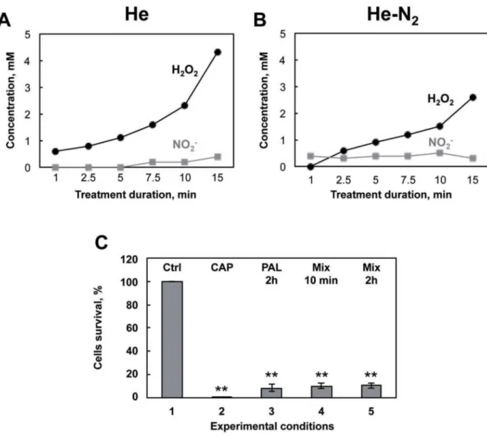
CAPPs are known to generate RONS in PBS [48,49]. It was therefore of interest to determine and quantify the species produced by the plasma in the liquid medium. To identify the species produced in PBS and their concentrations, PBS was treated as usual with plasma He and He-N2for times ranging from one to 15 min. At the end of the treatment, treated samples were
analyzed by cyclic voltammetry (Fig 4). Analysis indicated that two species are produced in the liquid medium, namely H2O2and NO2-. As expected,Fig 4A and 4B, indicate that He and
He-N2plasmas are able to form these two species in PBS in a time dependent manner. H2O2
pro-duced in the PBS is much higher with pure He plasma than with He-N2plasma (2.3 mM and
1.5 mM, respectively, both at 10 min) whereas concentrations in NO2-are roughly equivalent
for the two plasmas (0.5 mM at 10 min). In the medium treated with He-O2, species
consider-ably lower, being practically undetectable by cyclic voltammetry (detection limit of 200μM) were measured. Therefore, absorption and fluorescence spectrophotometry were used to mea-sure these species for He-O2plasma treated solutions. We measured 40μM H2O2and 5μM
NO2-at 10 min. H2O2and NO2-are species known to be chemically stable and potently toxic
to cells depending on their concentration level, which could be responsible for the PAL effects. If these species contributed to bacterial inactivation in PBS we should be able to mimic plasma treatment. As shown inFig 4C, H2O2and NO2-were added to non-plasma treated PBS at
con-centrations determined for 10 min of He plasma treatment and added to the bacteria for differ-ent conditions and compared to direct treatmdiffer-ent with He plasma (Fig 4C). The same results are obtained when the two species are added together. Finally, with 10 min contact with
bacteria, using this “mimicking medium”, the deleterious effect is already at a maximum, highlighting the toxicity of these two species and in line with what was observed inFig 3B. Col-lectively, these findings suggest that H2O2and NO2-are generated in significant amounts and
that they are the major RONS in He and He-N2plasma treated PBS.
Plasma treatment induced protein oxidation but no lipid peroxidation
Plasma treatment results in increased rates of RONS production in the PBS (Fig 4). It was therefore of interest to determine whether bacterial proteins were a target for oxidative modifi-cation. This was accomplished by evaluating changes in the relative levels of carbonyl groups
Fig 4. Nature, concentrations and toxicity of the species produced in PAL. A: Detection by cyclic voltammetry at a platinized
microelectrode of hydrogen peroxide and nitrites produced in PBS after 1 min to 15 min of He plasma treatment. B: He-N2treatment. C:
Model experiment mimicking PAL. 1: control bacteria incubated for 2 hours in PBS. 2: E. coli exposed to He plasma for 10 min and 2 hr storage and % of surviving cells evaluated by the CFU method. 3: Bacteria exposed to PAL (PBS treated for 10 min, He plasma) for 2 hours for 2 hours at 4˚C. 4: Bacteria exposed to a mix (2.3 mM Hydrogen peroxide and 0.5 mM nitrite) for 10 min or 5: for 2 hours for 2 hours at 4˚C and % of surviving cells was evaluated by the CFU method. The values are means±SEM of 3 separate experiments (**p<0.05) vs control).
present on the proteins. Carbonyl functional groups can be introduced into proteins by a vari-ety of oxidative processes including direct oxidation of amino acid with H2O2and reaction of
lipid peroxidation products from cellular membrane oxidation [50]. As shown inFig 5A, He, He-N2and He-O2, plasma treatment induced a distinct increase in levels of oxidized proteins.
Oxidative modifications were not global in nature but appeared specific to distinct low
Fig 5. Detection of oxidatively modified proteins following plasma exposure. A: Bacteria exposed to plasma treatment (He, He-O2
and He-N2) for 10 min with 2 hours post-treatment storage. To detect oxidatively, modified protein bacterial extracts were treated with
2,4-dinitorphenylhydrazine to derivatize protein carbonyls and then evaluated by SDS-gel electrophoresis using 2,4-dinitrophenyl antibodies. B: Detection of 4-hydroxy-2-nonenal protein modification by ELISA. The values are means±SEM of 3 separate experiments. C: Western blot analysis using nitrotyrosine specific antibodies. D: Bacterial extracts collected, lysed and detected by dot blot for lipid A content. Dot blot results were analyzed with a dot calibration curve and relative quantity of bacteria lipid A was estimated. The relative intensity of each spot was quantified (Image J).
molecular weight proteins and no high molecular weight aggregates. Surprisingly, the level of protein oxidation was the same for all three plasmas suggesting that a low level of H2O2or
other species produced in the plasma by He-O2such as•O2or1O2plasma were sufficient to
induce protein oxidation. 4-Hydroxy-2-nonenal (HNE), anα,β unsaturated aldehyde is a major product of lipid peroxidation and very toxic [51]. Utilizing antibodies specific to HNE-Michael adducts we did not detect an increase in the HNE content of protein after plasma treatment compared to control cells (Fig 5B) suggesting that membrane rupture and changes in cell permeability are not due to peroxidation and oxidation of the membrane lipid and may be due to a depolarization of the cell [29]. While protein carbonylation is an irreversible modi-fication due to H2O2interaction with cell components, nitrotyrosine in protein is the
detect-able marker for indirectly detecting peroxynitrite ONOO−. It is a highly reactive species, since once formed, it decomposes in aqueous solution into highly reactive hydroxyl•OH and nitroxyl NO2˚ [52]. Analysis of nitrated protein (Fig 5C) revealed no difference in
nitrotyro-sine amount on protein in plasma compared to control cells suggesting no formation of ONOO−or its rapid degradation in the PBS during plasma treatment. It has recently been shown thatE. coli cytochrome bd is able to catalyze the rapid degradation of ONOO−[53]. So we cannot rule out that the ONOO−degrading activity of cytochromebd in the bacteria resulted in the formation of nitrite preventing tyrosine nitration.
Lipid A is an essential component of Gram-negative bacteria membrane. It serves as an anchor for lipopolysaccharides, LPS, that constitute the outer monolayer of the outer mem-brane. The ability of LPS to elicit an immune response lies essentially with lipid A [54]. It is a very potent stimulant of the immune system, which activates the cells (monocytes, macro-phages) at concentrations of a few picograms per milliliter. At high concentration in the body in a gram-negative infection, it can trigger a systemic inflammatory response syndrome, which may cause sepsis [55]. In order to investigate if plasma treatment was able to degrade Lipid A, we used a dot blot method based on immuno-detection of Lipid A [38]. As shown inFig 5D, no change in the amount of Lipid A was detected after plasma treatment and the results are the same for the three plasmas used. Since SEM and cell viability (Fig 2) results indicated a strong alteration in theE. coli membrane integrity and structure, we did not expect this result but it was in line with the lack of membrane lipid peroxidation.
Nevertheless, we observed severely damaged membranes, after plasma exposure, seeFig 2. The membrane is a prime cellular target during plasma treatment. RONS emitted from the plasma are believed to be a key factor in bacterial inactivation [8,56], the results presented in this study confirm this hypothesis. Some authors have reported ROS induced loss of mem-brane integrity [18,23,29], but several stress factors occurred at the same time [57]. Free radi-cals and charged particles (electrons, ions) may act and cause surface lesions in membranes by direct bombardment [48,49]. So, in our conditions we hypothesize that, rather than oxidative stress, charges or electric field induce significant membrane damage. Localized lesions in the membrane allowed further penetration of toxic reactive plasma species into the cell and facili-tate the diffusion of RONS and other particles through the membrane, both causing severe damage to intracellular macromolecules [25]. Intracellular proteins are key regulators of bacte-rial function and are sensitive to oxidative stress [58].Fig 4Cshows that the three plasmas induced significant oxidative damage to intracellular proteins. This high level of protein oxidation participates in bacterial death by leading to dysregulation of cellular signalization [44]. This phenomenon might explain the delayed effect that was observed (Fig 1). It is also interesting to see that the He-O2plasma, which does not produce stable RONS in liquid
media, leads to the same level of oxidation as for the other plasmas. This could indicate that other oxidative species such as singlet oxygen are contained in the He-O2plasma. On the
significant concentrations of RONS produced in the liquid media. This is supported by studies indicating that bacteria suffer oxidative damage and protein oxidation during plasma treat-ment in liquid environtreat-ments [25].
Conclusion
In summary, our results provide support for the hypothesis that upon treatment with He and He-N2, free radicals generated in the PBS mediate bacterial inactivation thereby altering
pro-tein homeostasis and membrane integrity. However Lipid A is not degraded and remains toxic. Future studies are needed to identify plasma conditions to neutralize this cytotoxic toxin [38]. Their efficiency is dependent on treatment time and post-treatment storage duration because these parameters determine the concentration of reactive species essential for micro-bial inactivation. He-O2plasma is the most efficient plasma for bacterial inactivation and
seems to involve an electric field associated with the ionization front or, generated in the plasma environment. The inactivation of bacteria by electrochemical means has been well characterized [59]. It involves different mechanisms such as changes in the membrane poten-tial which surely may lead to local ion flux imbalances. Death occurs due to either the forma-tion of permanent pores and subsequent destabilizaforma-tion of the cell membrane, or loss of important cell components and destruction of chemical gradients via transport through tran-sient pores [60]. If reactive RONS are present, these pores may allow the oxidants to freely access to the interior of the cell thus improving at a platinized microelectrode the inactivation process [44]. Electric fields are also capable of destroying cells without destroying their mem-branes. Matsunaga et al. described a system in which cells were killed without rupturing, but rather with the electrochemical oxidation of intracellular coenzyme A [61]. Ongoing efforts to identify reactive species produced in the liquid, after treatment with He and He-O2, and the
impact of plasma generated electric field will enable the elucidation of the bacterial mechanism of inactivation.
Acknowledgments
The work was supported by the french ANR program through the grant "PLASMAVIV", ANR2010BLAN095001. The recipient of the grant was SC. Marlene Dezest is a recipient of a fellowship from French Ministere de l’Education Nationale de la Recherche et de Technologie (MENRT). Our thanks to Rosie Cox for improving the English, to conseil re´gional d’Aquitaine for the funding of Aquitraces facility and to Trigenotoul facility in Toulouse for microscopy analysis.
Author Contributions
Conceptualization: ALB SC SA FC. Formal analysis: ALB SA FC. Funding acquisition: SC ALB. Investigation: MD ALB LC MLB DQ.Methodology: ANS ALB MD SA DQ LC MLB FC. Project administration: SC ALB FC.
Resources: JPC SC. Supervision: ALB FC.
Validation: SA ALB FC MD ANS. Visualization: MD ALB.
Writing – original draft: MD ALB.
Writing – review & editing: SA FC ANS SC LC.
References
1. Ehlbeck J, Schnabel U, Polak M, Winter J, von Woedtke T, Brandenburg R, et al. Low temperature atmospheric pressure plasma sources for microbial decontamination. Journal of Physics D: Applied Physics. 2011; 44(1):013002.
2. Stoffels E, Kieft IE, Sladek REJ, Bedem LJMvd, Laan EPvd, Steinbuch M. Plasma needle for in vivo medical treatment: recent developments and perspectives. Plasma Sources Science and Technology. 2006; 15(4):S169–S80.
3. von Woedtke T, Reuter S, Masur K, Weltmann KD. Plasmas for medicine. Physics Reports. 2013; 530 (4):291–320.
4. Dobrynin D, Fridman G, Friedman G, Fridman A. Physical and biological mechanisms of direct plasma interaction with living tissue. New Journal of Physics. 2009; 11(11):115020.
5. Emmert S, Brehmer F, Ha¨nßle H, Helmke A, Mertens N, Ahmed R, et al. Atmospheric pressure plasma in dermatology: Ulcus treatment and much more. Clinical Plasma Medicine. 2013; 1(1):24–9.
6. Arndt S, Unger P, Wacker E, Shimizu T, Heinlin J, Li YF, et al. Cold atmospheric plasma (CAP) changes gene expression of key molecules of the wound healing machinery and improves wound healing in vitro and in vivo. PLoS One. 2013; 8(11):e79325.https://doi.org/10.1371/journal.pone.0079325PMID:
24265766
7. O’Connor N, Cahill O, Daniels S, Galvin S, Humphreys H. Cold atmospheric pressure plasma and decontamination. Can it contribute to preventing hospital-acquired infections? J Hosp Infect. 2014; 88 (2):59–65.https://doi.org/10.1016/j.jhin.2014.06.015PMID:25146226
8. Laroussi M. Low Temperature Plasma-Based Sterilization: Overview and State-of-the-Art. Plasma Pro-cesses and Polymers. 2005; 2(5):391–400.
9. Zimmermann JL, Dumler K, Shimizu T, Morfill GE, Wolf A, Boxhammer V, et al. Effects of cold atmo-spheric plasmas on adenoviruses in solution. Journal of Physics D: Applied Physics. 2011; 44 (50):505201.
10. Cooper M, Fridman G, Fridman A, Joshi SG. Biological responses of Bacillus stratosphericus to floating electrode-dielectric barrier discharge plasma treatment. J Appl Microbiol. 2010; 109(6):2039–48.
https://doi.org/10.1111/j.1365-2672.2010.04834.xPMID:20825520
11. Daeschlein G, von Woedtke T, Kindel E, Brandenburg R, Weltmann K-D, Ju¨nger M. Antibacterial Activ-ity of an Atmospheric Pressure Plasma Jet Against Relevant Wound Pathogens in vitro on a Simulated Wound Environment. Plasma Processes and Polymers. 2010; 7(3–4):224–30.
12. Klampfl TG, Isbary G, Shimizu T, Li YF, Zimmermann JL, Stolz W, et al. Cold atmospheric air plasma sterilization against spores and other microorganisms of clinical interest. Appl Environ Microbiol. 2012; 78(15):5077–82.https://doi.org/10.1128/AEM.00583-12PMID:22582068
13. Fricke K, Koban I, Tresp H, Jablonowski L, Schroder K, Kramer A, et al. Atmospheric pressure plasma: a high-performance tool for the efficient removal of biofilms. PLoS One. 2012; 7(8):e42539.https://doi. org/10.1371/journal.pone.0042539PMID:22880025
14. Pournaseh Y, Irani S, Atyabi S. Cold atmospheric plasma jet against Leishmania major in vitro study. Basis research journal of medicine and clinical sciences. 2014; 4.
15. Korachi M, Gurol C, Aslan N. Atmospheric plasma discharge sterilization effects on whole cell fatty acid profiles of Escherichia coli and Staphylococcus aureus. Journal of Electrostatics. 2010; 68 (6):508–12.
16. Mai-Prochnow A, Murphy AB, McLean KM, Kong MG, Ostrikov KK. Atmospheric pressure plasmas: infection control and bacterial responses. Int J Antimicrob Agents. 2014; 43(6):508–17.https://doi.org/ 10.1016/j.ijantimicag.2014.01.025PMID:24637224
17. Babaeva NY, Ning N, Graves DB, Kushner MJ. Ion activation energy delivered to wounds by atmo-spheric pressure dielectric-barrier discharges: sputtering of lipid-like surfaces. Journal of Physics D: Applied Physics. 2012; 45(11):115203.
18. Laroussi M, Leipold F. Evaluation of the roles of reactive species, heat, and UV radiation in the inactiva-tion of bacterial cells by air plasmas at atmospheric pressure. Internainactiva-tional Journal of Mass Spectrome-try. 2004; 233(1–3):81–6.
19. Boudam MK, Moisan M, Saoudi B, Popovici C, Gherardi N, Massines F. Bacterial spore inactivation by atmospheric-pressure plasmas in the presence or absence of UV photons as obtained with the same gas mixture. Journal of Physics D: Applied Physics. 2006; 39(16):3494–507.
20. Ferna´ndez A, Thompson A. The inactivation of Salmonella by cold atmospheric plasma treatment. Food Research International. 2012; 45(2):678–84.
21. Ziuzina D, Patil S, Cullen PJ, Keener KM, Bourke P. Atmospheric cold plasma inactivation of Escheri-chia coli in liquid media inside a sealed package. J Appl Microbiol. 2013; 114(3):778–87.https://doi.org/ 10.1111/jam.12087PMID:23190122
22. Boxhammer V, Li YF, Koritzer J, Shimizu T, Maisch T, Thomas HM, et al. Investigation of the mutagenic potential of cold atmospheric plasma at bactericidal dosages. Mutat Res. 2013; 753(1):23–8.https://doi. org/10.1016/j.mrgentox.2012.12.015PMID:23416235
23. Deng S, Cheng C, Ni G, Meng Y, Chen H. Bacillus subtilis devitalization mechanism of atmosphere pressure plasma jet. Current Applied Physics. 2010; 10(4):1164–8.
24. Uhm HS, Hong YC. Various microplasma jets and their sterilization of microbes. Thin Solid Films. 2011; 519(20):6974–80.
25. Lackmann JW, Bandow JE. Inactivation of microbes and macromolecules by atmospheric-pressure plasma jets. Appl Microbiol Biotechnol. 2014; 98(14):6205–13. https://doi.org/10.1007/s00253-014-5781-9PMID:24841116
26. Liu F, Sun P, Bai N, Tian Y, Zhou H, Wei S, et al. Inactivation of Bacteria in an Aqueous Environment by a Direct-Current, Cold-Atmospheric-Pressure Air Plasma Microjet. Plasma Processes and Polymers. 2010; 7(3–4):231–6.
27. Ikawa S, Kitano K, Hamaguchi S. Effects of pH on Bacterial Inactivation in Aqueous Solutions due to Low-Temperature Atmospheric Pressure Plasma Application. Plasma Processes and Polymers. 2010; 7(1):33–42.
28. Julak J, Scholtz V, Kotucova S, Janouskova O. The persistent microbicidal effect in water exposed to the corona discharge. Phys Med. 2012; 28(3):230–9.https://doi.org/10.1016/j.ejmp.2011.08.001PMID:
21925912
29. Joshi SG, Cooper M, Yost A, Paff M, Ercan UK, Fridman G, et al. Nonthermal dielectric-barrier dis-charge plasma-induced inactivation involves oxidative DNA damage and membrane lipid peroxidation in Escherichia coli. Antimicrob Agents Chemother. 2011; 55(3):1053–62.https://doi.org/10.1128/AAC. 01002-10PMID:21199923
30. Gazeli K, Svarnas P, Held B, Marlin L, Clement C. Possibility of controlling the chemical pattern of He and Ar "guided streamers" by means of N2 and O2 additives. Journal of Applied Physics. 2015; 117(093302).
31. Gazeli K, Svarnas P, Vafeas P, Papadopoulos PK, Gkelios A, Clement C. Investigation on streamers propagating into a helium jet air at atmospheric pressure: Electrical and optical emission analysis. Jour-nal of Applied Physics. 2013; 114(103304).
32. Gazeli K, Belmonte T, Clement C. A study of helium atmospheric-pressure guides streamers for poten-tial biological applications. Plasma sources science and technology. 2013; 22(025020).
33. Maheux S, Duday D, Belmonte T, Penny C, Cauchie HM, Clement F, et al. Formation of ammonium in saline solution treated by nanosecond pulsed cold atmospheric microplasma: a route to fast inactivation of E. coli bacteria. RSC Advances. 2015; 5:42135–40.
34. Virard F, Cousty S, Cambus JP, Valentin A, Kemoun P, Clement F. Cold Atmospheric Plasma Induces a Predominantly Necrotic Cell Death via the Microenvironment. PLoS One. 2015; 10(8):e0133120.
https://doi.org/10.1371/journal.pone.0133120PMID:26275141
35. Josset S, Keller N, Lett MC, Ledoux MJ, Keller V. Numeration methods for targeting photoactive materi-als in the UV-A photocatalytic removal of microorganisms. Chem Soc Rev. 2008; 37(4):744–55.https:// doi.org/10.1039/b711748pPMID:18362981
36. Amatore C, Arbault S, Bouton C, Coffi K, Drapier JC, Ghandour H, et al. Monitoring in real time with a microelectrode the release of reactive oxygen and nitrogen species by a single macrophage stimulated by its membrane mechanical depolarization. Chembiochem. 2006; 7(4):653–61.https://doi.org/10. 1002/cbic.200500359PMID:16502474
37. Arbault S, Pantano P, Jankowski JA, Vuillaume M, Amatore C. Monitoring an oxidative stress mecha-nism at a single human fibroblast. Anal Chem. 1995; 67(19):3382–90. PMID:8686890
38. Zerrouki H, Rizzati V, Bernis C, Negre-Salvayre A, Sarrette JP, Cousty S. Escherichia coli morphologi-cal changes and lipid A removal induced by reduced pressure nitrogen afterglow exposure. PLoS One. 2015; 10(3):e0116083.https://doi.org/10.1371/journal.pone.0116083PMID:25837580
39. Byrd JJ, Xu HS, Colwell RR. Viable but non culturable bacteria in drinking water. ApplEnvironmicrobiol. 1991; 57:875–8.
40. Colwell RR, Brayton P, Herrington D, Tall B, Huq A, Levine MM. Viable but non-culturable Vibrio cholera O1 revert to cultivable state in the human intestine. World JMicrobiolBiotechnol. 1996; 12:28–31.
41. Oliver JD. Recent findings on the viable but nonculturable state in pathogenic bacteria. FEMS Microbiol Rev. 2010; 34(4):415–25.https://doi.org/10.1111/j.1574-6976.2009.00200.xPMID:20059548
42. Frohling A, Schluter O. Flow cytometric evaluation of physico-chemical impact on Gram-positive and Gram-negative bacteria. Front Microbiol. 2015; 6:939.https://doi.org/10.3389/fmicb.2015.00939PMID:
26441874
43. Dolezalova E, Lukes P. Membrane damage and active but nonculturable state in liquid cultures of Escherichia coli treated with an atmospheric pressure plasma jet. Bioelectrochemistry. 2015; 103:7–14.
https://doi.org/10.1016/j.bioelechem.2014.08.018PMID:25212700
44. Han L, Patil S, Boehm D, Milosavljevic V, Cullen PJ, Bourke P. Mechanisms of Inactivation by High-Voltage Atmospheric Cold Plasma Differ for Escherichia coli and Staphylococcus aureus. Appl Environ Microbiol. 2015; 82(2):450–8.https://doi.org/10.1128/AEM.02660-15PMID:26519396
45. Robert E, Darny T, Dozias S, Iseni S, Pouvesle JM. New insights on the propagation of pulsed atmo-spheric plasma streams: From single jet to multi jet arrays. Physics of Plasmas. 2015; 22(122007).
46. Babaeva NY, Kushner MJ. Intracellular electric fields produced by dielectric barrier discharge treatment of skin. Journal of Physics D: Applied Physics. 2010; 43(185206):1–12.
47. Bourdon A, Darny T, Pechereau F, Pouvesle JM, Viegas P, Iseni S, et al. Numerical and experimental study of the dynamics of aμs helium plasma gun discharge with various amounts of N2 admixture. Plasma Sources Sci Technol. 2016; 25(035002):1–17.
48. Hensel K, Kucerova K, Tarabova B, Janda M, Machala Z, Sano K, et al. Effects of air transient spark dis-charge and helium plasma jet on water, bacteria, cells, and biomolecules. Biointerphases. 2015; 10 (2):029515.https://doi.org/10.1116/1.4919559PMID:25947389
49. Wende K, Williams P, Dalluge J, Gaens WV, Aboubakr H, Bischof J, et al. Identification of the biologi-cally active liquid chemistry induced by a nonthermal atmospheric pressure plasma jet. Biointerphases. 2015; 10(2):029518.https://doi.org/10.1116/1.4919710PMID:25947392
50. Weber D, Davies MJ, Grune T. Determination of protein carbonyls in plasma, cell extracts, tissue homogenates, isolated proteins: Focus on sample preparation and derivatization conditions. Redox Biol. 2015; 5:367–80.https://doi.org/10.1016/j.redox.2015.06.005PMID:26141921
51. Negre-Salvayre A, Auge N, Ayala V, Basaga H, Boada J, Brenke R, et al. Pathological aspects of lipid peroxidation. Free Radic Res. 2010; 44(10):1125–71.https://doi.org/10.3109/10715762.2010.498478
PMID:20836660
52. Slosky LM, Vanderah TW. Therapeutic potential of peroxynitrite decomposition catalysts: a patent review. Expert Opin Ther Pat. 2015; 25(4):443–66.https://doi.org/10.1517/13543776.2014.1000862
PMID:25576197
53. Borisov VB, Forte E, Siletsky SA, Sarti P, Giuffre A. Cytochrome bd from Escherichia coli catalyzes per-oxynitrite decomposition. Biochim Biophys Acta. 2015; 1847(2):182–8.https://doi.org/10.1016/j.bbabio. 2014.10.006PMID:25449967
54. Raetz CR, Whitfield C. Lipopolysaccharide endotoxins. Annu Rev Biochem. 2002; 71:635–700.https:// doi.org/10.1146/annurev.biochem.71.110601.135414PMID:12045108
55. Yamamoto M, Akira S. Lipid A receptor TLR4-mediated signaling pathways. Adv Exp Med Biol. 2010; 667:59–68.https://doi.org/10.1007/978-1-4419-1603-7_6PMID:20665200
56. Gaunt L, Beggs C, Georghiou G. bactericidal action of the reactive species produced by gas-discharge nonthermal plasma at atmospheric pressure: a review. IEEE transactions on plasma science. 2006; 34 (4):1257–69.
57. Sharma A, Collins G, Pruden A. Differential gene expression in Escherichia coli following exposure to nonthermal atmospheric pressure plasma. J Appl Microbiol. 2009; 107(5):1440–9.https://doi.org/10. 1111/j.1365-2672.2009.04323.xPMID:19426273
58. Winter T, Winter J, Polak M, Kusch K, Mader U, Sietmann R, et al. Characterization of the global impact of low temperature gas plasma on vegetative microorganisms. Proteomics. 2011; 11(17):3518–30.
https://doi.org/10.1002/pmic.201000637PMID:21751354
59. Guionet A, David F, Zaepffel C, Coustets M, Helmi K, Cheype C, et al. E. coli electroeradication on a closed loop circuit by using milli-, micro- and nanosecond pulsed electric fields: comparison between energy costs. Bioelectrochemistry. 2015; 103:65–73.https://doi.org/10.1016/j.bioelechem.2014.08.021
60. Drees KP, Abbaszadegan M, Maier RM. Comparative electrochemical inactivation of bacteria and bac-teriophage. Water Res. 2003; 37(10):2291–300.https://doi.org/10.1016/S0043-1354(03)00009-5
PMID:12727237
61. Matsunaga T, Nakasono S, Takamuku T, Burgess JG, Nakamura N, Sode K. Disinfection of drinking water by using a novel electrochemical reactor employing carbon-cloth electrodes. Appl Environ Micro-biol. 1992; 58(2):686–9. PMID:1610189