Peroxisome proliferator-activated receptor h/y activation inhibits
hypertrophy in neonatal rat cardiomyocytes
Anna Planavila
a, Ricardo Rodrı´guez-Calvo
a, Mireia Jove´
a, Liliane Michalik
b, Walter Wahli
b,
Juan C. Laguna
a, Manuel Va´zquez-Carrera
a,*
aPharmacology Unit, Department of Pharmacology and Therapeutic Chemistry, Faculty of Pharmacy, University of Barcelona, Diagonal 643,
E-08028 Barcelona, Spain
b
Center for Integrative Genomics, NCCR Frontiers in Genetics, University of Lausanne, Switzerland Received 29 September 2004; received in revised form 5 November 2004; accepted 10 November 2004
Available online 8 December 2004 Time for primary review 26 days
Abstract
Objective: Peroxisome proliferator-activated receptor B/D (PPARB/D) is the predominant PPAR subtype in cardiac cells and plays a prominent role in the regulation of cardiac lipid metabolism. However, the role of PPARB/D activators in cardiac hypertrophy is not yet known.
Methods and Results: In cultured neonatal rat cardiomyocytes, the selective PPARB/D activator L-165041 (10 Mmol/L) inhibited phenylephrine (PE)-induced protein synthesis ([3H]leucine uptake), induction of the fetal-type gene atrial natriuretic factor (ANF) and cardiac myocyte size. Induction of cardiac hypertrophy by PE stimulation also led to a reduction in the transcript levels of both muscle-type carnitine palmitoyltransferase (50%, Pb0.05) and pyruvatedehydrogenase kinase 4 (30%, Pb0.05), and these changes were reversed in the presence of the PPARh/y agonist L-165041. Stimulation of neonatal rat cardiomyocytes with PE and embryonic rat heart-derived H9c2 cells with lipopolysaccharide (LPS) enhanced the expression of the nuclear factor (NF)-nB-target gene monocyte chemoattractant protein 1 (MCP-1). The induction of MCP-1 was reduced in the presence of L-165041, suggesting that this compound prevented NF-nB activation. Electrophoretic mobility shift assay (EMSA) revealed that L-165041 significantly decreased LPS-stimulated NF-nB binding activity in H9c2 myotubes. Finally, coimmunoprecipitation studies showed that L-165041 strongly enhanced the physical interaction between PPARh/y and the p65 subunit of NF-nB, suggesting that increased association between these two proteins is the mechanism responsible for antagonizing NF-nB activation by PPARh/y activators.
Conclusion: These results suggest that PPARB/D activation inhibits PE-induced cardiac hypertrophy and LPS-induced NF-KB activation. D 2004 European Society of Cardiology. Published by Elsevier B.V. All rights reserved.
Keywords: Cardiac hypertrophy; NF-KB; L-165041; p65
This article is referred to in the Editorial by N.N. Petrashevskaya and A. Schwarz (pages 770–771) in this issue.
Cardiac hypertrophy is a response of the heart to a wide range of extrinsic stimuli, such as arterial hypertension, valvular heart disease, myocardial infarction and
hyper-trophic cardiomyopathy. Although this process is initially compensatory for an increase workload, its prolongation frequently results in congestive heart failure, arrhythmia and sudden death [1,2]. Among the signal transduction pathways involved in the hypertrophic growth of the myocardium, the nuclear factor (NF)-nB signaling pathway plays a pivotal role, since it has been shown that NF-nB inhibition blocks or attenuates the hypertrophic response of cultured cardiac myocytes [3–6]. In addition, cardiac hypertrophy is associated with an increase in glucose utilization and a decrease in fatty acid oxidation, which is
0008-6363/$ - see front matterD 2004 European Society of Cardiology. Published by Elsevier B.V. All rights reserved. doi:10.1016/j.cardiores.2004.11.011
* Corresponding author. Tel.: +34 93 4024531; fax: +34 93 4035982. E-mail address: mvazquezcarrera@ub.edu (M. Va´zquez-Carrera).
characteristic of fetal heart [7,8]. Defects in mitochondrial fatty acid oxidation enzymes cause childhood hypertrophic cardiomyopathy [9], and perturbation of fatty acid oxida-tion in animal models causes cardiac hypertrophy [10,11], demonstrating that substrate utilization is important in the pathogenesis of hypertrophy. Peroxisome proliferator-acti-vated receptors (PPARs) are ligand-actiproliferator-acti-vated transcription factors that regulate the expression of genes involved in fatty acid uptake and oxidation, lipid metabolism and inflammation [12]. The PPAR subfamily consists of three subtypes, PPARa (NR1C1 according to the unified nomenclature system for the nuclear receptor superfamily), PPARh (also known as PPARy) (NR1C2) and PPARg (NR1C3) [13]. PPARa is expressed primarily in tissues that have a high level of fatty acid catabolism such as liver, brown fat, kidney, heart and skeletal muscle [14]. PPARh/y is ubiquitously expressed, and PPARg has a restricted pattern of expression, mainly in white and brown adipose tissues, whereas other tissues such as skeletal muscle and heart contain limited amounts. In order to be transcriptionally active, PPARs need to heterodimerize with the 9-cis retinoic acid receptor (RXR) (NR2B). PPAR-RXR heterodimers binds to DNA specific sequences called peroxisome proliferator-response elements (PPREs), consisting of an imperfect direct repeat of the consensus binding site for nuclear hormone receptors (AGGTCA) separated by one nucleotide (DR-1). These sequences have been characterized within the promoter regions of PPAR target genes. However, the regulation of gene transcription by PPARs extends beyond their ability to transactivate specific target genes. PPARs are also able of regulating gene expression independently of binding to DNA through a mechanism termed receptor-dependent transrepression [15]. One of these mechanisms involves a physical interaction of PPARa with NF-nB, leading to suppression activity of the latter [16].
It has been demonstrated that, of the three PPAR subtypes, activation of both PPARa [17,18] and PPARg
[19,20] results in inhibition of cardiac hypertrophy.
However, the role of PPARh/y in the development of this process is unknown. The recent availability of specific synthetic ligands for PPARh/y, such as L-165041, now makes possible to study the role of this nuclear receptor in cardiac cells. Thus, recently, Gilde et al.[21], using neonatal rat cardiomyocytes as well as the embryonic rat heart-derived H9c2 cells, clearly demonstrated that PPARh/y is the predominant PPAR subtype in cardiac cells and plays a prominent role in the regulation of cardiac lipid metabolism, suggesting that PPARh/y, similarly to PPARa and g, may play an important role in cardiac disease.
In this study, we examined the role of PPARh/y activation in phenylephrine (PE)-induced hypertrophy in neonatal rat cardiac myocytes and in lipopolysaccharide (LPS)-estimu-lated H9c2 myotubes. We found that activation of PPARh/y inhibits PE-induced hypertrophy and LPS-induced NF-nB activation.
1. Methods 1.1. Materials
L-165041 was synthesized according to Berger et al. [22]. [g-32P]dATP (3000 Ci/mmol) and [3H]leucine (50 Ci/ mmol) were purchased from Amersham Pharmacia Biotech KK. Anti-atrial natriuretic factor (ANF) polyclonal anti-serum was from Peninsula Laboratories and Alexa flouro 488 goat anti-rabbit and 568 goat anti-mouse antibodies were from Molecular Probes. All other chemicals were purchased from Sigma.
1.2. Cell culture
Neonatal rat ventricular myocytes from 1- to 2-day-old Sprague–Dawley rats were prepared and cultured overnight in Dulbecco’s modified Eagle’s medium (DMEM) contain-ing 10% fetal bovine serum as described previously [23]. The media was changed to serum-free DMEM supple-mented with transferrin (10 Ag/mL), insulin (1 Ag/mL) and bromodeoxyuridine (0.1 mmol/L) 24 h before treatments. In this study, PE was used to stimulate neonatal rat cardio-myocytes. Animal handling and disposal were performed in accordance with NIH guidelines.
The embryonic rat-heart derived H9c2 cells (ATCC) were maintained in growth medium composed of DMEM supple-mented with 10% fetal bovine serum. H9c2 cells were plated at a density of 5000 cells/cm2and allowed to proliferate in growth medium. Medium was changed every 3 days. To induce differentiation of H9c2 myoblasts into myotubes, growth medium was replaced with differentiation medium (DMEM containing 2% horse serum) when cells had reached near confluence. For mRNA analysis H9c2 cells were treated with 10 Amol/L L-165041 and LPS (10 ng/mL) for 24 h. 1.3. Incorporation of [3H]leucine
To examine the effect of PE on protein synthesis, the incorporation of [3H]leucine was measured essentially by the method of Thaik et al. [24]. Cultured neonatal rat ventricular myocytes were treated with PE in the presence or in the absence of L-165041 and coincubated with [3 H]leu-cine (1 ACi/mL) for 24 h. The cells were washed with PBS and then treated with 10% trichloroacetic acid at 4 8C for 30 min to precipitate the proteins. The precipitates were then dissolved in NaOH (0.25 N). Aliquots were counted with scintillation counter.
1.4. Immunocytochemistry
Neonatal rat ventricular myocytes were fixed in ice-cold 100% methanol for 10 min. Anti-a-actinin antibody and anti-ANF polyclonal antiserum were added at dilutions 1:400 and 1:150, respectively, in PBS containing 1% BSA and incubated for 1 h at room temperature. Secondary antibodies,
Alexa flouro 488 goat anti-rabbit and Alexa flouro 568 goat anti-mouse, were used at a dilution of 1:300 in PBS containing 5% rat serum and incubated for 30 min at room temperature. Immunofluorescence was visualized using a confocal laser fluorescence microscope Olympus Fluoview FV500. Photographic images were taken from five random fields.
1.5. RNA preparation and analysis
Relative levels of specific mRNAs were assessed by the reverse transcription–polymerase chain reaction (RT– PCR) as previously described [25]. The sequences of the sense and antisense primers used for amplification were: ANF, TCCTCTTCCTGGCCTTTTGGC-3V and
5V-Fig. 1. The PPARh/y activator L-165041 inhibits PE-induced cardiac hypertrophy in neonatal rat cardiomyocytes. Cardiac myocytes were stimulated with 100 Amol/L PE in the presence or absence of 10 Amol/L L-165041 that was added 30 min before experiments. (A) [3H]leucine incorporation was determined by
coincubating cardiac myocytes with 1.0 ACi/mL [3H]leucine for 24 h. Data are expressed as meanFS.D. (n=6) of the treated-to-control ratio. (B) Analysis of
the mRNA levels of ANF in PE-stimulated cardiomyocytes in the presence or absence of 10 Amol/L L-165041. A representative autoradiogram is shown. (C) Effects of PE with and without L-165041 on cardiac myocyte ANF protein expression and cardiac myocyte size. Double immunofluorescent microscopy was performed using specific antibodies to a-actinin (upper panel, red color) and ANF (lower panel, green color). Experiments were performed three times with similar results. ***Pb0.001 vs. control.#
AGACGGGTTGCTTCCCCAGTC-3V; gp91, 5V-CAC-CTGCAGCCTGCCTGAATT-3V and 5V-ATGGTGTGAA-TGGCGGTGTGA-3V; inducible nitric oxide synthase (iNOS), 5V-GCATGGACCAGTATAAGGCAAGCA-3V and 5V-GCTTCTGGTCGATGTCATGAGCAA-3V; malonyl-CoA decarboxylase (MCD), 5V-TACGGTGAGAAGCACC-GAGGC-3V and 5V-GGGGCCTGTCTCCTCCAGGTA-3V; monocyte chemoattractant protein 1 (MCP-1), 5V-GGGCCG T T 5V-GGGCCG T T C A C A 5V-GGGCCG T T 5V-GGGCCG C - 3 V a n d 5 V- 5V-GGGCCG 5V-GGGCCG 5V-GGGCCG A C A C C T-GCTGCTGGTGAT-3V; muscle-type carnitine palmitoyltrans-ferase (M-CPT-I), 5V-TTCACTGTGACCCCAGACGGG-3V and5V-AATGGACCAGCCCCATGGAGA;pyruvatedehydro-genase kinase 4 (PDK-4), 5V-GAACACCCCTTCCGTC-CAGCT-3V and 5V-TGTGCCATCGTAGGGACCACA-3V; PPARg coactivator-1 (PGC-1), 5V-AGAAAGGGCCCGAG-CAATCTG-3V and 5V-AGATGTGCCCCTGCCAGTCAC-3V; p22, CCCCGGGGAAAGAGGAAAAAG-3V and
5V-GGATGGCTGCCAGCAGGTAGA-3V; and APRT (adenosyl phosphoribosyl transferase), 5V-GCCTCTTGGCCAGT-CACCTGA-3V and 5V-CCAGGCTCACACACTCCACCA-3V. Amplification of each gene yielded a single band of the expected size (ANF: 234 bp, gp91: 200 bp, iNOS: 198 bp, MCD: 231 bp, MCP-1: 157 bp, M-CPT-I: 222 bp, PDK-4: 168 bp, PGC-1: 234 bp, p22: 215 bp and APRT: 329 bp). The results for the expression of specific mRNAs are always presented relative to the expression of the control gene (aprt).
1.6. Immunoblotting
Cell lysates and nuclear extracts from H9c2 cells were obtained as previously described[25]. Proteins (50 Ag) were separated by SDS–PAGE on 10% separation gels and transferred to Immobilon polyvinylidene diflouride
mem-Fig. 2. L-165041 prevents downregulation of the expression of several genes involved in fatty acid lipid metabolism in PE-stimulated neonatal rat cardiomyocytes. Analysis of the mRNA levels of M-CPT-I (A), PDK-4 (B), MCD (C) and PGC-1 (C) in PE-stimulated cardiac myocytes in the presence or absence of 10 Amol/L L-165041. A representative autoradiogram and the quantification normalized to the APRT mRNA levels are shown. Data are expressed as meanFS.D. of five different experiments. *Pb0.05 and **Pb0.01 vs. control.###
branes (Millipore, Bedford, MA). Western blot analysis was performed using antibodies against InBa, InBh, p65 and PPARh/y (Santa Cruz Biotechnology) and h-tubulin (Sigma). Detection was achieved using the EZ-ECL chemiluminescence detection kit (Biological Industries, Beit Haemek, Israel). Size of detected proteins was estimated using protein molecular-mass standards (Life Technologies). 1.7. Electrophoretic mobility shift assay (EMSA)
H9c2 cells were pretreated with 10 Amol/L L-165041 for 24 h before stimulation with LPS (10 ng/ml) for 1 h. Isolation of nuclear extracts and EMSA were performed as previously described[25].
1.8. Coimmunoprecipitation
Cell nuclear extracts were brought to a final volume of 0.5 mL with buffer containing 10 mM PBS, 50 mM KCl, 0.05 mM EDTA, 2.5 mM MgCl2, 8.5% glycerol, 1 mM
dithiothreitol, 0.1% Triton X-100, BSA 2% and 1 mg/ml nonfat milk for 6 h at 4 8C and incubated with 4 Ag of anti-p65. Immunocomplex were captured by incubating the samples with protein A-agarose suspension overnight at 4 8C on a rocker platform. Agarose beads were collected by centrifugation and washed three times with PBS containing protease inhibitors. After microcentrifuga-tion, the pellet was washed with 60 Al of SDS–PAGE sample buffer and boiled for 5 min at 100 8C. An aliquot of the supernatant was subjected to electrophoresis on 10% SDS–PAGE and immunoblotted with an antibody against PPARh/y.
1.9. Statistical analyses
Results were obtained from at least four independent experiments and presented as meanFS.D. Comparisons between groups were performed with one-way ANOVA using the computer program GraphPad Instat (GraphPad Softwware, San Diego, CA). When significant variations were found, the Tukey–Kramer multiple comparisons test was performed. Differences were considered significant at Pb0.05.
2. Results
2.1. PPARb/d activation by L-165041 inhibits PE-induced cardiac hypertrophy in neonatal rat cardiomyocytes
Cardiac hypertrophy leads to significant increases in protein content (e.g., [3H]leucine uptake), induction of fetal-type genes (e.g., ANF) and cardiac myocyte size [26]. Therefore, we first examined the effects on these parameters of the specific PPARh/y agonist L-165041, which was previously shown to be selective for this PPAR-subtype at
10 Amol/L [27]. The cells were pretreated with either vehicle or L-165041 for 30 min and subsequently stimulated with 100 Amol/L PE for 24 h. As shown in Fig. 1A, [3H]leucine incorporation was significantly increased by PE (1.6-fold, Pb0.001) and this was inhibited by L-165041 ( 20%, Pb0.05). PE-induced cardiomyocyte hypertrophy also led to approximately twofold induction in the mRNA levels of the fetal cardiac gene ANF (Fig. 1B). In contrast, in the presence of L-165041 PE-induced ANF expression was abolished. Immunostaining of cardiac myocytes for the sarcomere-associated protein a-actinin and ANF clearly shown an increase in cardiac myocyte size and ANF protein expression following PE stimulation (Fig. 1C). These changes were blocked by the presence of the PPARh/y agonist L-165041.
2.2. Treatment with the PPARb/d agonist L-165041 prevents the reduction in the expression of genes involved in lipid metabolism caused by PE-induced cardiomyocyte
hypertrophy in neonatal rat cardiomyocytes
An important molecular adaptation in cardiac hyper-trophy is the increase in glucose utilization and decrease in fatty acid oxidation associated to a downregulation of the expression of the mRNA levels of genes involved in fatty acid metabolism [8]. Interestingly, PPARh/y activates the expression of several PPAR target genes involved in fatty acid utilization in cardiac myocytes [21], including M-CPT-I, which determines the flux of mitochondrial h-oxidation [28], and PDK-4, which suppresses glucose
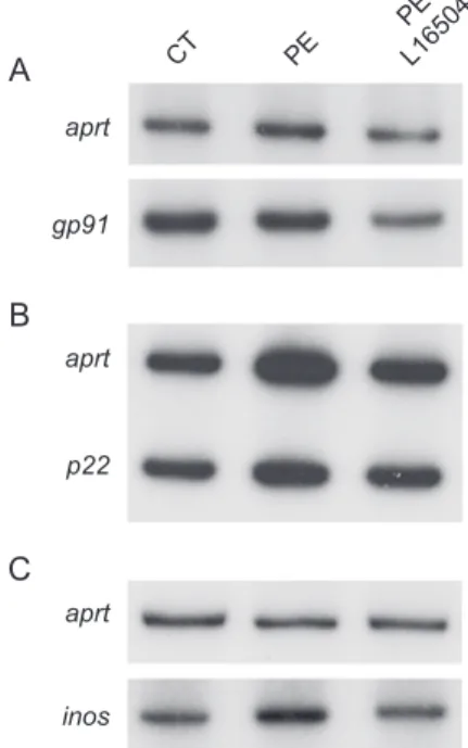
Fig. 3. Effects of L-165041 of the expression of NADPH oxidase subunits and iNOS. Analysis of the mRNA levels of gp91 (A), p22 (B) and iNOS (C) in PE-stimulated cardiac myocytes in the presence or absence of 10 Amol/L L-165041. A representative autoradiogram is shown. Experiments were performed three times with similar results.
oxidation by its inhibitory effect on the pyruvate dehydro-genase complex leading to an increase in fatty acid utilization [29]. Taking into account these data, we next assessed the effects of PE-induced cardiomyocyte hyper-trophy on the mRNA levels of these genes in the presence or in the absence of L-165041. Induction of cardiomyocyte hypertrophy by PE led to a reduction in the transcript levels of both M-CPT-I (50%, Pb0.05) and PDK-4 (30%, Pb0.05). In contrast, in the presence of the PPARh/y agonist, PE did not reduce the levels of these genes and even a robust induction (about fourfold, Pb0.001) was observed compared to control values (Fig. 2A and B). PE treatment did not affect the mRNA levels of MCD and PGC-1, two genes involved in lipid metabolism, whereas coincubation with L-165041 caused a significant increase in MCD expression (Fig. 2C and D).
2.3. Treatment with L-165041 inhibits the upregulation of MCP-1 caused by PE-induced cardiomyocyte hypertrophy in neonatal rat cardiomyocytes
PE-induced cardiomyocyte hypertrophy in neonatal rat cardiomyocytes is mediated through NF-nB activation via the generation of reactive oxygen species (ROS) [4]. NADPH oxidase is one of the systems generating ROS whose expression is increased in cardiac hypertrophy [30]. We therefore evaluated whether PPARh/y activation by L-165041 affected the expression of the NADPH oxidase subunits gp91 and p22 (Fig. 3A and B). No changes were observed in the mRNA levels of these genes in neonatal rat cardiomyocytes, making unlikely that reduced NADPH oxidase expression may account for the antihypertrophic effect of L-165041. Similarly, L-165041 treatment did not
Fig. 4. L-165041 inhibits the upregulation of MCP-1 caused by PE and LPS stimulation in myocytes. Analysis of the mRNA levels of MCP-1 in PE-stimulated in neonatal rat cardiomyocytes (A) in the presence or absence of 10 Amol/L L-165041. Analysis of the mRNA levels of MCP-1 (B), M-CPT-I (C) and PDK-4 (D) in LPS-stimulated H9c2 cells in the presence or absence of 10 Amol/L L-165041. A representative autoradiogram and the quantification normalized to the APRT mRNA levels are shown. Data are expressed as meanFS.D. of six different experiments. *Pb0.05 and ***Pb0.001 vs. control.###
Pb0.001 vs. either PE- or LPS-stimulated cells.
affect the expression of iNOS, which has been previously involved in maladaptive consequences of cardiac hyper-trophy [31] (Fig. 3C). Next, we determined the effects of L-165041 on the expression of MCP-1, a gene under the transcriptional control of NF-nB [32], in neonatal rat cardiomyocytes. Stimulation of cardiac myocytes with PE enhanced twofold the expression of this gene and this was abolished by L-165041 (Fig. 4A). This data suggests that prevention of NF-nB activation may be involved in the antihypertrophic effect attained by PPARh/y activation. Because of limited amount of mRNA and proteins obtained from neonatal rat cardiomyocytes, we continue our studies in the embryonic rat-heart derived H9c2 cells to confirm the involvement of NF-nB in the changes observed after L-165041 treatment. Gilde et al. [21] recently reported that H9c2 cells abundantly express the PPARh/y subtype, whereas PPARa and g were undetect-able. This fact converts H9c2 cells in a proper tool to investigate the role of PPARh/y activation without the potential interference of the other PPAR subtypes. In order to activate NF-nB, H9c2 cells were stimulated for 24 h with LPS, which has been reported to activate NF-nB in cardiomyocytes [33]. As expected, a robust induction (sevenfold, Pb0.001) was observed in the mRNA levels of the NF-nB target gene MCP-1 (Fig. 4B), that was significantly reduced ( 25%, Pb0.001) in the presence of L-165041. The stimulation of H9c2 cells with LPS also caused a similar pattern of changes in the expression of genes involved in fatty acid metabolism to those observed in PE-induced cardiomyocyte hypertrophy in neonatal rat cardiomyocytes. Thus, a 20% and a 40% reduction were observed in the mRNA levels of M-CPT-I and PDK-4, respectively, and these changes were prevented in the presence of L-165041 (Fig. 4C and D).
2.4. Treatment with the PPARb/d Activator L-165041 reduces LPS-induced NF-jB activation
Since activation of NF-nB is required for hypertrophic growth of cardiomyocytes [3–6] and MCP-1 transcription
is regulated by this transcription factor, we performed EMSAs to investigate whether the PPARh/y activator L-165041 inhibited LPS-induced NF-nB activation in H9c2 cells. EMSA studies shown that the NF-nB probe formed three complexes with cardiac nuclear proteins (complexes I to III, Fig. 5). Specificity of the three DNA-binding complexes was assessed in competition experiments by adding an excess of unlabeled NF-nB oligonucleotide to incubation mixtures (Fig. 5A). NF-nB binding activity, mainly of specific complex II, increased in cells stimulated with LPS for 1 h (Fig. 5B). In contrast, in the presence of L-165041 the LPS-induced increase in NF-nB binding activity was abolished. Characterization of NF-nB was performed by incubating nuclear extracts with an antibody directed against the p65 subunit of NF-nB. Addition of this antibody to incuba-tion mixtures resulted in complete supershift of complex II, thus showing that this complex contained p65. No changes were observed in the DNA binding of nuclear proteins to an Oct-1 probe, indicating that the increase observed for the NF-nB probe was specific (data not
Fig. 5. Treatment with the PPARh/y activator L-165041 reduces LPS-induced NF-nB activation in H9c2 myotubes. (A) Autoradiograph of EMSA performed with a32P-labeled NF-nB nucleotide and crude nuclear protein extract (NE) shows three specific complexes (I to III), based on competition with a molar excess
of unlabeled probe. (B) Autoradiograph of EMSA performed with a32P-labeled NF-nB nucleotide and NE from H9c2 myotubes stimulated with LPS for 1 h in
the presence or the absence of 10 Amol/L L-165041. (C) Supershift analysis performed by incubating NE with an antibody directed against the p65 subunit of NF-kB. Supershifted immune complex (IC) is denoted.
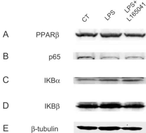
Fig. 6. Treatment with L-165041 does not affect the protein levels of InBa. Protein extracts from H9c2 myotubes stimulated with LPS for 1 h in the presence or the absence of 10 Amol/L L-165041 were assayed for Western blot analysis with PPARh/y (A), p65 (B), InBa (C) InBh (D) and h-tubulin (E) antibodies.
shown). Overall, these data demonstrate that PPARh/y activation by L-165041 inhibits LPS-induced NF-nB activation.
2.5. Treatment with the PPARb/d activator enhances its interaction with the p65 subunit of NF-kB
Finally, we sought to determine the molecular mecha-nism by which the PPARh/y activator L-165041 inhibits LPS-induced NF-nB activation. Since it has been proposed that PPARh/y and PPARa share similar biological roles [21], we studied whether PPARh/y inhibited NF-nB signaling through mechanisms similar to those reported for PPARa. Activation of PPARa may result in inhibition of NF-nB signaling through different mechanisms. First, PPARa activators have been reported to induce the expression of InBa, which forms a cytoplasmic inactive complex with the p65–p50 heterodimeric complex[34,35]. We did not observe significant changes in the protein expression of PPARh/y, the p65 subunit of NF-nB, InBa or InBh after L-165041 treatment (Fig. 6), suggesting that PPARh/y activation did not act through this mechanism. In addition, PPARa activators may act through DNA-binding independent mechanisms that may involve a physical interaction with NF-nB. This association prevents NF-nB from binding to its response element and thereby inhibits its ability to induce gene transcription [16]. In order to evaluate whether PPARh/y activation acts through a similar mechanism, we performed coimmunoprecipitation studies with isolated nuclear extracts using antibodies against the p65 subunit of NF-nB and PPARh/y. Data shown inFig. 7demonstrate that addition of the PPARh/y agonist L-165041 strongly enhanced the physical inter-action between p65 and PPARh/y, suggesting that increased association between these two proteins is the mechanism through which PPARh/y activation prevents NF-nB activation.
3. Discussion
In the present study, we demonstrate that activation of PPARh/y by the specific ligand L-165041 inhibits PE-induced cardiomyocyte hypertrophy in neonatal rat cardio-myocytes. Treatment with L-165041 also inhibited PE-induced expression of the NF-nB-target gene MCP-1, suggesting that the antihypetrophic effect of this compound involves downregulation of NF-nB signaling pathway. Further, it is shown that L-165041 may inhibit LPS-induced NF-nB activation through enhanced physical interaction of PPARh/y with the p65 subunit of NF-nB.
Several studies have reported that both PPARa and PPARg activators inhibit cardiac hypertrophy [36–39]. In contrast, the biologic role of PPARh/y activation in cardiac hypertrophy was unknown. The availability of selective PPARh/y ligands, such as L-165041, opened the possibility of studying the role of this PPAR subtype in cardiac cells. Thus, previous data of a recent study [21] pointed to an important function of PPARh/y in the heart. The authors demonstrated that both PPARa and PPARh/y were expressed in comparable levels in heart, whereas PPARg was barely detectable. Further, PPARh/y was fatty acid inducible and activated the expression of PPARa target genes involved in fatty acid utilization in cardiac myocytes. The authors of this study suggested that PPARa and PPARh/y shared similar functions in cardiac cells regarding cardiac fatty acid metabolism. In agreement with this idea, Muoio et al. [40] shown that fatty acid oxidation in skeletal muscle of PPARa / mice was not impaired, probably because of PPARh/y compensated for the lack of PPARa in these mice. In the present study, we define a new role for PPARh/y activation, inhibition of cardiomyocyte hyper-trophy. Therefore, given the abundant expression of both PPARa and PPARh/y in heart and the fact that PPARa activation also inhibits cardiac hypertrophy [41,42], these PPAR subtypes may also share similar roles in the development of cardiac hypertrophy.
It is still a matter of controversy whether changes in intracellular substrate and metabolite levels in cardiomyo-cytes are the consequence or the reason for cardiac hypertrophy. However, several factors support a role for cardiac metabolism in the development of cardiac hyper-trophy. Thus, an increase in the activities of several glycolytic enzymes has been reported prior to cardiac hypertrophy [7]. Moreover, the fact that PPARa gene influences human left ventricular growth in response to exercise and hypertension, indicates that maladaptative cardiac substrate utilization can play a causative role in the pathogenesis of left ventricular hypertrophy [43]. In the present work, stimulation of rat neonatal cardiomyocytes with PE, which leads to NF-nB activation [4], caused cardiomyocyte hypertrophy that was accompanied by a fall in the expression of genes involved in fatty acid metabo-lism, such as M-CPT-I and PDK-4. This effect was abolished by the addition of the PPARh/y activator
Fig. 7. L-165041 enhances PPARh/y association with the p65 subunit of NF-nB. Nuclear extracts from H9c2 myotubes stimulated with LPS for 1 h in the presence or the absence of 10 Amol/L L-165041 were subjected to immunoprecipitation using anti-p65 antibody coupled to protein-A agarose beads. Immunoprecipitates were subjected to SDS–PAGE and immuno-blotted with an anti-PPARh/y antibody. The blot data are representative of three separate experiments.
L-165041, which strongly induced the expression of these genes. Further studies are necessary to clearly establish whether pharmacological modulation of cardiac fatty acid metabolism with either PPARa or PPARh/y activators is enough to alleviate or inhibit cardiac hypertrophy. However, it is worth noting that treatment of H9c2 cells with LPS for 24 h caused a similar pattern of changes in the expression of M-CPT-I and PDK-4 to those observed in PE-induced cardiomyocyte hypertrophy. Since both PE-induced cardi-omyocyte hypertrophy and LPS lead to NF-nB activation, these data point to the involvement of this transcription factor in the downregulation of genes involved in fatty acid metabolism. Activation of PPARh/y would inhibit NF-nB signaling pathway avoiding both cardiomyocyte hyper-trophy and downregulation of genes involved in fatty acid metabolism. Furthermore, and although, it is not the objective of this study, apoptosis is considered an important factor in the progression from cardiac hypertrophy to heart failure. Activation of NF-nB is involved in direct regulation of both anti- and proapoptotic effects [44] and the latter maybe stimulated by LPS.
Interestingly, L-165041 reduced the induction of the NF-nB target gene MCP-1 in cardiac cells stimulated by either PE or LPS, suggesting that PPARh/y may antag-onize NF-nB activation. Enhanced myocardial MCP-1 has been described in the hypertrophied and failing heart [45] and may lead to the infiltration and activation of inflammatory cells, such as monocytes/macrophages and lymphocytes. In addition, it has been reported that activation of MCP-1 expression contributes to left ventricular remodeling and failure after myocardial infarc-tion [46]. Therefore, PPARh/y activainfarc-tion may become a therapeutic option to reduce the expression of MCP-1 in heart. It is important to note that the inhibitory effect of L-165041 on LPS-induced MCP-1 expression was of lower intensity that the observed for PE. This probably reflects the higher induction achieved by LPS stimulation (sevenfold induction) compared to PE (twofold induction) and/or differences in the two cell system used. The use of H9c2 myotubes, which only express PPARh/y, offers the advantage of avoiding the interference of other PPAR subtypes and, therefore, permits to adscribe the changes observed to this transcription factor. However, LPS-treat-ment of H9c2 myotubes was performed to achieve NF-nB activation, but not cardiac hypertrophy, and consequently the findings observed in these cells should be limited to the activation of NF-nB by LPS.
PPARa activators may inhibit NF-nB signaling through different mechanisms [16,47,48]. One of these mechanisms involves physical interaction of PPARa and the p65 subunit of NF-nB [16]. Here, we demonstrate that PPARh/y activation by L-165041 enhances the protein-protein association between PPARh/y and p65, indicating that this mechanism may interfere NF-nB transactivation capacity. Therefore, PPARa and PPARh/y may also share similar mechanisms of action inhibiting NF-nB signaling.
Further studies are necessary to investigate whether PPARh/y activation may inhibit the NF-nB signaling pathway through additional mechanisms or affects the activity of other transcription factors involved in cardiac hypertrophy, such as nuclear factor of activated T lymphocyte (NFAT).
In summary, in the present study, we show that PPARh/y activation inhibits PE-induced cardiomyocyte hypertrophy in neonatal rat ventricular cardiomyocytes. PPARh/y acti-vation also inhibits LPS-induced NF-nB actiacti-vation through a mechanism that may involve enhanced protein–protein interaction between this PPAR subtype and the p65 subunit of NF-nB. These data indicate that inhibition of the NF-nB signaling pathway may be the underlying mechanism responsible for the inhibition of cardiomyocyte growth.
Acknowledgments
This study was partly supported by grants from the Fundacio´ Privada Catalana de Nutricio´ i Lipids, Fundacio´n Ramo´n Areces, Ministerio de Ciencia y Tecnologı´a of Spain (SAF2003-01232) and the European Union FEDER funds. We also thank the Generalitat de Catalunya for grant 2001SGR00141. Anna Planavila was supported by a grant of the Divisio´ IV from the University of Barcelona. Mireia Jove´ was supported by a grant from the Ministerio de Ciencia y Tecnologı´a of Spain.
References
[1] Levy D, Garrison RJ, Kannel WB, Castelli WP. Prognostic implications of echocardiographically determined left-ventricular mass in the Framingham Heart-Study. N Engl J Med 1990; 322:1561 – 6.
[2] Lorell BH, Carabello BA. Left ventricular hypertrophy—patho-genesis, detection, and prognosis. Circulation 2000;102:470 – 9. [3] Purcell NH, Tang GL, Yu CF, Mercurio F, DiDonato JA, Lin AN.
Activation of NF-kappa B is required for hypertrophic growth of primary rat neonatal ventricular cardiomyocytes. Proc Natl Acad Sci U S A 2001;98:6668 – 73.
[4] Hirotani S, Otsu K, Nishida K, Higuchi Y, Morita T, Nakayama H, et al. Involvement of nuclear factor-kappa B and apoptosis signal-regulating kinase 1 in G-protein-coupled receptor agonist-induced cardiomyocyte hypertrophy. Circulation 2002;105:509 – 15. [5] Higuchi Y, Otsu K, Nishida K, Hirotani S, Nakayama H, Yamaguchi
O, et al. Involvement of reactive oxygen species-mediated NF-kappa B activation in TNF-alpha-induced cardiomyocyte hypertrophy. J Mol Cell Cardiol 2002;34:233 – 40.
[6] Gupta S, Purcell NH, Lin AN, Sen S. Activation of nuclear factor-kappa B is necessary for myotrophin-induced cardiac hypertrophy. J Cell Biol 2002;159:1019 – 28.
[7] Taegtmeyer H, Overturf ML. Effects of moderate hypertension on cardiac-function and metabolism in the rabbit. Hypertension 1988;11:416 – 26.
[8] Sack MN, Rader TA, Park SH, Bastin J, Mccune SA, Kelly DP. Fatty acid oxidation enzyme gene expression is downregulated in the failing heart. Circulation 1996;94:2837 – 42.
[9] Kelly DP, Strauss AW. Mechanisms of disease—inherited cardiomyo-pathies. N Engl J Med 1994;330:913 – 9.
[10] Binas B, Danneberg H, McWhir J, Mullins L, Clark AJ. Requirement for the heart-type fatty acid binding protein in cardiac fatty acid utilization. FASEB J 1999;13:805 – 12.
[11] Chiu HC, Kovacs A, Ford DA, Hsu FF, Garcia R, Herrero P, et al. A novel mouse model of lipotoxic cardiomyopathy. J Clin Invest 2001;107:813 – 22.
[12] Kersten S, Desvergne B, Wahli W. Roles of PPARs in health and disease. Nature 2000;405:421 – 4.
[13] Auwerx J, Baulieu E, Beato M, Becker-Andre M, Burbach PH, Camerino G, et al. A unified nomenclature system for the nuclear receptor superfamily. Cell 1999;97:161 – 3.
[14] Braissant O, Foufelle F, Scotto C, Dauca M, Wahli W. Differential expression of peroxisome proliferator-activated receptors (PPARs): tissue distribution of PPAR-alpha, -beta, and -gamma in the adult rat. Endocrinology 1996;137:354 – 66.
[15] Daynes RA, Jones DC. Emerging roles of PPARs in inflammation and immunity. Nat Rev Immunol 2002;2:748 – 59.
[16] Delerive P, De Bosscher K, Besnard S, Vanden Berghe W, Peters JM, Gonzalez FJ, et al. Peroxisome proliferator-activated receptor alpha negatively regulates the vascular inflammatory gene response by negative cross-talk with transcription factors NF-kappa B and AP-1. J Biol Chem 1999;274:32048 – 54.
[17] Liang FQ, Wang F, Zhang SM, Gardner DG. Peroxisome proliferator activated receptor (PPAR)alpha agonists inhibit hypertrophy of neonatal rat cardiac myocytes. Endocrinology 2003;144:4187 – 94. [18] Irukayama-Tomobe Y, Miyauchi T, Sakai S, Kasuya Y, Ogata T,
Takanashi M, et al. Endothelin-1-induced cardiac hypertrophy is inhibited by activation of peroxisome proliferator-activated receptor-alpha partly via blockade of c-Jun NH2-terminal kinase pathway. Circulation 2004;109:904 – 10.
[19] Yamamoto K, Ohki R, Lee RT, Ikeda U, Shimada K. Peroxisome proliferator-activated receptor gamma activators inhibit cardiac hyper-trophy in cardiac myocytes. Circulation 2001;104:1670 – 5. [20] Asakawa M, Takano H, Nagai T, Uozumi H, Hasegawa H, Kubota N,
et al. Peroxisome proliferator-activated receptor gamma plays a critical role in inhibition of cardiac hypertrophy in vitro and in vivo. Circulation 2002;105:1240 – 6.
[21] Gilde AJ, van der Lee KAJM, Willemsen PHM, Chinetti G, van der Leij FR, van der Vusse GJ, et al. Peroxisome proliferator-activated receptor (PPAR) alpha and PPAR beta/delta, but not PPAR gamma, modulate the expression of genes involved in cardiac lipid metabo-lism. Circ Res 2003;92:518 – 24.
[22] Berger J, Leibowitz MD, Doebber TW, Elbrecht A, Zhang B, Zhou GC, et al. Novel peroxisome proliferator-activated receptor (PPAR) gamma and PPAR delta ligands produce distinct biological effects. J Biol Chem 1999;274:6718 – 25.
[23] Kimura Y, Otsu K, Nishida K, Kuzuya T, Tada M. Thyroid-hormone enhances Ca2+ pumping activity of the cardiac sarcoplasmic-reticulum by increasing Ca2+ Atpase and decreasing phospholamban expression. J Mol Cell Cardiol 1994;26:1145 – 54.
[24] Thaik CM, Calderone A, Takahashi N, Colucci WS. Interleukin-1-beta modulates the growth and phenotype of neonatal rat cardiac myocytes. J Clin Invest 1995;96:1093 – 9.
[25] Cabrero A, Alegret M, Sanchez RM, Adzet T, Laguna JC, Va´zquez-Carrera M. Increased reactive oxygen species production
down-regulates peroxisome proliferator-activated alpha pathway in C2C12 skeletal muscle cells. J Biol Chem 2002;277:10100 – 7.
[26] Takemoto M, Node K, Nakagami H, Liao YL, Grimm M, Takemoto Y, et al. Statins as antioxidant therapy for preventing cardiac myocyte hypertrophy. J Clin Invest 2001;108:1429 – 37.
[27] McGarry JD, Brown NF. The mitochondrial carnitine palmitoyltrans-ferase system—from concept to molecular analysis. Eur J Biochem 1997;244:1 – 14.
[28] Wu PF, Sato J, Zhao Y, Jaskiewicz J, Popov KM, Harris RA. Starvation and diabetes increase the amount of pyruvate dehydrogen-ase kindehydrogen-ase isoenzyme 4 in rat heart. Biochem J 1998;329:197 – 201. [29] Li JM, Gall NP, Grieve DJ, Chen MY, Shah AM. Activation of
NADPH oxidase during progression of cardiac hypertrophy to failure. Hypertension 2002;40:477 – 84.
[30] Sadoshima J, Jahn L, Takahashi T, Kulik TJ, Izumo S. Molecular characterization of the stretch-induced adaptation of cultured cardiac-cells—an invitro model of load-induced cardiac-hypertrophy. J Biol Chem 1992;267:10551 – 60.
[31] Shin WS, Szuba A, Rockson SG. The role of chemokines in human cardiovascular pathology: enhanced biological insights. Atheroscle-rosis 2002;160:91 – 102.
[32] Takano H, Nagai T, Asakawa M, Toyozaki T, Oka T, Komuro I, et al. Peroxisome proliferator-activated receptor activators inhibit lipopoly-saccharide-induced turner necrosis factor-alpha expression in neonatal rat cardiac myocytes. Circ Res 2000;87:596 – 602.
[33] Delerive P, Gervois P, Fruchart JC, Staels B. Induction of I kappa B alpha expression as a mechanism contributing to the anti-inflamma-tory activities of peroxisome proliferator-activated receptor-alpha activators. J Biol Chem 2000;275:36703 – 7.
[34] Delerive P, De Bosscher K, Vanden Berghe W, Fruchart JC, Haegeman G, Staels G. DNA binding-independent induction of I kappa B alpha gene transcription by PPAR alpha. Mol Endocrinol 2002;16:1029 – 39.
[35] Muoio DM, MacLean PS, Lang DB, Li S, Houmard JA, Way JM, et al. Fatty acid homeostasis and induction of lipid regulatory genes in skeletal muscles of peroxisome proliferator-activated receptor (PPAR) alpha knock-out mice—evidence for compensatory regu-lation by PPAR delta. J Biol Chem 2002;277:26089 – 97. [36] Jamshidi Y, Montgomery HE, Hense HW, Myerson SG, Torra IP,
Staels B, et al. Peroxisome proliferator-activated receptor a gene regulates left ventricular growth in response to exercise and hyper-tension. Circulation 2002;105:950 – 5.
[37] van Empel VPM, De Windt LJ. Myocyte hypertrophy and apoptosis: a balancing act. Cardiovasc Res 2004;63:487 – 99.
[38] Shioi T, Matsumori A, Kihara Y, Inoko M, Ono K, Iwanaga Y, et al. Increased expression of interleukin-1 beta and monocyte chemotactic and activating factor monocyte chemoattractant protein-1 in the hypertrophied and failing heart with pressure overload. Circ Res 1997;81:664 – 71.
[39] Hayashidani S, Tsutsui H, Shiomi T, Ikeuchi M, Matsusaka H, Suematsu N, et al. Anti-monocyte chemoattractant protein-1 gene therapy attenuates left ventricular remodeling and failure after experimental myocardial infarction. Circulation 2003;108:2134 – 40.