A REVISION OF EUROPEAN SAXICOLOUS
SPECIES OF THE GENUS BUELLIA DE NOT. AND
FORMERLY INCLUDED GENERA
Christoph SCHEIDEGGER*
Abstract: A detailed taxonomic survey of the saxicolous European species ofBuellia
based on a detailed survey and assessment of the important features of the genus is presented. These include the conidia, the anatomy of the exciple, the spore wall pigmentation, ornamentation and internal wall thickening, as well as analysis of the lichen substances. As a result, 36 saxicolous species are recognized, of which Buellia griseosquamulata and B. longispora are new taxa and B. atrocinerella and B. parvula are new combinations. Buellia coniops, B, lecideina and B. punclata are transferred to the validated genus Amandinea. A key to 43 accepted species of Buellia, Amandinea and Hafellia is included.
Introduction
The genus Buellia was described by De Notaris (1846), who named it after his friend, Esuperanzo Buelli (Leunis 1877). The genus was soon accepted by many early lichenologists (Massalongo 1852; Korber 1855; Fries 1860). Zahlbruckner (1926) subsequently delimited the genus from Rinodina on account of its lecideine apothecia and included both of the crustose genera in the Buelliaceae (Zahlbruckner 1907). Hafellner et al. (1979) distinguished
Buellia from Rinodina by differences in the spore type and apothecial margin.
Following his circumscription, Buellia was characterized by having spores with-out any internal wall thickening and possessing lecideine or cryptolecanorine apothecia. Poelt (1973), Henssen & Jahns (1973) and Hafellner et al. (1979) included the family Buelliaceae in the Physciaceae, thus indicating the close affinities between crustose, foliose and fruticose genera therein (Mayrhofer 1982). Miiller & von Arx (1962) designated B. disciformis as the type species of the genus, which was later accepted and further discussed by Hafellner (1979). Aptroot (1987) outlined a proposal to conserve Buellia against Gassicurtia Fee (Fee 1824).
Buellia remains the least well studied genus of the Physciaceae in Europe,
and, with few exceptions, is very poorly known in other parts of the world (North America, Imshaug 1951, 1955; South America, Magnusson 1955; Antarctica, Lamb 1968; India, Singh & Awasthi 1981; New Zealand, Galloway 1985). Probably because the genus has not less than about 400 (Hawksworth
et al. 1983) to 600 taxa (Grummann 1963), a worldwide taxonomic study has
not been attempted and even in Central Europe only the corticolous species have been studied in relatively recent times (Schauer 1965).
*Swiss Federal Institute for Forest, Snow and Landscape Research, CH-8903 Birmensdorf, Switzerland.
316 THE LICHENOLOGIST Vol.25 Because many of the classical species characteristics, for example the iodine reaction of the medulla, have been shown to vary greatly, sometimes even within a single thallus (Scheidegger 1987), the aim of this paper was to assess the taxonomic relevance of a range of morphological, anatomical, as well as chemical, characteristics within the genus. A key is presented, followed by concise descriptions of 36 species, which include chorological and ecological data.
Materials and Methods
Extensive field studies were undertaken in order to study most of the species in nature and to assess variation in their ecology and morphology.
Morphological measurements of external characters were investigated with a binocular Wild M5 with a measuring eyepiece. Internal features, including spore ornamentation were examined at x 1000 (aperture 1-32) with a Leitz Dialux with a drawing tube. Sections c. 15-um thick were cut with a freezing microtome Leitz-1310 with cryomat. Sections of exciple were mounted in an aqueous solution of trichloroacetic acid.
Spore ornamentation was additionally investigated with a Jeol JSM-T 300 scanning electron microscope. A hand section or an entire apothecium was macerated by two tweezers in a small watch glass containing water. Single spores were then separated with a needle from the adhering hymenial gelatinous substance and subsequently transferred in water to an aluminium stub with a small pipette. The aluminium stub was previously coated with a very thin layer of fusion adhesive (Bosch). Following air-drying of the spores, the stubs were heated at 50°C for 20 min to guarantee a good adhesion of the spores to the adhesive. After the stubs had cooled to room temperature, the specimens were sputter-coated with gold and examined at 12—15 kV. The descriptions of the spore ornamentation follow Erdtman (1943, 1956) and Kremp (1968).
Most specimens investigated were analysed by thin layer chromatography (TLC) following the methods described by Culberson & Ammann (1979), Culberson et al. (1981), Leuckert (1984), and White & James (1985). Xanthone-containing species were analysed by double-focusing mass spectrometry (Scheidegger & Ruef 1988; Ruef 1990). Great care was taken to avoid contamination from adjacent lichen thalli. For all analyses only selected areoles were removed with tweezers from the substratum.
Herbarium specimens were requested from the following institutional and private herbaria (abbreviations according to Holmgren et al. 1990): ANGUC, BERN, BC, BCC, BG, BM, C, CANL, E, FH, GB, GZU, H, HBG, LAUS, LD, LISU, M, MAF, MARSJ, MUB, O, PC, S, STR, STU, SZU, TO, UPS, TUR, TSB, VER, W, WU, Z, ZT, Aptroot (Utrecht), Clerc (Geneva), Hafellner (Graz), Kalb (Neumarkt), Mayrhofer (Graz), Mies (Koln), Poelt (Graz), Renobales (Bilbao), Scheidegger (Birmensdorf), Ullrich (Goslar), Vezda (Brno) and Wunder (Berchtesgaden). A list of the specimens investigated has been lodged at BERN. The abbreviations of the geographical areas from where specimens have been analysed follow Tutin et al. (1964): Al, Albania; Au, Austria; Az, Azores; Be, Belgium and Luxembourg; Bl, Balearic Islands; Br, Britain; Bu, Bulgaria; Co, Corsica; Cr, Crete; Cz, Czechoslovakia; Da, Denmark; Fa, Faeroe Islands; Fe, Finland; Ga, France; Ge, Germany; Gr, Greece; Hb, Ireland; He, Switzerland; Ho, Netherlands; Hs, Spain; Hu, Hungary; Is, Iceland; It, Italy; Ju, Jugoslavia; Lu, Portugal; No, Norway; Po, Poland; Rm, Romania; Rs, former Soviet Union; Sa, Sardinia; Sb, Svalbard (Spitsbergen); Si, Sicily; Su, Sweden; Tu, Turkey.
Results Life strategy
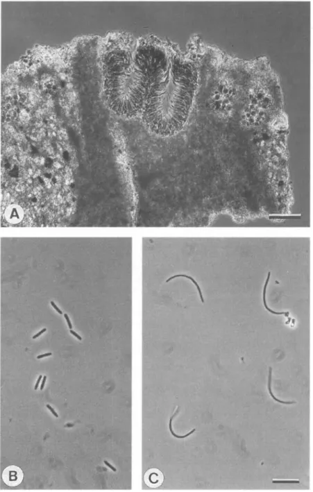
Saxicolous taxa of Buellia show a remarkable variation in their life strategies. Many species are autonomous lichens, 11 European species are lichenicolous lichens (Table 1) and B. adjuncta is a parasymbiont on Lecanora straminea (Hafellner 1979). Only a small number of species are parasites for their entire
''/•
FIG. 1. Buellia griseosquamulata (above) growing as a lichenicolous lichen on B. tirolensis (below). Scale = 1 mm.
(Fig. 1), but some others are only parasitic during a juvenile stage and grow autonomously when forming larger thalli (Table 1). Parasitism can therefore be considered to be a not unusual biological aspect of this genus, at least for the saxicolous species.
Substrata
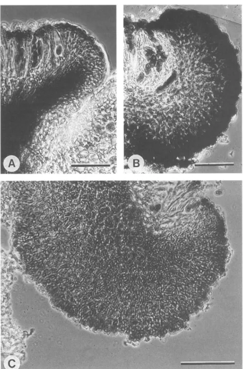
Buellia almeriensis is restricted to gypsum, and B. caldesiana, B. dispersa and B. stellulata may grow on more or less calcareous rocks; all other saxicolous
species are confined to siliceous substrata. Buellia dispersa and B. stellulata grow on basic and acid substrata and for these species the influence of a calcareous substratum on the morphology of the thallus is clearly demonstrated by B.
dispersa (Fig. 2). On siliceous rock, the thallus is areolate to squamulose and
slightly placodioid and ochre-coloured (Fig. 2B); when on calcareous rock, the thallus is rimose-cracked to areolate and chalk-white and is never squamulose
318 THE LICHENOLOGIST Vol.25
TABLE 1. Parasymbiontic species of Buellia and their hosts
Species B. adjuncta* B. badia* B. concinna B. griseosquamulata* B. imshaugii* B.jugorum B. longispora B. rniriquidica* B. sequax B. uberior B. uberiuscula* Hosts Lecanora straminea
Acarospora, Aspicilia, Caloplaca, Diploschistes, Parmelia s. lat., Rinodina, Umbilicaria
Amandinea coniops, B. uberior, Physcia sp. Protoparmelia badia B. tirolensis Dimelaena oreina Placynthiella sp. Aspicilia sp. Schaereriafuscocinerea
Caloplaca, unidentified crustose lichens
Schaereria fuscocinerea A ca rospora fus cat a
* = regularly parasitic.
(Fig. 2A). The respective thallus anatomies differ mostly in the occurrence of K-insoluble crystals (probably oxalate) in the epinecral layer of thalli on calcareous rocks. K-soluble crystals, probably the lichen products (see below), occur in thalli on both substrata (Figs 2C-E).
Thallus
In the species investigated, chasmolithic, granular, rimose, areolate, bullate, squamulose and placodioid thalli are distinguished. In chasmolithic lichens the thalli are restricted to very narrow cracks in the substratum, whereas the others are epilithic. Granular thalli have disjunct phycobiont-containing areas on a continuous hypothallus. Rimose thalli have a continuous phycobiont-containing thallus with irregularly arranged, non-reticulate cracks, whereas areolate thalli always have regularly arranged cracks resulting in a reticulate pattern.
Bullate thalli develop from areolate thalli if the areoles grow vertically and finally become stalked, as is regularly found in luxuriant thalli of Amandinea
coniops. Squamulose thalli have thalline parts that have only very loose
con-tact with the substratum or that are slightly ascending; placodioid thalli have elongate marginal areoles.
Buellia vilis is mostly chasmolithic and was observed with a thin, superficial,
rimose-cracked thallus only once. Buellia leptocline and B. sequax are usually epilithic but when on very porous substrata, such as schists, they regularly have chasmolithic thalli. All the other species have epilithic, mostly heteromerous thalli (Fig. 3). Amandinea punctata has a thin granular thallus with isolated to confluent, flat granules. Most species are areolate or rimose-cracked. Due to a great variability in thallus thickness in most species, areolate or rimose thalli often reflect developmental stages, particularly for species that usually have thin thalli. For instance, lime-containing substrata may change an areolate
dispersa. A, Rimose thallus on lime-containing sandstone (isotype of B. tergestina). B, Areolate to squamulose and slightly placodioid thallus on lime-free siliceous rock (topotype of B. squamulata). C, Section through thallus of A after K treatment. High amounts of insoluble crystals, presumably of calcium oxalate, are localized in the phaenocortex. Lichen substances were removed by the K treatment. D, Section through untreated thallus of B. High amounts of crystalline lichen substances are localized in the phenocortex. E, Same section as in D, after K treatment. All crystals were removed by the K treatment; no insoluble crystals are present in thalli on lime-free substrata. C-E
320 THE LICHENOLOGIST Vol. 25
FIG. 3. Different cortical types in Buellia: A, Phenocortex of B. badia with epinecral and thin cortical layer with pigmented and incrassate terminal cells. B, Phenocortex of B. dispersa with thick epinecral and thin cortical layer. C, Phenocortex of B. spuria with thick cortical and thin epinecral
thallus to rimose, or an areolate thallus may regenerate to rimose after feeding by herbivores. Despite this variability, B. sequax, B. subdisciformis and Hafellia
leptoclinoides regularly have rimose-cracked thalli; B. aethalea, B. ocellata, B. miriquidica and B. uberior usually have areolate thalli.
The epinecral layer consists of dead and collapsed hyphae in which the cell lumina do not stain with cotton blue. Collapsed algal cell walls can also be observed in this layer in varying amounts in all the species. Although the thickness of this layer is very variable and is often thinned by damage, very thick and cartilaginous epinecral layers, up to 50 \im, occur in B. badia (Fig. 3A), B. dispersa (Fig. 3B) and related species, whereas in B. spuria (Fig. 3C),
B. leptocline and related species the epinecral layer is much thinner and always
interspersed with very small crystals.
The cortical layer is characterized by anticlinal, cellular hyphae that stain with cotton blue. In some species (Amandinea coniops, A. punctata, Buellia.
aethalea (p.p.), B. atrocinerella, B. badia, B. concinna (p.p.), B. fusca, B. miriquidica, B. tirolensis, and B. uberior) the terminal cells have
brown-pigmented caps and are clearly swollen. The other species always have unpigmented terminal cells or only a few pigmented cells around apothecia. Both types of cortex belong to the phenocortex (Poelt 1958, 1989) because collapsed algal cells are always found in the epinecral layer (see above). It is possible that particular chemical substances may be an influence in the forma-tion of pigmented cells, as B. ocellata and B.jugorum, which contain xanthones, are the only species of the B. aethalea group in which these pigmented cells are absent.
The iodine reaction of the medulla was formerly a widely used taxonomic character at the species level in Buellia (Erichsen 1930, 1957). In some cases this reaction is very constant and closely correlated with other characters, e.g. the chemistry of B. leptocline (I +), B. saxorum (I +) and B. subdisciformis (I —); in this and some other species groups this character is taxonomically very reliable. However, in other cases, especially in B. aethalea and related species, the reaction may vary greatly, even within a single thallus (Scheidegger 1988), and is therefore of little taxonomic value in these groups.
Pycnidia
Conidia are always single-celled and thin-walled. They are formed within completely immersed, flask-shaped pycnidia. In B. saxorum and B. subdisciformis, two to a few pycnidia may be regularly confluent (Fig. 4A). The conidiophores are of the Roccella-type (Vobis & Hawksworth 1981) in A. coniops, A. lecideina and
A. punctata, and of the Anaptychia-type in all the species of Buellia and Hafellia where pycnidia have been observed.
The conidia of the last two genera are bacilliform and less than or about 10 um long and 0-7-1 (xm wide (Figs 4B, 5). The length of the bacilliform conidia is a valuable character for separating some closely related species, for example, B. ocellata and B.jugorum (Scheidegger & Ruef 1988) or taxa with an exciple of leptocline-type (Fig. 5) (see below). By contrast, the three species of
Amandinea treated have long, filiform conidia, up to 30 (im long (Fig. 4C).
322 THE LICHENOLOGIST Vol. 25
FIG. 4. Conidiomata and conidia of Buellia and Amandinea: A, Confluent pycnidia of B. subdisciformis. B, Bacilliform conidia of B. dispersa. C, Filiform conidia of A. lecideina. Scales:
A. coniops A. lecideina A. punctata B. subdisciformis B. fimbriata B. jugorum B. longispora B. sardiniensis B. saxorum B. indissimilis H. leptoclinoides B. dispersa B. almeriensis B. aethalea B. spuria B. concinna B. ocellata B. badia B. leptocline B. uberior B. miriquidica B. stellulata B. vilis — "II , 10 15 20 conidium length (urn)
25 30
FIG. 5. Range of conidial length in Amandinea, Buellia and Hafellia.
this conidial character that the genus Amandinea (Choisy 1950) is proposed. Only the three European taxa are treated here, but other, non-European species will be discussed elsewhere (Matzer et al. 1994).
Ascomata
The current delimitation of the genus Buellia includes species with various types of apothecial margins. Cryptolecanorine apothecia are completely immersed in the thallus and have no exiple; lecanorine, biatorine and lecideine apothecia are according to Hawksworth et al. (1983) and Hafellner et al. (1979). The different exciple types discussed below can be examined by the use of cryotome or well-prepared hand sections of younger apothecia. Only exciples that are not coincident with the edge of an areole may be used. The descriptions of plectenchyma follow Korf (1973): textura angularis consists of short-celled, isodiametric hyphae without intercellular spaces, textura intricata has long-celled, interwoven hyphae, whereas in textura oblita the long-celled hyphae are parallel, strongly agglutinated and thick-walled.
324 THE LICHENOLOGIST Vol.25
Pigments
The exciple, hypothecium, paraphysis tips and, in some species, the terminal cells of the thallus cortex, are pigmented. They are distinguished below by their colour in water mounts, their colour changes and solubility in K and HNO3, and their localization.
Pigment A: green to aeruginose, K —, HNO3+ red. Always diffuse around pigment B. Rarely localized in the cortex of B. aethalea and B. uberior, fre-quently in exciples of the aethalea-type and on paraphysis tips. It is also typical for the subhymenium of B. ocellata and B.jugorum. This pigment is probably identical to pigment A of Coppins (1983).
Pigment B: dull brown, K —, HNO3 —. In or on the hyphal wall of the paraphysis tips of all species, in the hypothecium of all species, except that of
B. vilis, also in all coloured cortical cells and in exciples of aethalea- and
dispersa-types. This pigment is probably identical to pigment F of Coppins (1983).
Pigment C: dull brown-red to black, K —, HNO3 + intensifying purple, plus a diffusing brown-red solution. Only in the outer part of the exciple of B. vilis.
Pigment D: dull brown, K + brownish solution, HNO3 —. In dispersa-type exciples.
Pigment E: orange-red, K + red solution, HNO3 —. In leptocline-type exciples. In necrotic parts of thalli further brown pigments may occur but they are not discussed in detail here.
Aethalea-type
The apothecia are completely immersed to sessile. The disc of the apothecium is plane to hemispherical. The width varies from 0-3 mm (e.g. B. stellulata) in most cryptolecanorine to zeorine apothecia to about 1 mm in some species with lecideine apothecia (B. subsquamosa). The margin may be lecanorine (B. ocellata,
B. parvula and B. uberiuscula), zeorine (B. aethalea, B.jugorum, B. miriquidica, B. stellulata and B. uberior) (Fig. 6C) or lecideine (B. atrocinerella, B. badia, B. fusca, B. griseosquamulata, B. indissimilis, B. spuria, B. subsquamosa, B. sequax)
(Fig. 6A, B & D). It is mostly prominent and persistent, narrow (40 |am) to broad (100 um) and in most species, black. In B. atrocinerella, B. fusca and B.
indissimilis the margin is brown. The plectenchyma of the zeorine apothecia is
textura oblita and is anticlinal to the thallus surface. In lecideine apothecia the exciple is radially formed by a textura prismatica or angularis.
The colour of the exciple is the result of pigment A and, in varying amounts, also pigment B; this pigmentation is often restricted to the outer part of the exciple only.
Lecideine, cryptolecanorine and zeorine apothecia with the above-mentioned characters represent only different forms of the aethalea-type. and formation of a proper margin is probably also regulated in several species by factors such as thallus thickness and damage to the thallus, e.g. by browsing invertebrates (molluscs, insects, mites).
1993
FIG. 6. Exciple of aethalea-type: A, Lecideine apothecium of Amandinea lecideina with hyaline inner part. B, Zeorine apothecium ofB.jugorum. C, Lecideine apothecium of Buellia spuria with hyaline
FIG. 7. Exciple-types: A, Dispersa-type of Buellia dispersa with dark inner part and less pigmented outer part of exciple. B, Vilis-xype of B. vilis with very dark pigmented outer part and almost hyaline inner part. C, Leptocline-type of B. leptodine with intricate hyphae homogeneously pigmented
Vilis-type
The apothecia are sessile and constricted at their base. The lecideine margin is prominent and persistent. The exciple is radially composed of textura oblita and up to 60 um broad. The outer part of the exciple is markedly coloured with pigment C. The inner part is unpigmented and is very strongly amyloid, I + violet (Fig. 7B); B. vilis is unique to this type.
Dispersa-type
The apothecia are sessile with a constricted base. The margin is dark brown to black, 60-100 um wide. The disc is mostly plane but in B. excelsa it may become hemispherical. The exciple is radially formed, the inner part of textura oblita, the outer of textura angularis (Fig. 7A). Whereas in younger apothecia only the outer part is coloured with pigments D and B, older exciples are equally pigmented in their inner and outer parts; B. excelsa, B. dispersa, B. longispora and
H. leptoclinoides have this type. Leptocline-type
The apothecia are sessile and constricted at their base. The margin is black, about 100 um broad (in B. almeriensis, 60 um) and mostly prominent. The disc is plane to strongly convex. The inner part of the exciple is textura intricata, the outer part textura oblita (Fig. 7C). Pigment E is distributed throughout the exciple; B. almeriensis, B. leptocline, B. saxorum, B. sardiniensis, and B.
subdisciformis belong here.
Hymenium
A pigmented epihymenium is present in every species. According to the presence or absence of pigment B and/or A the colour may vary from brown to olive or green. The apical cells of the paraphyses are always incrassate and coloured with pigment B ('Pigmentkappe' in Kilias 1981). Pigment A, diffusing into the hymenial gelatine ('Pigmenthaube' in Kilias 1981) occurs only in some of the species with apothecia of aethalea-, leptocline- and wiVw-types. The amount of pigment A, if present, always varies greatly within individual species (Scheidegger 1987).
The hymenium is colourless in all species except B. ocellata and B. jugorum, in which the subhymenium is slightly coloured with pigment A. Hafellia
leptoclinoides and B. excelsa are the only species that have numerous oil
droplets in the hymenium. The oil droplets often exceed 2 um in diameter and are therefore much coarser than those in Lecidella elaeochroma (Ach.) Choisy.
The paraphyses are 1-7-2-5 um broad in all species. Anastomoses are few and occur mostly in the lower part of the hymenium, whereas branching occurs in or just below the epihymenium. In B. dispersa and B. excelsa the paraphyses may be easily separated in a squash preparation in water but in the other species they are strongly conglutinate.
The asci are clavate and 30-100 um long. They always belong to the Lecanora-type (Honegger 1978a,fc). They are usually 8-spored, but in B. concinna a few asci with only four spores may be regularly observed.
FIG. 8. Mature ascospores of two newly described species. A, Spores of Buellia-type of B. longispora. B, Spores of Physconia-type of B. griseosquamulata. Scale = 10 (jm.
10.5 •=• 9.5 £ 8.5 1 7.5 » 6.5 1 , 5 4.5 A
* A* * *
0 0 o • a a + • • * B o B o B D + • D -aethalea 1 + aethalea 1 -uberior 10 12 14 16 18 20 10 9 8 7 6 B 0 x x xX X*? X " x*a£x * x * x I,1 "?,* x X x x . x X 0 o » B. longispora x B. dispersa 11 14 17 20 23 26 B. concinna l+ B. concinna I-12 13 14 15 16 17 18 19 20 21mean spore length (nm)
9.1 •g- 8.6 £ 8.1 1 7 . 6 o « 7.1 c E 6.6 6.1 9.1 x B. excelsa o H. leptodinoides 11 14 17 20 I 8.1 7.1 § 6-1 E 5.1 E 0 -, o o 0 a °D» 0 o l * 0 ° o ° ° D X a o % X O o B x x x X 0 subsquamosa -B. caldesiana B spuria 10 12 14 16 18 dt h I spor e w i
s
E 0.5 9.5 8.5 7.5 6.5 5.5 F 0 °° oV
o + o o o c +++ a a B * B o B + + ^+ ^ a + D ocellata + ocellata -jugorum • stictic acid . stictic acid 10 11 12 13 14 15 16 17 18 19 20 mean spore length (urn)FIG. 9. Mean spore length and width of approx. 30 measurements of selected species of Buellia and
Hafellia. A, B. aethalea and B. uberior. B, B. longispora and B. dispersa. C, B. concinna. D , B. excelsa
and H. leptodinoides. E, B. subsquamosa, B. caldesiana and B. spuria. F , B. ocellata and B. jugorum.
Spores
The ascospores of the species discussed here are predominantly one-septate; in most species two- or more-septate spores are exceptional. Only H. leptodinoides,
B. longispora (Fig. 8A) and B. concinna regularly have spores with 2, or even 4,
additional, relatively thin trans-septa that only appear in the very late stages of spore ontogeny. Spore length varies from 8 \ixn in B. uberior to about 30 (xm in B.
longispora (Fig. 8). Variation in spore length and width for a single species is
sometimes unusually high, as in B. aethalea (Fig. 9A) or B. concinna (Fig. 9C), whereas it is rather small in species with small spores, such as B. uberior (Fig. 9A).
330 THE LICHENOLOGIST Vol.25
FIG. 10. Spore types of the genera Buellia and Hafellia. Wl (outermost layer) and W3 are dark and W2 and W4 (innermost layer) are not or only slightly pigmented. A-B, Buellia-iypt without internal wall thickenings. A, B. spuria with diffuse torus. B, B. uberior with intensely pigmented torus. Pigmented wall layers are visible on each side of the septum; the pigmentation is more intense at the periphery, where the two layers fuse. C, Spore of Physconia-type. Juvenile spore of B. dispersa with the beginning of pigmentation. Median internal thickening of endospore and rela-tively thick intermediary layer are visible. D, Spore of Callispora-type. Premature, relarela-tively dark pigmented spore of H. leptoclinoid.es with median and lateral thickenings of the spore wall and the
endospore. Scale =10 um
Only in a few cases can spore measurements be used alone to separate taxa, such as H. leptoclinoides from B. excelsa (Fig. 9D) and B. longispora from B.
dispersa (Fig. 9B). In other related species the spore lengths generally overlap
and can therefore be considered only in combination with other characters, such as chemical compounds, for example B. subsquamosa from B. spuria (Fig. 9E) and B. ocellata from B.jugorum (Fig. 9F). In the chemotypes of B. ocellata (stictic acid present or absent) (Fig. 9F), amyloid or non-amyloid forms of B.
concinna (Fig. 9C) and B. stellulata (not shown) with rugulate or psilate spore
ornamentation, no significant differences in spore length are present and the respective pairs of taxa are here considered to be conspecific. Amyloid and non-amyloid specimens of B. aethalea differ significantly in their respective spore length (Fig. 9 A) but are considered here to be conspecific because of numerous intermediate forms with varying amyloid reaction of the thallus medulla.
Various distinctive spore types have been described within the Physciaceae (Poelt & Mayrhofer 1979; Hafellner et al. 1979; Mayrhofer 1982,1984a,b) that have proved to be of significant taxonomic value, particularly in the modern revisions of the genus Rinodina. The two major characteristics used for this delimitation of spore type are the internal wall thickening of the immature spore and the nature of the torus. The spore wall oiBuellia, as in other genera of the Physciaceae, consists of four layers, as observed by the light microscope (Fig. 10). The outermost layer, Wl, is faintly pigmented, W2 is uncoloured, and W3 is pigmented but with greater intensity than Wl. These three layers are
M
FIG. 11. Spore ontogeny of B. leptocline. Non-septate stages are completely unpigmented. After septation, pigmentation of the perispore and the spore wall starts. Short-lived apical thickenings of the endospore and median thickenings of the premature spore are regularly found. The endospore
of mature spores is uniformly thick. Scale = 10 um.
very thin and may be resolved with light microscopy only after pretreatment of the spores with K. Only B. dispersa (Fig. IOC), B. excelsa and B. longispora have a somewhat wider W2 layer. The innermost layer, W4, is uncoloured and distinctly thicker than all of the outer layers. The septum as well as the internal wall thickening are formed by layers W3 and W4. Comparisons with TEM illustrations (Bellemere & Letrouit-Galinou 1987) show that Wl corresponds to the perispore, W2 to the intermediate layer, W3 to the spore wall, and W4 to the endospore. At the beginning of their ontogeny the spores are completely colourless and non-septate. Subsequently, a median septum is formed and eventually, pigmentation of both the perispore and the spore wall starts and continues until the spore is mature (Fig. 11).
The intensity of pigmentation of the spores permits a comparison of the different ontogenetic stages of spore development. The torus is a dark belt in the region of the septum and is considered the principal character in the delimitation of spore types in the Physciaceae by Mayrhofer (1982). No TEM photographs have been published in which a torus-like structure could clearly
be seen. Based on light microscopy, I consider the torus to be the peripheral part of the spore wall, which belongs to the septum. It is therefore evident that the appearance of the torus during the ontogeny of the spore is highly depen-dent on the development of the pigmentation of the spore wall. Buellia uberior and B. miriquidica both have a very conspicuous and intensely pigmented torus (Fig. 10B). In these species, pigmentation of the spore wall starts in the torus region. First, two faintly coloured rings appear, one at each side of the septum. In the course of further pigmentation the torus region remains the most intensely pigmented part of the spore wall until the mature stage of the spore is reached. At this stage, the two rings are fused at their peripheral part, a feature that has been observed only in these two species but not in A. punctata, B. badia or B. saxorum, which also have an intensely pigmented torus. On the other hand, in H. leptoclinoides (Fig. 10D), B. dispersa (Fig. IOC), B. spuria (Fig.
10A) and others, the torus region is not more intensely pigmented than the lateral part of the spore wall. In these cases, the torus has a diffuse appearance during the whole process of spore ontogeny.
The relative intensity of the torus compared to the lateral spore wall and its shape varies greatly within the genus but is constant for the species if similar stages of ontogeny are compared. As even closely related species, for instance B.
aethalea and B. miriquidica, may differ considerably in their torus (Scheidegger
1987) and because species with only slight affinities to each other, such as B.
saxorum and B. badia, have a similar torus, it is concluded that this character may
have some taxonomic value at species level but not at a higher rank. Therefore we do not distinguish between Buellia- and Beltraminea- or between Physconia-,
Dubyana- and Sicula-types in the following descriptions of spore types.
The spore wall thickening is reported to be the most important character separating Rinodina from Buellia. Therefore, it was surprising during the course of this study to find thickening of the endospore and, to a lesser extent, also of the spore wall in some species regarded as typical taxa belonging to
Buellia: A. coniops, B. dispersa, B. excelsa, B. leptocline, B. sardiniensis, B. saxorum, B. subdisciformis, H. leptoclinoides and the newly described B. griseosquamulata. All these taxa have median wall thickening, at least during
early stages of spore ontogeny, which may be observed with or without pre-treatment with K. Only H. leptoclinoides has additional lateral thickenings. The median wall thickenings are often much less obvious than those of typical
Rinodina species, e.g. R. oxydata (Massal.) Massal. or R. atrocinerea (Hook.)
Korber. They are also restricted to a rather short period during spore ontogeny and may, therefore, not be observed in all apothecia of these species. In B.
leptocline (Fig. 11) the unpigmented spore wall is uniformly thin until the
septum is formed. Soon after, very short-lived apical thickenings appear and later disappear, while median thickenings are built up at the same time as spore wall pigmentation commences. The median thickenings are present during the subsequent stages of spore ontogeny and disappear only just before the spores are fully pigmented.
FIG. 12. Ascospore ornamentations. A, psilate spores of B. miriquidica. B, striate spores of B. uberior. C, microfoveate spores of B. caldesiana. Scale = 1 nm.
334 THE LICHENOLOGIST Vol. 25
FIG. 13. Ascospore ornamentations. Rugulate ornamentation of B. leptocline (A) and B.fimbriata (B). Scale =1 |im
Considering the wall differences in thickenings described above, but ignoring the different forms of torus, the following spore types are distinguished:
Buellia-type (incl. Beltraminea-type)
Spores without any wall thickening during their ontogeny belong to this type (Fig. 10A & B). Most of the European saxicolous species are included here, including almost all species with an exciple of the aethalea- and vilis-types, as well as B. concinna and B. longispora with dispersa-type and B. almeriensis with
leptocline-type; A.punctata also belongs here. Also the corticolous B. disciformis,
the type species of the genus, has spores of this type. Regarding spore shape, constriction of the spore at the septum, spore wall thickness, and torus, this group is very variable. Nevertheless, further division of this type is not justified because these various characteristics form a continuum between well-defined extremes.
Physconia-type (incl. Dubyana- and Sicula-types)
All species with apothecia of leptocline-type (except of B. almeriensis), as well as
A. coniops and A. lecideina, have spores with faint median thickening. This is also
true for B. dispersa (Fig. IOC) and B. excelsa but they, in contrast to the above-mentioned species, have a distinctly thicker intermediary layer and may therefore also have some relationship to the tunicata-type ot Rinodina. However, in contrast to R. tunicata Mayrhofer & Poelt, B. dispersa and B. excelsa lack apical thickening.
TABLE 2. Spore ornamentation in Amandinea, Buellia and Hafellia Ornament Psilate Striate Microrugulate Rugulate Microfoveate Description Without spore ornamentation Sculptures elongate, parallely arranged Sculptures < 1 um, circular to elongate irregularly arranged As microrugulate but sculptures about 1 um Cavities round, < 1 urn scattered
Examples
A. punctata, B. almeriensis, B. atrocinerella, B. badia, B. excelsa, B.fusca,
B. griseosquamulata, B. uberior
A. lecideina, B. aethalea, B. concinna p.p., B. dispersa, B. ectolechioides, B. imshaugii, B.jugorum, B. ocellata, B. parvula, B. sardiniensis, B. saxorum, B. subdisciformis, B. subsquamosa p.p., B. uberiuscula A. coniops, B. concinna p.p., B.fimbriata B. leptocline, B. longispora, B. stellulata p.p., B. subsquamosa p.p., B. tesserata
B. caldesiana
Callispora-type
The spores of//, leptoclinoides belong here (Fig. 10D), although the lateral thickening is less pronounced than in the type species of its genus, H. parastata (Nyl.) Kalb (Kalb 1986).
Spore ornamentation
The nature of spore ornamentation is constant for many taxa and, for several species, has proved to have important taxonomic value at species level (Scheidegger 1987). Spore ornamentation can be distinguished with a light microscope equipped with a lens with an aperture of not less than 1 -32 (Table 2). The most remarkable type is probably the striate ornamentation of B. uberior (Fig. 12B), but also (micro)rugulate (Fig. 13A & B) and psilate (Fig. 12A) ornamentation can easily be recognized with light microscopy. The micro-foveate ornamentation of the rare B. caldesiana (Fig. 12C) can be distinguished from the microrugulate by carefully focusing through the spore wall and observing the change in brightness of the elements of the ornamentation (Erdtman 1943, 1956). Buellia concinna, B. spuria and B. stellulata have variable ornamentation ranging from rugulate to psilate.
Chemistry
(3-Orcinol para-depsides
Atranorin. Very constant in H. leptoclinoides and in all species of Buellia
with leptocline-type exciples (except the non-European B. halonia (Ach.) Tuck, with xanthones). It also occurs in most species with dispersa-type. (except B.
concinna with xanthones), and to a lesser extent also those with aethalea-type
336 THE LICHENOLOGIST Vol.25
TABLE 3. Lichen products detected in Amandinea, Buellia and Hafellia
Substances Taxon 1 2 3 4 5 6 7 8 9 10 11 12 13 14 15 16 A. coniops + A. lecideina + A. punctata + B.aethalea + ( + ) B. almeriensis + + + B. atrocinerella + + B. badia B. caldesiana + + B. concinna -+• B. dispersa + + + + B. excelsa + + B. ectolechioides + B.fimbriata + B.fusca + B. griseosquamulata + B. imshaugii + B. indissimilis + + B.jugorum + B. leptocline + B. longispora + + + B. miriquidica + B.ocellata ( + ) + B. parvula + B. sardiniensis + + + B. saxorum + + B.sequax ( + ) ( + ) + B.spuria ++ ( + ) ( + ) B.stellulata + + + B. subdisciformis + + B. subsquamosa + ( + ) B. tesserata + B. tirolensis + ( + ) B.uberior ( + ) + B. uberiuscula + B. vilis + H. leptoclinoides + +
1, Atranorin; 2, chloratranorin; 3, barbatic acid; 4, 3-chlorodivaricatic acid; 5, norstictic acid; 6, psoromic acid; 7, stictic acid; 8, gyrophoric acid; 9, confluentic acid; 10, 2'-O-methyl perlatolic acid; 11, miriquidic acid; 12, placodiolic acid; 13, arthothelin; 14, dichlorolichexanthone; 15, not analysed; 16, no substances found; + , substance present; ± , present in small amounts; ( + ), present in a part of the samples.
Chloratranorin. Only occurs with atranorin. It is found in species with an
exciple of dispersa- or aethalea-type but seems to be lacking in species with the
leptocline-type.
Barbatic acid. Buellia tesserata contains a substance that is probably barbatic
acid. Co-chromatography with barbatic acid was not possible due to the scanty material of B. tesserata, which is known only from its type collection.
3-Chlorodivaricatic acid. The Rf values are similar to diffractaic acid but the
spot of 3-chlorodivaricatic acid is 2 mm lower in A, 2 mm higher in B and identical in C. Mass spectrometry showed the following significant peaks m/e: 300, 298, 263, 244, 228, 226, 196, 178 (base peak). This substance is regularly found in European and American samples of B. fimbriata and Dimelaena
radiata (Tuck.) Hale & Culb.
P-Orcinol depsidones
Norstictic acid. Connorstictic acid is always found with this substance.
Norstictic acid may occasionally be replaced by stictic acid, e.g. in badly damaged thalli of B. aethalea, and also in B. tirolensis and B. spuria. In B. sequax norstictic acid is found in one of the two respective chemotypes.
Stictic acid. Cryptostictic acid, constictic acid and menegazziaic acid are
found with stictic acid in variable relative concentrations. In B. uberior (Scheidegger 1987) and in B. ocellata (Scheidegger & Ruef 1988) stictic acid occurs only in one of the two respective chemotypes and no taxonomic value is given to the occurrence of this substance in these species.
Psoromic acid. Only in B. subsquamosa, occurring in about 50% of the
specimens investigated. (3-Orcinol tridepside
Gyrophoric acid. Highly constant in B. saxorum, B. sardiniensis and B. uberior. Lecanoric acid is always an accessory but in minor amounts.
Orcinol /xzra-depsides
Confluentic acid. This substance always occurs with 2'-O-methylperlatolic
acid in various relative amounts. Whereas the spots on the chromatograms of both substances have the same intensity in B. stellulata, the spot of confluentic acid is much less intense in B. dispersa.
2'-O-methylperlatolic acid. Highly constant in B. stellulata and occurs in most specimens of B. dispersa. In the latter species it is absent in badly damaged specimens and in the samples from Switzerland.
Miriquidic acid. This rather rare substance is constant in B. miriquidica.
Usnic acid and related compounds
Placodiolic acid. Constant in H. leptoclinoides. Pseudoplacodiolic acid has
not been detected in this species. Xanthones
Dichlorlichexanthone. Found only in B. indissimilis (Scheidegger & Ruef
338 THE LICHENOLOGIST Vol.25
Arthothelin. This always occurs with tetrachlornorlichexanthone and
dichlornorlichexanthone. In a few specimens additional trichlor-O-methylnorlichexanthone was detected but found to be of no taxonomic value (Scheidegger & Ruef 1988).
Discussion
This study has revealed many morphological and chemical characters that are valuable for the circumscription of taxa at various hierarchical levels. A cladistic analysis of the species discussed has not yet been carried out as numerous taxa from other continents have not yet been critically studied. However, several characters and/or combinations have led to the acceptance here of a recently described and a neglected genus and to a division of the genus
Buellia into species groups.
Long filiform conidia and conidiophores of Roccella-type are known in the Physciaceae from the foliose genus Hyperphyscia (Vainio 1890; Moberg 1977) and now from some elements of the crustose genera Buellia and Rinodina. Based on these characters the genus Amandinea was proposed by Choisy (1950) for B. coniops and B. punctata. This genus is accepted and validated here, and in addition includes those species of Rinodina with filiform conidia, for example
R. lecideina. Non-European species in this resurrected genus will be discussed
elsewhere.
Hafellia is defined by the callispora spore-type and the presence of placodiolic
acid or diploicin.
Exciple types are important for the delimitation of species groups within
Buellia. All of these exciple types are also found in non-European species and are
therefore likely to be of overall importance in the definitive subdivision of the genus. The aethalea-type is typical for the core of the genus, including the type species of the genus. Furthermore, there is acceptance here of stirps leptocline Th. Fr., stirps vilis (Imshaug 1951) and the species group surrounding B.
dispersa, which is probably related to stirps retrovertens (Imshaug 1951). The
genus Amandinea, as well as species groups of Buellia with leptocline- and dispersa-type exciples, includes taxa with spores both of Physconia- and Buellia-dispersa-types (e.g.
Amandinea coniops and A. punctata, B. dispersa and B. concinna, B. leptocline and B. almeriensis). From this it is concluded that these two spore types have a
significant taxonomic value only at the species level in the genus Buellia. Owing to the fact that several species groups include both spores with and without internal wall thickenings, the distinction between Rinodina (with thickenings) and Buellia (without thickenings) is no longer tenable and a new circumscription of these two currently highly heterogeneous genera is now needed.
Key to the Saxicolous Species with One-septate Spores
1 Conidia filiform, > 15 urn long 2 Conidia bacilliform, < 12 um long 4 2(1) Thallus areolate, sometimes becoming bullate, > 0-5 mm thick, brownish. Spores rugulate. Maritime Amandinea coniops Thallus granular to rimose, <0-3 mm thick, whitish to greyish. Spores microrugulate or psilate. Maritime or not 3
3(2) Spores with median wall thickenings, microrugulate. Mostly maritime, rarely alpine A. lecideina Spores without median wall thickenings, psilate. Widely distributed . .
A. punctata s.lat.
4(1) Parasymbiont on Lecanora straminea. [Not treated here, see Hafellner (1979)] Buellia adjuncta Th. Fr. Lichenized, not on Lecanora straminea 5 5(4) Spore wall with median and lateral thickenings
Hafellia leptoclinoides
Spore wall without lateral thickenings, with or without apical and median thickenings 6 6(5) Apothecia cryptolecanorine, or if lecideine, then exciple of aethalea-type 7 Apothecia lecideine, excipula of leptocline-, dispersa- or vilis-type .. 34 7(6) Thallus yellowish, C + persistent orange (with xanthones) 8
Thallus whitish, greyish or brownish, C- or C + red (without xanthones) 11 8(7) Medulla amyloid, I + blue 9 Medulla not amyloid, I — 10 9(8) Spore ornamentation microfoveate. Thallus K 4- yellow B. caldesiana Spore ornamentation psilate. Thallus K + yellow to red (norstictic acid)
B. indissimilis
10(8) Conidia 4-5-5-5 yon long. Thallus rimose to areolate, marginal areoles uniform, not elongate. Widely distributed lowland species in Central Europe B. ocellata Conidia 6-9 um long. Thallus of scattered areoles or continuously areolate, marginal areoles enlarged, elongate. Alpine . . B. jugorum 11(7) Thallus parasitic on Schaereriafuscocinerea 12 Thallus not associated with Schaereriafuscocinerea 13 12(11) Spore ornamentation striate; gyrophoric acid B. uberior
Spore ornamentation psilate; miriquidic acid B. miriquidica 13(11) Thallus K + yellow, orange or red 14 Thallus K — or thallus not visible 26 14(13) Atranorin present. Thallus whitish, apothecia often >0-5 mm diam. or
if <0-5 mm diam., then immersed; epihymenium green 15 Atranorin absent. Thallus brownish or greyish, if whitish then K —
(without lichen acids) or K + red (norstictic acid). Apothecia often <0-5 mm diam 21 15(14) Thallus lobate 16 Thallus not lobate 19 16(15) Thallus sorediate, spore wall with apical and median thickening [Not treated here, see Llimona et al. (1976)] Diploicia Thallus not sorediate, spore walls without thickenings. Terricolous species of B. epigaea group, sometimes also on rocks. [Not treated here, see Poelt & Sulzer (1974)] 17 17(16) Asci 4-spored, spore ornamentation psilate
B. asterella Poelt & M. Sulzer
340 THE LICHENOLOGIST Vol.25 18(17) Spore ornamentation psilate. On gypsum in the Mediterranean area ..
B. zoharyi Galun
Spore ornamentation microrugulate. On slightly calcareous soil in alpine regions B. elegans Poelt 19(15) Medulla I-. Apothecia <0-5 mm, thallus K + yellow (atranorin and 2'-O-methylperlatolic acid) B. stellulata Medulla I + violet. Apothecia >0-5mm diam. or if <0-5mm, thallus K + orange (stictic and/or norstictic acid) 20 20(19) Spores 10-15 |xm long, spore wall brown, psilate to microrugulate. Medulla K + yellow to orange or red, P + orange (atranorin and stictic or norstictic acid) B. spuria Spores 13-17 um, long spore wall very dark brown, rugulate. Medulla K —, P + red (atranorin, psoromic acid) B. subsquamosa 21(15) Spores psilate 22 Spores microrugulate 23 22(21) Thallus areolate, dark brown, often glossy. Areoles often white marginate. Apothecia cryptolecanorine B. tirolensis Thallus rimose, pale brown, matt. Apothecia lecideine B. atrocinerella 23(21) Apothecia sessile, constricted at the base B. sequax p.p. Apothecia innate (B. aethalea s. lat.) 24 24(23) Areolae scattered, conical B. ectolechioides
Areolae crowded, thallus rimose, areolate or slightly squamulose, flattened or somewhat convex 25 25(24) Thallus squamulose. On Acarospora fuscata in nutrient-rich alpine
habitats B. uberiuscula Thallus not squamulose; evenly areolate or + rimose, not on A. fuscata
B. aethalea
26(13) Thallus brown or greyish 27 Thallus whitish 29 27(26) Thallus squamose, overgrowing other lichens B. badia Thallus areolate, not overgrowing other lichens 28 28(27) Apothecia sessile, lecideine Amandinea punctata s. lat. Apothecia immersed, biatorine or cryptolecanorine B. fusca 29(26) On rocks incrustated with calcareous soil. [Not treated here, see Poelt &
Sulzer (1974)] B. epigaea (Pers.) Tuck. On siliceous rocks, never on soil 30 30(29) Thallus whitish, apothecial disc convex to hemispherical 31 Thallus greyish or endolithic, apothecial disc plane 32 31(30) Barbatic acid present B. tesserata 3-Chlorodivaricatic acid present B. fimbriata 32(30) Apothecia lecanorine or cryptolecanorine. Alpine B. parvula Apothecia lecideine, sessile 33 33(32) Spores 9-11 |im long, with median wall thickenings. Thallus areolate to
slightly squamulose. Growing over B. tirolensis
B. griseosquamulata
Spores 11-13-5 urn long, without median wall thickenings. Thallus rimose to areolate. Autonomous lichen or growing over Caloplaca ..
34(6) Outer part of exciple reddish black, K —, hyaline in inner part. Hypothecium pale, strongly amyloid, I + blue-violet B. vilis Exciple not hyaline in inner part and reddish black in outer part, hypothecium dark or if pale then not amyloid, I — 35 35(34) Exciple of intricate hyphae, orange-brown in thin sections, pigment
soluble in K, diffusing orange (except for B. almeriensis) (leptocline-type) 36 Exciple of hyphae of textura oblita or textura angularis, without orange
pigment (dispersa-type) 40 36(35) Medulla amyloid, 1+ blue 37 Medulla not amyloid, I — 39 37(36) Thallus C + red (gyrophoric acid) 38 Thallus C - B. leptocline 38(37) Thallus K + red crystals (atranorin and norstictic acid)
B. sardiniensis
Thallus K + yellow (atranorin) B. saxorum 39(36) On gypsum B. almeriensis On siliceous rocks B. subdisciformis 40(35) Thallus on Dimeleana oreina B. imshaugii
Thallus autonomous or growing over various lichens 41 41(40) Hymenium with numerous oil droplets B. excelsa Hymenium without oil droplets 42 42(41) Thallus K + red crystals (norstictic acid and atranorin), spores rugulate, 17-25 \im long B. longispora Thallus K + yellow (2'-O-methylperlatolic acid and atranorin), spores psilate to microrugulate, 11-16 |xm long B. dispersa
Short descriptions of the taxa I. Amandinea
Amandinea Choisy ex Scheidegger & Mayrh. gen. nov.
Amandinea Choisy, Bull. Mens. Soc. Linn. Lyon 19: 16 (1950), nom. inval. (Art. 36.1). Ascomycetes lichenisati. Thallus crustaceus algas chlorococcales continens. Apothecia lecideina vel lecanorina. Sporae 8nae, fuscae, 1-septatae. Pycnidia immersa, innata vel sessilia. Conidia filiformia, arcuata.
Typus: Lecidea coniops Wahlenb. in Ach.
Thallus crustose, rimose, areolate to bullate. Medulla I — , not amyloid. Apothecia lecanorine or lecideine, innate to sessile with broad or constricted base. Ascospores brown, one-septate, with or without median spore wall thickenings,
often with rugulate ornamentation. Pycnidia often present; conidia filiform, curved, up to 30 nm long.
Chemistry: Rarely norstictic acid, more often no chemical compounds
342 THE LICHENOLOGIST Vol.25
Amandinea coniops (Wahlenb. in Ach.) Choisy ex Scheidegger & Mayrh. comb. nov.
Basionym: Lecidea coniops Wahlenb. in Ach., Meth. Lick., Suppl.: 8 (1803).—Buellia coniops (Wahlenb. in Ach.) Th. Fr., Nova Ada Reg. Soc. Scient. Upsal. Ser. 3,3:331 (1860). Type: Norway. Finmark. Altenfjord, Baskop. April 1802, Wahlenberg (UPS!—holotypus).
Thallus areolate to bullate, brown, medulla I — , not amyloid. Apothecia
biatorine to lecideine, 0-5-0-8 mm diam., broadly sessile, disc plane to convex, margin thick, persistent. Exciple aethalea-type. Hymenium without oil droplets, 58-80 urn high, epihymenium brown, hypothecium dark brown. Ascospores broadly oblong, constricted at septum, 13-18 x 7—9-5 urn, Physconia-type, ornamentation rugulate. Conidia filiform, curved, 15-30 (im long.
Chemistry: No substances found.
Taxonomy: Amandinea coniops differs from A. lecideina in the thicker thallus,
the bigger apothecia and slightly longer and broader spores. In most cases
A. coniops is easily recognizable by its bullate thallus and filiform conidia. In
southern Scandinavia, however, the species may resemble A. lecideina and could be confused with this species. Collections with both species growing side by side would be useful for delimiting the two taxa.
Ecology and distribution: Confined to granitic rocks in boreal to arctic sea
coasts. The species is widely distributed in Northern Europe south to Ireland, but also occurs in Siberia, Greenland, Alaska (Imshaug 1951), Canada and Antarctica (Lamb 1968). Collections from inland habitats are incorrect determinations. European specimens have been studied from: Br, Fe, Hb, No, Sb, Su.
Amandinea lecideina (Mayrh. & Poelt) Scheidegger & Mayrh. comb, nov.
Rinodina lecideina Mayrh. & Poelt, Bibl. Lich. 12:112 (1979). Type: Eire, Co. Ciarrai/Kerry, Corca Dhuibhne/Dingle-peninsula,Umgebung des Weilers Ballyoughteragh N Baile an Fheirtearaigh/ Ballyferriter, an Weidemauern, August 1978, Poelt (GZU!—holotype).
Buelliapunctata f. crassior (Erichs.) Zahlbr., Cat. Lich. Univ. 8: 591 (1932).—Buellia myriocarpa f. crassior Erichsen, Das linke Untertraveufer: 151 (1932). Type: Deutschland, Schleswig-Holstein, Liibeck, Dummersdorfer Ufer, Strandblocke unterhalb Stulperbank. April 1928, Erichsen (HBG!—holotype).
Buellia punctata f. litoralis (Erichsen) Zahlbr., Cat. Lich. Univ. 7:397 (1931).—Buellia myriocarpa var. litoralis Erichsen, Verh. Bot. Ver. Provinz Brandenburg 72: 48 (1930). Type: Deutschland, Schleswig-Holstein, Insel Alsen, Ostkiiste bei Kettingholz, an Strandblocken der supralitoralen Zone, July 1932, Erichsen (HBG!—holotype).
Thallus rimose, whitish to brownish, medulla not amyloid, I —. Apothecia
lecideine, 0-3-0-6 mm diam., broadly sessile, disc plane, margin thin, persistent. Exciple aethalea-type. Hymenium without oil droplets, 70-90 um high, epihymenium brown, hypothecium brown. Ascospores oblong, not constricted at septum, Physconia-type, 10-15 x 6-5-8-5 um, ornamentation microrugulate.
Conidia filiform, curved, 15-30 um long. Chemistry: No substances found.
Taxonomy: Differs from A. punctata by the median spore wall thickening,
rimose thallus, ecology, and possibly, by the frequent presence of pycnidia. The three collections from the Eastern Alps included here require further critical study.
Ecology and distribution: Mediterranean and Western Europe, North Africa
(Morocco). Appears not to occur in Scandinavia. On siliceous rocks and pebbles, mostly near the sea. European specimens have been studied from: Au, Ga, Ge, Br, Hb, Hs, It, Lu.
Amandinea punctata (Hoffm.) Coppins & Scheidegger comb. nov. Basionym: Verrucaria punctata Hoffm., Deutschlands Flora: 192 (1796) Type: not seen.—B.
punctata (Hoffm.) Massal., Ricerch. Auton. Lich.: 81 (1852).
Buellia cupreola Mull. Arg., Memoir. Soc. Phys. et Hist. Natur. Geneve 16: 405 (1862). Type:
Erratische Blocke am Saleve, 16 September 1860, J. Miiller Arg. (G!—holotype).
? Buellia vagans Mull. Arg., Flora, Jena 55: 501 (1872). Type: in thallo et subinde in apotheciis
Lecanorae polytropae et L. Hageni b. umbrinae, nee non juxta thallum in saxi nudi fragmentulis
dispersa, J. Miiller Arg. (G!—holotype).
Buellia ocellata f. depauperata Anzi ex Arnold, Flora, Jena 53: 215 (1870). Type: Ad murum ex
saxis micaceis absque cemento prope Bormium (Piazza): 1200 m supra mare. (G!—isotype).
Thallus rimose, very thin, greyish. Medulla not amyloid, I —. Apothecia
lecideine, 0-2-0-6 mm diam., broadly sessile or slightly constricted at the base. Disc plane to convex, margin thin, persistent. Exciple aethalea-type. Hymenium without oil droplets, 70 um high, epihymenium brown, hypothecium brown.
Ascospores oblong, constricted or not at the septum, Buellia-typc, 12-15 x
6-8 um, ornamentation psilate. Pycnidia very rare and inconspicuous. Conidia filiform, curved, up to 15 |im long.
Chemistry: No substances found.
Taxonomy: Saxicolous material, usually placed under this name, is possibly
not homogeneous and is not yet completely understood by the author; the relationship between corticolous and saxicolous populations, in particular, needs to be studied. Many of the maritime specimens cited under this name in the literature belong to A. lecideina; Mediterranean specimens, often contain-ing stictic and/or norstictic acid, belong to B. sequax. Pycnidia in A. punctata are usually rare and are difficult to locate, compared to those of other species of
Amandinea.
Ecology and distribution: Corticolous, muscicolous and saxicolous. Specimens
from all of these substrata have identical conidia. Reported from most parts of Europe as well as from other continents. European specimens have been studied from: Au, Ga, Ge, He, Hu, It, Lu, Su.
II. Buellia
Buellia aethalea (Ach.) Th. Fr.
Lichenogr. Scand. 1: 604 (1874).—Gyalecta aethalea Ach., Lichenogr. Univ.: 669 (1810). Type:
344 THE LICHENOLOGIST Vol.25
Buellia aethaleoides (Nyl.) Oliv., Bull. Acad. Intern. Geogr. Bot. 12: 176 (1903).—Lecidea aethaleoides Nyl., Flora, Jena 68: 42 (1885) Type: France, Pyrenees orientales, Amelie, 11 June 1884, Nylander (H-NYL 9280!—holotype).
Rinodina atropallidula (Nyl.) Arnold, Flora, Jena 68: 236 (1885).—Lecanora atropallidula Nyl., Flora, Jena 55: 428 (1872). Type: France, Pyrenees orientales, Forca Real, alt. 400 m., 16 July
1872, W. Nylander (H-NYL 28570!—holotype).
Lecidea nigerrima Nyl. in Sandst., Abh. naturw. Ver. Bremen 14: 491 (1898) Type: Oldenburg, auf Dachziegeln der beiden Ziegeleien an der Chaussee Zwischenahn-Edewecht, H. Sandstede (H-NYL 5795!—isotype?).
Buellia ocellata var. tenella Mull. Arg., Flora, Jena 58: 62 (1875). Type: Switzerland, Valais, Distelgrat, 1874, A. Brun (G!—holotype).
Buellia baltica Erichsen, Verh. Bot. Ver. Prov. Brandenburg 72: 46 (1930). Type: Schleswig-Holstein, Kreis Plon, Hohwacht, an eingebetteten Steinen der Gerolldiinen bei Strandesberg; supralitorale Zone, 29 August 1933, Erichsen, (HBG!—holotype).
B. sororia Th. Fr., Lichenogr. Scand. 1: 603 (1874). Type: Sweden, Sodermanland, Vastermo prastgard, 1872, Blomberg (UPS!—lectotype, selected here).
Rinodina ocellulata Bagl. & Carest., Atti Soc. Crittog. Ital. 2:210 (1880). Type: Valsesia, Varallo su di un muro a secco fatto con pietre dioritiche, 1877, Carestia [Erb. Critt. Ital. Ser 2: Nr. 721] (MOD!—isotypes).
Buellia sororioides Erichsen, Verhand. Bot. Ver. Prov. Brandenburg 11: 49 (1930). Type: Schleswig-Holstein, Kreis Lauenburg, an erratischen Blocken bei Buchhorst, 3 October 1926, Erichsen (HBG!—holotype).
Buellia sororioides f. dendritica Erichsen, Verhand. Bot. Ver. Prov. Brandenburg 72: 49 (1930). Type: Schleswig-Holstein, Kreis Flensburg, Angeln. an Geroll am Strande bei Birknach, 21 September 1914, Erichsen (HBG!—holotype).
Buellia subatra Erichsen, Hedwigia 70:218 (1930). Type: Schleswig-Holstein, Kreis Lauenburg, an einem Blockwall westl. von Kasseburg, am Wege nach Friedrichsruh, in Menge, 1 April 1927, Erichsen (HBG!—holotype).
Thallus areolate, greyish to brownish, often exceeding 1 cm diam. Medulla
amyloid or not. Apothecia cryptolecanorine or zeorine, 0-2-0-4 mm diam., innate, often immarginate. Exciple aethalea-type. Hymenium without oil droplets, 60-90 um high, epihymenium brown to green, hypothecium colour-less to dark brown. Ascospores broadly oblong, slightly constricted at the septum, of Buellia-type, 13-18 x 8-10-5 um, ornamentation microrugulate.
Conidia bacilliform, 5-5-5 um long.
Chemistry: Norstictic acid with connorstictic acid; rarely with stictic,
cryptostictic, constictic and menegazziaic acids.
Taxonomy: Buellia aethalea is a very variable species with a wide ecological
amplitude. Specimens with a non-amyloid medulla, previously named as B.
sororia, have consistently longer spores than those thalli with an amyloid
medulla. Nevertheless, amyloid and non-amyloid specimens are united here under a single taxon, B. aethalea, particularly as the amyloid reaction of the medulla has been shown to be very variable even within a single thallus (Scheidegger 1987). The species described under the genus Melanaspicilia are very closely related to B. aethalea and are in need of careful revision.
Ecology and distribution: On horizontal to vertical siliceous substrata from
the coast to the alpine zone, from the Mediterranean to the subarctic zone. European specimens have been studied from: Au, Br, Bu, Cz, Da, Fe, Ga, Ge, Gr, He, It, Ju, No, Sa, Su, Tu.
Buellia almeriensis Llimona in Vezda
Lichenes selecti exsiccati Fasc. 48: Nr. 1199 (1973). Type: Spain, Almeria, Cuevas de los Medinas, in
collibus gypsaceis, 31 December 1973, Llimona (BCC 600!—holotype).
Buellia heliophila Llimona, Univ. de Barcelona, Fac. de Ciencias: 11 (1974). Type: Spain,
Almeria, Canada de Miralles, Llimona (BCC 609!—holotype).
Thallus endolithic to superficial and rimose, medulla not amyloid, I — . Apothecia lecideine, 0-3-O-7mm diam., sessile, constricted at the base. Disc
plane to slightly convex, margin thin, persistent. Exciple leptocline-type, pig-ment E lacking. Hymenium without oil droplets, c. 95 um high, epihymenium brown, hypothecium dark brown. Ascospores Buellia-type, narrowly oblong, not constricted at septum, 13-18 x 5-5-6-5 um, ornamentation psilate. Conidia bacilliform, 5 um long.
Chemistry: Atranorin, norstictic and connorstictic acids.
Taxonomy: Differs from B. subdisciformis by the thinner exciple, shorter
conidia (Fig. 5) and thin-walled spores without median thickening.
Ecology and distribution: On gypsum in semi-desert regions in south-eastern
Spain.
Buellia atrocinerella (Nyl.) Scheidegger comb. nov.
Lecanora atrocinerella Nyl., Flora, Jena 55:428 (1872). Type: France, Pyrenees orientales,
Forca-Real, 16 July 1872, W. Nylander (H-NYL 28564!—holotype).
Thallus rimose to areolate, marginal areolae elongate, brownish grey, matt,
medulla not amyloid, I —. Apothecia biatorine, 0-2-0-3 mm diam., innate, with a narrow, brown margin. Exciple aethalea-type. Hymenium without oil drop-lets, 75 um high, epihymenium brown, hypothecium dark brown. Ascospores oblong, not constricted at the septum, Buellia-type, 10-12 x 7-8 um, ornamentation psilate. Conidia not found.
Chemistry: Norstictic and connorstictic acids.
Taxonomy: Buellia atrocinerella appears similar to B. tirolensis and differs
mainly in the elongate marginal areoles. The author is convinced of the taxonomic status of this taxon by observing adjacent specimens of both species in the field.
Ecology and distribution: On hard siliceous rocks in xerothermic habitats in
the Mediterranean region in Europe and North Africa (Morocco). European specimens have been studied from: Ga, Hs, It, Lu.
Buellia badia (Fr.) Massal.
Memor. Lichenogr. 1853:124 (1853).—Lecidea badia Fr., Syst. Orb. Veget. 1:287 (1825). Type: not
seen.
Buellia conioptiza (Nyl.) B. de Lesd., Bull. Soc. Bot. France 53: 685 (1907).—Lecidea conioptiza
Nyl., Flora, Jena 61: 244 (1878). Type: Sur un rocher schisteux pres de Chalucet, espece tres rare, 28 July 1877, Lamy (H-NYL 9537a!—holotype).
346 THE LICHENOLOGIST Vol.25
Buellia pernigrans (Nyl. in Sandst.) Sandst., Abh. naturw. Verein Bremen 21: 225 (1912).— Lecidea pernigrans tiy\. in Sandst., Abh. naturw. Verein Bremen 14: 491 (1898). Type: An einem Granitblock der Glaner Braut, (H-NYL 10394!—isotype).
Buellia schisticola Magnusson, Bot. Not. 108: 306 (1955). Type: Italy, Liguria, Inter Vesima et Arenzano, June 1951, Sbarbaro (UPS!—holotype).
Thallus areolate to squamulose, brown, growing over other lichens, medulla
I—, not amyloid. Apothecia lecideine, 0-3—0-7 mm diam., sessile with con-stricted base, disc plane to slightly convex, margin thin, persistent. Exciple
aethalea-type. Hymenium without oil droplets, c. 80 |xm high, epihymenium
brown, hypothecium dark brown. Ascospores broadly oblong, slightly con-stricted at the septum, Buellia-type, 12-14 x 7-8 (xm, ornamentation psilate.
Conidia not found.
Chemistry: No substances found.
Taxonomy: In Central and Northern Europe this species has a host range
restricted to certain brown and yellow species of Parmelia s. lat. However, in the Mediterranean region B. badia also grows on Acarospora, Aspicilia,
Caloplaca, Diploschistes, Rinodina and Umbilicaria and its morphology may vary
considerably and seems to be dependent on the thallus of the host. Spores with marked median thickenings, should be compared with Rinodina interjecta (Mull. Arg.) Mayrh., Scheidegger & Sheard (Mayrhofer et al. 1992).
Ecology and distribution: Buellia badia is a widespread and often common
species from lowlands up to alpine regions in Europe, North Africa (Morocco), North America (Egan 1987) and New Zealand (Triebel 1987). European specimens have been studied from: Au, Br, Cz, Ga, Ge, He, Hs, It, No, Su, Tu.
Buellia caldesiana Bagl.
Comm. Soc. Crittogam. Hal. 1(1): 19 (1861). Type: Italy, Liguria, nella Valletta di S. Tecla, presso Genova, Baglietto (TO!—isotype).
See also Scheidegger & Ruef (1988)
Thallus rimose to areolate, chalky, yellowish, medulla faintly amyloid, I +
violet. Apothecia cryptolecanorine or lecideine, 0-4—0-6mm diam., innate, immarginate to thinly marginate. Disc plane, slightly whitish-pruinose. Exciple
aethalea-type., 40-60 um broad. Hymenium without oil droplets, 58-70 um high,
epihymenium olive, hypothecium dark brown. Ascospores oblong, slightly constricted at the septum, Buellia-typc, 12-5-14 x 6-5-7-5 urn, ornamentation microfoveate. Conidia not observed.
Chemistry: Atranorin and arthothelin.
Taxonomy: This species could be confused with B. epipolia (Ach.) Mong. but
differs in the one-septate spores and the presence of xanthones.
Ecology and distribution: Known only from two localities (map in Scheidegger
Buellia concinna Th. Fr.
Nova Acta Reg. Soc. Scient. Upsal. Ser.3, 3: 332 (1860). Type: Norway, Finmark, Varanger, Nesseby, 30 August 1857, Th. M. Fries, (UPS!—holotype; BM, GZU, M, O, PC!-isotypes). See also Scheidegger & Ruef (1988).
Thallus areolate, yellowish, medulla I + violet, amyloid or not. Apothecia
lecideine, 0-5-1 mm diam., sessile constricted at the base. Disc plane to markedly convex, margin thick, prominent, often disappearing. Exciple
dispersa-type, 40-70 [im broad. Hymenium without oil droplets, 60-90 um
high, epihymenium brown, hypothecium dark brown. Asci mostly 8- but sometimes 4-spored. Ascospores oblong, often curved, Physconia-type, 13-5-18 x 7-8-5 (im, ornamentation microrugulate to rugulate. Conidia bacilliform, 4-5-5-5 urn long.
Chemistry: Arthothelin, trichlor-O-methyl norlichexanthone. Taxonomy: Well characterized by the exciple-type and the chemistry. Ecology and distribution: On hard siliceous rocks, often on perpendicular
sites. Widely distributed from the Arctic (Greenland) to Mediterranean mountains. European specimens have been studied from: Au, He, Ga, Hs, Fe, It, No, Rs, Su.
Buellia dispersa Massal.
Schedul. Critic. 8:150 (1856). Type: Nel fossato di Granarolo, 22 January 1853, Baglietto (VER!— lectotype, selected here).
B. italica var. tumida Massal., Sched. Critic. 9: 163 (1856).—B. tumida Bagl., Mem. Reale Acad. Sci. Torino, Ser. 2, 17: 423 (1857). Type: Ad saxa micaceo-schistosa Liguriae prope oppidum Voltri (Bosco dell' aqua Santa), Baglietto [Lich. exs. Ital. no. 303] (TO!—isotypes).
Lecidea squamulata Nyl. Flora, Jena 56: 201 (1873), Bull. Soc. Linn. Normandie, Ser. 2, 6: 311 (1873). Type: France, Pyrenees orientales, Collioure, 4 July 1872, Nylander (H-NYL 9229b!— holotype).
Lecidea dispersa var. subeffigurans Nyl. in Lamy, Bull. Soc. Bot. Fr. 30:421 (1883). Type: France, sur du schiste a Lourdes, E. Lamy (H-NYL 9228!—isotype).
Buellia tergestina Steiner et Zahlbr. in Zahlbr., Annal. Naturhist. Hofmus. Wien 9: 134 (1894). Type: Litorale austriacum, ad saxa arenaria in agro tergestina, Schuler [Krypt. Exs. Vindob. no. 58] (GZU, W!—isotypes).
Buelliaduartei Samp., Liquen. Ined.: 1 (1920). Type: Portugal, Povoa de Lanhoso, 29 September 1919, Sampaio (W!—isotype).
Buellia subsquamosa sensu Buschardt (1979), non Steiner (1907).
Thallus rimose, areolate to squamulose, often slightly placodioid, whitish to
ochre, medullanot amyloid, I —. Apothecia lecideine, 0-4—0-7 mm diam., sessile with constricted base, disc plane to convex. Exciple dispersa-typt. Hymenium without oil droplets, 70 um high, epihymenium brown, hypothecium dark brown. Ascospores of Physconia-type, narrowly oblong, 12-14 x 6-5-8 um, ornamentation microrugulate. Conidia bacilliform, 5-6 um long.