. . . .
. . . .
Risk stratification in patients with acute chest pain
using three high-sensitivity cardiac troponin assays
Philip Haaf
1, Tobias Reichlin
1,2, Raphael Twerenbold
1,3, Rebeca Hoeller
1,
Maria Rubini Gimenez
1,4, Christa Zellweger
1, Berit Moehring
1, Catherine Fischer
1,
Bernadette Meller
1, Karin Wildi
1, Michael Freese
1, Claudia Stelzig
1,
Tamina Mosimann
1, Miriam Reiter
1, Mira Mueller
1, Thomas Hochgruber
1,
Seoung Mann Sou
1, Karsten Murray
1, Jan Minners
1, Heike Freidank
5, Stefan Osswald
1,
and Christian Mueller
1*
1
Department of Cardiology, University Hospital Basel, Petersgraben 4, CH-4031 Basel, Switzerland;2
Cardiovascular Division, Department of Medicine Brigham and Women’s Hospital and Harvard Medical School, Boston, MA, USA;3
Universita¨res Herz-Zentrum Bad Krozingen, Bad Krozingen, Germany;4
Pneumology Department, Parc de Salut Mar-IMIM-UPF, CIBERES (ISC iii), Barcelona, Spain; and5
Department of Laboratory Medicine, University Hospital Basel, Basel, Switzerland Received 18 December 2012; revised 19 May 2013; accepted 27 May 2013; online publish-ahead-of-print 2 July 2013 See page 338 for the editorial comment on this article (doi:10.1093/eurheartj/eht357)
Aims Several high-sensitivity cardiac troponin (hs-cTn) assays have recently been developed. It is unknown which hs-cTn
pro-vides the most accurate prognostic information and to what extent early changes in hs-cTn predict mortality.
Methods and results
In a prospective, international multicentre study, cTn was simultaneously measured with three novel [high-sensitivity cardiac Troponin T (hs-cTnT), Roche Diagnostics; hs-cTnI, Beckman-Coulter; hs-cTnI, Siemens] and a conventional assay (cTnT, Roche Diagnostics) in a blinded fashion in 1117 unselected patients with acute chest pain. Patients were fol-lowed up 2 years regarding mortality. Eighty-two (7.3%) patients died during the follow-up. The 2-year prognostic accur-acy of hs-cTn was most accurate for hs-cTnT [area under the receivers operating characteristic curve (AUC) 0.78 (95% CI: 0.73 – 0.83) and outperformed both hs-cTnI (Beckman-Coulter, 0.71 (95% CI: 0.65 – 0.77; P ¼ 0.001 for comparison), hs-cTnI (Siemens) 0.70 (95% CI: 0.64 – 0.76; P , 0.001 for comparison)] and cTnT 0.67 (95% CI: 0.61 – 0.74; P , 0.001 for comparison). Absolute changes of hs-cTnT were more accurate than relative changes in predicting mortality, but inferior to presentation values of hs-cTnT. Combining changes of hs-cTnT within the first 6 h with their presentation values did not further improve prognostic accuracy. Similar results were obtained for both hs-cTnI assays regarding the incremental value of changes. Hs-cTn concentrations remained predictors of death in clinically challenging subgroups such as patients with pre-existing coronary artery disease, impaired renal function, and patients older than 75 years.
Conclusion High-sensitivity cardiac Troponin T is more accurate than hs-cTnI in the prediction of long-term mortality. Changes of
hs-cTn do not seem to further improve risk stratification beyond initial presentation values.
-Keywords High-sensitivity cardiac troponin † Risk stratification † Acute chest pain † Acute myocardial infarction
Introduction
Identifying acute chest pain patients at high risk for death remains a clinical challenge. Cardiac troponin is the preferred cardiac biomark-er quantifying cardiomyocyte damage for diagnosis and risk assess-ment in patients presenting with suspected acute myocardial
infarction (AMI).1 The early diagnosis of AMI has been markedly
improved by the introduction of novel high-sensitivity cardiac
Tropo-nin (hs-cTn) assays in comparison with prior generation assays.2,3
Rise and fall of cTn is a prerequisite for the diagnosis of AMI.4
Recent-ly, absolute (vs. relative) changes in the diagnosis of AMI have been
shown to further improve diagnostic accuracy.5,6
Numerous studies have demonstrated a strong independent
rela-tionship between cTn and prognosis.7Novel hs-cTn assays are able
*Corresponding author. Tel:+41 613286549, Fax: +41 612655353, Email:christian.mueller@usb.ch
to measure around the 99th percentile with high precision. This has led to an increase in the number of patients detected to have mild ele-vations in hs-cTn. Many of these patients are finally found to have cardiac disorders other than AMI. Frequently, clinicians are unsure about the prognostic clinical relevance of low elevations of hs-cTn and quantifiable values below the 99th percentile and are inclined to ascribe (minor) elevations of hs-cTn values to a patient’s advanced age, impaired renal function, or pre-existing coronary artery disease. In contrast to diagnostic considerations, the relevance of early changes of hs-cTn for prognosis is unknown.
We performed a large prospective, observational, international, multicentre study to examine the prognostic performance of novel biomarkers in unselected patients presenting to the emergency de-partment (ED) with acute chest pain. In this analysis, we scrutinized the early and long-term prognostic accuracy of three novel hs-cTn assays in comparison with a conventional cTn assay. We studied the incremental value of early changes of hs-cTn for prognosis and evaluated the prognostic accuracy of hs-cTn assays in important sub-groups.
Methods
Study design and population
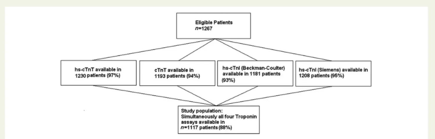
Advantageous Predictors of Acute Coronary Syndrome Evaluation (APACE) is an ongoing prospective international multicentre study designed and coordinated by the University Hospital Basel, Switzerland. From April 2006 to June 2009, a total of 1267 consecutive patients pre-senting to the ED with symptoms suggestive of AMI of ,12 h were en-rolled.3To reach a high rate of comparability, patients were included if simultaneous measurements of three hs-cTn assays [high-sensitivity cardiac Troponin T (hs-cTnT), Roche Diagnostics; hs-cTnI, Beckman-Coulter; and hs-cTnI Siemens] and conventional cTnT (Roche Diagnos-tics) were performed at presentation and serially thereafter, yielding to a study population of 1117 patients (Figure1). Baseline characteristics did not differ significantly between patients included and those not consid-ered for the respective analyses. There were no technical errors with any of the assays studied. The majority of patients were enrolled by the University Hospital of Basel, Switzerland (74.9%), followed by Hospital del Mar, Barcelona, Spain (13.2%), Hospital of Limmattal, Switzerland
(7.2%), Cantonal Hospital of Olten, Switzerland (4.7%). Patients with ter-minal kidney failure requiring dialysis were excluded. The study was carried out according to the principles of the Declaration of Helsinki and approved by the local ethics committees at each institution. Written informed consent was obtained from all patients. The authors designed the study, gathered and analysed the data, vouch for the data and analysis, wrote the paper, and decided to publish. The sponsors had no role in conducting the study or analysing the data.
Routine clinical assessment
All the patients underwent an initial clinical assessment that included clinical history, physical examination, 12-lead ECG, continuous ECG-monitoring, pulse oximetry, standard blood tests and chest radiography. Cardiac troponin, the MB fraction of creatine kinase and myoglobin were measured at presentation and after 6 – 9 h as long as clinically indicated. Treatment of patients was left to the discretion of the attending physi-cians who were unaware of the centrally measured hs-cTn values and only aware of the locally available conventional troponin results. All 12-lead ECGs were assessed as recommended in current guidelines4in
a core lab by internal medicine specialists blinded to patient details.
Adjudicated final diagnosis
Adjudication of final diagnoses was performed centrally in the core lab (University Hospital Basel) for all patients twice: Once according to con-ventional cTn levels used onsite (this method was used in the initial ana-lyses to examine the performance of hs-cTn assays8–12) and once including levels of Roche hs-cTnT in order to also take advantage of the higher sensitivity and higher overall diagnostic accuracy offered by hs-cTn assays6(this allows the additional detection of small AMIs that were missed by the adjudication based on conventional cTn assays). Two independent cardiologists reviewed all available medical records— patient history, physical examination, results of laboratory testing (including hs-cTnT levels), radiologic testing, ECG, echocardiography, cardiac exercise test, lesion severity, and morphology in coronary angiography—pertaining to the patient from the time of ED presentation to 90-day follow-up. In situations of disagreement about the dia-gnosis, cases were reviewed and adjudicated in conjunction with a third cardiologist.
Acute myocardial infarction was defined and cTn levels interpreted as recommended in current guidelines.13,14In brief, AMI was diagnosed when there was evidence of myocardial necrosis in association with a
Figure 1 Flow diagram displaying the proportions of patients with troponin measurements available from all eligible patients and the resulting study population with simultaneously all four troponin measurements available.
P. Haaf et al.
366
clinical setting consistent with myocardial ischaemia. Myocardial necrosis was diagnosed by at least one cTn value above the 99th percentile (or for the conventional cTn assays above the 10% imprecision value if not ful-filled at the 99th percentile) together with a significant rising and/or falling.11,14,15The criteria used to define rise and/or fall in conventional cTn and hs-cTnT are described in detail in Supplementary material online, Method.
Unstable angina was diagnosed in patients with normal cardiac tropo-nin levels and typical angina at rest, a deterioration of a previously stable angina and in cases of positive cardiac exercise testing or cardiac catheter-ization with coronary arteries found to have a stenosis of 70% or greater. As we adjudicated the cause of the presentation to the ED (¼acute chest pain) and not the cause of elevations of hs-cTnT, ‘stable coronary artery disease (CAD)’ was not a diagnostic group: a patient with ‘stable CAD’ with acute myocardial ischaemia at rest (acute chest pain) would there-fore be classified as either ‘unstable angina’ or ‘acute myocardial infarc-tion’. A further category was non-cardiac chest pain (such as musculoskeletal pain, gastroesophageal disorder). If no sufficient conclu-sive diagnostic procedures were performed, symptoms were classified as to be of unknown origin.
Follow-up and clinical endpoints
After hospital discharge patients were followed after 3, 12, and 24 months by telephone or in written form. Any clinical (cardiovascular) events since presentation to the ED were collected by establishing contact with the patient and his family physician. Information regarding death was also obtained from the national registry on mortality. The primary endpoint was all-cause mortality.
Investigational high-sensitivity cardiac
troponin analysis
Blood samples for determination of hs-cTn were collected at presenta-tion to the ED and serially thereafter at 1, 2, 3, and 6 h. Serial sampling was discontinued when the diagnosis of AMI was certain and treatment required transferring the patient to the catheter laboratory. All four troponin samples were frozen at 2808C until assayed in a blinded fashion in a dedicated core laboratory. High-sensitivity cardiac Troponin T was measured on the Elecsys 2010 (Roche Diagnostics), limit of blank and limit of detection (LoD) have been determined to be 3 and 5 ng/L, an imprecision corresponding to 10% coefficient of variation was reported at 13 ng/L and the 99th percentile of a healthy reference population at 14 ng/L,12cTnT (4th generation) was measured on the Elecsys 2010 (Roche Diagnostics); LoD of 0.01 mg/L, a 99th percentile cut-off value of 0.01 mg/L and a coefficient of variation of ,10% at 0.035 mg/L. Beckman-Coulter hs-cTnI was measured on the Access 2 analyser using an investigational prototype assay. According to the manufacturer, LoD is 2 ng/L, the 99th percentile of a healthy reference population is 9 ng/L with a 10% CV lower than the 99th percentile. For Siemens hs-cTnI, LoD is 5 ng/L, the imprecision level corresponding to 10% CV is found at 3 ng/L and the 99th percentile of a healthy reference popula-tion is 9 ng/L (all data according to the manufacturer).
Statistical analysis
Comparisons between groups were made using thex2method, Mann – Whitney U, or Kruskal – Wallis test. Receivers operating characteristic (ROC) curves were constructed to assess the sensitivity and specificity of hs-cTn assays and compared as recommended by DeLong et al.16 For the ROC analysis for 730-day mortality, patients who were alive at last patient contact and who had an observation time of slightly ,730 days were not excluded but counted as survivors (as this was the latest information available). Optimal cut-offs of ROC curve analysis were
chosen by scrutinizing Youden indices. Correlations between continuous variables were assessed using the Spearman rank-correlation method.
For comparisons of nested models likelihood-ratios were used. The Kaplan – Meier method was employed to analyse the timing of events during the follow-up. Statistical assessment was performed using the log-rank test.
Patients were categorized in three groups both below and above the 99th percentile of each respective troponin assay. This was done by both minimizing differences in the size of groups of one hs-cTn assay and to reach comparable group sizes for all hs-cTn assays. Besides, patients were also categorized in equally large hexiles.
Maximum, numerical, absolute changes were calculated for all patients within the first 6 h after presentation compared with the first value at presentation (0 h value). All serial measurements available (see also Sup-plementary material online, Table S1) were used for this calculation for each patient. The percentage change between the 0 h value of hs-cTnT and the respective 1h-value was calculated and the numerical change used for all calculations and illustrations.
All hypothesis testing was two-tailed and a P-value of ,0.05 was considered statistically significant. All statistical analyses were performed using SPSS for Windows 19.0 (IBM) and MedCalc 9.6.4.0 (MedCalc Software).
Results
Characteristics of patients
Baseline characteristics of the 1117 patients are shown in Table1. The
adjudicated final diagnosis was AMI in 215 (19.2%) patients (21% STEMI, 79% NSTEMI), unstable angina in 11.3%, cardiac symptoms of origin other than coronary artery disease in 14.0%, non-cardiac symptoms in 46.3%, and symptoms of unknown origin in 9.2%. All the patients were followed up with a median follow-up time of 798 days (IQR: 738 – 926). Ninety-two patients (8%) who did not die during follow-up had a follow-up period shorter than 730 days [median follow-up of 483 days (IQR: 399 – 631) in this patient group] (Supplementary material online, Table S2). Since APACE is an on-going study, 730 days of follow-up are not yet available in all patients enrolled in the study.
Levels of high-sensitivity cardiac troponin
in survivors and non-survivors
Levels of hs-cTn for patients who died within 30 days, respectively,
730 days in comparison with survivors are displayed in Figure2A
and B and Supplementary material online, Figure S1A and B. There was no significant difference in hs-cTnT concentrations of patients with STEMI [median 116 ng/L (IQR 26 – 579 ng/L)] and patients with NSTEMI [median 80 ng/L (IQR: 30 – 196 ng/L)] (P ¼ 0.254 for comparison). The median time to presentation did not differ signifi-cantly between patients with STEMI and NSTEMI.
High-sensitivity cardiac Troponin T had a moderately high correl-ation with hs-cTnI (Beckman-Coulter) (0.769) and hs-cTnI (Siemens) (0.758), and a low correlation with cTnT (0.376). The correlation between both hs-cTnI assays amounted to 0.785. When the analysis was carried out in groups according to the final diagnosis of AMI, dif-ferent results were observed: inter-assay correlations in the non-AMI group were much lower than in all patients and assays of patients with AMI at presentation displayed very high correlation numbers, with
near-perfect correlation between the two cTnT assays (Supplemen-tary material online, Figure S2).
Prognostic accuracy of high-sensitivity
cardiac troponin
During a follow-up time of 730 days 82 patients (7.3%) died with a median time to death of 189 days (IQR: 40 – 473). Of the 82 patients who died within the first 730 days, 31 patients (38%) suffered from cardiac death, 13 patients (16%) pulmonic death, 8 patients (10%) had other causes of death, and 30 patients (37%) unknown cause of death. Forty patients with AMI (18.6%) and 42 patients without AMI (4.7%) died during the first 730 days of follow-up.
The diagnostic accuracy did not differ significantly between the three hs-cTn assays, and only hs-cTnT outperformed the
conven-tional cTnT in the first 30 days (Table 2). No patient with an
hs-cTnT level ,9 ng/L died in the first 30 days. Both hs-cTnI assays did not differ significantly from the AUC of cTnT (AUC: 0.76; 95%
CI: 0.64 – 0.89) (Figure3A).
Long-term mortality (730 days) was most accurately predicted by hs-cTnT (Roche Diagnostics) expressed by an AUC of 0.78 (95% CI:
0.73–0.83), sensitivity of 70.7%, specificity of 74.8%, negative predictive value (NPV) 97.0%, and PPV of 18.2%. Both hs-cTnI assays did not differ significantly from the AUC of cTnT (AUC: 0.67; 95% CI: 0.61–0.74) and
were inferior to hs-cTnT (Figure3B). The risk of patients with negative
cTnT values, but elevated hs-cTn values at presentation is displayed in
Figure3C. Fifty-four patients in this analysis had normal (i.e. below the
99th percentile) inaugural hs-cTnT values and simultaneously con-verted to an elevated (.99th percentile) value in the first 6 h there-after. Of these 54 patients, only 2 patients (4%) died during the first 730 days. There were no statistically significant differences for any of the investigated assays between the centres providing internal valid-ation of our results.
The prognostic accuracy for the prediction of myocardial infarc-tion during the first 30 days of follow-up was not significant for any of the four assays; the AUC for the prediction of myocardial infarction in the first 730 days during the follow-up amounted to 0.64 (95% CI: 0.57 – 0.71) for both hs-cTnT and hs-cTnI (Beckman-Coulter); AUC of 0.63 (95% CI: 0.56 – 0.70) for hs-cTnI (Siemens), and an AUC of 0.58 (95% CI: 0.50 – 0.66) for cTnT; P-values for comparison were
not significant between all four assays (Table2).
. . . .
. . . .
. . . .
. . . . Table 1 Baseline characteristics of the patients
Characteristics All patients (n 5 1117) Non-Survivors (2 year) (n 5 82) Survivors (2 year) (n 5 1035) Age, year 64 (51 – 75) 80 (73 – 86) 62 (50 – 74) Male gender, n (%) 743 (67) 59 (72) 684 (66)
Body mass indexa 26.4 (24.0 – 29.7) 25.4 (22.9 – 27.7) 26.5 (24.0 – 29.8)
Hypertension, n (%) 746 (67) 73 (89) 673 (65) Hypercholesterolaemia, n (%) 497 (44) 43 (52) 454 (44) Diabetes, n (%) 214 (19) 24 (29) 190 (18) Current smoking, n (%) 271 (24) 13 (16) 258 (25) History of smoking, n (%) 387 (35) 35 (43) 352 (34) Pack years 30 (15 – 45) 40 (23 – 57) 30 (15 – 42) Family history (18 grade) , n (%) 430 (38) 24 (29) 406 (39) History, n (%)
Coronary artery disease 402 (36) 57 (70) 345 (33) Previous myocardial infarction 273 (24) 43 (52) 230 (22) Vital status
Heart rate, b.p.m. 76 (66 – 89) 83 (71 – 99) 75 (65 – 88) Systolic blood pressure (mmHg) 142 (127 – 160) 131 (113 – 155) 143 (128 – 160) Diastolic blood pressure (mmHg) 84 (74 – 93) 75 (67 – 88) 85 (75 – 93) Glomerular filtration rate (mL/min/1.73 m2)b 89 (71 – 106) 65 (46 – 85) 90 (73 – 107) Adjudicated final diagnosis, n (%)
STEMI 45 (4) 10 (12) 35 (3)
NSTEMI 170 (15) 30 (37) 140 (14)
Unstable angina 126 (11) 7 (9) 119 (11)
Cardiac, non-coronary disease 156 (14) 10 (12) 146 (14)
Non-cardiac symptoms 517 (46) 20 (24) 497 (48)
Unknown origin 103 (9) 5 (6) 98 (9)
a
The body mass index is the weight in kilograms divided by the square of the height in meters.
b
Glomerular filtration rate was calculated using Modification of Diet in Renal Disease formula (MDRD).
P. Haaf et al.
368
Intriguingly, in patients without AMI at presentation the prognostic accuracy of hs-cTnT for the prediction of long-term mortality
out-performed all other assays significantly (Figure3D); in patients with
AMI at presentation hs-cTnT and cTnT displayed the same high prog-nostic accuracy, whereas both hs-cTnI assays did not even reach
stat-istical significance (Figure3E). In patients with STEMI, neither any of
the four troponins studied (Table 4), nor TIMI flow (AUC: 0.59,
95% CI: 0.34 – 0.85, P ¼ 0.433), nor left-ventricular ejection fraction (AUC: 0.69, 95% CI: 0.49 – 0.90, P-value 0.095) provided a significant prognostic accuracy as to 730-day mortality.
The varying prognostic accuracy of all four troponin assays studied
stratified into the six main diagnostic groups is displayed in Table4for
730-day mortality and in Supplementary material online, Table S3 for 30-day mortality. The prognostic accuracy of all four troponin assays studied as to the prediction of cardiac and non-cardiac mortality is provided in Supplementary material online, Table S4. A gender-specific prognostic analysis is provided in Supplementary material online, Table S5.
Use of early and maximum changes
of high-sensitivity cardiac troponin
for the prediction of long-term
mortality (730 days)
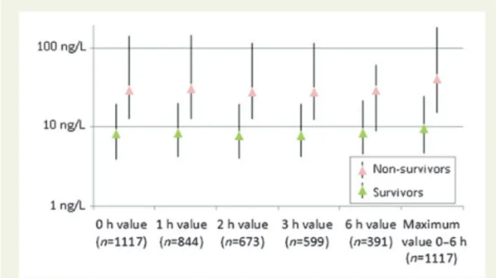
Values of hs-cTnT have been determined serially within the first 6 h
after presentation at the ED (Figure4; Supplementary material online,
Table S2 and Figure S3 and S4A – C). The prognostic accuracy of abso-lute and relative changes of hs-cTnT in the first hour and maximum
changes in the first 6 h are displayed in Figure5A. Absolute changes
outperformed relative changes in the accuracy of predicting long-term mortality (730 days) but were inferior to presentation values. Highest prognostic accuracy regarding changes of hs-cTnT was
achieved by the numerical absolute change in the first hour (|0–1 h
abs. change|) with an AUC of 0.66 (95% CI: 0.59–0.73) and by
|0–6 h abs. change| with an AUC of 0.69 (95% CI: 0.63–0.76). Relative changes—both in the first h and in the first 6 h including
their respective numerical values (|value|) did not yield significant
AUC (Figure5A).
The prognostic accuracy of the presentation value of hs-cTnT for long-term mortality (AUC: 0.78; 95% CI: 0.73 – 0.83) could not be improved by adding any changes of hs-cTnT within the first 6 h. Using maximum values of hs-cTnT measured in the first 6 h yielded to an AUC of 0.77 [95% CI: 0.72 – 0.82; P ¼ 0.051 for comparison
with presentation value (0 h value)] (Figure5A).
Mortality rates for hs-cTnT values at presentation are displayed in
Figure5B and Supplementary material online, Figure S5A and B for the
other troponin assays; mortality rates for maximum hs-cTnT values, (numerical) absolute, and relative changes of hs-cTnT within the first 6 h are displayed in Supplementary material online, Figure S6.
Similar results were obtained when both hs-cTnI assays were studied: neither changes nor maximum values of hs-cTnI occurring in the first 6 h could outperform or further improve the prognostic accuracy of their respective values at presentation (0 h value).
Prognostic accuracy of high-sensitivity
cardiac troponin in patients with
pre-existing coronary artery disease,
impaired renal function, and patients older
than 70 years
All three hs-cTn assays yielded significant AUC values for the predic-tion of long-term mortality (730 days) in the three subgroups
(Table3). Highest prognostic accuracy was achieved by hs-cTnT for
patients with pre-existing coronary artery disease (AUC: 0.74; 95% CI: 0.67 – 0.81), and patients older than 70 years (AUC: 0.68; 95% CI: 0.61 – 0.75) in comparison with both hs-cTnI assays. In patients Figure 2 (A and B) Presentation values of high-sensitivity cardiac troponins and cardiac troponin T in survivors and non-survivors during the first (A) 30 days and (B) 730 days; all values are displayed in nanogram per litre and as median values with inter-quartile ranges and outliers.
. . . .
. . . .
. . . .
Table 2 A prognostic performance of cardiac troponin assays during 2-year follow-up
n 5 1117 AUC (95% CI) Optimal cut-off Sensitivity (%) specificity (%) NPV (%) PPV (%) hs-cTnI (BC) hs-cTnI (S) c-TnT (R4)
Death in the first 30 days P-value for comparison
hs-cTnT,Roche Diagnostics 0.85 (0.78 – 0.91) 19.4 88.9 72.4 99.7 5.0 0.667 0.051 0.033
hs-cTnI, Beckman-Coulter (BC) 0.83 (0.74 – 0.92) 20.5 88.9 76.4 99.8 5.8 — 0.146 0.132
hs-cTnI, Siemens (S) 0.75 (0.63 – 0.87) 21.6 72.2 73.7 99.4 4.3 0.146 — 0.798
cTnT, Roche, 4th generation (R4) 0.76 (0.64 – 0.89) 13.0 61.1 82.9 99.2 5.5 0.132 0.798 —
Death in the first 730 days P-value for comparison
hs-cTnT, Roche Diagnostics 0.78 (0.73 – 0.83) 19.4 70.7 74.8 97.0 18.2 0.001 ,0.001 ,0.001
hs-cTnI, Beckman-Coulter (BC) 0.71 (0.65 – 0.77) 9.9 68.3 65.6 96.3 13.6 — 0.655 0.272
hs-cTnI, Siemens (S) 0.70 (0.64 – 0.76) 14.6 62.2 68.9 95.8 13.7 0.655 — 0.391
cTnT, Roche, 4th generation (R4) 0.67 (0.61 – 0.74) 28.0 43.9 88.2 95.2 22.8 0.272 0.391 —
MI in the first 730 days P-value for comparison
hs-cTnT, Roche Diagnostics 0.64 (0.57 – 0.71) 11.2 71.2 58.4 97.6 7.7 0.920 0.708 0.127
hs-cTnI, Beckman-Coulter 0.64 (0.57 – 0.71) 5.5 80.8 47.8 98.1 7.0 — 0.760 0.083
hs-cTnI, Siemens 0.63 (0.56 – 0.70) 6.3 73.1 53.1 97.6 7.1 0.760 — 0.155
cTnT, Roche, 4th generation (R4) 0.58 (0.50 – 0.66) 9.0 75.0 37.0 96.8 5.5 0.083 0.155 —
Optimal cut-off values are displayed as nanogram per litre.
P .Haaf et al .
370
Figure 3 (A and B) Area under the receivers operating characteristic curve displaying prognostic accuracy of high-sensitivity cardiac troponin assays for (A) early (30 days) and (B) long-term (730 days) all-cause mortality in comparison with cTnT4 (Roche Diagnostics). (C) Kaplan – Meier curves displaying cumulative survival during the follow-up in all acute chest pain patients stratified by presentation values of high-sensitivity cardiac troponin above (+) and below (2) the respective 99th percentile. (D and E) Area under the receivers operating characteristic curve dis-playing prognostic accuracy for long-term (730 days) all-cause mortality of high-sensitivity cardiac troponin assays for patients without (D) and with (E) acute myocardial infarction at presentation in comparison with cTnT4 (Roche Diagnostics); the area under the receivers operating characteristic curve of high-sensitivity cardiac Troponin T (Roche Diagnostics) was significantly higher than for both hs-cTnI assays in both patients with and without acute myocardial infarction at presentation; the area under the receivers operating characteristic curve for both hs-cTnI assays in patients with acute myocardial infarction did not reach statistical significance. Using either of the two hs-cTnI assays for the adjudication of acute myocardial infarction did not change the above results significantly.
with impaired renal function (,60 mL/min/1.73 m2glomerular filtra-tion rate) the prognostic accuracy of hs-cTnT (AUC: 0.69; 95% CI: 0.59 – 0.80) outperformed only hs-cTnI (Siemens). The prognostic accuracy of conventional cTnT did not differ significantly in all
three subgroups in comparison with all hs-cTn assays (Table3).
Discussion
In this prospective, observational, international, multicentre trial of 1117 unselected patients presenting with acute chest pain to the ED, we scrutinized the prognostic value of presentation values and serial measurements of three novel hs-cTn assays and compared them with a conventional cTn assay. We report five major findings.
First, hs-cTnT at presentation outperformed both hs-cTnI assays and conventional cTnT regarding the accuracy to predict 2-year mor-tality. This finding extends previous analyses that found similar high
diagnostic accuracy for the hs-cTn assays.3,8,9 High-sensitivity
cardiac Troponin T outperformed hs-cTnI in its prognostic accuracy
both in all patients and in important subgroups such as patients with AMI at presentation, pre-existing coronary artery disease, impaired renal function, or patients older than 70 years. Optimal prognostic cut-off values in these subgroups were only slightly higher than derived from all patients. In patients with AMI at presentation only hs-cTnT and cTnT yielded statistical significance. Secondly, novel hs-cTn assays moderately predict mortality in patients with pre-existing CAD, impaired renal function, and in patients older than 70 years. Thirdly, unlike in the diagnosis of AMI, neither serial mea-surements of hs-cTn nor changes in the first 6 h provided important additional information alone or in combination with presentation values of hs-cTn. To the best of our knowledge, this is the first analysis to clearly show that—in contrast to diagnostic
considera-tions5,6,17–21—for prognostic purposes serial measurements,
abso-lute, or relative changes of hs-cTn do not provide relevant added value regarding risk stratification of acute chest pain patients. This novel finding seems at least partly explained by the fact that most patients with acute chest pain show little change in their hs-cTn con-centration. In addition, those who do show a change are predomin-ately those with AMI, who already have the highest concentrations at presentation. It is important for clinicians to know that a patient’s risk of death can reliably be estimated already with the hs-cTn value at presentation. Fourthly, the prognostic benefit of the novel hs-cTn assays was pronounced in acute chest pain patients without AMI
(Figure 3D), a group of patients whose troponin concentrations
often cannot be detected and quantified by conventional cTn assays. This clinically highly relevant large group of patients can now be better risk-stratified with hs-cTn assays. It is important to em-phasize that in acute chest pain patients with AMI the prognostic
ac-curacy of hs-cTn assays was only moderate (Figure3E) and other
biomarkers22,23might be better (additional) prognosticators of
mor-tality in this patient group. Fifthly, the accuracy of the hs-cTnI assays to predict 2-year mortality did neither differ significantly between each other nor in comparison with conventional cTnT.
The reasoning behind the varying prognostic accuracy of the three hs-cTn assays studied remains speculative: the range of hs-cTn values Figure 4 Serial median high-sensitivity cardiac Troponin T
(Roche diagnostics) values in patients surviving and dying within 730 days. All values are displayed as median value (with inter-quartile range) in nanogram per litre.
Figure 5 (A) Area under the receivers operating characteristic curve displaying prognostic accuracy of high-sensitivity cardiac Troponin T and its early changes within the first 6 h for long-term (730 days) all-cause mortality; (B) mortality rates at 730 days for patients subdivided in six groups according to presentation values of high-sensitivity cardiac Troponin T.
P. Haaf et al.
372
. . . .
. . . .
. . . .
Table 3 Prognostic performance of cardiac troponin assays during 2-year follow-up for subgroups
Death in the first 730 days AUC (95% CI) Optimal cut-off Sensitivity (%) Specificity (%) NPV (%) PPV (%) hs-cTnI (BC) hs-cTnI (S) c-TnT (R4)
Pre-existing CAD (n ¼ 402) P-value for comparison
hs-cTnT, Roche Diagnostics (RD) 0.74 (0.67 – 0.81) 25.6 64.9 77.1 93.0 31.9 0.037 0.001 0.142
hs-cTnI, Beckman-Coulter (BC) 0.69 (0.61 – 0.76) 11.2 70.2 62.6 92.7 23.7 — 0.062 0.936
hs-cTnI, Siemens (S) 0.64 (0.56 – 0.72) 21.2 54.4 72.2 90.5 24.4 0.062 — 0.175
cTnT, Roche, 4th generation (R4) 0.69 (0.61 – 0.77) 13.0 50.9 84.0 91.2 34.5 0.936 0.175 —
Impaired renal functiona(n ¼ 160) P-value for comparison
hs-cTnT, Roche Diagnostics (RD) 0.69 (0.59 – 0.80) 29.3 73.5 62.7 89.8 34.7 0.104 0.002 0.363
hs-cTnI, Beckman-Coulter (BC) 0.63 (0.52 – 0.74) 15.4 67.6 57.1 86.7 29.9 — 0.241 0.509
hs-cTnI, Siemens (S) 0.58 (0.46 – 0.70) 379.8 35.3 88.9 83.6 46.2 0.241 — 0.075
cTnT, Roche, 4th generation (R4) 0.66 (0.55 – 0.77) 106.0 41.2 88.9 84.8 50.0 0.509 0.075 —
Older than 70 years (n ¼ 406) P-value for comparison
hs-cTnT, Roche Diagnostics (RD) 0.68 (0.61 – 0.75) 23.1 68.2 63.8 91.2 26.8 0.027 0.008 0.118
hs-cTnI, Beckman-Coulter (BC) 0.61 (0.53 – 0.69) 19.8 54.5 67.9 88.5 24.8 — 0.927 0.545
hs-cTnI, Siemens (S) 0.61 (0.53 – 0.69) 29.5 56.1 68.2 88.9 25.5 0.927 — 0.518
cTnT, Roche, 4th generation (R4) 0.63 (0.55 – 0.71) 29.0 45.5 82.1 88.6 33.0 0.548 0.518 —
Optimal cut-off values are displayed as nanogram per litre. CAD, coronary artery disease.
a
All patients with a renal function ,60 mL/min/1.73 m2
. cardiac troponins for mortality
373
in general and the overlap between survivors and non-survivors was highest for the hs-cTnI (Siemens) assay, thus resulting in a lower dis-criminatory power. Besides, hs-cTn assays measure tiny amounts of protein: It is currently unknown whether the incidence of disturbing phenomena such as fibrin interference or interference due to
anti-bodies varies significantly among novel hs-cTn assays.24 Ungerer
et al.25scrutinized four similar contemporary cTnT and cTnI assays
and concluded that the difference in cTnT and cTnI might in parts be explained by the fact that cTnT and cTnI measure different moieties, albeit that they are supposedly released in equimolar amounts via the same pathological process. Furthermore, it is known that haemolysis of even modest extent reduces cTnT and increases cTnI with some
assays.26Further studies need to re-evaluate the possible impact of
bio-logical variability, assay interference such as from nonspecific antibody binding in recently introduced hs-cTn assays and detect potential further confounding factors influencing their prognostic accuracy.
Bonaca et al.27showed that even small increases above the 99th
percentile of a sensitive cTn assay were associated with a significantly higher risk of death in both short-term (30 days) and long-term (12 months) perspective and proposed the use of the 99th percentile also for prognostic analyses. In this study, we have been able to dem-onstrate that mortality differs considerably even among patients with values below the 99th percentile. Detectable hs-cTn concentrations in the ‘normal range’ represent subclinical cardiomyocyte injury and
thus may have important clinical implications.28–30A dichotomous
outcome stratification seems to be hardly possible with any of the
four troponin assays studied. As illustrated in Figure5B any
measur-able amount of hs-cTnT seems to be associated with a higher risk of mortality, even at concentrations below the 99th percentile. Owing to the higher accuracy of hs-cTn assays at the 99th percentile in comparison with conventional assays, we believe that hs-cTn yields incremental prognostic benefit over the conventional cTnT assay. In particular, the large group of acute chest pain patients without AMI (and mainly low troponin concentrations) seems to benefit from the higher sensitivity of novel troponin assays and improved selection
of patients at risk (Table4; Figure3D and E).
All new hs-cTn assays permit a quantification of cTn concentra-tions in significant numbers of patients without coronary obstruction, such as patients with heart failure, hypertensive crisis, patients with subclinical heart disease, and even in apparently healthy subjects.
Our data as well as findings in other cohorts suggest that levels of hs-cTn should be considered quantitative markers of cardiomyocyte damage. The higher the hs-cTn level, the more extensive is the cardi-omyocyte damage. The association between hs-cTn ¼ cardiomyo-cyte damage and mortality seems to be linear. When comparing hs-cTnT with the fourth generation cTnT, the ROC curves for 720-day mortality are superimposable for the measureable range of the fourth generation cTnT. This indicates that both assays provide identical sensitivity for any given specificity. In addition to these values that provide a rather high specificity (1-specificity ¼ 0,2; therefore specificity ¼ about 80%), hs-cTn assays provide mea-surements in the undetectable range with the fourth generation assay that indicate that those patients labelled undetectable with the fourth generation represent a mixed group of patients: some with actually also very low levels of hs-cTn and therefore a very low risk of dying, but also some patients detected to have mildly elevated levels of hs-cTn indicating a risk of mortality that is much higher when compared with those with really normal hs-cTn levels.
The reasoning behind the good performance of hs-cTnT in patients with cardiac, non-coronary artery disease, and patients with unknown cause of chest pain remains speculative: Yet, it seems that any quantifiable amount of cardiomyocyte damage and therefore cTn release seems to be correlated with a worse prognosis
(Figure5B). Owing to the fact that the hs-cTnT assay is highly precise
. . . . . . . . . . . . . . . . . . . . . . . . . . . . Table 4 Area under the receivers operating
characteristic curve for presentation values of four cardiac troponin assays for the prediction of 730-day mortality
AUC 95% CI P-value
Patients with AMI (40/215 non-survivors)
hs-cTnT (Roche Diagnostics) 0.64 (0.55 – 0.73) 0.006 hs-cTnI (Beckman-Coulter) 0.57 (0.46 – 0.67) 0.189 hs-cTnI (Siemens) 0.56 (0.45 – 0.66) 0.265 cTnT (Roche Diagnostics) 0.64 (0.55 – 0.73) 0.006 Patients with STEMI (10/45 non-survivors)
hs-cTnT (Roche Diagnostics) 0.64 (0.46 – 0.83) 0.172 hs-cTnI (Beckman-Coulter) 0.57 (0.35 – 0.79) 0.495 hs-cTnI (Siemens) 0.59 (0.38 – 0.80) 0.397 cTnT (Roche Diagnostics) 0.61 (0.42 – 0.81) 0.281 Patients with NSTEMI (30/170 non-survivors)
hs-cTnT (Roche Diagnostics) 0.63 (0.53 – 0.74) 0.023 hs-cTnI (Beckman-Coulter) 0.56 (0.45 – 0.68) 0.288 hs-cTnI (Siemens) 0.54 (0.42 – 0.66) 0.510 cTnT (Roche Diagnostics) 0.64 (0.54 – 0.74) 0.015 Patients with Unstable Angina (7/126 non-survivors)
hs-cTnT (Roche Diagnostics) 0.66 (0.40 – 0.91) 0.168 hs-cTnI (Beckman-Coulter) 0.61 (0.42 – 0.80) 0.335 hs-cTnI (Siemens) 0.53 (0.37 – 0.70) 0.766 cTnT (Roche Diagnostics) 0.66 (0.46 – 0.87) 0.147 Patients with cardiac, non-coronary artery disease (10/156
non-survivors)
hs-cTnT (Roche Diagnostics) 0.8 (0.71 – 0.89) 0.001 hs-cTnI (Beckman-Coulter) 0.54 (0.41 – 0.68) 0.649 hs-cTnI (Siemens) 0.66 (0.55 – 0.78) 0.086 cTnT (Roche Diagnostics) 0.56 (0.34 – 0.79) 0.517 Patients with non-cardiac chest pain (20/517 non-survivors)
hs-cTnT (Roche Diagnostics) 0.74 (0.64 – 0.83) ,0.001 hs-cTnI (Beckman-Coulter) 0.63 (0.51 – 0.75) 0.049 hs-cTnI (Siemens) 0.61 (0.49 – 0.73) 0.095 cTnT (Roche Diagnostics) 0.47 (0.34 – 0.60) 0.636 Patients with unknown cause of chest pain (5/103 non-survivors)
hs-cTnT (Roche Diagnostics) 0.79 (0.61 – 0.97) 0.032 hs-cTnI (Beckman-Coulter) 0.84 (0.73 – 0.96) 0.01 hs-cTnI (Siemens) 0.69 (0.38 – 0.99) 0.16 cTnT (Roche Diagnostics) 0.56 (0.29 – 0.84) 0.64
All P-values refer to the respective ROC curve itself.
P. Haaf et al.
374
around the 99th percentile (,10% coefficient of variation) and is able to quantify troponin in the majority of healthy individuals, especially patients without AMI (and low concentrations of troponin) seem to benefit from the increased prognostic accuracy of hs-cTnT
(Figure3C). Nevertheless, the prognostic value in these subgroups
needs to be confirmed in further prospective studies, not least because the number of events (patients dying) in these subgroups was relatively small in this analysis.
The prognostic accuracy as to future myocardial infarction in the first 730 days did not differ significantly between the four cTn assays studied and was only modest for all four assays. Nevertheless, high NPVs could be reached by using optimal cut-off values slightly
below the 99th percentile of the respective assay (Table2).
There was an intriguing difference in correlation between hs-cTn
assays in patients with and without AMI at presentation. Lippi et al.31
in a small study of 47 patients reached similar conclusions as in this study: assays of hs-cTnT and hs-cTnI display much higher values for correlation in patients with than without AMI. The near-perfect correl-ation of hs-cTnT and cTnT in patients with AMI in contrast to a low cor-relation of the two assays in all patients (and not significant corcor-relation in patients without AMI) might be explainable by the fact that the same protein has been measured (with different assays) and the low sensitiv-ity of cTnT leading to non-detectable levels of cTnT in most patients without AMI. The near-perfect correlation of the two cTnT assays in
patients with AMI has also been shown by Lindahl et al.32in a large
study of patients with acute coronary syndrome. But, the fact that the correlation between the two cTnT assays in patients with AMI was much higher than between hs-cTnI assays and between hs-cTnT and the two hs-cTnI assays is more surprising and suggests a higher prognostic validity of hs-cTnT than hs-cTnI. This is also expressed by the AUC analyses: cTnT—although much less sensitive than hs-cTnI—was not inferior as to the prognostic accuracy than hs-cTnI assays; in patients with AMI it even outperformed the prognostic accur-acy of hs-cTnI assays. Based on our data, although cTnI and cTnT reflect the same pathological process, they do not seem to be interchangeable with each other as to their prognostic accuracy. Furthermore, correl-ation analyses of cTn after mixing patients with and without AMI might be misleading.
Limitations
First, as a prospective observational study, we cannot quantify exactly the clinical benefit associated with improved risk stratification. Sec-ondly, we cannot comment on prognostic accuracy among patients with terminal kidney failure requiring dialysis, since such patients were excluded from our study. Thirdly, due to the size of the study and limited number of events that occurred during the follow-up we cannot fully exclude a minor additional prognostic benefit of early changes of hs-cTn. Fourthly, patients with STEMI [whose diag-nosis is mainly based on the ECG, clinical presentation and not on (serial) troponin measurements] are underrepresented in this ana-lysis since their diagnosis required prompt transfer to the catheter la-boratory, often before a 1-h value of hs-cTn could be obtained. Fifthly, we cannot exclude the probability that cTn measurements and changes after the 6-h period would have improved the prognostic accuracy since no such study samples were taken. Sixthly, patients in cardiogenic shock might be underrepresented in this study, since their inclusion is hindered by proper informed consent. Seventhly,
due to inter-individual difficulties in taking blood samples and varying patients’ willingness to accept repetitive blood withdrawals not all serial measurements have been able to be determined in all patients. Therefore, the statistical power of our data set may have been too small to detect a small prognostic benefit of serial measure-ments.
Conclusion
High-sensitivity cardiac Troponin T predicts mortality more accur-ately than hs-cTnI assays in patients with suspected AMI. Unlike in the diagnosis of AMI, serial measurements and changes of hs-cTn do not seem to further improve the prognostic accuracy of presen-tation values of hs-cTn.
Supplementary material
Supplementary material is available at European Heart Journal online.
Acknowledgements
We thank the patients who participated in the study, the staff of the emergency department, the laboratory technicians, and particularly Kirsten Hochholzer, Esther Garrido, Irina Klimmeck, Melanie Wieland, and Fausta Chiaverio for their most valuable efforts, and we thank Drs C. Schindler and K. Denhaerynck for expert statistical advice.
Funding
We disclose that C.M. has received research support from the Swiss Na-tional Science Foundation (PP00B-102853), the Swiss Heart Foundation, the Stanley Thomas Johnson Foundation, Abbott, ALERE, Brahms, Nano-sphere, Roche, Siemens, and the Department of Internal Medicine, Uni-versity Hospital Basel, as well as speaker honoraria from Abbott, ALERE, Brahms, Roche, and Siemens. T.R. has received research grants from the University of Basel and the Department of Internal Medicine, University Hospital Basel as well as speaker honoraria from Brahms, and Roche. Conflict of interest: none declared.
References
1. Morrow DA, Cannon CP, Jesse RL, Newby LK, Ravkilde J, Storrow AB, Wu AH, Christenson RH, Apple FS, Francis G, Tang W, National Academy of Clinical B. Na-tional academy of clinical biochemistry laboratory medicine practice guidelines: Clin-ical characteristics and utilization of biochemClin-ical markers in acute coronary syndromes. Clin Chem 2007;53:552 – 574.
2. Keller T, Zeller T, Peetz D, Tzikas S, Roth A, Czyz E, Bickel C, Baldus S, Warnholtz A, Frohlich M, Sinning CR, Eleftheriadis MS, Wild PS, Schnabel RB, Lubos E, Jachmann N, Genth-Zotz S, Post F, Nicaud V, Tiret L, Lackner KJ, Munzel TF, Blankenberg S. Sen-sitive troponin i assay in early diagnosis of acute myocardial infarction. N Engl J Med 2009;361:868 – 877.
3. Reichlin T, Hochholzer W, Bassetti S, Steuer S, Stelzig C, Hartwiger S, Biedert S, Schaub N, Buerge C, Potocki M, Noveanu M, Breidthardt T, Twerenbold R, Winkler K, Bingisser R, Mueller C. Early diagnosis of myocardial infarction with sen-sitive cardiac troponin assays. N Engl J Med 2009;361:858 – 867.
4. Thygesen K, Alpert JS, White HD, Jaffe AS, Apple FS, Galvani M, Katus HA, Newby LK, Ravkilde J, Chaitman B, Clemmensen PM, Dellborg M, Hod H, Porela P, Underwood R, Bax JJ, Beller GA, Bonow R, Van der Wall EE, Bassand JP, Wijns W, Ferguson TB, Steg PG, Uretsky BF, Williams DO, Armstrong PW, Antman EM, Fox KA, Hamm CW, Ohman EM, Simoons ML, Poole-Wilson PA, Gurfinkel EP, Lopez-Sendon JL, Pais P, Mendis S, Zhu JR, Wallentin LC, Fernandez-Aviles F, Fox KM, Parkhomenko AN, Priori SG, Tendera M, Voipio-Pulkki LM, Vahanian A, Camm AJ, De Caterina R, Dean V, Dickstein K, Filippatos G, Funck-Brentano C, Hellemans I, Kristensen SD, McGregor K, Sechtem U, Silber S, Tendera M, Widimsky P, Zamorano JL, Morais J, Brener S, Harrington R, Morrow D, Lim M, Martinez-Rios MA, Steinhubl S, Levine GN,
Gibler WB, Goff D, Tubaro M, Dudek D, Al-Attar N. Universal definition of myocar-dial infarction. Circulation 2007;116:2634 – 2653.
5. Reichlin T, Irfan A, Twerenbold R, Reiter M, Hochholzer W, Burkhalter H, Bassetti S, Steuer S, Winkler K, Peter F, Meissner J, Haaf P, Potocki M, Drexler B, Osswald S, Mueller C. Utility of absolute and relative changes in cardiac troponin concentrations in the early diagnosis of acute myocardial infarction. Circulation 2011;124:136 – 145. 6. Haaf P, Drexler B, Reichlin T, Twerenbold R, Reiter M, Meissner J, Schaub N, Stelzig C, Freese M, Heinzelmann A, Meune C, Balmelli C, Freidank H, Winkler K, Denhaerynck K, Hochholzer W, Osswald S, Mueller C. High-sensitivity cardiac troponin in the distinction of acute myocardial infarction from acute cardiac noncor-onary artery disease. Circulation 2012;126:31 – 40.
7. Heidenreich PA, Alloggiamento T, Melsop K, McDonald KM, Go AS, Hlatky MA. The prognostic value of troponin in patients with non-ST elevation acute coronary syn-dromes: a meta-analysis. J Am Coll Cardiol 2001;38:478 – 485.
8. Reiter M, Twerenbold R, Reichlin T, Haaf P, Peter F, Meissner J, Hochholzer W, Stelzig C, Freese M, Heinisch C, Breidthardt T, Freidank H, Winkler K, Campodarve I, Gea J, Mueller C. Early diagnosis of acute myocardial infarction in the elderly using more sensitive cardiac troponin assays. Eur Heart J 2011;32: 1379 – 1389.
9. Reiter M, Twerenbold R, Reichlin T, Benz B, Haaf P, Meissner J, Hochholzer W, Stelzig C, Freese M, Heinisch C, Balmelli C, Drexler B, Freidank H, Winkler K, Campodarve I, Gea J, Mueller C. Early diagnosis of acute myocardial infarction in patients with pre-existing coronary artery disease using more sensitive cardiac troponin assays. Eur Heart J 2012;33:988 – 997.
10. Reichlin T, Schindler C, Drexler B, Twerenbold R, Reiter M, Zellweger C, Moehring B, Ziller R, Hoeller R, Rubini Gimenez M, Haaf P, Potocki M, Wildi K, Balmelli C, Freese M, Stelzig C, Freidank H, Osswald S, Mueller C. One-hour rule-out and rule-in of acute myocardial infarction using high-sensitivity cardiac troponin T. Arch Intern Med 2012;172:1211 – 1218.
11. Apple FS, Jesse RL, Newby LK, Wu AH, Christenson RH, National Academy of Clin-ical B, Damage ICfSoMoC. National academy of clinClin-ical biochemistry and IFCC com-mittee for standardization of markers of cardiac damage laboratory medicine practice guidelines: analytical issues for biochemical markers of acute coronary syn-dromes. Circulation 2007;115:e352 – e355.
12. Giannitsis E, Kurz K, Hallermayer K, Jarausch J, Jaffe AS, Katus HA. Analytical valid-ation of a high-sensitivity cardiac troponin T assay. Clin Chem 2010;56:254 – 261. 13. Thygesen K, Alpert JS, White HD, Joint ESCAAHAWHFTFftRoMI. Universal
defin-ition of myocardial infarction. J Am Coll Cardiol 2007;50:2173 – 2195.
14. Thygesen K, Mair J, Giannitsis E, Mueller C, Lindahl B, Blankenberg S, Huber K, Plebani M, Biasucci LM, Tubaro M, Collinson P, Venge P, Hasin Y, Galvani M, Koenig W, Hamm C, Alpert JS, Katus H, Jaffe AS, the Study Group on Biomarkers in Cardiology of the ESCWGoACC. How to use high-sensitivity cardiac troponins in acute cardiac care. Eur Heart J 2012;33:2252 – 2257.
15. Apple FS, Wu AH, Jaffe AS. European society of cardiology and American college of cardiology guidelines for redefinition of myocardial infarction: how to use existing assays clinically and for clinical trials. Am Heart J 2002;144:981 – 986.
16. DeLong ER, DeLong DM, Clarke-Pearson DL. Comparing the areas under two or more correlated receiver operating characteristic curves: a nonparametric ap-proach. Biometrics 1988;44:837 – 845.
17. Keller T, Zeller T, Ojeda F, Tzikas S, Lillpopp L, Sinning C, Wild P, Genth-Zotz S, Warnholtz A, Giannitsis E, Mockel M, Bickel C, Peetz D, Lackner K, Baldus S, Munzel T, Blankenberg S. Serial changes in highly sensitive troponin I assay and early diagnosis of myocardial infarction. JAMA 2011;306:2684 – 2693.
18. Giannitsis E, Becker M, Kurz K, Hess G, Zdunek D, Katus HA. High-sensitivity cardiac troponin T for early prediction of evolving non-ST-segment elevation myocardial in-farction in patients with suspected acute coronary syndrome and negative troponin results on admission. Clin Chem 2010;56:642 – 650.
19. Hamm CW, Bassand JP, Agewall S, Bax J, Boersma E, Bueno H, Caso P, Dudek D, Gielen S, Huber K, Ohman M, Petrie MC, Sonntag F, Uva MS, Storey RF, Wijns W, Zahger D, Bax JJ, Auricchio A, Baumgartner H, Ceconi C, Dean V, Deaton C, Fagard R, Funck-Brentano C, Hasdai D, Hoes A, Knuuti J, Kolh P, McDonagh T, Moulin C, Poldermans D, Popescu BA, Reiner Z, Sechtem U, Sirnes PA, Torbicki A, Vahanian A, Windecker S, Achenbach S, Badimon L, Bertrand M, Botker HE, Collet JP, Crea F, Danchin N, Falk E, Goudevenos J, Gulba D, Hambrecht R, Herrmann J, Kastrati A, Kjeldsen K, Kristensen SD, Lancellotti P, Mehilli J, Merkely B, Montalescot G, Neumann FJ, Neyses L, Perk J, Roffi M, Romeo F, Ruda M, Swahn E, Valgimigli M, Vrints CJ, Widimsky P. ESC guidelines
for the management of acute coronary syndromes in patients presenting without persistent ST-segment elevation: The task force for the management of acute cor-onary syndromes (ACS) in patients presenting without persistent ST-segment ele-vation of the European Society of Cardiology (ESC). Eur Heart J 2011;32:2999 – 3054. 20. Thygesen K, Mair J, Katus H, Plebani M, Venge P, Collinson P, Lindahl B, Giannitsis E, Hasin Y, Galvani M, Tubaro M, Alpert JS, Biasucci LM, Koenig W, Mueller C, Huber K, Hamm C, Jaffe AS. Recommendations for the use of cardiac troponin measurement in acute cardiac care. Eur Heart J 2010;31:2197 – 2204.
21. Thygesen K, Alpert JS, Jaffe AS, Simoons ML, Chaitman BR, White HD, the Writing Group on behalf of the Joint ESCAAHAWHFTFftUDoMI, Authors/Task Force Members C, Thygesen K, Alpert JS, White HD, Biomarker S, Jaffe AS, Katus HA, Apple FS, Lindahl B, Morrow DA, Subcommittee ECG, Chaitman BR, Clemmensen PM, Johanson P, Hod H, Imaging S, Underwood R, Bax JJ, Bonow RO, Pinto F, Gibbons RJ, Classification S, Fox KA, Atar D, Newby LK, Galvani M, Hamm CW, Intervention S, Uretsky BF, Gabriel Steg P, Wijns W, Bassand JP, Menasche P, Ravkilde J, Trials, Registries S, Ohman EM, Antman EM, Wallentin LC, Armstrong PW, Simoons ML, Heart Failure S, Januzzi JL, Nieminen MS, Gheorghiade M, Filippatos G, Epidemiology S, Luepker RV, Fortmann SP, Rosamond WD, Levy D, Wood D, Global Perspective S, Smith SC, Hu D, Lopez-Sendon JL, Robertson RM, Weaver D, Tendera M, Bove AA, Parkhomenko AN, Vasilieva EJ, Mendis S, Guidelines ESCCfP, Bax JJ, Baumgartner H, Ceconi C, Dean V, Deaton C, Fagard R, Funck-Brentano C, Hasdai D, Hoes A, Kirchhof P, Knuuti J, Kolh P, McDonagh T, Moulin C, Popescu BA, Reiner Z, Sechtem U, Sirnes PA, Tendera M, Torbicki A, Vahanian A, Windecker S, Document R, Morais J, Aguiar C, Almahmeed W, Arnar DO, Barili F, Bloch KD, Bolger AF, Botker HE, Bozkurt B, Bugiardini R, Cannon C, de Lemos J, Eberli FR, Escobar E, Hlatky M, James S, Kern KB, Moliterno DJ, Mueller C, Neskovic AN, Pieske BM, Schulman SP, Storey RF, Taubert KA, Vranckx P, Wagner DR. Third universal definition of myocardial infarction. Eur Heart J 2012;33:2551 – 2567.
22. Haaf P, Twerenbold R, Reichlin T, Faoro J, Reiter M, Meune C, Steuer S, Bassetti S, Ziller R, Balmelli C, Campodarve I, Zellweger C, Kilchenmann A, Irfan A, Papassotiriou J, Drexler B, Mueller C. Mid-regional pro-adrenomedullin in the early evaluation of acute chest pain patients. Int J Cardiol; doi: 10.1016/ j.ijcard.2012.10.025. Published online ahead of print 27 November 2012. 23. Meune C, Balmelli C, Twerenbold R, Reiter M, Reichlin T, Ziller R, Drexler B,
Stelzig C, Freese M, Wolf C, Haaf P, Osswald S, Mueller C. Utility of 14 novel biomar-kers in patients with acute chest pain and undetectable levels of conventional cardiac troponin. Int J Cardiol; doi: 10.1016/j.ijcard.2012.03.117. Published online ahead of print. Published online ahead of print 14 April 2012.
24. Jaffe AS. The 10 commandments of troponin, with special reference to high sensitiv-ity assays. Heart 2011;97:940 – 946.
25. Ungerer JP, Marquart L, O’Rourke PK, Wilgen U, Pretorius CJ. Concordance, vari-ance, and outliers in 4 contemporary cardiac troponin assays: Implications for har-monization. Clin Chem 2012;58:274 – 283.
26. Bais R. The effect of sample hemolysis on cardiac troponin i and t assays. Clin Chem 2010;56:1357 – 1359.
27. Bonaca M, Scirica B, Sabatine M, Dalby A, Spinar J, Murphy SA, Jarolim P, Braunwald E, Morrow DA. Prospective evaluation of the prognostic implications of improved assay performance with a sensitive assay for cardiac troponin I. J Am Coll Cardiol 2010;55:2118 – 2124.
28. Zethelius B, Johnston N, Venge P. Troponin I as a predictor of coronary heart disease and mortality in 70-year-old men: a community-based cohort study. Circulation 2006; 113:1071 – 1078.
29. Wallace TW, Abdullah SM, Drazner MH, Das SR, Khera A, McGuire DK, Wians F, Sabatine MS, Morrow DA, de Lemos JA. Prevalence and determinants of troponin T elevation in the general population. Circulation 2006;113:1958 – 1965. 30. Latini R, Masson S, Anand IS, Missov E, Carlson M, Vago T, Angelici L, Barlera S,
Parrinello G, Maggioni AP, Tognoni G, Cohn JN, Val-He FTI. Prognostic value of very low plasma concentrations of troponin T in patients with stable chronic heart failure. Circulation 2007;116:1242 – 1249.
31. Lippi G, Cervellin G, Aloe R, Montagnana M, Salvagno GL, Guidi GC. Non-commutability of results of highly sensitive troponin i and t immunoassays. Biochem Med (Zagreb) 2012;22:127 – 129.
32. Lindahl B, Venge P, James S. The new high-sensitivity cardiac troponin t assay improves risk assessment in acute coronary syndromes. Am Heart J 2010;160: 224 – 229.
P. Haaf et al.