Université de Montréal
i7i/t.
3/92.
)
Hydrophobically
-Modified Hydroxypropyl Celluloses:
Synthesis and Self
-Assembly in Water
Par
Mariella Piredda
Sciences Pharmaceutiques
Faculté de Pharmacie
Mémoire présenté à la Faculté des études supérieures
En vue de l’obtention du grade de
Maître ès Sciences (M. Sc.)
En sciences pharmaceutiques
Option Chimie médicinale
Avril, 2004
\f.
Université
14b
de Montré al
Direction des bibliothèques
AVIS
L’auteur a autorisé l’Université de Montréal à reproduire et diffuser, en totalité ou en partie, par quelque moyen que ce soit et sur quelque support que ce soit, et exclusivement à des fins non lucratives d’enseignement et de
recherche, des copies de ce mémoire ou de cette thèse.
L’auteur et les coauteurs le cas échéant conservent la propriété du droit
d’auteur et des droits moraux qui protègent ce document. Ni la thèse ou le
mémoire, ni des extraits substantiels de ce document, ne doivent être imprimés ou autrement reproduits sans l’autorisation de l’auteur.
Afin de se conformer à la Loi canadienne sur la protection des renseignements personnels, quelques formulaires secondaires, coordonnées
ou signatures intégrées au texte ont pu être enlevés de ce document. Bien
que cela ait pu affecter la pagination, il n’y a aucun contenu manquant.
NOTICE
The author of this thesis or dissertation has granted a nonexclusive license allowing Université de Montréal to reproduce and publish the document, in part or in whole, and in any format, solely for noncommercial educational and
research purposes.
The author and co-authors if applicable retain copyright ownership and moral rights in this document. Neither the whole thesis or dissertation, nor
substantial extracts from it, may be printed or otherwise reproduced without
the author’s permission.
In compliance with the Canadian Privacy Act some supporting forms, contact information or signatures may have been removed from the document. While this may affect the document page count, it does not represent any loss of
Université de Montréal
Faculté des études supérieures
Ce mémoire intitulé
$Hydrophobically
-Modifted Hydroxypropyl Cellulose:
Synthesis and Self- Assembly in Aqueous Media
Présenté par:
Mariella Piredda
A été évalué par un jury composé des personnes suivantes:
Suzanne Giasson
Président-rapporteur Géraldine Bazuin Membre du Jury Françoise M. Winnik Directeur de recherche111
RÉSUMÉ
Une série de polymères semi flexibles greffés dans lesquels la partie hydrophobe est distnbuée d’une façon statistique le long de l’unité de Ï’anhydroglucose- a été synthétisée à partir de l’hydroxypropyl cellulose (HPC), du polyoxyéthylène (10) cétyl éther (POEï0-Ci6; Brij-56®), du polyoxyéthylène (20) cétyl éther (POE20-C16; Brij-58®) et du polyoxyéthylène (20) stéaryle éther (POE20-Cig; Brij-78®). Les caractérisations physiques et chimiques des polymères modifiés hydrophobiquement sont décrites. La spectroscopie RMN du proton a été utilisée pour déterminer la quantité de Bnj conjugué avec les polymères hydrophiles.
Ce type de polymères comportant une partie hydrophile et une partie hydrophobe peut former des micelles, ceci étant dû à leur caractère amphiphule. Ainsi la partie hydrophobe forme le coeur de la micelle et la partie hydrophile entourant ce coeur lui permet de s’hydrater en milieu hydrophile. Ce système permet à la miceLle de transporter et de délivrer des médicaments pauvrement solubles dans l’eau. Tant que le coeur hydrophobe peut servir comme micro réservoir pour le médicament, ce dernier sera séparé du milieu par les parties hydrophiles de la micelle. La spectroscopie par fluorescence et la technique de la diffusion de la lumière ont permis la détermination de la dimension des agrégats des micelles polymériques en milieu aqueux. Le fait que les micelles polyménques soient solubles dans l’eau nous a permis d’étudier, avec succès, par DSC (microcalorimétrie thermique différentielle) les propriétés en solution de ces polymères hydrophobiquement modifiés, ainsi que de déterminer leur point de turbidité en fonction du taux de Brij. Du fait que l’HPC poile des groupements fonctionnels, elle pourrait avoir différentes applications dans les domaines biomédicaux comme la libération des médicaments, les diagnostics et la modification des surfaces à travers le couplage des substances bioactives.
Mots clés: HPC, Polysacchandes, micelles polyménques, polymères-HM, libération des médicaments, spectroscopie, diffusion de la lumière, calorimétrie.
ABSTRACT
A series of grafted semiflexible polymers, in which the hydrophobic side chains were located randomly along the anhydroglucose unit, were synthesized by reacting Hydroxypropyl cellulose (HPC) with polyoxyethylene (10) cetyl ether (P0E10-C16; Brij-56®), polyoxyethylene (20) cetyl ether (POE20-C16; Brij-5$®) and polyoxyethylene (20) stearyl ether (POE20-C18; Brij-78®). The physical and chemical characterization of Hydrophobically Modified (HIvI) - Polymers is described. 1H-NMR spectroscopy was used for a quantitative determination of the level of Brij®
conjugation onto water-soluble polymers. Such systems composed of hydrophilic and hydrophobic segments can form a micellar structure as a resuit of their amphiphilic character. The hydrophobic segment forms the hydrophobïc core of the micelle, and the hydrophilic segment surrounds this core as a hydrated outer shell. This core-sheil structure provides polymenc micelles with the potential for use as vehicles for drug delivery, since the hydrophobic core may serve as a microreservoir for drugs, which are segregated from the outer environment by hydrophilic segments. Considerable discussions are made on the physicochemical characteristic of polymeric micelles and dimension of the aggregates in aqueous milieu by Fluorescence spectroscopy and Dynamic Light Scattenng techniques. One of the favourable characteristics of polymenc micelles is the water-solubility: Differential Scanning Calonmetry (DSC) was successfully employed to study the solution properties of the HIVI-polymers to determine their cÏoud points and elucidate the effects of Brij® content. Functionalized
HPC is expected to have a wide utility in biomedical applications such as drLlg
delivery, diagnosis, and surface modification through the coupling of bioactive substances
Keywords: HPC, Polysaccharides, Polymeric micelles, HM-polymers, Drug deÏivery,
V
TABLE 0F CONTENTS
II’4StJ1’IIF
..iii
i%.13S1Ij\CT
iv
0F CONTENTS
‘Vl_
ISnI ()1
r1l/.iiÀES
...‘vii
LIST
0F’
viii
L IST O1’
1I1—N1’II.
S1ET1i
x
L IST 0F FLUO! ESCEN(E PlOTS
xi
ABBIATIONS
iii
C I1AlT1I 1
1
Introduction 1
1.1 Polymeric Micelles 2
1.2 Hydrophobïcally Modified (HM)-Cellulose Ethers 4
1.3 Hydroxypropyl cellulose (HPC) 6
1.4 Polyoxyethylene alcohols (Brij®) $
1.5 Cyclosporin A, (CsA), used as drug model 9
1.6 Characterization Methods of HM-Celluloses 11
1.6.1 Fluorescence spectroscopy 11
1.6.2 Dynamïc Light Scattering (DLS) 15
1.6.3 Microcalonmetry 17 1.6.4 Objectives 19 1.7 References 21
C E1APTEI.
2....
... ... .... ... .28
2.1 Expenmental Section 28 2.1.1.Materials 282.1.2 Synthesis of polyoxyethylene (20) cetyl ether tosylate1’2 2$ 2.1.3 Synthesis of HPC-g-polyoxyethylene (20) cetyl ether 29 2.1.4 GPC (Gel Permeation Chromatography) analysis 30
2.1.5 Steady-state fluorescence 30
2.1.6 Dynamic Light Scattering 32
2.2 References.33
jj/\prJj3j
3
34
3.1 Results and Discussion 34
3.1.1 Fluorescence 36
3.1.2 Dynamic Light Scattering 42
3.1.3 Microcalorimetry 42 3.2 References 46
z
4
48
4.1 Conclusions 48 4.2 References 50i\cknoiv1edgiients
51
F’I_tJ()JE S(11N(E1 PL
()s
xii
vii
LIST 0F TABLES
Table I. Cac values and particle mean diameters of aqueous solution of HPC
-POEnCm;
Table II. Hydrophobic content in RM-Polymers expressed as mrnoles of POE1C per litre cf solution
Table III. Cloud points and heat of transition (AH) of aqueocts solution of HM polymers
LIST 0F FIGURES
Figure 1.1. Cellulose hydrogen bonds keep adjacent chains together
Figure 1.2. Hydroxypropyl cellulose reported as an ideal structure with MS = 3
Figure 1.3. Synthetic scheme
Fïgtire 1.4. Structures of Polyoxyethylene(1O) cetyl ether (Brij®-56), Polyoxyethylene(20) cetyl ether (Brij®-58) and Polyoxyethylene(20) stearyl ether (Brij®-78).
Figure 1.5. Cyclosponn A (CsA); C62H111N11012; MW= 1202.61
Figttre 1.6. One form ofJabIoiiski diagram
Figtire 1.7. Representative excitation and emission spectrum Figtire 1.8. Common fluorescent and phosphorescent dyes Figure 1.9. Emission spectrum of pyrene in pure water at 25°C
FigtLre 2.1 Change in the fluorescence characteristic of pyrene as a fctnction of HPC-P0E20C18 (MS 3.1 mol%) concentration, below (À) and above
(.)the cac.
Figure 2.2 2 Emission spectrum of pyrene in pure water with the labelled peaks Ii and I.
Fïgtire 3.1. ‘H-NMR spectra of Brij-58®, HPC and HPC-P0E70C16; the former in CDCI3 and the others in DMSO-d6.
Figure 3.2 Emission spectra of pyrene in aqueous solutions of HPC-g-POE20-C16 (ca. 3.9%) at different polymer concentrations [pyrene in pure
water
(u), HM-polymer 0.006mgImL
(.).
HM-polymer 0.6 mg/rnL (À)].2ex 333nm
Figtcre 3.3 Excitation spectra of pyrene in aqueous solutions of HPC-g-POE2o-Ci (ca. 3.1%) at different polymer concentrations [HM-polyrner 0.006 mg/mL
(.),
HM-polymer 0.6mgImLt.),
HM-polymer 0.06 mg/mL (À)]. 2ern 390nmix
Figure 3.4. Plots of pyrene intensity ratio ii/Im as a function of HM-polymers
concentration.
(.)
HPC-P0E10C16 4.7 moÏ%,(.)
HPC-P0E10C16 5.4mol%, (Â) HPC-P0E10C16 0.9 mol%; for ail cases, as the polyrner
concentration increases above the cac, the ‘i”mratio decreases
Figure 3.5. Plots of pyrene intensÏty ratio 133J1333 as a function of HN’I-poiymers
concentration.
(.)
HPC-P0E20C18 3.1 mol%,(.)
HPC-P0E70C18 1.1mol%, (Â) HPC-P0E20C16 1.1 moi%; the ai-mw shows the CAC
value for HPC-P0E20C18 1.1 moÏ% sample
Figtire 3.6. Plots of pyrene intensity ratio 133611333 as a function of mmoi POEnCm
per litre of solution.
(.)
HPC-P0E20C15 3.1 mol%, (Â) HPCP0E20C18 1.1 mol%,
(.)
HPC-P0E20C16 1.1 moi%Figure 3.7. Microcaiorimetnc endotherm of aqueous solution of HPC-P0E10C16
with different MS.
(.)
0.9 mol%, (V) 4.7 mol%,(.)
5.4 rnoi%. 30°CIhr from 20 oc to $0 °C
LIST 0F ‘H-NMR SPECTRA
n
n
Spectrum 1. Spectrum 2. Spectrum 3. Spectriim 4. Spectrum 5. Spectrucn 6. Spectrum 7. Spectrum 8. Spectrum 9. Spectrum 10. Spectrum 11. Spectrum 12. Spectrum 13. Spectrum 14. HPC in DMSO-d6 POE]0C16 in CDCI3 P0E20C16 in CDC13 P0E20C18 in CDC13POE10C16-Tosyiate in CDC13 +TMS as internai standard
POE20C16-Tosyiate in CDC13 +TMS as internai standard
POE20C18-Tosylate in CDC13 +TMS as internai standard
HPC-g-POE10C16 (0.9 % moi) in DMSO-d6
HPC-g-POE10C16 (4.7 % moi) in DMSO-d6; here POEnCm =
P0E10C16, 2(m-3)= 13, la= 1.23, Ia/2(m-3)H x 100 4.7%
HPC-g-POE10C16 (5.4 % mol) in DMSO-d6 HPC-g-P0E20C16 (3.9 % moi) in DMSO-d6 NPC-g-POE20C16 (1.1 % mol) in DMSO-d6 HPC-g-POE20C18 (3.1 % moi) in DMSO-d6 HPC-g-P0E20C18 (1.1 % mol) in DMSO-d6
xi
LIST 0F FLUORESCENCE PLOTS
Fluo 1.Plots of pyrene intensity ratio ‘333/1336 as a function of HM-g-polymer
concentration:
(.)
HPC-g-POE20C16 3.9%, (.)HPC-g-POE20C16 1.1%.Fitto 2.Plots of pyrene intensity ratio I333336 as a function of HM-polymer
concentration:
(.)
HPC-g-POE20C18 3.1%, (•)HPC-g-POE0C18 1.1%. 2.Plots of pyrene intensity ratio 1333/1336 as a function of HM-polyrner concentration : (.)IIPC-g-POE20C18 3.1%, (.)HPC-g-POE70C18 1.1%.
Fltio 3.Plots of pyrene intensity ratio I333336 as a function of HM-polymer
concentration:
t.)
HPC-g-POE10C16 0.9%, (À)HPC-g-POE10C16 5.4%.(.)HPC-g-POE10C16 4.7%.
r)
n
fl
LIST 0F DSC THERMOGRAMS
DSC 1. Mïcrocalorimetric endotherms of aqueous solution of HPC-P0E10C16 wïth
different MS.
(.)
0.9 mol%, (V) 4.7 mol%,(.)
5.4 mol%. 30 °C/hr from20 °C to 80 °C.
DSC 2. Microcalonmetric endotherms of aqueous solution of HPC-P0E20C16 wïth
different MS.
(.)
1.1 mol%, (Â) 3.9 mol%, 30 °C/hr from 20 oc to $0 °cDSC 3. Microcalorimetric endotherms of aqueous solution of HPC-P0E20C18 with
xiii
ABBREVIATIONS
‘H-NMR Proton Nuclear Magnetic Resonance
AH Enthalpy of transition
CAC Critical Aggregation Concentration
CH2CÏ2 Methylene chloride
CDCI3 ChÏoroform deuterated
CMC Critical Micellar Concentration
Molar Heat Capacity
CsA Cyclosponn A
DLS Dynamic Light Scattering
DMF Dimethylformamide
DMSO Dimethylsulfoxide
DS Degree of Substitution
DSC Differential Scanning Calorimetry
GPC Gel Permeation Chromatography
HC1 Chloridnc acid
HM-HPC Hydrophobically ModifiedHPC
HPC Hydroxypropyl cellulose
NPLC High Performance Liquid Chromatography
LCST Lower Cntical Solution Temperature
MS Molar Substitution
Nag Aggregation Number
NaH Sodium hydroxyde
PAHs Polycyclic Aromatic Hydrocarbons
Py Pyrene
POE Polyoxyethylene
RES Reticuloendothelial System
5D Standard Deviation
TEA Triethylamine
TsC1 Tosyl chloride
Tos Tosylate
UV Ultraviolet
Ï
CHAPTER 1
Introduction
With the rapid progress of biotechnology, peptide drugs have become important therapeutic agents. A wide variety of peptides have been used as drugs, including hormones, synthetic peptides, enzymes substrates, and inhibitors1. Although they are highly potent and specific in their physiological functions, most peptides are difficuit to administer orally because of their unique physicochemical propeiÏies. These include large molecular weight, poor solubility in aqueous media, short plasma haif life, requirement for unique mechanisms for membrane transport, and susceptibility to enzymatic breakdown2. Many different approaches have been used to improve the oral absorption and enhance the bioavailability of peptide drugs. In recent years, enhanced bioavailability after oral administration has been observed upon entrapping the drug in a wide variety of polymer particulate carriers which protect the active molecules against inactivation by the host and control drctg release in body fluids, eg., blood, lymph and digestive juice3.
Microspheres4’5 and liposomes67 have been devised as delivery systems for the controlled release of vaccines, cytostatics and insulin with speciaÏ attention to
achieving long circulation in the blood stream, and avoiding the RES
(reticuloendothelial system) scavenging mechanism. However, the
reticuloendothelial system (mainly liver and spleen) actively removes a great majonty of these spheres when they are injected. Due to this scavenging, effective delivery of microspheres to organs other than those of the RES is often difficult to achieve.
Thus, several problems, such as biodistribution of drugs, drug solubility, undesirable side effects, rapid clearance by the RES, thermal instability, structurally fragile and low-loading efficiency stiil exist8. New technologies for the preparation of nanosized biodegradable polymenc particles are still required for providing more effective and selective drug delivery systems9’4
1.1 Polymeric Micelles
Graft and block copolymers under selective solvent conditions (good solvent for one block and non-solvent for the other block) adopt various organized structures known as “polymeric micelles”521. Micelles formed by block or graft copolymers belong to the family of colloidal systems.
Polymer micelles have been the subject of growing scientific and ïndustrial interest for their potential biomedical and pharmaceutical applications, such as drcig delivery’8’2231, diagnostics32, separation technology33, as well as electronics and optical technologies34’35. In particular, in the field of drug delivery, extensive studies have been carried out by many research groups, proposing to use polymer micelles as a novel carrier system for hydrophobic drugs. Many kinds of polymers that form various aggregates in water have been used for this purpose3640.
Polyrnenc micelles result from the association of amphiphilic polymers in water. They are in a mesoscopic size range (several tens of nanometers to 100 nanometers) having a characteristic core-sheli architecture, in which the solvophilic segments of the copolymer form an outer sheli surrounding the inner core composed of solvophobic segments4144. The core may be utilized as a reservoir of solvophobic compounds which are segregated from the outer environment. This hydrophobic environment possesses potential uses, for example it suppresses the inactivation of drugs molecules by decreasing contact with inactivating species in the aqueous phase
(e.g. water, specific enzymes), and it controls the drug release by micelle stability and micelle-core hydrophobicity45. The outer shell is responsible for the interaction with biocomponents such as proteins and cells, which may determine the pharmacokinetic
behaviour and the biodistribution of drugs. Thus, in vivo delivery of drugs may be
controlled by the outer-sheli segment independently of the inner core of the micelle, which expresses pharmacological activities.
By utilizing polymeric micelles, a unique delivery system may be constructed.
3
pharmacokinetic behaviour and biodistribution of polymeric micelles are the chemical charactehstics of the outer sheli, the water solubility and stability of the micelles.
Because a large number of drugs have a hydrophobic character, a concept very simple and fascinating was to coupling the drug with the carrier in order to deliver the drug by a carrier that had a specific affinity to the organs, tissues, and ceils. But conjugation of the drug with a polymer easily leads to precipitation because of the high and localized concentration of the hydrophobic drug molecules bound along the polymer chains. Thus, polymeric micelles were developed with a core-sheli structure, which may maïntain their water solubility by inhibiting intermicellar aggregation of the hydrophobic cores, irrespective of high hydrophobicity of the inner cores. Furthermore, since interactions among the hydrophobie segments are the driving force in the formation of micelles, the strength of that interaction determines their stability. Compared to low-molecular weight surfactant micelles, polymenc micelles are generally more stable because they self-assembly at much lower concentration, and show slower dissociation which allows longer retention of loaded drugs and higher drug accumulation at the target site22’46.
The use of polymenc micelles as oral delivery carriers systems has two specific advantages: the first is their size, with diameters in the range of approximately 10-100 nm, which is considered appropriate for evading renal excretion and non-specific capture by the RES. The second advantage includes the protection of the incorporated drug from the aggressive conditions present in the gastrointestinal tract.
In summary, drugs with a hydrophobie character can be easily incorporated into the inner core by physical entrapment475’ using experimental methods such as dialysis, salting out process, or solvent evaporation method3. Physical entrapment utilizing hydrophobie interaction can be applied to many ldnds of drugs, since most drugs possess hydrophobie moiety(ies) in their chemical structure52. Some reported advantages of these systems include reduced toxic side effects, solubilization of
hydrophobic drugs, stable storage, bio-distribution, and lower interactions with RES’6.
Based on these considerations, our hypothesis was to create an array of materials with controllable, predictable charactenstics such as water solubility and abillty to encapsulate or solubilize hydrophobic drugs. We expected that water solubility and encapsulation ability would be determined by the ratio of hydrophobic, aikyl chains located in the polymer core relative to the hydrophilic segments at the polymer sheil. Since the structural design of the amphiphilic graft copolymer is key in determining the stability of the micelles and their drug-releasing properties, modifying the hydrophobic charactenstics of the copolymer may provide micelles with high stability and the most desirable drug-releasing profile37;
1.2 HydrophobicalÏy Modified (HM)-Cellulose Ethers
Cellulose is a natural polymer and the chief component of wood and plant fibres. Cotton, for example, is almost pure cellulose. Cellulose is a polysaccharide composed of individual anhydroglucose (AHG) units which are held together by B 1,4 glycoside linkages which make cellulose a longrigid molecule.
Pure cellulose bas large crystalline regions due to hydrogen bonds between the -OH groups on different chains (Figure 1.1).
w I • I É I b j • j t I _I fl j — ,II t
ou
o
uo
I H0 I fl j, I t 2o
t II t -n
0 0H 0H Ho
U - U - O *4 0 Hu
o
jo
H Hu
Ii U H O OH HQH HFigtire 1.1. Cellulose hydrogen bonds keep adjacent chains together
In the manufacturing of cellulose ethers, purified cellulose is activated in a first step
with sodium hydroxide solution. In this aikalisation reaction, the structures of the crystalline areas are expanded allowing the hydroxyl groups to be transformed into alcoholate (this cellulose alcoholate is termed aikali cellulose). The strong attractive forces between cellulose chains due to interchain hydrogen bonds will be greatly reduced by alkylating the greater portion of the -OH groups, thereby preventing hydrogen bonding. Such chemical modification results in significantly changed charactenstics with regard to solubility, surface activity, chemical resistance and enzyme resistance. In this way it is possible to produce sets of cellulose ethers with carefully calculated performance characteristics. The properties of the cellulose ether are determined by the type of substituents, and also by their number and distribution along the molecule chain.
Cellulose ethers have a broad range of industrial applications. They are used as additives in materials such as paints, inks, papers, cosmetics, pharmaceuticals, foods and ceramics. Their solution properties are dictated mostly by the chemical structure
of the ether moiety and by the degree of substitution, e.g., the number of ether groups
per glucose unit, and to a lesser extent by the molecular weight of the polymer and by the fabrication process58.
Methylcellulose, hydroxypropylcellulose, and hydroxyalkylmethylcelÏulose, for example, at modest concentrations are soluble in cold water. The incorporation of low levels of hydrophobie modifiers (HM-polymers) on nonionic cellulose ethers resuits in polymers having highly unusual solution properties. As reported in detail from Landoll59, this behaviour is thought to be due to interchain association induced by hydrophobie moieties, analogous to the aggregation of Iow-molecular-weight amphiphatic species into mïce]les, but the resultïng aggregates are three-dimensïonai polymer networks. As a consequence, he found that the associative tendency (i.e. hydrophobie strength) of the modifying group governs both the viscosity and the onset of association (CMCy’ in solution. Particularly it was found that the concentration of hydrocarbon portion in solution, which depends on the size of the long-chain alkyl group, is closely related to the CMC, suggesting that it represent a cntical value for the onset of intermolecular association. Also Winnik et al.6° demonstrated that the solution properties of HfVT-cellulose ethers can be explained in terms of interchain polymer associations giving rise to aggregates, which influence the phase behaviour of the polymer solution61’62.
1.3 Hydroxypropyl cellulose (HPC)
Nearly ail polymers, both natural and synthetic, are polydisperse in nature, and cellulose is no exception. Depending on its source and processing, purified cellulose may contain fragments varying in molecular weight from a few thousand to a few million daltons. Usually the molecular size of cellulose and its derivatives is expressed in terms of the degree of polymenzatïon (DP), the average number of anhydroglucose monomer units in polymer chains. Cellulose derivatives, ethers or
esters, are formed by appropnate reaction with the three available hydroxyl groups on
each anhydroglucose unit; the extent of reaction is described as degree of substitution
“The critical aggregation concentration” or CAC is the concentration at which the interaction between surfactant micelles and polymer chains starts. “The critical micellization concentration” or CMC refers to a polymer-free surfactant solution. In our case surfactant and polymer are linked, thus the two terms refer to as the concentration below which virtually no micelles exist and above which almost ail additional copolymer goes into the micellar phase101
7
(DS), the average number of the three hydroxyls which are substituted. The substitution of such polymers is expressed in terms of the molar substitution (MS), the average number of moles of reactant combined per mole of monorner units. Whereas the maximum possible DS of a cellulose denvative is three, there is no theoretical limit to the MS63.
Hydroxypropyl cellulose (HPC), Figure 1.2, is formed from a base-catalyzed reaction of propylene oxide with “aikali cellulose”, whereby anionic nng-opening reaction (oligomenzation) occurs with scission of the methylene-oxygen bond, the product is ethenfied by a mixture of “oligo-propylene oxides” having vanous degrees of
polymerization64. The properties of HPC are severely influenced by the extent and uniformity of substitution, and much less by the poÏydispersity of their molecular weight distribution.
HPC is a nonionic semiflexible water-soluble polymer. Its characteristics were
reported primanly by the Hercules researchers around 197O6566; it is widely used as
an excipient in oral solid dosage fomis, in which it acts as a disintegrant67, and as a binder in granulation68. Essentially a non-toxic and non-irritant polysaccharide69, FIPC is recognized for its bioadhesive properties, and it is has been shown to promote drug absorption70.
As most cellulose ethers, HPC precipitates from water upon heating. When an
aqueous solution is heated it suddenly becomes cloudy at a characteristic
temperature, referred to as the “cloud point” or lower cntical solution temperature
OH OH
CH2OCH2H-CH3 OCH2-CH-CH3
(LCST). The phenomenon is reversible: upon cooling, the cloudy suspension becomes clear at the same temperature61. It has been demonstrated that even a very small level of substitution of hydrophobic groups affects the solubility of the polymer in water.
For this study we prepare hydrophobically-modified HPC graft copolymers (RPC-g POEiCm) of vanous compositions by attaching hexadecyl or octadecyl residues to the
hydrophilic HPC backbone via short POE linkers of different Iengths (Figure 1.3),
and we characterize them from a physicochemical point of view by fluorescence spectroscopy, dynamic light scattering and microcalorimetry techniques.
R= hydrophobic nx)difier
HPC
[e.g. R = POEnCmI
Figure 1.3. Synthetic scheme
1.4 Polyoxyethylene alcohols (Brij®)
Nonionic suffactants containing POE chains as hydrophulic moieties (C1E) are well known and have been widely used for decades. They have found applications in many fields, such as cosmetics, pharmaceuticals, paints, and cleaning agents. Moreover, they are routinely used for the solubilization of membrane proteins71. These types of compounds are generally prepared commercially by the condensation of ethylene oxide with fatty alcohols to yield a product in which the hydrophilic portion has a Poisson distribution of chains lengths72. These commercial products have been extensively studied7375; they show very nch phase behaviour in aqueous solutions, including phases from isotropic micellar to various ordered liquid crystalline ones. Their general structure is R(OCH2CH2)OH, where R is a long chain aikyl group or mixture of aikyl groups (Figure 1.4).
—o
9
n = 9; m = 13 P0E10C16 = Brij-56
n= 19;m= 13 P0E20C16=Brij-58
n = 19; m = 15 P0E20C18 = Brij-78
Figtire 1.4. Structures and commercial names of Alkylpolyoxyethylenes
POE(fl+l )C(m+3) Polyoxyethylene( 10) cetyl ether (Bnj ®-56), Polyoxyethylene(20)
cetyl ether (Brij®-58) and Polyoxyethylene(20) stearyl ether (Brij®-78).
Like HPC, the poly(ethyleneoxide) type nonionic surfactants possess a concentration and structure-dependent lower critical solution temperature.
1.5 Cyclosporin A, (CsA), used as drug model
Cyclosporin A (CsA) is a poorly water-soluble nonpolar cyclic peptide (Figure 1.5) comprising 11 aminoacids. It is produced from Tolypocladium inflatum Gams (formerly designated as Trichoderma polysporum) and other fungi imperfecti76. It is used clinically for immunomodulation, such as the prevention of xenograft rejection following transplantation of lddney, liver, bone marrow and pancreas7981, and it also applied in the treatment of patients with selected autoimmune diseases82. It inhibits the T-cell receptor signal transduction pathway via the formation of cyclosporïn A cyclophilin that inhibits calcineurin (protein phosphatase 2B)83. It inhibïts nïtric oxide synthesis induced by interleukin la, lipopolysaccharides and TNFa. and can block cytochrome c release from mitochondria. This drug has a great importance for achieving successful resuits of organ transplantation, but it must be carefully monitored due to the associated high risk of nephrotoxicity and hepatotoxicity reported in lïterature84.
[3
CH3 O
N— C,
CAbu—MeGly-MeLeu—D-Ala—Ala—MeLeu—Val—MeLeu
Figure 1.5. Cyclosporin A (CsA); C62H111N11012; MW= 1202.6 1
CsA is usually administered by the oral route, but it is reported as a typical drug with a non-regular absorption77’78. For this reason, this mode of administration presents
major problems related to highly variable and incomplete absorption from its
conventional oral formulation, leading to tremendous variation in drug
pharmacokinetics and, consequently, to an uncertain relation between drug dosage andin vivo exposure8587. The absorption of CsA is determined by the biliary flow, by intestinal integnty and motility, and by dietary intake8891, and its metabolism is
97
directly related to the cytochrome P-450-ffl-A in the reticuloendoplasmic system
Due to such characteristics, drug absorption and bioavailability may vary
considerably in the same patient and from one patient to another.
It is weIl known that the absolute bioavailability of CsA is low due to the poor absorption, which is related to the relatively high molecular weight, very high
lipophilicity°3 and poor solubility in aqueous fluids94; a recent study has
demonstrated that low bioavailability is due to the extensive CsA metabolism in the
11
1.6 Characterization Methods of HM-Celluloses
1.6.1 Fluorescence spectroscopyFluorescence spectroscopy is extensively used in physical chemistry and
biochemistry96. It is a valuable tool for the investigation of many micellar properties, including micelle formation, micelle-unimer equilibrium, micelle structure as weII as chain dynamics, and ldnetics of micelle formation and dissociation.
Luminescence is the emission of light from any substance and occurs from electronically excited states. Luminescence is formally divided in two categories, fluorescence and phosphorescence, depending on the nature of the excited state. For fluorescence phenomena, in excited singlet states the electron in the excited orbital is paired (of opposite spin) to the second electron in the ground-state orbital. Consequently, retum to the ground state is spin-allowed and occurs rapidly by emission of a photon.
Phosphorescence is emission of light from triplet excited states, in which the electron in the excited orbital has the same spin orientation as the ground-state electron.
Transitions to the ground state are forbidden and the emission rates are siow.
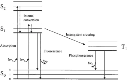
A Jabloiiski diagram, Figure 1.6, illustrates the processes that occur between the absorption and emission of light.
s2
si
nteystesystemcrossing
Fluorescence
0
The singlet ground, and first and second electronic states are depicted by So, S1, and S2, respectively. At each of these electronic energy levels, the fluorophores (fluorescent substances) can exist in a number of vibrational energy leveis, denoted byO, 1, 2, etc.
The transitions between states are depicted as vertical unes to illustrate the instantaneous nature of light absorption or emission. Transitions occur in about 1015 sec, a time too short for significant displacement of nuclei (Frank-Condon principie). Following light absorption, several processes can occur. A fluorophore is usually
excited to some higher vibrational level of either S or 52. Molecules in condensed
phases rapidly relax to the lowest vibrational level of Si. This process is called internai conversion and generally occurs in 1O2 sec or iess.
Since fluorescence lifetimes are typically near to iO sec, internal conversion is generaiiy complete prior to emission. Hence, fluorescence emission generally results from the thermally equilibrated excited state, that is, the lowest-energy vibrational state Si.
Retum to ground state typically occurs to a higher excited vibrational ground-state level, which then quickly (1OE12 sec) reaches thermal equilibrium. Molecuies in the S state can also undergo a spin conversion to the first triplet state, T1. Emission from T1 is termed phosphorescence and is generally shifted to longer wavelengths (iower energy) reiative to the fluorescence. Conversion of S to T1 is called intersystem crossing. Transition from T1 to the singlet ground state is forbidden, and as a resuit, rate constant for triplet emission is several orders of magnitude smaller than those for fluorescence98.
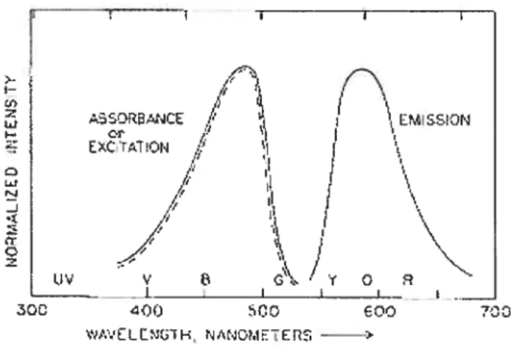
In a fluorescence spectrum excitation vs emission (Figure 1.7), the fluorescence intensity is plotted versus wavelength (nm).
t o o CHO Pyrene (Py) — H 3C 3H
2-Naphthalenesulfonate (2-NpS) ti&N__1___l,_i
H 13 f? ORe.NCE ExCrïON ‘ tiv c\.
/
‘\ EMSSONj
,Y O — f300 O0 400 SCO wAVELLN(iH, NA40METERS -——Figttre 1.7. Representative excitation and emission spectra
Since the polarity of the environment immediately suffounding a molecctle often
determines its basic properties, e.g. solubility or optical response, it is possible to use
fluorescence spectroscopy to detect the formation of the microenvironment within a macroscopically homogeneous solution and to correlate changes in photophysical parameters as a function of various stimuli to structural modifications within the self assemblies. Fluorescence typically occurs from aromatic molecules; thus, pyrene and
other polycyclic aromatic hydrocarbons (PHA) are used extensively for probing local
polarity (Figure 1.8).
1-Pyrenesulfonate (1 -PyS)
Pyrene- I -carboxaldehyde (PCA)
1 -Methylpyrene
H Profiavine (PF)
6-p-Toluidino-2-naphtalene-sulfonoc acid (TNS)
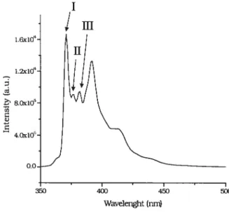
Pyrene is by far the most frequently used dye in fluorescence studies, and its spectroscopy is well documented99. It lias a long singlet lifetime; it readily forms excimers and the vibronic hand structure of its emission is sensitive to the environment; it is a strongly hydrophobic probe with low solubility (ca. 31OE7 M) in water.
Pyrene shows significant fine structure (vibronic hands) in its monomer emission spectrum. In the absence of any solvent interactions with the solute (either individually or collectively), the relative intensities of these vibronic bands in the fluorescence spectrum are govemed, as in a UV absorption spectrum, by the relative positions of the potential energy surfaces of the excited singlet states relative to the ground state singlet and by the Franck-Condon principle’°°. At low concentrations (< 1J06 M) in homogeneous solution, the ratio of the fluorescence intensity of the highest energy vibration hand (I) to the fluorescence intensity of the third highest energy vibrational hand (ifi) undergoes significant perturbation upon going from polar to non-polar solvents’°’’°4 (Figure 1.9). The I peak, which arises from the (0,0) transition from lowest excited electronic state, is a “symmetry-forbidden” transition that can be enhanced by the distortion of the electron cloud. On the other hand, the peak is forbidden and thus is relatively solvent-insensitive.
I
1m
Wavelenght (nn 1.6x1O 0.0. 400 450 50015
The fluorescence emission probe technique has rather microscopic and dynamic character. It utilizes the information from fluorescence emission spectra of probes incorporated into the polymer. The spectra are sensitive to the change in the environment around the probes; i.e., the change of the conformation of the polymer
and the chain dynamics are reflected by the change in spectra. For example, in a plot
of copolymer concentration against the ratio from excitation spectra, the
phenomenon of miccilization is accompanied by an abrupt decrease in 11/11,1 (over haif
a magnitude of concentration). The concentration where this sharp transition occurs is denoted as the critical micelle concentration.
1.6.2 Dynamic Light Scattering (DLS)
Following the pïoneering use of lasers and photon beating for the development of dynamic light scattering about thirty years ago, there have been numerous studies using light scattering techniques to investigate liquid dispersions in vanous research fields. Light scattering is one of the most important ways to charactenze new
polymeric and colloidal materials’°5’°8. Using modem instrumentation, one can
readily obtain the weight average molecular weight and radius of gyration from total intensity (static) light scattering, and the hydrodynamic (Stokes) radius from dynamic light scattering that is a non-invasive technique109 for investigation of dynamic processes by measuring the temporal fluctuations of the scattered light’10 In general,
the scatteming and the hydrodynamic behaviour are strongly nonlinear functions of
size, due to hydrodynamic interaction if the particles are composed of multiple subunits, and intra-particle interference if they are comparable in size to the wavelength of scattered light’
The general equation for the photoelectron count time coffelation function is”2
where g’2(t) is a normalized second-order correlation function, is the opticai constant of the instrument, and g1(T) is a normalized first-order colTelation function and is given by formula (2). F is a characteristic une width. For spherical particies, f
is given by a function of the translationai diffusion coefficient DT provided that
internai motions are negligible as shown in formulas (3) and (4).
g(l)
()
=exp(—Fr) (2)FrDq2 (3)
q=4nn0sin(I2)/%0 (4)
where O is the scattering angle, is the waveiength of the laser, n0 is the refractive
index of the solvent, and q is the magnitude of the scattering vector. In the low
concentration region, concentration dependence of the translation diffusion
coefficient DT can be expressed by a linear expression:
=
R
(1+k1c) (5)where D0 is the translation diffusion coefficient at infinite dilution, kd is the diffusion second virai coefficient, and c is the concentration. The hydrodynamic diameter (Stokes diameter) dh is given by the Stokes-Einstein equation:
= kBTI(3J»rDO) (6)
where kB is the Boltzman constant, T is the absolute temperature, and 1 the solvent viscosity.
17
gm()
= G(f)exp(—f)dF (7)
where the G(F) is the distribution function of f. In the cumulant method, autocolTelation functions are analyzed using an approximate equation
g(r)= exp[_fr+
2,22 (p /3!)3
+...]
(8)yielding an average characteristic une width (f) and a variance (polydispersity
index), i I12 . In the histogram method, estimation of the size distribution is caiiied
out by a correlation function profile of the histogram analysis software, and eq. (7) is rep!aced by
gt1r)=G(F)exp(—f)Af (9)
and G(f) is detenumned using the Marquart non-linear !east-square routine. G(f), which is a distribution function according to the ratio of light scattering by the particles with f, is then converted into the particle size distribution G(d) using eqs. (3) and (6). The distribution according to the weight ratio and the number ratio is then determined from G(cf).
The dynamic light scattering instrument requires a high-power laser, typica!ly an Argon gas laser, a temperature contro!led sample ce!!, a sensitive detector such as a photomultip!ier tube, and a time correlator capable of recording intensity (or cuiTent) from the photomultipler tube on an extremely short time scale (nanoseconds).
1.6.3 Microcalorimetry
Almost ai! physica! and chemical processes have an associated heat effect, and this can be used as the basis of a number of analytical techniques as well as for the
determination of absolute thermodynamic quantities. Differenti al scanning calorimetry (DSC) is an experimental technique to measure the heat energy change that takes place in a sample during controlled increase (or decrease) in temperature.
At the simplest level, it may be used to determine thermal transition (e.g. “melting”)
temperatures for samples in solution, solid, or mixed phases (e.g. suspensions). With
a sensitive apparatus and careful experimentation, it may be used to determine absolute thermodynamic data for thermally-induced transitions of various kinds. Formerly this was more the reaim of the dedicated specialist, but now with the ready availability of sensitive, stable, user-fnendly DSC instruments, microcalorimetry bas become part of the standard repertoire of methods available to the biophysical chemist for the study of macromolecular conformation and interactions in solution at reasonable concentrations. The advantages of calorimetric techniques arise because they are based on direct measurements of inthnsic thermal properties of the samples, and are usually non-invasive and require no chemical modifications or extnnsic
probes. Furthermore, with careful analysis and interpretation, calorimetric
experiments can directly provide fundamental thermodynamic information about the processes involved.
A calonmeter measures the energetics (enthalpy changes) associated with processes that occur, usually, during a linear heating temperature ramp. Measunng the heat flow into (endothermic) or out of (exothermic) a material as it undergoes a phase change yields a plot of temperature-dependent molar heat capacity, Cp.
One of the most fascinating properties exhibited by many water-soluble polymers is reverse temperature-dependent phase behaviour. They form isotropic, one-phase systems in water at or below room temperature, but when heated above a critical temperature (often called the cloud point), they separate into two phases, as indicated visually by a sudden transition from clear solutions to opaque suspensions. The phase transition is reversible: cooling the suspension below the cloud point resuits in a sudden clarification of the two-phase system”3.
19
As mentioned earlier, hydroxypropyl cellulose in water exhibits this phenomenon.
Functionalization of HPC, e.g. by attaching hydrophobic groups, even at a small level
of substitution, affects the temperature at which the phase transition occurs114 In
general, the LCST ofHM-HPC is lower than that recorded for a solution of HPC of
identical concentration (ca. 45°C for HPC 5.0 mglmL)”5; i.e. the LCST should
decrease with increasing hydrophobicity of the polymer’16”7.
1.6.4 Objectives
Many potent drug candidates fail in preclinical studies because of limited solubility, stability and toxicity. Thus, a number of drug delivery systems have been developed to overcome these transport problems. These include physical aggregates of amphiphilic molecules, such as polymeric micelles.
Major charactenstics of these systems are a highly water-soluble core (hydrophobic) sheil (hydrophilic) structure, high carrying capacity of hydrophobic drugs, prolonged storage stability, low viscosity and low interaction with biological components. It is well known that graft copolymers are able to form micelles by themselves in a common good solvent. These phenomena obviously result from the amphiphilic character of the compounds, which contain two groups or sequences greatly different in their solubility behaviour in the same molecule. In aqueous media, formation of hydrophobic regions is due to the hydrophobic interaction between the nonpolar groups, while the sheli consists of a dense brush of the hydrophilic segments. Polymeric micelles are an effective vehicle for the solubilization of hydrophobic drugs: the drug can be covalently coupled to the grafted copolymer or physically incorporated into the hydrophobic core of micelles. In contrast to micelles of small surfactant molecules, polymeric micelles are generally more stable and can retain a loaded drug for a longer penod of time.
This thesis focuses on the synthesis and physicochemical charactenzation of new polymeric micelles, which offer potential as carrier systems for hydrophobic drugs and other active molecules. The grafted-polymers were designed to ensure water
solubility and promote hydrophobic associations by a balance between hydrophilic
group and hydrophobie moieties, by grafting hydroxypropyl cellulose with
hydrophobic hexadecyl and octadecyl residues (HPCPOEnCm). Hydroxypropyl cellulose (HPC) was chosen to provide a hydrated steric barrier; POEnCm (n = 10/20 and m = 16/18) was chosen as hydrophobic segment, to both maintain the solubility and promote hydrophobic association in water of the novel HM-polymers.
We expected the HM-polymers to form micelles in water solution, exhibiting a strong tendency to self-associate via inter or intrapolymeric interactions of the hydrophobic side chains when the concentration of the amphiphilic graft copolymer is above the critical micelle concentration. We studied aggregation processes of graft-copolymers in dilute solution with fluorescence emission and excitation probe techniques and with microcalorimetry, while the size of these grafted-copolymer micelles was determined by dynamic light scattenng.
21
1.7 References
n
(1) Lee, V. H. L., J ControÏÏedReÏ. 1990, 13, 213
(2) Lee, V. H. L.; Dodda-Kashi, S.; Grass, G. M.; Rubas, W. In Lee, V. H. L. (Ed.),
Protein andPeptide Drug Delivery. Marcel Dekker, New York, 1991, pp. 691
(3) Kim, S. Y.; Shin, I. G.; Lee Y. M. Biomaterials 1999, 20, 1033
(4) Couvreur, P.; Vauthier, C. I ControlledRel. 1991, 17, 187
(5) Kreuter, J. I ControlledRel. 1991, 16, 169
(6) Weinstein, J. N. In Ostro, M. J. (Ed.), Lzosornes. from Biophysics to
Therapeutics, Marcel Dekker, New York, 1987, pp. 277
(7) Tardi, P. G.; Boman, N. L.; Cullis, P. R. I Drug Targeting 1986, 4, 129
(8) Nah, J.-W.; Jeong, Y.-I.; Cho, C.-S. I Polym. Sel.: Part B. Polym. Phys. 1998,
t
36,415
(9) Kreuter, J. I ControïledRet. 1991, 16, 169
(10) Seijo, B.; Fattal, E.; Treupel, L.; Couvreur, P. Int. I Pharm. 1990, 62, 1 (11) Peppas, L. Int. J Pharm. 1995, 116, 1
(12) Cavallaro, G.; Fresta, M.; Giammona, G.; Puglisi, G.; Villari, A. Int. I Pharm. 1994, 111,31
(13) Schoiz, G.; lijima, M.; Nagasaki, Y.; Kataoka, K. Macromoleciiles 1995, 28, 7295
(14) La, S.; Okano, T.; Kataoka, K. I Pharm. Sel. 1996, 85 (1), 85
(15) Riess, G.; Hurtrez, G.; Bahadur, P. In EncycÏopedia ofFolymer Science and
Engineering, Ed.; Mark, H. F.; Bikales, N. M.; Overberger, C. G.; Menges,
G. Eds. Wiley-Interscience: New York, 1985, 2, 324
(16) La, S. B.; Okano, T.; Kataoka, K. I Pharm. Sci. 1996, 85 (1), 85
(17) Riess, G.; Hurtrez, G.; Bahadur, P. In EncycÏopedia ofPolymer Science and Technology; Wiley: New York, 1985
(18) Bader, H.; Ringsdorf, H.; Schmidt, B. Angew. Makromol. Chem. 1984, 123/124, 457
(19) Xu, R.; Winnik, M. A.; Riess, G.; Chu, B.; Croucher, M. D. MacromoÏecuÏes 1992, 25, 644
(20) Kwon, G.; Naito, M.; Yokoyama, M.; Okano, T.; Sakurai, Y.; Kataoka, K. Langmïtir 1993, 9, 945
(21) Gao, Z.; Eisenberg, A. Macromotecules 1993, 26, 7353
(22) Kataoka, K.; Kwon, G. S.; Yokoyama, M.; Okano, T.; Sakuraï, Y. J ControÏled
ReÏ. 1993, 24, 119
(23) Zhang, L.; Eisenberg, A. Science 1995, 268, 1728
(24) Nakamura, K.; Endo, R.; Takeda, M. I Folym. Sci. FoÏym. Phys. Ed. 1976, 14, 1287
(25) Yokoyama, M.; moue, S.; Kataoka, K.; Yui, N.; Okano, T.; Sakurai, Y. MakromoÏ. Chem. 1989, 190, 2041
(26) Nishimura, T.; Sato, Y.; Yokoyama, M.; Okuya, M.; moue, S.; Kataoka, K.; Okano, T.; Sakurai, Y. Makromol. Chem. 1984, 185, 2109
(27) Piskin, E.; Kaitian, X.; Denkbas, E. B.; Kiçûkyavuz, Z. J. Biornat. $ci. Polym. Ed. 1995, 7, 359
(28) Tobio, M.; Gref, R.; Sanchez, A.; Langer, R.; Alonso, M. J. Pharin. Res. 199$, 15, 270
(29) Soma, C. E.; Dubemet, C.; Barratt, G.; Nemati, F.; Appel, M.; Benita, S.;
Couvreur, P. Pharm. Res. 1999, 16, 1710
(30) Cammas-Marion, S.; Béar, M.-M.; Harada, A.; Guérin, P.; Kataoka, K. MakromoÏ. Chem. Phys. 2000, 201, 355
(31) Kwon, G. S.; Kataoka, K. Adv. Drug. Delivery Rev. 1995, 16, 295
(32) Trubetskoy, V. S.; Frank-Kamenetsky, M. D.; Whiteman, K. R.; Wolf, G. L.;
Torchilin, V. P. Acad Radio!. 1996, 3, 232
(33) Nagarajan, R.; Barry, M.; Ruckenstein, E. Langmuir, 1986, 2, 210
(34) Moffitt, M.; McMahon, L.; Pessel, V.; Eisenberg, A. Chem. Mater. 1995, 7,
1185
(35) Spatz, J. P.; Roesher, A.; Môller, M.Adv. Mater. 1996, 8, 337
(36) Gast, A. P.; Vinson, P. K.; Cogan-Farinas, K. A. Macromolecutes 1993, 26, 1774
(37) Antonietti, M.; Heinz, S.; Schmidt, M.; Rosenauer, C. MacromoÏecuÏes 1994,
23
(38) Zhao, J.
Q.;
Pearce, E. M.; Kwei, T. K.; Jeon, H. S.; Kesani, P. K.; Balsara, N.P. Macromolecules 1995, 28, 1972
(39) Kidchob, T.; Kimura, S.; Imanishi, Y. J ControïtedRel. 1998, 51, 241
(40) Wang, L-Q.; Tu, K.; Li, Y.; Zhang, J.; Jiang, L.; Zhang, Z. React. Funct.
Polym. 2002, 53, 19
(41) Tuzar, Z.; Kratochvil, P.Adv. Colloidlnterf Sel. 1976, 6, 201
(42) Halperin, A.; Tiffe!!, M.; Lodge, T. P. Adv. FoÏym. Sci. 1992, 100, 31
(43) Tuzar, Z.; Kratochvil, P. Surf Colloids Sel.; Plenum Press: New York 1993, 15,
1
(44) Webber, S. E. J Phys. Chem. B 1998, 102, 261$
(45) Yokoyama, M. Crit. Rev. Ther. Drug Carrier Syst. 1992, 9 (3, 4), 213
(46) Kwon, G. S.; Suwa, S.; Yokoyama, M.; Okano, T.; Sakurai, Y.; Kataoka, K. J ControlledRel. 1994, 29, 17
(47) Kwon, G. S.; Naito, M.; Yokoyama, M.; Okano, T.; Sakurai, Y.; Kataoka, K. Pharm. Res. 1995, 12, 192
(4$) Cammas, S, Suzuki, K.; Sone, Y.; Sakurai, Y.; Kataoka, K.; Okano, T. J
ControÏÏes ReÏ. 1997, 48, 157
(49) Chung, J. E.; Yokoyama, M.; Aoyagi, T.; Sakurai, Y.; Okano, T. J ControÏÏed
ReÏ. 1998, 53 (1-3), 119
(50) Kohori, F.; Sakai, K.; Aoyagi, T.; Yokoyama, M.; Yamato, M.; Sakurai Y.; Okano, T. CoÏÏoids Surf B: Biointerfaces 1999, 16 (1-4), 195
(51) Chung, J. E.; Yokoyama, M.; Okano, T. J ControlledReÏ. 2000, 65, 93
(52) Yokoyama, M.; Okano, T.; Sakurai, Y.; Fukushima, S.; Okamoto, K. Kataoka, K. J Drug Targeting 1999, 7 (3), 171
(53) Gref, R.; Minamitake, Y.; Peracchia, M. T.; Trubetskoy, V.; Torchilin, V.;
Langer, R. Science, 1994, 263, 1600
(54) Kwon, G. S.; Yokoyama, M.; Okano, T.; Sakurai, Y.; Kataoka, K. Fharm. Res. 1993, 11, 970
(55) Kwon, G. S.; Suwa, S.; Yokoyama, M.; Okano, T.; Sakurai, Y.; Kataoka, K. J
(56) Liu, L.; Li, C.; Li, X.; Yuan, Z.; An, Y.; He, B. I Appl. PoÏyrn. Sci. 2001, 80, 1976
(57) Gorshkova, M. Y.; Stotskaya, L. L. Polym. Adv. Tech. 1998, 9, 362
(58) Just, E. K.; Majewicz, T. G.; EncycÏopaedia of Folyrner Science and
Engineering, 2ndEd.; John Wiley: New York, 1985; Vol. 3, pp 226
(59) Landoll, L. M. I Polym. Sci.: Polym. Chem. Ed, 1982, Vol. 20, 443
(60) Winnik, F. M.; Regismond, S. T. A; Goddard, E. D.Langmuir 1997, 13, 111
(61) Winnik, F. M. Macromolecules 1987, 20, 2745
(62) Thuresson, K.; Niisson, S.; Lindman, B.Langmuir 1996, 12, 2412
(63) Wirick, M. G.; Waldman, M. H.I Appi. Polym. Sci. 1970, 14, 579
(64) Tezuka, Y.; 1mai, K. Carb. Res. 1990, 196, 1
(65) Aqualon; Technical Literature: Klucel, Hydroxypropyl cellulose, a nonionic
water-soluble polymer: physical and chemical properties, 1987
(66) Salamon, J. C.; Polymeric Materials Encyclopedia, Vol. 5, CRC Press, Boca
Raton, 1996, 3167
(67) Machida, Y.; Nagai, T. Chem. Pharm. Buil. 1974, 22, 2346
(6$) Skinner, G. W.; Harcum, W. W.; Bamum, P. E.; Guo, J. H. Drïtg Dcliv. md
Pharm. 1999, 25, 1121
(69) Final report on the safety assessment of hydroxyethyl cellulose, hydroxypropyl cellulose, methylcellulose, hydroxypropyl methylcellulose and cellulose gum J. Am. Cou. Toxicol. 1986, 5, 1
(70) Suzuki, Y.; Makino, Y. I ControlledRel. 1999, 62, 101
(71) The Merck Index; Monography no. 07659
(72) Flory, P. I Am. Chem. Soc. 1940, 62, 1561
(73) Kushner, L. M.; Hubbard, W. D. I Phys. Chem. 1954, 58, 1163 (74) Becher, P.; Clifton, N. K. I CoÏloidSci. 1959, 14, 519
(75) Heush, R. Prog. ColloidPoÏym. Sci. 1978, 65, 186
(76) The Merck Index; Monography no. 02781
(77) Klompmaker, I. J. Ther. DrugMonit. 1993, 15,60
25
(79) Matzke, G. R.; Luke, D. R. In Herfindal, E. T.; Gourley, D. R.; Hart, L. L. (Eds.), Clinical Fharmacy and Therapeutics. William and Willdns, Baltimore, 1988, pp. 229
($0) Lee, M.-K.; Choi, L.; Kim, M.-H.; Kim, C.-K. Inter. I Fharm. 1999, 191, 87 (81) Mcevoy, G. K. Cyclosporine. In Mcevoy G. K.; Litvak, K.; Welsh, O. H.
(Eds.), AHFS Drug Information. The Amencan Society of Health System Pharmacists, Inc., Bethesda, 1995, pp. 2570
(82) Martindale, in: K. Parfitt (Ed.), The Complete Drug Rejèrence, 32,
Pharmaceutical Press, London, 1999, pp. 519
(83) Gref. R.; Quellec, P.; Sanchez, A.; Calvo, P.; Dellacherie, E.; Alonso, M. J. Eur. I Fharm. Biopharm. 2001, 51, 111
(84) Molpeceres, J.; Aberturas, M. R.; Guzman, M. I Microencapsulation 2000, 17 (5), 599
($5) Kahan, B. D. Pharmacokinetic Profiles of Sandinnum and Neoral. XVth World Congress of Transplantation $ociety, Kyoto, Japan 1994
(86) Kahan, B. D. Kidney Inter. 1993, 44 (43), 12
($7) Tufverson, G.; Odlind, B.; Sjôberg, A.; Lindberg, A.; Gabrielsson, J.; Lindstrôm, B.; Lithell, H.; Selinus, I.; Tôtterman, T.; Wahlberg, J. Transplant
Froc. 1986, 18 (5), 16
(88) Grevel, J. Transplant Froc. 1986, 18 (5), 9
(89) Drewe, J.; Berlinger, C.; Kissel, T. Br. I Clin. Fharmacol. 1992, 33 (90) Reymond, J. P.; Sucker, H.; Vonderscher, I Fharm. Res. 1988, 5, 677 (91) Wassef, R.; Cohen, Z.; Nordgren, S.; Langer, B. Kidney Int. 1985, 27, 593 (92) McCaughan, G. W.; Bookallilm Sheil, A. Transplant Froc. 1994, 26, 2674 (93) Taylor, N. E.; Mark, A. E.; Vallat, P.; Brunne, R. M.; Testa, B.; Van Gunteren,
W. F. I MecL Chem. 1993, 36, 3752
(94) Ismailos, G.; Reppas, C.; Dressman, J. B.; Macheras, P. I Fharrn. Fharrnacol. 1991, 43, 287
(95) Wu, C. Y.; Bennet, L. Z.; Bebert, M. F.; Gupta, S. K.; Rowiand, M.; Gomez, D. Y.; Whacher, V. I Clin. Fharm. Ther. 1995, 58, 492
(96) W. R. G. Baeyens, D. De Keukeleire, K. Korkidis (Eds.), Luminescence techniques in Chemical and Biochemical Analysis, M. Dekker, New York, 1991
(97) Atldns, P. W. Physical Chemistry, 3idEd., 1986, Oxford University Press
(98) Lakowicz, J. R. Principles of fluorescence Spectroscopy, 2nd Ed, Kiuwer
Academic, Plenum Publisher, New York
(99) a- Winnik F. M. Chem. Rev. 1993, 93, 587; b-Turro, N. J.; Baretz, B. H.; Kuo
P.-L. Macromolecules 1984, 17, 1321; e- Thomas K. J. I Phys. Chem 1987, 91,
267; d- Fender J. H. Chem. Rev. 1987, 87, 877; e- Grieser F.; Drummond C. J.
I Phys. Chem. 198$, 92, 5580; f- Ananthapadmanabhan, K. P.; Goddard, E. D.;
Turro, N. J.; Kuo P. L. Langmuir 1985, 1, 352; g- Winnik, F. M.; Winnik, M.
A.; and Tazuke S. MacromoÏecuÏes 1987, 20, 3$; h- Riegelman, S.; Aelawala,
N. A.; Hrenogg, M. K.; Strait L. A. I CoÏloid Inteif $ci. 195$, 13, 20$; j
Almgren, M.; Grieser, F.; Thomas J. K. I Am. Chem. $oc. 1979, 101, 279;
j
Anghel, D. F.; Toca-Herrera, J. L.; Winnik, F. M.; Rettig, W.; Klitzing R. V. Langmïtir 2002, 18, 5600; k- Karpovich, D. S.; Blanchard G. J. I Phys. Chem.
1995, 99, 3951; 1- Winnik, F. M.; Winnik, M. A.; Tazuke S. I Phys. Chem.
1987, 91, 594; m- Turro, N. J.; Chung C-J. Macromolecules 1984, 17, 2123
(100) Kalyanasundaram, K.; Thomas J. K. I Am. Chem. Soc. 1977, 99, 2039
(101)Reeves, R. L. I Am. Chem. Soc. 1975, 97, 6019; 1975, 97, 6025
(102)Shaikh, S.; Asrof Ah, S. K.; Hamad, E. Z.; A1-Nafaa, M.; A1-Jarallah, A.; Abu
Sharkh B. I Appl. Polym. Sci. 1998, 70, 2499
(103)Thomas, J. K. Chem. Rev. 1980, 80 (4), 283
(104) Munch, M. R.; Gast, A. P. Macromolecules 1988, 21, 1360
(105)Doi, M. Introduction to Polymer Physics, 3 Ed., Oxford University Press,
2001
(106)Schmitz, K. S. Introduction to Dynamic Light Scattering by MacromolecuÏes, Academic Press, Boston, 1990
(107)Chu, B. Laser Light Scattering: Basic Princzples and Practice, 2nd Ed.,
27
(10$)Bee, M. J. Y. Quasielastic Neutron Scattering: Princtles and Applications in Solid State Chemistry, Biology and Materials Science, Université des Science et des Techniques de Lille Flandres, France, 198$
(109)Nishiyama, N.; Yokoyama, M.; Aoyagi, T.; Okano, T.; Sakurai, Y.; Kataoka, K. Langmuir 1999, 15, 377
(110)G. Popescu, A. DogariuAppÏied Optics 2001, 24,40
(11 1)V. A. BloomfieÏdBiopolymers 2000, 54, 16$
(112)Zhao, C.-L.; Winnik, M. A. Langmuir 1990, 6, 514
(113) Water-Soluble Polymers; J. Glass, Ed.; Advances in Chemistry Series 1986,
213; American Chemical Society: Washington, DC
(114) Winnik, F. M. Macromolecules 1987, 20, 2745
(115) Webowyj, R. S.; Gray, D. G.Macromolecules 1984, 17, 1512
(116) Taylor, L. D.; CerankowskiI Polym. Sci., Polym. Chem. Ed. 1975, 13, 2551
CHAPTER 2
2.1 Experimental Section2.1.1. Materials
Hydroxypropyl cellulose (HPC) with an average molecular weight M of ca. $0,000 and a MS of 3.7 was purchased from Aldrich Chemical Co. Polyoxyethylene (10) cetyl ether (POE10-C16; Brij-56®), polyoxyethylene (20) cetyl ether (POE20-C16; Brij
5$®) and polyoxyethylene (20) stearyl ether (POE20-C1g; Brij-7$®) and
trimethylamine hydrochioride were purchased from Aldnch Chemical Co. and were dried to constant weight under reduced pressure over P205 before use. Triethylamine (TEA) and methylene chloride (from Aldrich) were dned over sodium hydride (NaH). p-toluenesulfonyl chloride (Aldrich) was used as received. Spectral grade solvents were used. HC1 was purchased from Merck KGaA. Sodium hydride (NaH) for the synthesis was purchased as a 60 wt % dispersion in mineral ou (Aldnch). Ion exchange resins IRA-67 (Aldrich) were regenerated before use with NH3 2N. Water was distilled and deionized with a Millipore Milli-Q water purification system. Cellulose dialysis tubing (no. 6) from Spectrum Laboratories mc, had molecular weight cut-off of $000.
2.1.2 Synthesis of polyoxyethylene (20) cetyl ether tosylate1’2
To a solution (cooled in an ice-sait bath) of Brij-58® (6.06 g, 5.40 mmol) in 45 mLof
dry CH2C12 were added Me3N hydrochloride (266 mg, 2.7$ mmol) and TEA (1.5 mL,
1.10 g, 10.83 mmol), under nitrogen. Then a solution of p-toluenesulphonyl chloride (1.59 g, 8.34 mmol) in 15 mL of dry CH2CI2 was added dropwise over a period of 10 min. The solution was stiffed at ca. O °C ovemight. To the reaction mixture was added water (twice, 100 mL), the two phases were separated and to the organic layer was added IRA-67. The slurry was kept (without stirring) at room temperature for 1 h. Filtration and then extraction with water (twice, 5OmL) yielded a white product
(with a low melting point) (5.5 g; yield = 81%; purity = 85 %). ‘H-NMR (CDCI3, E)
0.9 (t, 3 H), 1.2 (b s, 26 H), 1.6 (quint, 2 H), 2.5 (s, 3 H), 3.4 (t, 2 H), 3.5-3.8 (m, 7$
29
Similarly, tosylation of Brij-56® and Brij-78® was performed.
2.1.3 Synthesis of HPC-g-poiyoxyethylene (20) cetyl ether
To prepare the sample with higher Brij-58® content, DMSO was used as solvent reaction. To a solution of HPC (1.02 g) in 30 mL of DMSO, under nitrogen, was added a suspension of NaH (400 mg, wt 60% dispersion in ou, washed twice with dry hexanes) in 30 mL of DMSO. The reaction was stirred mechanically at room temperature for 1 h, and then a solution of Brij-5$®-Tos (476 mg, purity 85 %) in 20 mL of DMSO was added drop wise over a period of 15 min.
The reaction was stirred at room temperature for two days. A yeliow solution was formed. To the reaction mixture 100 mL of water were added and the excess of base was neutralized with diluted HCI (5 mL, 2 M), and it was extracted with 300 mL of CH2CI2. The two phases were separated and dialysed extensively (SpectralPor® 6 membranes, MWCO 2.000).
The organic layer was freeze-dried and the crude product (1,24 g) was purified through soxhiet extraction with 500 mL of n-hexanes for 1 day. The final product was dried in vacuo for 24 h to yield 1.06 g of a white amorphous solid (purification
yield = 85%). 1.04 g of pure product were solubiiized in 50 mL of water; the
suspension was then fiitered with 0.2 im Whatman filters. The filtrate was freeze dned to yieid 713 mg of product. By 1H-NMR, the Brij-58® content was found to be 3.9 mol% (ail signals were normalized to the HPC anomeric proton signai); (DMSO
c16, 3)1.2 (s, 1.0 H), 4.4 (b, 1 H).
Samples with lower Brij-5$® content were prepared using tetrahydrofuran (THF) as soivent, and similarly, HPC-g-POE10C16, and HPC-g-POE20C18 with various moiar contents were synthesized using both TUF and dimethylformamide (DMF).
1H-NMR spectra were recorded at 400 MHz with a Bmker Avance-400 spectrometer. Spectra were run in chloroform-d containing 1 % v/v Me4Si as internai standard, and in methyi sulfoxide-d6.
2.1.4 GPC (Gel Permeation Chromatography) analysis
The purity of the HM-po!ymers and their molecular weight was ascertained by using a refractive index detector for GPC analysis. The GPC instrument was equipped with a Waters 510 NPLC pump, a differential refractometer (Waters 410) and a tunable absorbance detector (Waters 486). The mobile phase was THF, previously degassed. The columns (TSK-GEL Aipha-Guard, 6.0 mm x 4.0 cm, 13 mm; TSK-GEL Alpha-M, 7.8 mm x 30 cm, 13 mm; TSK-GEL Alpha-3000, 7.8 mm x 30 cm, 7 mm) were thermostatted at 40 °C; the flow rate was 0.5 mL/min and the injection volume 30 mL; the UV wavelength was 254 nm and the analysis time was 60 min. The samples were prepared by dissolving HIVI-polymers, POEnCm and HPC separately at a concentration of 2.5 mglmL. GPC technique cannot detects littie differences of molecular weights: due to the low percentage of substitution, the HN/1-polymer chromatograms were flot expected to change from HPC ones; for the grafted polymers we observed the same molecular weight of HPC (80.000).
2.1.5 Steady-state fluorescence
Pyrene was used as the hydrophobic fluorescent probe (99 %, Aldrich); it was
purified by repeated crystallization from absolute ethanol and subsequent
sublimation. Measurements were canied out at room temperature on a Fluorolog-3 Mode! FL3-11 contro!!ed by DataMax for Windows® software. Emission spectra
were recorded at Xex = 333 nm. Excitation was carried out at 2em = 390 nm. Emission
and excitation slits were set at 0.5 mm and 1.0 mm, respectively. From the pyrene
excitation spectra, the ratio 1336 /1333 was analysed as a function of concentration of
the po!ymer solution. Values for critical aggregation concentration were determined
from the onset of the plot for1336/ 1333 changes (see Resuits, pag.38).
Stock solutions of HPC-g-POE10C16, HPC-g-POE20C16 and HPC-g-POE20C18, with different MS (Table I, chapter 3), were prepared at room temperature by dissolving the polymer in pyrene-saturated Mil!i-Q water, previously filtered to remove pyrene microcrysta!s3. It is desirable to achieve molecular solubilization of pyrene in the
31
given system, i.e. conditions where pyrene groups do flot exist as dimersa or aggregates4. The solutions were allowed to equïlibrate for 24 h. HM-polymer solutions of vanous concentrations were obtained by dilution of the stock solution in pyrene-saturated Milli-Q water. Solutions for the analyses were prepared 24 h before spectroscopic measurements.
The critical aggregation concentration (cac) was obtained from the analysïs of the fluorescence excitation spectra of pyrene. With increasing concentration of the HM polymer, a red shift was observed in the excitation spectrum; that is, the (0,0) band of pyrene shifted from 333 nm (in pure water) to 336 nm in the presence of the polymer (Figure 2.1) at a concentration higher than cac.
1 .E*07 1 .L*07 ‘ 8.E*06 6.E*06 U 2E-t-06 0.E+00
Figure 2.1 Change in the fluorescence characteristic of pyrene as a function of HPC P0E20C18 (MS 3.1 mol%) concentration, below (Â) and above (.)the cac.
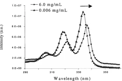
The ratio 11/1111 of the first (371 nm) and the third (382 nm) vibronic peaks of the fluorescence emission spectrum of micelle-solubilized monomeric pyrene was also determïned. It provided an estimate of the polarity sensed by pyrene in its micellar solubilization site (Figure 2.2).
An excimer, as defined by Birks, is a dimer which is associated in an electronic excited state and which is dissociated in its ground state. The formation of a pyrene excimer requires the encounter of an electronically excited pyrene with a second pyrene in its ground electronic state. According to this definition, the two pyrenes must be sufficiently far apart when light is absorbed, so that the excitation is localized on one of them; Birks, J. B. Rep. Prog. Phys. 1975, 38,903
——6.0 mg/mL —“--0.006 mg/mL
290 310 330 350
32 II . III 2x10⁶ .‘ . 6 1x10 • . .. .
J
I • I • I • I 350 400 450 500 Wavelength (nrn)Figtire 2.2 Emission spectrum of pyrene in pure water with the labelled peaks Ii and
III’.
2.1.6 Dynamic Light Scattering
The hydrodynamic diameter of vanous HPC-based polymenc micelles in aqueous solution (5 mg/mL) was evaluated by dynamic laser light scattering (DLS) using a Brookhaven system with an uniphase Blue laser at wavelength of 532 nm and a
scattering angle of 90 0
AIl measurements were performed in triplicate at 25 °C; the
data presented are the mean ±SD (Standard Deviation).
2.1.7 Microcalonmetry
A VP-differential scanning calorimeter from MicroCal (Northampton, MA) was used. The volume of the cell was about 0.52 n±. Samples for cloud point and microcalorimetric measurements were prepared by dilution of 10 mg of HPC-g
P0E10-C16, HPC-g-POE20-C16 and HPC-g-POE20-Cis, with different molar
substitutions, in 2 mL of Milli-Q water, at room temperature (microcalorimetric technique is very sensitive to sample concentration; after several trials, the sample concentration that gave the best endotherm was SmgImL). Samples were degassed and transferred to the sample celI with a calibrated synnge. Milli-Q water was similarly placed in the reference ce!!. Samples were heated at 30 °C/hr starting from
33
20 oc up to 80 °C. Four scans were done for each analysis. Baseline subtraction and normalization with respect to the scan rate and mM concentration were performed,
yielding the temperature-dependent molar heat capacity,
cp.
The molecular weight of the anhydroglucose unit conesponding to MS = 3.7 is 3775•
2.2 References
(1) Yoshida, Y.; Sakakura, Y.; Aso, N.; Okada, S.; Tanabe, Y. Tetrahedron 1999,
55, 2183
(2) cnstea, M. BiomacromoÏecuÏes (2003), submitted
(3) Winnik, F. M.; Regismond, S. T. A.; Goddard, E. D. Colloids Surf A:
Fhysicochem. Eng. Aspects 1996, 106, 243
(4) Winnik, F. M. Chem. Rev. 1993, 93, 587
(5) Conio, G.; Bianchi, E.;

![Figure 3.3 Excitation spectra of pyrene in aqueous solutions of HPC-g-POE20-C18 (ca. 3.1%) at different polymer concentrations [HIvI-polymer 0.006 mglmL (u), HM polymer 0.6mgImL (.), HM-polymer 0.06 mg/mL (Â)]](https://thumb-eu.123doks.com/thumbv2/123doknet/8263146.278207/53.918.301.701.117.401/figure-excitation-spectra-aqueous-solutions-different-polymer-concentrations.webp)